Chapter 061. Disorders of Granulocytes and Monocytes (Part 1) pps
Bạn đang xem bản rút gọn của tài liệu. Xem và tải ngay bản đầy đủ của tài liệu tại đây (74.32 KB, 5 trang )
Chapter 061. Disorders of Granulocytes
and Monocytes
(Part 1)
Harrison's Internal Medicine > Chapter 61. Disorders of Granulocytes
and Monocytes
Disorders of Granulocytes and Monocytes: Introduction
Leukocytes, the major cells comprising inflammatory and immune
responses, include neutrophils, T and B lymphocytes, natural killer (NK) cells,
monocytes, eosinophils, and basophils. These cells have specific functions, such as
antibody production by B lymphocytes or destruction of bacteria by neutrophils,
but in no single infectious disease is the exact role of the cell types completely
established. Thus, whereas neutrophils are classically thought to be critical to host
defense against bacteria, they may also play important roles in defense against
viral infections.
The blood delivers leukocytes to the various tissues from the bone marrow,
where they are produced. Normal blood leukocyte counts are 4.3–10.8 x 10
9
/L,
with neutrophils representing 45–74% of the cells, bands 0–4%, lymphocytes 16–
45%, monocytes 4–10%, eosinophils 0–7%, and basophils 0–2%. Variation among
individuals and among different ethnic groups can be substantial with lower
leukocyte numbers for certain African-American ethnic groups. The various
leukocytes are derived from a common stem cell in the bone marrow. Three-
fourths of the nucleated cells of bone marrow are committed to the production of
leukocytes. Leukocyte maturation in the marrow is under the regulatory control of
a number of different factors, known as colony-stimulating factors (CSFs) and
interleukins (ILs). Because an alteration in the number and type of leukocytes is
often associated with disease processes, total white blood count (WBC) (cells per
µL) and differential counts are informative. This chapter focuses on neutrophils,
monocytes, and eosinophils. Lymphocytes and basophils are discussed in Chaps.
308 and 311, respectively.
Neutrophils
Maturation
Important events in neutrophil life are summarized in Fig. 61-1. In normal
humans, neutrophils are produced only in the bone marrow. The minimum number
of stem cells necessary to support hematopoiesis is estimated to be 400–500 at any
one time. Human blood monocytes, tissue macrophages, and stromal cells produce
CSFs, hormones required for the growth of monocytes and neutrophils in the bone
marrow. The hematopoietic system not only produces enough neutrophils (~1.3 x
10
11
cells per 80-kg person per day) to carry out physiologic functions but also has
a large reserve stored in the marrow, which can be mobilized in response to
inflammation or infection. An increase in the number of blood neutrophils is
called neutrophilia, and the presence of immature cells is termed a shift to the left.
A decrease in the number of blood neutrophils is called neutropenia .
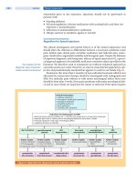
Figure 61-1
Schematic events in neutrophil production, recruitment, and
inflammation. The four cardinal signs of inflammation (rubor, tumor, calor,
dolor) are indicated, as are the interactions of neutrophils with other cells and
cytokines. PMN, polymorphonuclear leukocytes; G-CSF, granulocyte colony-
stimulating factor; IL, interleukin; TNF-α, tumor necrosis factor α.
Neutrophils and monocytes evolve from pluripotent stem cells under the
influence of cytokines and CSFs (Fig. 61-2). The proliferation phase through the
metamyelocyte takes about 1 week, while the maturation phase from
metamyelocyte to mature neutrophil takes another week. The myeloblast is the
first recognizable precursor cell and is followed by the promyelocyte. The
promyelocyte evolves when the classic lysosomal granules, called the primary, or
azurophil, granules are produced. The primary granules contain hydrolases,
elastase, myeloperoxidase, cathepsin G, cationic proteins, and
bactericidal/permeability-increasing protein, which is important for killing gram-
negative bacteria. Azurophil granules also contain defensins, a family of cysteine-
rich polypeptides with broad antimicrobial activity against bacteria, fungi, and
certain enveloped viruses. The promyelocyte divides to produce the myelocyte, a
cell responsible for the synthesis of the specific, or secondary, granules, which
contain unique (specific) constituents such as lactoferrin, vitamin B
12
–binding
protein, membrane components of the reduced nicotinamide-adenine dinucleotide
phosphate (NADPH) oxidase required for hydrogen peroxide production,
histaminase, and receptors for certain chemoattractants and adherence-promoting
factors (CR3) as well as receptors for the basement membrane component,
laminin. The secondary granules do not contain acid hydrolases and therefore are
not classic lysosomes. Packaging of secondary granule contents during
myelopoiesis is controlled by CCAAT/enhancer binding protein-ε. Secondary
granule contents are readily released extracellularly, and their mobilization is
important in modulating inflammation. During the final stages of maturation, no
cell division occurs, and the cell passes through the metamyelocyte stage and then
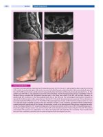
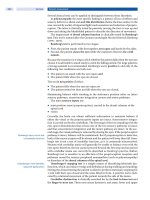
to the band neutrophil with a sausage-shaped nucleus (Fig. 61-3). As the band cell
matures, the nucleus assumes a lobulated configuration. The nucleus of
neutrophils normally contains up to four segments (Fig. 61-4). Excessive
segmentation (more than five nuclear lobes) may be a manifestation of folate or
vitamin B
12
deficiency and the congenital neutropenia syndrome of warts,
hypogammaglobulinemia, infections, and myelokathexis (WHIM) described
below. The Pelger-Hüet anomaly (Fig. 61-5), an infrequent dominant benign
inherited trait, results in neutrophils with distinctive bilobed nuclei that must be
distinguished from band forms. Acquired bilobed nuclei, pseudo Pelger-Huet
anomaly, can occur with acute infections or in myelodysplastic syndromes. The
physiologic role of the normal multilobed nucleus of neutrophils is unknown, but
it may allow great deformation of neutrophils during migration into tissues at sites
of inflammation.