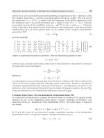
- Trang chủ >>
- Khoa Học Tự Nhiên >>
- Vật lý
INTERFACIAL AND CONFINED WATER Part 6 pdf
Bạn đang xem bản rút gọn của tài liệu. Xem và tải ngay bản đầy đủ của tài liệu tại đây (357.01 KB, 32 trang )
This page intentionally left blank
6 Role of interfacial water in
biological function
Importance of water in biology is well known: life on the earth cannot
exist without water. There is a large amount of water in living organisms
(about 60% by weight in human body), both inside and outside the bio-
logical cells. Water is involved in various biochemical reactions and acts
as a solvent for biomolecules. Despite the relatively high water content
in living organisms, pure liquid water is practically absent in biosystems.
Both intracellular and extracellular liquids consist mainly of water, but
the concentration of organic compounds, including large biomolecules,
is very high (about 20 to 30%). The central role of water in biological
function is recognized [442, 443], but the numerous questions concern-
ing the physical mechanisms behind the importance of water for life
remain unanswered. There are several important physical phenomena,
which should be taken into account when considering water properties in
biosystems and the role of water in biological function.
First phenomenon is related to the bulk phase transitions in aqueous
mixtures. In biosystems, water is a component of a multicomponent fluid
mixture with various biomacromolecules, small organic molecules, ions,
etc. This complex mixture unavoidably possesses a rich phase diagram
with numerous phase transitions and respective critical points, which may
occur close to the thermodynamic conditions typical of living organisms
on the earth. The general features of these phase transitions are similar
to the ones of the liquid–liquid transitions of binary mixtures of small
organic molecules with water. However, there are several factors that
make the phase transitions in biological liquids much more complex.
Multiplicity of the transitions in a multicomponent mixture assumes mul-
tiplicity not only of the stable but also of the metastable states, which may
exist during a long period of time. Phases enriched with macromolecules
are usually not liquids but solid-like structures with some level of order-
ing at the mesoscopic or macroscopic scales (micelles, fibrils, etc.).
Biomolecules have variety of conformational states, which are strongly
coupled with the phase state of a system. Strictly speaking, conforma-
tional transition of a single biomolecule and the phase transition, which
151
152 Interfacial and confined water
involves an ensemble of such molecules, cannot be considered separately.
Finally, situation is complicated by the possible chemical reactions in
complex biosystems.
The phase state of the aqueous mixture, in particular its location with
respect to the phase transitions, governs the clustering of both water and
organic molecules. For example, being inside the two-phase region, two
phases may appear as two macroscopic clusters of like molecules. In the
system being in the one-phase region, the clustering of like molecules
(water or biomolecules) is determined by the proximity to the phase
transition. When the phase transition is approached, clustering of the
minor component enhances. This approaching may be achieved by vary-
ing temperature, pressure, pH and by adding some cosolvents, ions, etc.
Majority of aqueous solutions of organic molecules show a closed-loop
phase diagram, which terminates by the lower critical solution tempera-
ture (LCST) and upper critical solution temperature from low and high
temperature sides, respectively. For example, the system in a one-phase
region below LCST separates into two phases upon heating. Accordingly,
the trend of the biomolecules to form clusters intensifies when the system
approaches solution temperature upon heating. In chemical literature,
clustering of solute molecules in water is often described as a manifes-
tation of “hydrophobic interactions.” Note that the phase transition and
related clustering of biomolecules inside the relatively small biological
cells may be affected by the finite size effect [332], which should suppress
aggregation of biomolecules [444].
Second phenomenon is related to the surface phase transitions. It is
natural to expect preferential adsorption of water or another component
of the biological liquids on the cell wall or other biosurfaces. Obviously,
this adsorption strongly affects the properties of biological liquids near
the walls. In particular, adsorption of biomolecules may facilitate forma-
tion of their ordered aggregates. If the effective attraction of biomolecules
to a surface is strong enough, we may expect a surface phase transition,
which results in the formation of a specific surface phase. Description
of the biological fluids based on the statistical theory of the bulk and
surface phase transitions should be very useful for understanding their
properties. Due to the extremely complex character of these systems, full
application of such approach seems to be possible in the long-term per-
spective only. However, the phase behavior and properties of water in
Role of interfacial water in biological function 153
biosystems may be studied by the experimental and simulation methods
available.
Biological liquids contain small solvent molecules (water) and high
concentration of large solute molecules (biomolecules). Due to the strong
difference in the sizes of typical biomolecules and water molecules,
a high fraction of water molecules belongs to the hydration shells of
biomolecules, as just one to three water layers separate biomolecules in
living cells. Accordingly, water in biosystems exists mainly as interfa-
cial (hydration) water, which is located in a close vicinity of the surfaces
of biomolecules, cell walls, etc. This emphasizes the role of interfacial
water in biological function. To describe the properties of interfacial
water in a systematic way, we have to characterize its possible states,
taking into account the effect of the phase transitions. For example, lay-
ering transition of hydration water (Section 2.2) is closely related to the
formation of the hydrogen-bonded water network, which covers some
surface homogeneously (Section 5.1). This network breaks upon heating
or upon dehydration, indicating qualitative changes of the state of hydra-
tion water. Liquid–liquid transition(s) of hydration water (Sections 1 and
4.2) may affect its properties upon cooling and pressurization. Analy-
sis of the possible states of hydration water should help clarify how the
presence of water makes the biological function possible. In this section,
we consider how biological function depends on hydration level, tem-
perature, and pressure. Formation of the spanning water network upon
hydration and its effect on the properties of biosytsems are analyzed in
Section 7. Properties of hydration shell in fully hydrated biosystems are
considered in Section 8.
To clarify the role of water in biofunction, it is reasonable first to con-
sider the relation between the hydration level and various manifestations
of biological activity. Experimental studies of some biosystems show
that their physiological activity appears rapidly at some critical hydra-
tion level. At the cellular and multicellular levels, biological function of
living organisms appears as metabolism, which includes a set of chemi-
cal reactions and transport of metabolites. The possibility to study these
processes upon dehydration/hydration of living organisms is limited by
the fact that most of them die when the water loss exceeds some critical
level. For most organisms, this level is 50% of body water (about 14% for
humans). However, some unicellular organisms, plants, and invertebrates
154 Interfacial and confined water
(seeds of plants, fungal spores, lichens, cysts of embryos, nematodes,
rotifers, tardigrades, etc.) remain viable after almost complete dehydra-
tion (95 to 99%) [445–451]. After dehydration, metabolism is completely
shutdown and organisms can stay in such state of a temporary death
for many years, but they cannot function untill some hydration level is
restored. The first observation of this phenomenon was described by the
pioneering microscopist Antony van Leeuwenhoek in 1702 [452]. The
ability of organisms to survive in anhydrobiotic state may be explained
by the water-replacement hypothesis [453]. This hypothesis assumes that
under dehydration, some polyhydroxyl compounds, such as glycerol,
cucrose, and theralose, substitute intracellular water, preserving macro-
molecular integrity and preventing cells from destruction. Experimental
studies of the dehydration/hydration processes of anhydrobiotic organ-
isms give unique possibility to follow decline/restoration of metabolism
in living organisms with hydration level. Understanding of the micro-
scopic mechanisms of these “hydration-dependent metabolic transitions”
should clarify the role of water in biofunctions [453].
There is a clear correlation between the water content and metabolism
in living organisms. For example, the metabolism of tardigrades dras-
tically declines with decreasing humidity, and when humidity is below
48%, oxygen consumption is below 0.035% of its value for hydrated
animals [456]. The most detailed experimental studies of the interrela-
tionship between hydration and metabolism in a living organism were
performed for Artemia salina cysts [453–455, 457–462]. Biological acti-
vity of these cysts develops upon hydration in a stepwise fashion. There are
no emergence of larvas below the hydration level h (gram of water per
gram of organics) of about 0.46 g/g, whereas at h = 0.72 g/g, already
22% of cysts produce swimming larvas [454] (see Fig. 90). The onset
of various important biochemical processes is seen in the vicinity of
this interval of hydrations. At the critical hydtation level h ≈ 0.60 g/g,
conventional cellular metabolism develops in a stepwise fashion. In par-
ticular, mass of the cysts starts to decrease, indicating oxidation of
their endogeneous reserves of carbohydrate [457]; cellular respiration
appears [455] (Fig. 90); amount of adenosine triphosphate starts to
increase and the total content and composition of free amino acids start
to change [461]; and incorporation of CO
2
into proteins and RNA begins
[460]. Another critical hydration level h ≈ 0.30 g/g indicates initiation
Role of interfacial water in biological function 155
emergence
respiration
into amino acids
and nucleotides
into proteins
and RNA
respiration (arb. units)
emergence of cysts (%)
incorporation of
14
CO
2
(arb. units)
80
60
40
20
0
0.2 0.4 0.6 0.8
h (g/g)
Figure 90: Hydration-induced metabolic transition of Artemia cysts. Upper
panel: emergence of cysts [454] and respiration [455]. Lower panel: incorpora-
tion of radioactivity into amino acids, nucleotides, proteins, and RNA [453].
of intermediary metabolism, which involves some particular amino acids
[461] and causes incorporation of CO
2
into amino acids and nucleotides
[459, 460] (Fig. 90).
Respiration rate of the yeast cells linearly decreases with water content
upon dehydration and apparently stops at hydration level h ≈ 0.20 g/g
[463] (Fig. 91). For lichens, two “switching points” in the hydration-
induced metabolism were found [464]. Limited metabolism appears
when water content is below 10% of the fully hydrated samples, and
at hydrations above 20%, another class of enzymes becomes active.
Seeds of plants may stay for years in dehydrated state but germinate
promptly upon hydration. This makes the analysis of the evolution of
physiological activities of seeds with increasing hydration possible. The
rate of O
2
consumption and the rate of CO
2
evolution by dry seeds
are very low, indicating an absence of mitochondrial metabolism. It
156 Interfacial and confined water
1.0
0.8
0.6
0.4
0.2
20 40 60 80
H
2
O (%)
O
2
uptake rate
Figure 91: Respiration rate of partially dried yeast cells. Oxygen uptake rates
at 30
◦
C are plotted in relative units. The closed circle represents the internal
respiration rate of the native cells. Reprinted, with permission, from [463].
increases dramatically in a stepwise manner at some critical hydration
level [465–468]. This level is h ≈ 0.14 g/g for apple, 0.20 g/g for corn,
0.24 g/g for soybean, and 0.26 g/g for pea. Additionally, some other
physiological activities (photosynthetic electron transport, transfer of
light-excited states) start at lower hydration levels (about two times lower
than those given above).
At molecular level, the manifestations of the biological activity appear
in specific biochemical reactions, conformational behavior, and dynami-
cal properties of biomolecules. Experimental studies of various partially
hydrated enzymatic proteins show that their activity accelerates rapidly
at some critical hydration levels. Onset of the enzymatic activity of ure-
ase occurs at h ≈ 0.15 g/g [469]. In the presence of chymotrypsin, the
acylation reaction is undetectable at hydrations h<0.12 g/g, but its
rate grows sharply above this critical hydration level [470]. The rate
of enzymatic activity of glucose-6-phosphate dehydrogenase, hexoki-
nase, and fumarase becomes detectable and start to increase sharply
at h ≈ 0.20 g/g, whereas this critical hydration is about 0.15 g/g for
phosphoglucose isomerase [471]. Enzymatic activity of lysozyme can
be detected only when hydration level achieves h ≈ 0.20 g/g [472, 473]
(see Fig. 92).
Existence of the critical hydration level h
c
for enzymatic activity may
reflect the fact that hydration water can serve as a transport media for
the substrates and/or for the products of the reactions only above h
c
[471]. This possibility was explored by the experiments with gas-phase
Role of interfacial water in biological function 157
enzymatic activity, log(a)
0.2 0.4 0.6 0.8
h (g/g)
Figure 92: The rate a of the enzymatic activity of lysozyme at various
hydration levels [473].
substrates [474–476] and by the experiments with enzymes in nonaque-
ous fluid environment [477, 478]. Activity of alcohol dehydrogenase from
bakers yeast with respect to substrate vapor appears when hydration level
reaches 0.16 g/g [474]. In other studies, nonzero enzymatic activity of
lipase and esterase was detected for gas-phase substrates at extremely low
hydrations [475, 476]. However, in these cases, a noticeable increase of
enzymatic activity is also seen in the hydration range 0.10 to 0.20 g/g.
Activity of laccase [478] and subtilisin [477] in organic solvents appears
only at some critical hydration level of added water, which depends on
solvent. Obviously, in experiments with enzymes in organic solvents, the
critical water level is determined by the miscibility of water and solvent
and by the difference in the water–protein and solvent–protein interac-
tions. Clearly, less water amount is necessary to provide the same cov-
erage of protein molecules in hydrophobic solvents. When the enzymatic
activity is analyzed as a function of water bound to enzyme, the critical
water level does not depend noticeably on the solvent and is close to about
0.10 g/g for yeast alcohol oxidase in various solvents [479].
Bacteriorhodopsin is an intramembrane protein, which uses adsorbed
light energy to transfer a proton through the membrane. The microscopic
mechanism of the proton pumping is based on the set of isomerization
processes initiated by the light adsorption. Upon dehydration, photoiso-
merization of bacteriorhodopsin reduces [480–484] and proton pumping
stops below 60% relative humidity [483–486]. The above examples show
158 Interfacial and confined water
direct correlation between the hydration level and the biological activity
of biomolecules.
In most cases, it is not easy to get explicit dependence of some form
of biological activity on the hydration level even at molecular level.
However, we may consider effect of hydration on the properties of
biomolecules, which are known to be necessary for their functionality.
Biomolecules in biologically active state are characterized by the specific
conformation and by some level of internal conformational dynam-
ics. Conformational stability of DNA double helix strongly depends on
hydration water. DNA exists in biologically relevant B-form until the
hydration Γ, measured as a number of water molecules per nucleotide,
exceeds Γ ≈ 20 [487, 488]. In the B-form, DNA is a right-handed dou-
ble helix, which makes a turn every 34
˚
A, and the distance between two
neighboring base pairs is 3.4
˚
A. At lower hydrations, DNA undergoes
different conformational transitions depending on its sequence, bound
metal ions, and other environmental conditions. The most studied is the
transition from B- to A-form [489], with the midpoint at about Γ=15
[487, 490, 491]. In the A-form, DNA helix remains right handed but
becomes shorter and broader (Fig. 93). Dehydration of B-DNA may be
achieved not only in the vapor phase by decreasing the relative humidity
but also in a liquid phase by adding some organic solvent. For instance,
B-DNA, G518 A-DNA, G512
dehydration
Figure 93: DNA exists in a biologically relevant B-form at high hydrations
and undergoes conformational transition into A-form upon dehydration.
Role of interfacial water in biological function 159
B to A transition was also observed in concentrated solutions of some
nonelectrolytes miscible in water [492, 493].
Proteins and polypeptides also undergo conformational changes upon
dehydration [494]. For example, a Raman spectrum of a dry lysozyme
powder differs from a spectrum of a solution. The parameters of the
main structure-sensitive spectral bands achieve their values in solu-
tion at hydration h ≈ 0.20 g/g [495, 496], which coincides with the
onset of the enzymatic activity of lysozyme [472, 473]. Experimental
studies of NMR spectra of a lysozyme powder also evidence confor-
mational changes within the hydration range from 0.1 to 0.3 g/g [497].
The hydration-induced conformational changes of lysozyme are fully
reversible, whereas in some other proteins, these changes are stronger and
only partially reversible [498]. Lyophilized subtilisin undergoes confor-
mational transition in organic solvent, when water content increases from
0.15 to 0.35 g/g [499]. Conversion of hemichrome to methemoglobin
with increasing water content shows sigmoid dependence on hydration
level, with an inflection point at about 0.25 g/g [500, 501]. It is well
known that conformation of a biomolecule may be strongly affected
when it is adsorbed on the surface (for example, on the surface of a
membrane). Apart from various factors that affect conformation of a
biomolecules in this case, “dehydration” due to the direct contact with
a surface should also play a role. Similar effect may result from the
crowding of biomolecules in a cell.
A biologically relevant lamellar phase of biomembranes exists only
when hydration level exceeds some critical value, typically about h ≈
0.20 to 0.30 g/g [502–504]. For example, this hydration level is required
to suppress the leakage from seeds and pollen [502, 505]. Neutron scat-
tering studies evidence “hydration-induced flexibility” of biomembranes
[484, 506, 507]. Slower motions are more strongly influenced by the
hydration level, and for the purple membrane samples, they increase
when hydration increases from about 0.3 to 0.4 g/g.
Internal dynamics of biomolecules is practically frozen without water.
Upon increasing hydration level, it develops in a stepwise fashion
[508]. At h ≈ 0.15 g/g, internal protein motion, monitored by hydro-
gen exchange, achieves its solution rate [509]. Full internal dynamics
of lysozyme is restored at h ≈ 0.38 g/g [510]. Mossbauer spectroscopy
studies evidence restoration of the internal dynamics of lysozyme
160 Interfacial and confined water
molecules when hydration level achieves 0.1 to 0.2 g/g [511]. Neu-
tron and light scattering experiments indicate the appearance of a slow
relaxation process in lysozyme powder at about 0.20 g/g [512, 513].
Experiments with lysozyme in glycerol show the onset of its dynam-
ics at about 0.1 g/g of water with saturation at ≈ 0.4 g/g [514–516].
Elastic properties of elastin strongly depends on its hydration. At room
temperature, its elongation under constant load increases drastically at
h ≈ 0.25 g/g [517]. Upon hydration to 0.2 g/g, only backbone motion
of elastin slightly increases, whereas above 0.3 g/g hydration there are
large-amplitude motions of both the backbone and the side-chains [518].
Importance of hydration water in the dynamics and functions of
biomolecules is also seen from the studies of hydrated biomolecules at
low temperatures. In the temperature interval from about 180 to 230 K,
dynamics of biomolecules show rapid increase. Experimental studies
show dynamic transition of crystalline ribonuclease A at about 220 K
[519], and this temperature corresponds to the onset of its enzymatic
activity upon heating [520]. Approximately at the same temperature,
enzymatic activity of elactase [521] and myoglobin [522] starts to
develop upon heating. The dynamic transition of chromatophore mem-
brane occurs at about 180 K, and at the same temperature, the efficiency
of the photoinduced electron transfer starts to increase upon heating
[523]. The dynamic transition of biomolecules was detected by var-
ious experimental methods: Mossbauer scattering [523, 524], neutron
scattering [525–528], X-ray crystallography [519, 529], infrared spec-
troscopy [530], etc. Besides, this transition is clearly seen in computer
simulations of hydrated biomolecules [531–537]. The temperature of the
dynamic transition is not very sensitive to the biomolecular structure and
for various biomolecules (ribonuclease [519, 520], DNA [526–528, 535],
bacteriorhodopsin [484, 486, 507, 538], myoglobin [524, 525, 530, 531,
533, 534, 536, 537], lysozyme [514, 515, 528, 539], carbohydrates [540],
etc.) varies within relatively narrow temperature interval.
Dynamic transition does not occur when biomolecules are dry as it
requires some minimal amount of water [514, 527, 528, 530, 538, 540–
542] and may be strongly affected by the presence of cosolvents [514,
515, 539]. The apparent temperature of the dynamic transition increases
with the lowering of hydration level or with adding of cosolvents [514,
539, 540, 542–545]. The most drastic increase in this temperature occurs
Role of interfacial water in biological function 161
when hydration level increases from 0.1 to 0.3 g/g [514, 543–545].
These facts indicate that the temperature-induced dynamic transition of
biomolecules is governed by hydration water.
There is some upper temperature limit for life. Some microorganisms
remain viable at 121
◦
C [547], but in most cases, this temperature is below
100
◦
C. This upper limit is closely related to the loss of the ordered struc-
tures of biomolecules upon heating. Activity of biomolecules depends
on their flexibility, and a less flexible biomolecule should be more sta-
ble against heating [548]. Dehydration of biomolecules or removing of
water by adding some organic solvents increases their thermal stability
[549–552]. Irreversible thermal inactivation of trypsin and ribonucle-
ase is strongly suppressed by drying [549]. Thermal stability of some
enzymes is enhanced when they are suspended in anhydrous organic
solvents [550, 551]. The denaturation temperature of bacteriorhodopsin
increases by more then 50
◦
C upon dehydration [552]. For lysozyme, an
increase in the denaturation temperature exceeds 90
◦
C [546, 553, 554]
and becomes noticeable when the hydration level h is below 0.4 g/g
[515, 546] (see Fig. 94). The temperature width of the denaturation peaks
20
15
10
400
380
360
340
0 0.2 0.4 0.6 0.8 1.0 1.2 1.4 1.6
Water content (g/g)
T
d
(K) DT
d
(K)
Figure 94: The temperature of denaturation, T
d
, and temperature width of
denaturation peak, ΔT
d
, of lysozyme as function of water content. Reproduced,
with permission, from [546].
162 Interfacial and confined water
in the different scanning calorimetry thermogram of lysozyme starts to
increase when h is below 0.2 g/g [546, 553]. Similar to lysozyme, the
denaturation temperature of ovalbumin starts to increase at h<0.4g/g
[555]. The denaturation temperature of elastin and collagen increases
upon dehydration by more than 150
◦
C, and this effect is noticeable when
water weight concentration is below 50% [556]. The chain melting tem-
perature of biological membranes increases by more than 30
◦
C upon
dehydration, and this increase starts when the hydration level is below
about 15 water molecules per lipid molecule [557].
Temperature-induced unfolding of fully hydrated biomolecules is usu-
ally accompanied by their aggregation. Upon heating, aqueous solutions
of some polypeptides (for example, large ELP [558]) separate into water-
rich and organic-rich phases, thus possessing a LCST. The temperature
of this phase transition depends on the peptide composition, its concen-
tration, addition of cosolvents, pH, etc. Similar to other macromolecules
whose aqueous solutions show an LCST [559–561], polypeptides dras-
tically change their conformational distribution when crossing the phase
separation temperature. The origin of the LCST in aqueous solutions is
often considered in relation to the ordered character of the hydration shell,
surrounding the solute molecule [64, 562–565]. At low temperatures, a
solute molecule is covered by ordered hydration shell, which promotes
its solubility in water. This shell becomes less ordered upon heating, that
causes demixing at some temperature [566]. So, even in the case of a fully
hydrated biomolecule, the state of the hydration shell can noticeably affect
its properties.
Biosystems and their functions can be strongly affected also by pres-
sure [567–570]. Activity of some microorganisms may increase upon
applying pressure and reach a maximum in some pressure range [569].
However, above some pressure, activity of all living organisms decays.
For example, a noticeable decay of cellular activity of yeasts starts at P ≈
1 kbar, and yeast cells are killed at P ≈ 2 kbar [571]. Upon pressurization
to about 6 kbar, DNA molecules undergo the conformational transition
from the native B-form to left-handed double-helical Z-form [572]. The
chain melting temperature of biological membranes increases by about
22
◦
C, when pressure increases by 1 kbar [570]. The melting tempera-
ture of DNA is also sensitive to pressure and may increase or decrease
upon pressurization [573]. There were extensive experimental studies of
Role of interfacial water in biological function 163
the pressure-induced protein unfolding (see [568–570] and references
therein). In the temperature–pressure plane, there is a closed-loop region
of the protein stability. Inside this region, proteins mainly preserve their
native conformations and are miscible with water, whereas outside this
region, they undergo unfolding/denaturation, accompanied by the protein
aggregation. For example, staphylococcal nuclease (S Nase) undergoes
unfolding at about 50
◦
C at ambient pressure. At T = 25
◦
C, the quali-
tatively similar unfolding transition occurs at about 2 kbar [574, 575].
Biomolecules, which are insoluble in water and form aggregates at ambi-
ent pressure, may dissolve with increasing pressure. For example, pressure
of about 300 bar is sufficient to prevent aggregation of insulin [576].
Closed-loop temperature–pressure stability diagram of some protein
in water should be directly related to the phase diagram of the protein/
water mixture. It is well known that the phase diagrams of aqueous
solutions are highly sensitive to pressure [63, 577–580], which may
either promote miscibility or induce demixing. For some aqueous solu-
tions, which show immiscibility gap at zero pressure, increasing pressure
causes extension of this gap in concentration and temperature range
(some pyridines [577]). Similar trend can be seen for some solutes, which
are completely misscible with water at zero pressure: upon pressuriza-
tion, aggregation of solute molecules enhances, indicating approaching
immiscibility (methanol [581]). For other solutes, the effect of pressure
is opposite (tetrahydrofuran [579], alkanes, noble gases). In some solu-
tions, changes of the solubility with pressure are even nonmonotonous
[579, 580]. Therefore, various evolutions of the phase diagrams with
pressure can be expected for aqueous solutions of various biomolecules.
When considering the effect of pressure on hydrated biomolecules, we
have to take into account possible changes of the phase state and ther-
modynamic properties of a bulk liquid water upon pressurization (see
Section 2), which should also affect hydration water at biosurfaces.
We have considered various manifestations of the importance of water
in biological function. In most cases, there are clear indications on the
crucial role of interfacial water in life. Two main aspects of the phase
behavior of interfacial water can be distinguished: a) condensation of a
layer of hydration water at biosurfaces and b) effect of temperature and
pressure on the state and properties of this hydration layer. These two
aspects are considered in the Sections 8 and 9, respectively.
This page intentionally left blank
7 Water in low-hydrated
biosystems
A step-like growth of various forms of biological activity occurs, when
the coverage of a biosurface by water approaches about one layer. Forma-
tion of a condensed water monolayer on the surface of various biosystem
may be expected at the hydration below about 0.4g/g. It is natural to
relate appearance of a biological function to some qualitative change
in the state of the interfacial water, which causes essential changes in
water properties. Formation of a condensed water monolayer indicates
transition of a hydration water from the gas-like state, where only small
hydrogen-bonded water clusters are present, to the condensed state. In
the case of idealized smooth surfaces, this transition may occur via a first-
order layering transition (Section 2.2) or continuously via a percolation
transition (Section 5.1). On strongly hydrophilic and heterogeneous bio-
surfaces, the critical temperature of the layering transition may be below
the ambient temperatures. Besides, this transition may be smeared out
if the surface heterogeneity is strong enough. In both cases, at ambient
temperatures, we may expect the formation of a condensed water layer at
biosurfaces via a percolation transition. In the Section 7.1, we consider
percolation transition of water in low-hydrated biosystems, and its effect
on the properties of the system is analyzed in Section 7.2.
7.1 Percolation transition of water in low-hydrated
biosystems
Formation of the hydrogen-bonded water networks may affect conductiv-
ity of a system in a drastic way, as these networks provide the paths for the
conduction of protons, ions, or other charges in the system. So, the qualita-
tive changes in the conductivity may be expected at hydrations, close to the
percolation transition of water. Surface conductivity of quartz increases
relatively slowly with increasing hydration level until the completion of
the adsorbed water monolayer, but much faster at higher hydrations [582].
The hydration dependence of the dielectric losses of hydrated collagen
165
166 Interfacial and confined water
[583] may be described by a power law (equation 24) with h
c
= 0 and
exponent t ≈ 6. Dependence of such kind was observed also for cellu-
lose (t ≈ 9.3), silk (t ≈ 16.0), and wood (t ≈ 16.4) [404–406], where it
was attributed to the developing network of conducting water chains. The
qualitative changes in the conductivity of a biosystem at some thresh-
old hydration level were first reported for albumin [584, 585] (Fig. 95).
Steady-state conductivity σ strongly depends on hydration level, and
this dependence changes at h ≈ 0.07 g/g. Change in the conductivity–
hydration relationship at this particular hydration was attributed to the
change in the “configuration of the water molecules bound to the protein
surface” [585]. Note that the use of the power-law hydration dependence
(equation 24) with h
c
= 0 to describe the data, shown in Fig. 95, gives
high values (6 to 9) of the exponent t. Sharp increase in the conductiv-
ity of melanin upon hydration is observed at h
c
≈ 0.11 g/g [586]. This
increase may be described by a power law with t ≈ 11. The conductivity
exponents, obtained in the considered cases, exceed noticeably the values
1.3 and 2.0 expected for 2D and 3D percolation [416]. This may be related
to the underestimation of the threshold hydration level or to the setting it
to zero in the studies described above. DC conductivity of hydrated triti-
cale seed samples and their counterparts increases in a stepwise manner
1E-7
1E-9
1E-11
1E-13
1E-15
0.05
log(ր⍀
–1
m
–1
)
0.10
h (g/g)
0.15 0.20
Figure 95: Variation of the steady-state conductivity σ with hydration of
albumin [585].
Water in low-hydrated biosystems 167
by about 4 orders of the magnitude within hydration range h = 0.15 to
0.30 g/g [587, 588].
Capacitance measurements of hydrated lecithin, cytochrome-c, and
hemoglobin at low frequencies (from 10
3
to 10
5
Hz) show its strong
increase at h ≈ 0.04, 0.06 and 0.12 g/g, respectively. These values are
close to the hydration levels, corresponding to monolayer coverage, which
were estimated from adsorption isotherms [589]. In the case of a lysozyme
powder, increase in the dielectric losses with increasing hydration was
attributed to the protonic conductivity, which is “restricted to the surface
of individual macromolecules and involves shifting of protons between
ionizable side chain groups of the protein” [590]. So, this conductiv-
ity appears due to the formation of mesoscopic water networks, which
spread over the distances comparable with the size of one macromolecule.
Experimental studies of the capacitance C of a lysozyme powder at vari-
ous hydration levels h allowed approximate estimations of the threshold
hydration, corresponding to the percolation transition of water [591]. The
threshold hydration h
c
was estimated by the extrapolation of the derivative
dC/dh to zero. The average value of h
c
, obtained from the measurements
in the frequency range from 10 kHz to 4 MHz and for pH from 3.11 to 7.0,
was 0.152 ± 0.016. The critical hydration practically does not depend on
pH = 3 to 8 but increases to 0.24 at pH = 9.9.
Experimental measurements of the capacitance of the hydrated
lysozyme powder were used to calculate the hydration dependence of
the dc protonic conductivity [592]. The obtained dependence is shown
in Fig. 96 in the linear (left panel) and in the double-logarithmic (right
panel) scales. Analysis of the conductivity in the vicinity of the perco-
lation threshold allows its localization and also gives information about
the dimensionality of the percolating cluster [416] (see equation (24)). In
accordance with the results, shown in the right panel of Fig. 96, just above
the percolation threshold, water molecules form 2D networks, which
are characterized by the conductivity exponent t of about 1.3. These
networks are mesoscopic and provide possibility of the long-range pro-
tonic displacements along the surfaces of lysozyme molecules. When h
exceeds h
c
+ 0.03, 2D water networks transform into 3D networks and
conductivity exponent crosses over to the value of about 2.0.
For hydrated sample of purple membrane, the percolation threshold of
water was reported at h = 0.0456 g/g [593] and at h = 0.06 g/g [594].
168 Interfacial and confined water
/
0
h (g/g)
log
10
[(2
c
)/
c
]
log
10
(h 2 h
c
)
t 5 2.0
t 5 1.3
0.0 0.1 0.2 0.3 0.4
0
100
200
300
400
1.0
0.0
21.0
23.0 22.0 21.0
Figure 96: DC conductivity, calculated from capacitance data measurements,
vs hydration level h of the lysozyme powder. Left panel: dc conductivity σ
normalized by the dc conductivity σ
0
of the dry sample. Right panel: dc con-
ductivity vs hydration level in a double-logarithmic scale. Solid lines show
the slopes, corresponding to the critical exponents 1.3 and 2.0 for 2D and 3D
percolation, respectively. Reprinted, with permission, from [592].
The conductivity exponent of about 1.23 indicates 2D character of the
percolation transition. Similar values of the conductivity exponent were
obtained for the hydration dependence of the conductivity of embryo
and endosperm of maize seeds [595, 596], where the percolation thresh-
old is h = 0.082 and 0.127 g/g, respectively. In hydrated bakers yeast,
protonic conductivity evidences 2D percolation transition of water at
h = 0.163 g/g, and the value of the conductivity exponent is about 1.08
[597]. In this system, increase in conductivity due to 3D water percola-
tion is observed at essentially higher hydration level h = 1.47 g/g, where
conductivity exponent is about 1.94, i.e., close to the 3D value t = 2.0.
Conductivity measurements of Artemia cysts at various hydrations show
strong increase in conductivity starting from the threshold hydration h =
0.35 g/g [598] (see Fig. 97). The conductivity exponent in this system is
1.635, which is in between the values expected for 2D and 3D systems.
DC conductivity of lichens, evaluated from the dielectric studies at fre-
quencies between 100 Hz and 1 MHz [599], shows strong enhancement
at some hydration level. Fit of the conductance–hydration dependence to
equation (24) gave the following parameters: h
c
= 0.0990 g/g, t = 1.46
for Himantormia lugubris and h
c
= 0.0926 g/g, t = 1.18 for Cladonia
Water in low-hydrated biosystems 169
mitis. So, percolation transition of water in lichens has 2D character and
reflects the formation of a spanning network of hydration water.
Low-frequency dielectric measurements (0.1 Hz–1 MHz) of hydrated
lysozyme, ovalbumin, and pepsin were used to estimate the fractal
dimension for the random walk of protons through the hydrogen-bonded
network of water molecules by fitting the shape of the dielectric loss
peak [600, 601]. In all three systems, a crossover from 2D to 3D water
network occurs within the interval of hydration from 0.05 to 0.10 g/g.
For lysozyme, these values are noticeably below the value of about
0.17 g/g, reported in [592]. This difference may be attributed to the pres-
ence of about 0.07 g/g of strongly bound water, which presumably was
not taken into account in [601]. When hydration exceeds the threshold
value, dielectric losses increase almost linearly with temperature. With
approaching T ≈ 310 to 330 K, this increase slows down and dielectric
losses turn to decrease with temperature. This behavior may reflect ther-
mal break of the spanning hydrogen-bonded water network, which will
be considered in Section 8.1.
At low temperatures, radiation-induced conductivity critically depends
on the water content and appears only above the critical hydration lev-
els 0.41 and 0.79 g/g for collagen and DNA, respectively [602, 603].
The critical hydration level for DNA corresponds to about 15 water
molecules per phosphate group (Γ=15). The effects of various additives
on the conductivity evidence charge migration in the hydration shell of
DNA [604]. At much lower hydrations (0.12 to 0.22 g/g), conductiv-
ity of hydrated DNA shows exponential dependence on h [605], which
may be attributed to the intrinsic semiconductivity of the DNA backbone.
More detailed experimental studies of DNA hydration [606] show that
radiation-induced conductivity starts not strictly at Γ=15 [602, 603] but
via a sigmoid-like increase within hydration range from Γ=11 to Γ=16
with subsequent stepwise increase at Γ ≈ 24.
The above experimental studies of the conductivity of hydrated biosys-
tems directly evidence the formation of a spanning network of hydration
water via percolation transition. The charge transfer itself may play a
crucial role in biofunction [607]. In most of the cases, described above,
the percolation transition of water occurs at the hydration level, where
various forms of biological activity develop in a stepwise manner (see
Section 6). In particular, the following biological processes starts close
170 Interfacial and confined water
h (g/g)
10
9
/⍀
Ϫ1
m
Ϫ1
800
600
400
200
0
0.0 0.1 0.2 0.3 0.4 0.5
Figure 97: The dc conductivity of Artemia cysts as a function of hydration
level h. Reprinted, with permission, from [598].
to the percolation transition of water: premetabolism of Artemia cysts,
enzymatic activity of lysozyme, respiration of yeasts, photoelectric
response of purple membrane, germination of seeds, conformational
transition of DNA to biologically relevant B-form. Besides, various con-
formational and dynamical properties of biomolecules necessary for bio-
logical function also develop in the hydration range, where percolation
transition is seen or may be expected in biosystems.
Below we show how the appearance of spanning water networks may
be detected in computer simulations. In particular, a percolation transition
of water upon hydration was studied by simulations in model lysozyme
powders and on the surface of a single lysozyme molecule. In protein
crystals, increase in hydration of a biomolecular surface may be achieved
by applying pressure. In some hydration range, pressurization leads to
the formation of spanning water networks enveloping the surface of each
biomolecule. Finally, the formation of the spanning water network is
shown for the DNA molecule at various conformations and for different
forms of DNA.
a) Percolation transition of water in lysozyme powders
The structure of amorphous lysozyme powder, used for experimen-
tal studies, is not available. In order to do simulations, which may
be at least qualitatively related to the experiment, the density of the
lysozyme powder should be as close to the experiment as possible.
Water in low-hydrated biosystems 171
In low-humidity tetragonal crystal with the partial density of lysozyme
of about 0.80 g/cm
3
, approximately 120 water molecules are in the first
hydration shell of lysozyme molecule. In order to explore a wide range of
hydration level up to monolayer coverage (about 300 water molecules),
partial density of lysozyme in powder should be < 0.80 g/cm
3
.In
Ref. [401], two models for protein powder were studied: densely packed
powder with the density of dry protein 0.66 g/cm
3
and loosely packed
powder with a density 0.44 g/cm
3
. In loosely packed powder, the per-
colation transition of water was noticeably (by a factor of two) shifted
to higher hydration levels compared with experiment. The fractal dimen-
sion of the water network at the percolation threshold as well as other
properties evidenced that the percolation transition of water in this model
was not two dimensional. The spanning water network consists of the
2D sheets at the protein surface as well as of the 3D water domains,
formed due to the capillary condensation of water in hydrophilic cavities.
The latter effect causes essential distortion of various distribution func-
tions of water clusters in loosely packed powder. Therefore, below we
present an overview of the results obtained for the densely packed model
powder.
Spanning probability R, defined as a probability to observe a water
cluster that crosses the model system at least in one dimension, shows
sigmoid dependence on the mass fraction C of water (Fig. 98, upper
panel). At ambient temperature (T = 300 K), the inflection point of this
dependence corresponding to R = 50% is located at about C = 0.122.
This hydration level is close to that where the mean cluster size S
mean
passes through a maximum (Fig. 98, middle panel). Fractal dimension
of the largest water cluster achieves the value d
2D
f
at C ≈ 0.155 (Fig. 98,
lower panel). Summarizing, the percolation transition of water may be
attributed to the hydration level C ≈ 0.155. The cluster size distribution
n
S
supports this conclusion [401].
The percolation threshold of water found in simulations should be
compared with experiments performed in the lysozyme powder [591].
At ambient temperature, it was observed at the hydration level h =
0.152 ± 0.016 g of water per gram of dry lysozyme, which corresponds to
the water mass fraction C ≈ 0.132 [591]. The percolation threshold seen
in simulations thus occurs at h ≈ 0.183, i.e., at slightly higher hydration
level than in experiment. This should be considered as rather good
172 Interfacial and confined water
10
0.10
1.0
1.5
2.0
2.5
15
30
45
0.0
0.2
0.4
0.6
0.8
1.0
T 5 300 K
T 5 400 K
0.15
C
d
f
d
f
2D
R
S
mean
C
0.20 0.15 0.20
15
20
25
Figure 98: 2D percolation transition of water in the hydrated densely packed
powder of lysozyme at two temperatures. Spanning probability R (upper panel),
mean cluster size S
mean
(middle panel), and fractal dimension of the largest clus-
ter d
f
(lower panel) are shown as functions of a mass fraction of water C. The
dashed lines are guides for eyes only. Vertical lines indicate the 2D percolation
threshold. Reprinted, with permission, from [401].
agreement between experiment and simulations in view of rather crude
model of lysozyme powder, which was imposed to have a rather arbitrary
density. Probably, the more densely packed model powder may show
better agreement with experiment. Besides, in experiment, lysozyme
molecules expose to water more and more their surfaces upon increasing
hydration [508]. That is not the case for the simulated powder, which was
frozen so that the rearrangement of lysozyme molecules upon hydration
was not allowed.
Water in low-hydrated biosystems 173
Simulations enable to explore the arrangement of water molecules near
hydrated proteins and their evolution during the percolation transition.
Experimental studies provide the average number of water molecules
per one protein (N
w
/N
p
∼120 [591]) at the percolation threshold. How-
ever, this number could not be equal to the average number of water
molecules N
1
w
in the first hydration shell of each lysozyme in powder. In
the model powder, N
w
/N
p
∼146 and N
1
w
∼149 at the percolation thresh-
old at 300 K. Close values of N
w
/N
p
and N
1
w
could mean that most
water molecules belong to the hydration shell of one protein molecule
only. These numbers are significantly smaller than numbers N
w
= 450
and N
1
w
≈ 336 in the case of the percolation threshold at the surface of
a single lysozyme molecule at the same temperature (see below). Such
strong difference could be attributed to the significant decrease in the
accessible surface area of proteins in powder due to close contacts. So,
the 2D percolation transition of water in protein powder appears as a for-
mation of water network, which spans the extended “collective” surface,
created by aggregated protein molecules, covering each protein molecule
only partially.
Temperature affects strongly the percolation threshold of hydration
water. Changes of the various cluster properties with hydration are com-
pared for T = 300 and 400 K in Fig. 98. Increasing temperature notably
shifts the percolation threshold to higher hydration level. It was found at
C = 0.175 (h = 0.212 g/g) at T = 400 K. The hydration dependence of
the spanning probability R becomes more rounded with increasing tem-
perature. Cluster size distributions n
S
of water in lysozyme powder at
T = 400 K are shown in Fig. 99 at some hydration levels. A hump of n
S
at large S appears at hydration where R ≈ 50% and reflects a cut in the
water clusters spanning the simulated system (see Section 5.1). Right at
the percolation threshold the cluster size distribution n
S
should follow
the power law (19) in a widest range of cluster sizes. At T = 400 K and
C = 0.173, n
S
follows a power law in the range of cluster sizes up to
200 molecules (see squares in Fig. 99). Wave-like deviations from the
power-law behavior could not be eliminated by improving statistics and
should be attributed to the peculiar arrangement of lysozyme molecules
in the model powder. Note that deviations of n
S
from the power-law
behavior become larger when the temperature decreases to the ambient.
This makes the use of n
S
distribution for location of a water percolation
174 Interfacial and confined water
1 10 100
S
n
S
n
S
~ S
22.05
1000
10
28
10
27
10
26
10
25
10
24
10
23
10
22
10
21
10
0
10
1
10
2
10
3
Figure 99: Distributions n
S
of clusters with S water molecules in densely
packed lysozyme powder at T = 400 K. Mass fraction of water increases from
C = 0.128 (top) to 0.201 (bottom). Circles represent n
S
at C = 0.151, when the
spanning cluster exists with probability of about 50%, while squares correspond
to C = 0.173, when the fractal dimension of the largest cluster is close to the 2D
percolation threshold value. The distributions are shifted consecutively by one
order of magnitude each, starting from the bottom. Reprinted, with permission,
from [401].
threshold in powders to be not very fruitful at low temperatures. For
large S left to the hump, the distribution n
S
deviates from the power
law upward below the percolation threshold and downwards above the
percolation threshold. So, the cluster size distributions indicate the per-
colation threshold at T = 400 K at water mass fraction C
p
≈ 0.17, that is
quite close to the threshold value C
p
≈ 0.175 estimated from the behavior
of the fractal dimension d
f
of the largest cluster (Fig. 98, lower panel).