Atomic Force Microscopy in Cell Biology Episode 1 Part 9 doc
Bạn đang xem bản rút gọn của tài liệu. Xem và tải ngay bản đầy đủ của tài liệu tại đây (525.98 KB, 20 trang )
146 Langer and Koitschev
of parallel oriented actin filaments, called the cuticular plate, forming a core and a root
embedded in a platform made of actin on top of the cell (Tilney and Tilney, 1986). At the
base, the number of actin filaments is reduced from a few hundred to about a dozen form-
ing a point of flexing. The actin filaments are crosslinked by smaller filaments, probably
fimbrin, preventing bending of stereocilia along their length. This construction allows
stereocilia to flex around their pivot point located at the bottom where actin filaments
penetrate the cuticular plate. A fibrous layer, called the tectorial membrane, overlies the
organ of Corti. The tips of the tallest stereocilia of the OHC but not of the IHC touch the
bottom side of the tectorial membrane. The origin of stereocilia displacement is a rela-
tive displacement between the basilar and the tectorial membrane forming together with
the hair cells a layered or sheet-like structure. Bending of the stereocilia results in stretch-
ing the tip links and opening of the transduction channel allowing an influx of cations into
the hair cell (Gillespie, 1995; Markin and Hudspeth, 1995). The ultrastructure of sensory
hair bundles of the mammalian inner ear has been extensively studied by scanning and
transmission electron microscopy (SEM and TEM) (Pickles et al., 1984; Lenoir et al.,
1987; Russell and Richardson, 1987). These methods are restricted to fixed and dehy-
drated specimens, however. Structural details of living hair cells have been visualized
by light microscopy with a limited spatial resolution due to the limitation of the optical
system. Investigation of hair cells with atomic force microscopy (AFM) seems attractive
since this method may combine three advantages: the high spatial resolution of scanning
probe microscopy (Binnig et al., 1986), the possibility to work under physiological con-
ditions (H¨orber et al., 1992), and the opportunity to record mechanical properties of the
structures being imaged (Hoh and Schoenenberger, 1994). AFM has already been used
for simultaneous imaging and elasticity measurements on living cells under physiologi-
cal conditions (H¨orber et al., 1992; Hoh and Schoenenberger, 1994; Shroff et al., 1995;
Radmacher et al., 1996). Therefore, it seems to be an appropriate technique for studying
the mechanical characteristics of the hair bundle under physiological conditions.
B. Preparation
Organs of Corti were isolated from 4-day-old rats (Wistar, Interfauna, Germany)
according to the methods used by Sobkowicz et al. (1975) and by Russell and Richardson
(1987) for mice. They were cut into three segments (basal, medial, apical) and placed
with the basilar membrane onto Cell-TAK-coated glass coverslips with a diameter of
10 mm. The sample was transferred into Ø 35-mm Falcon dishes filled with 3 ml culture
medium MEM D-VAL with 10% heat-inactivated FCS and 10 mM Hepes buffer, (pH 7.2)
supplemented at 37
◦
C and 5% CO
2
. Using this preparation, hair bundles are oriented
perpendicular to the surface of the supporting glass coverslip. For experiments,specimens
were transferred from the incubator to a specimen chamber. Cochlear cultures were
bathed at room temperature (22
◦
C) in a solution containing (millimolar concentrations)
144 NaCl, 0.7 NaH
2
PO
4
, 5.8 KCl, 1.3 CaCl
2
, 0.9 MgCl
2
, 5.6 D-glucose, 10 Hepes–
NaOH, pH 7.4, osmolarity 304 mOsmol. The solution was exchanged before and after
investigation using a conventional gravity-driven perfusion system to keep cells in good
condition. At 4 to 6 days after birth the tectorial membrane of rats is already partially
developed. In some preparations, the tectorialmembranedirectlytouchedthehairbundles
7. Sensory Cells of Inner Ear Examined by AFM 147
of OHC. To guarantee free access of the AFM tip to the hair bundles, the tectorial
membrane was removed with a cleaning pipette under the light microscope. Cultures
were investigated for a maximum of 2 h.
III. AFM Technology
A. Short Introduction to AFM
The theories of AFM, the technological aspects, imaging techniques, and applications,
have already been the topic of numerous publications (e.g., Meyer, 1992). Therefore,
only the aspects necessary for understanding the contents of this chapter, not aspects
just briefly described in other publications, will be discussed. The AFM experiments
described here concentrate on force spectroscopy at the extracellular surface of hair
cells. The capability of imaging is only used for correlation of force and position of the
sensor tip on the specimen. As described by Binnig et al. (1986), AFM locally measures
the force between a small tip and the specimen surface using a free-moving cantilever
with the force constant k
L
. Scanning the cantilever line for line across the sample while
the tip interacts with the surface, we can obtain an image of the sample topography.
The vertical deflection of the cantilever can be assigned to each point of the object. The
AFM tip is scanned with a piezo-electric tube. The outer electrode is segmented into
eight commensurate electrodes electrically connected in a way that the walls at the end
of the piezo-electric tube remain perpendicular to the direction of motion (Siegel et al.,
1995). The AFM tip consequently moves on a plane surface in contrast to piezo-electric
tube scanners with only four outer electrodes scanning on a curved surface. Typically
the scan range was between 1 and 8 μm. The force F exerted to the sample depends on
the vertical cantilever deflection z and the force constant k
L
as follows:
F = k
L
· z. [1]
Typical force constants of commercially available cantilevers vary from 10
−3
to 10
2
N/m.
Different methods for detection of the cantilever deflection such as heterodyne interfero-
meters (Martin et al., 1987), capacitive detection (Neubauer et al., 1990; G¨oddenhenrich
et al., 1990; Miller et al., 1990), and tunneling current detection (Binnig et al., 1986)
have been reported. In our instrument, the most common detection method was used:
the optical lever method (Meyer and Amer, 1988, Alexander et al., 1989). The light of
a laser diode is focused on the gold-coated surface of the AFM cantilever. The reflected
light is detected on a position-sensitive four-segmented photo-diode. Deflection of the
cantilever beam results in displacement of the laser spot on the photo-diode. The dif-
ference in intensity between the pairs of upper and lower segments encodes the vertical
cantilever deflection. Torsional forces are detected subtracting the intensity at the left
pair of segments from the intensity detected at the right pair of segments. AFM pro-
vides a wide range of microscopic techniques, which allow the measurement of different
surface and material properties. These techniques can briefly be classified into two dif-
ferent operation modes: the contact and noncontact mode. Both principles, discussing
the measurement of a force versus distance curve, can easily be explained (F versus d).
148 Langer and Koitschev
Fig. 3 Principle of force versus distance measurements. The deflection z of the cantilever beam is recorded
versus elongation of the piezo-electric scanner vertically moving the sample. The force corresponds to the
product of cantilever deflection and force constant. Before examination the sample is fully retracted, not
interacting with the AFM cantilever (inset: free-moving cantilever). As the scanner extends (arrows), the tip
and sample surface attract each other (position z
1
) and the cantilever starts to bend downward. This attraction
decreases with increasing scanner position. The force goesto zero (equilibrium between attractive and repulsive
forces, piezo position 0). As the scanner continues to extend the total force becomes positive (inset: repulsive
forces). At the maximum of the curve the scanner stops and retracts. With increasing contraction of the piezo-
electric scanner (dashed lines with arrowhead) the cantilever follows this motion beyond zero position and
loses contact at position z
2
. At this point the tension of the cantilever beam exceeds the attractive forces (see
inset), thereby losing contact with the sample surface.
A F versus d is a plot of the deflection of the cantilever versus the extension of the
piezo-electric scanner (Fig. 3). On the left side of the curve, the scanner is fully retracted
and the cantilever is in its resting position since the tip does not touch the sample. As
the scanner moves, the cantilever remains undeflected until it comes close enough to the
sample surface. As the atoms of tip and surface are gradually brought together, they first
weakly attract each other. This attraction increases until the atoms are so close together
that their electron clouds begin to repel each other electrostatically (position z
1
). This
electrostatic repulsion progressively weakens the attractive force as the interatomic sep-
aration continues to decrease. The force approaches zero when the distance between the
atoms reaches a couple of angstroms, about the length of a chemical bond (position 0).
As the scanner continues to extend, the total van der Waals force becomes positive
(repulsive), and the atoms are in contact. This situation corresponds to the contact mode.
For the noncontact mode AFM measurements have to be performed in the attractive
regime (position z
1
).
B. Principle of Hair Cell Measurements
Normally, force versus distance curves are recorded perpendicular to the sample sup-
port. In our experiments the cantilever scans in constant height exerting a horizontal
force to the mechanosensory structures of outer hair cells. The applied force results in a
horizontal displacement of the stereocilia along the axis of symmetry of V-shaped hair
7. Sensory Cells of Inner Ear Examined by AFM 149
bundles. This kind of measurement (Fig. 4A) corresponds to a force versus distance
curve performed in the horizontal direction. The feedback electronics normally keeping
the measured force at a constant level was switched off during investigation. Every data
point in an AFM image represents a spatial convolution (in the general sense, not in the
sense of Fourier analysis) of the shape of the tip and the shape of the feature imaged.
For most imaging applications this convolution leads to unwanted artifacts. In our mea-
surements, we took advantage of the convolution of AFM tip and rod-like stereocilia
geometry permitting us to calculate the stiffness of stereocilia. The van der Waals force
curve (Fig. 3) represents just one contribution to the cantilever deflection. Local varia-
tions in the form of the F versus d curve indicate variations in the local elastic properties.
These local variations in the slope of F versus d were used as a measure for the elasticity
of the investigated sample. Langer et al. (2000) showed for stereocilia that a steep slope
of the force curve indicates a high stiffness of the sample, while a shallow slope indicates
a soft sample. For examination, the AFM tip was adapted to the axis of symmetry of
the stereociliary bundles of OHC rotating the specimen chamber. A certain hair bundle
was visually selected in the light microscope and directly positioned below the AFM tip
adjusting the specimen stage. For the vertical fine approach, the specimen chamber was
elevated with the help of a piezo-electric stack while scanning the cantilever tip (scan
range: 4.5 μm). The approach is stopped as soon as stereocilia of the OHC cause signif-
icant deflection of the AFM cantilever (about 20 to 50 nm). For stiffness measurements,
the AFM tip successively scans each stereocilium within a hair bundle (Fig. 4A). Only
the tallest stereocilia of the hair bundle are touched with the frontal lateral face of the
pyramidal tip and deflected in excitatory and inhibitory directions (Langer et al., 2000).
Fig. 4 Principle of stereocilia examination by AFM. (A) The AFM tip scans with the feedback electronics
switched off across the hair bundle, thereby deflecting a stereocilium of the tallest row of stereocilia (see dashes
stereocilium). Relative displacement between this taller and the adjacent shorter stereocilium (smaller dashes
stereocilium) results in a strain of the connecting tip link possibly pulling at a transduction channel. Interaction
with the stereocilium results in vertical deflection of the AFM cantilever beam indicated as a broken line for
the excitatory scan direction (arrow). (B) Representative example of an AFM curve. The vertical deflection of
the AFM cantilever was recorded versus time while scanning an individual stereocilium. The scan frequency
was set to 2 Hz. The peak represents a force interaction between AFM tip and stereocilium (between 1700 and
2250 nm).
150 Langer and Koitschev
Figure 4B shows a typical example of an AFM trace. The ordinate displays the vertical
deflection of the AFM cantilever while scanning an individual stereocilium. An increase
in stiffness as a result, for example, of chemical fixation (Fig. 6 in Langer et al., 2000)
leads to an increase in the positive slope of the force curve. This increase in slope reflects
that for the same horizontal force F
L
exerted by an AFM tip, a stiff stereocilium does
not horizontally move as much as a soft stereocilium. Assuming that friction is very low,
F
L
can be calculated from the measured vertical deflection “a” (Fig. 4B) using
F
L
= F
N
· tan α = k
Cant
· a · tan α, [2]
where α is the angle between the lateral face of the pyramidal-shaped AFM tip and the
scan direction, “a” is the vertical deflection of the cantilever beam, and k
Cant
is the force
constant of the AFM cantilever.
It was shown (Langer et al., 2001) that the lateral force constant k
L
of a stereocilium
is given by
k
L
= F
L
/(c − (a/ tan α)), [3]
where c is the distance the AFM tip moves from the first point of contact to the point
where the AFM cantilever reaches deflection “a”. The denominator in Eq. [3] corre-
sponds to the horizontal displacement of the tip of the stereocilium. Using the horizontal
deflection method described earlier, it is essential to know the angle α between the lateral
face of the AFM tip and the scan plane. Therefore, pyramidal-shaped Si
3
N
4
tips with a
well-defined tip angle of 70
◦
were used. For calculation of the force constant the scan
line with maximum peak amplitude was chosen. At this particular scan line the AFM
tip is in contact with the center of the stereocilium. Contaminants and lubricants may
affect our measurements by inducing adhesion and friction forces that distort the stiff-
ness measurements. Such adhesive and friction forces were detected using a horizontal
modulation technique.
The investigated hair bundles were scanned adding a sinusoidal modulation signal
(peak-to-peak amplitude: 100 nm; frequency: 98 Hz) to the fast scan signal of the piezo-
electric tube scanner of the AFM. This results in an additional horizontal forward and
backward movement of the AFM tip at higher frequency. When being in contact with
a stereocilium, this technique (G¨oddenhenrich et al., 1994) allows the detection of fric-
tional and attractive forces. For negligible friction, the AFM tip slides up and down
on identical paths while scanning the tip forward and backward at this fast modulation
frequency. If frictional forces acting between AFM tip and sample surface increase, the
cantilever moves on small loops indicating a speed- and direction-dependent interaction
between tip and sample.
C. Basic Requirements for an AFM/Patch-Clamp Setup
Section II reported the unique features of hair cells. Not even their functional prop-
erties, as the possibility to transform a mechanical into an electrical signal, mani-
fest not only their extraordinary characteristics but also their high degree of structural
organization within the organ of Corti. The orientation of hair bundles of isolated hair
7. Sensory Cells of Inner Ear Examined by AFM 151
cells may vary in a wide range when being attached to a supporting glass coverslip,
thereby complicating the detection of the hair bundle orientation in the light microscope
and access with the AFM tip. In contrast, cultures of the organ of Corti offer a nice
way to hold hair cells in upright position. Accordingly, the instrument used to study
the mechanical properties of hair bundles must support the identification of individual
hair bundles of about 4 to 5 μm in width within the whole organ of Corti (diameter:
about 300 μm). The identification exclusively by AFM at big scan ranges is insufficient.
Contact between the AFM tip and the microvilli, situated between the hair cells (see
Fig. 2A), promptly results in adsorption of contaminants at the tip surface. Accordingly,
both proper selection of individual hair bundles under light microscopic control and
precise adjustment of the AFM tip above the selected bundle are inevitable. The exami-
nation of stereocilia stiffness and gating of the transduction channel require acquisition
of all physical values such as force and current as a function of time. In contrast to
most commercial AFM instruments, our setup is able to record the correlation in time
of all recorded signals and not only the spatial position of the AFM tip. Therefore,
the AFM/patch-clamp hardware was combined with data acquisition hardware allowing
the simultaneous generation of up to four stimulation patterns such as the scan signal
for AFM and time-correlated acquisition of data such as the transduction current. For
maximum flexibility, the data acquisition software provides the possibility of generating
user-defined stimulation patterns.
D. AFM Setup
Attachment of the organ of Corti with the basilar membrane to the supporting glass
coverslip results in orientation of hair cell bodies and stereocilia perpendicular to the
specimen support. The identification of stereociliary bundles requires high optical res-
olution in both vertical and horizontal directions. Stereocilia are about 1.5 to 3.0 μm
in length (postnatal rat; age: 3 days) located on top of a 40- to 50-μm-thick cell layer.
To allow stable patch-clamp measurements simultaneously to AFM scans, as soon as
the microelectrode has sealed to the lipid membrane of a hair cell, we must prevent
relative movement between the cell body and the pipette. Therefore, the AFM tip,
rather than the sample, is scanned. Figure 5 shows the three major units of the instru-
ment: optical microscope, AFM, and patch-clamp device. The whole setup is mounted
on a regulated air-damped table isolating mechanical vibrations at low frequencies
(15–100 Hz). An upright optical microscope is mounted on a custom-made xy-translation
stage allowing two-dimensional horizontal adjustment. This translation stage provides
an independent positioning of the objective with respect to the separately mounted AFM
cantilever and the patch-clamp pipette. Optical imaging is done using a water immersion
objective (40×/0.75, Achroplan, Zeiss) with a working distance of 1.92 mm. The outside
is coated with a nonconductive material allowing electrical recordings at low noise.
Mechanical noise of the experimental setup was reduced by mounting AFM and the
patch-clamp head stage on a separate platform. For additional higher mechanical
stability eyepiece and CCD-camera were separated from the optical microscope. Cells
were investigated in a liquid chamber filled with physiological solution. A micrometer
152 Langer and Koitschev
Fig. 5 Mechanical components of the experimental setup. It consists of three major units: the optical
microscope, the AFM, and the patch-clamp head stage. 1, xyz-translation device allowing precise positioning
of the AFM sensor; 2, electrically shielded piezoelectric tube scanner of the AFM equipped with a cantilever
holder made of titanium; 3, patch-clamp headstage mounted on a xyz-translation device; 4, microelectrode
holder; 5, halogen light source illuminating the sample from below. The liquid chamber includes a piezo-
electric actuator allowing fine approach of the sample to the AFM tip.
screw-driven xy translation device allows horizontal displacement of the specimen
chamber in two dimensions. The chamber is additionally adjustable in a vertical direction
using a piezo-electric stack providing a resolution of better than 1 nm and a maxi-
mum vertical displacement of 20 μm. The patch-clamp setup, the object chamber, and
the AFM head are fixed on a separate support, at a height of 190 mm, made of alu-
minum with a 20-mm-thick U-shaped steel plate on top to prevent mechanical vibrations.
The AFM cantilever is mounted on a piezo-electric tube scanner introduced by Binnig
and Smith (1986). The maximum horizontal scan size at maximum driving voltage of
±160 V is 6.5 μm. The maximum elongation is 3.7 μminthez direction. The AFM
cantilever is attached to the end of a rigid titanium beam, which is small enough to po-
sition the cantilever between objective and specimen. In the coarse approach, the whole
scanning unit is adjustable in the horizontal plane and vertical direction by a xy- and
z-translation stage. The light microscope (Axioskop FS I, Zeiss) uses infrared differential
interference contrast (DIC) and a water immersion objective providing information from
a thin optical plane of the organ of Corti. The combination of an AFM with such optics
is essential for the precise vertical approach of the AFM tip to the top of hair bundles
of postnatal rats. Due to the limited working distance between objective and speci-
men, bending of the AFM cantilever had to be detected through the objective (Langer
et al., 1997). Detection from below through the condenser of the optical microscope was
7. Sensory Cells of Inner Ear Examined by AFM 153
critical because the laser beam would have to penetrate the optically quite inhomoge-
neous cell tissue. Moreover, it was necessary to use a collimated instead of a focused
laser beam for detection of the AFM cantilever deflection. A collimated laser beam ex-
clusively allows detection of changes in angle rather than in linear movements of the
cantilever. Movement of the cantilever along the optical axis of the collimated laser beam
does not affect the electrical signal of the photodiode. The reflected beam propagates the
same way back to the AFM detector. Only a deflection of the AFM cantilever results in
an off-axis angle of the reflected beam shifting the laser spot on the photodiode. Never-
theless, AFM cantilevers had to be controlled in the light microscope for contaminants
potentially causing changes in the detector signal.
E. Patch-Clamp Setup
The patch-clamp technique is an electrophysiological method that allows the recording
of whole-cell or single-channel currents flowing across biological membranes through
ion channels. Using patch clamp we have the possibility to control and manipulate the
voltage (voltage clamp) of membrane patches or whole cells such as hair cells. The basic
approach to measuring small ionic currents in the picoampere range as the transduction
currents in hair cells requires a low-noise-recording technique combined with a precise
mechanical positioning of the patch-clamp microelectrode. This paragraph concentrates
on the AFM-specific requirements for a patch-clamp setup. For a detailed and general
description of the patch-clamp technology, see, e.g., Sakmann and Neher (1995). The
electrical connection between microelectrode and preamplifier headstage is kept as small
as possible keeping the noise at a low level. Patch-clamp experiments require a precise
and drift-free control of the movement of the microelectrode. Therefore, the head stage
is attached to the top of a three-dimensional micrometer screw-driven translation stage
with a spatial resolution of about 1 μm in each direction. For vertical fine position-
ing of the patch pipette, the same piezo-electric device as that used for the specimen
chamber was implemented into this translation stage. Thus, the vertical movement of the
patch pipette and the investigated specimen are synchronized. A Faraday cage made of a
metal grate shielding the microelectrode from electrical noise surrounds the whole setup.
The piezo-electric tube scanner of the AFM is separately shielded preventing electro-
magnetic cross-talk between scanner and patch-clamp microelectrode. In voltage-clamp
experiments the voltage across the membrane is measured with respect to a bath elec-
trode. Unfortunately, the AFM cantilever holder consists of metal (titanium) which in
principle could lead to interfering potentials between the bath electrode and the AFM
holder. Therefore, the AFM holder and the bath electrode were electrically connected
with a small cable. Supporting cells surrounding the outer hair cells had to be removed
using a big cleaning pipette (diameter: 10–15 μm) allowing free access to the cell bodies
with the patch-clamp microelectrode. Cleaning pipettes were fabricated ofØ2mm
borosilicate glass and mounted on a hydraulic-driven three-axis manipulator providing
an adjustment range of 10 × 10 ×10 mm. Patch-clamp pipettes were fabricated of Ø
1-mm quartz glass using a laser-based puller. Stimulating voltage steps and intracellular
DC potentials were generated using custom-made software.
154 Langer and Koitschev
7. Sensory Cells of Inner Ear Examined by AFM 155
IV. Applications
Although hair bundles and their cross-links have been extensively examined by SEM
and TEM many questions about their function remain unanswered. Filamentous links
were identified in SEM and TEM, but their elastic and functional properties have not yet
been directly measured. Local AFM measurements at individual stereocilia are presented
providing information on the mechanical properties of side links and their possible func-
tion for hearing. For the further understanding of the mechanoelectrical transduction of
stereociliary bundles in the inner ear, two experiments were performed. In the initial ex-
periment, we examined the force transmitted by side-to-side links connecting stereocilia
of the same row. The strength of side links determines the magnitude of displacement of
adjacent stereocilia not directly interacting with the AFM tip. Results should allow the
number of stereocilia displaced when an individual stereocilium is stimulated by AFM
to be determined. In a second set of experiments the mechanical properties of stereocilia
were correlated to the gating properties of the transduction channel.
A. Effect of Lateral Links on Hair Bundle Mechanics
Force transmission between adjacent stereocilia was examined deflecting one or two
stereocilia of a hair bundle with a fine modulating glass fiber tip (diameter: 229 ±
21 nm (mean ±SD)) while scanning the entire stereociliary bundle with the AFM tip.
The force transmitted via the side links was measured at different stereocilia detecting
the magnitude of the AFM signal at the modulation frequency with a lock-in amplifier.
Organs of Corti were rotated around their vertical axis until the direction of motion of
AFM tip and stimulator were aligned parallel to the axis of symmetry of OHC bundles.
Stimulation fibers were attached to a piezo-electric tube actuator mounted on a three-
axis translation stage tilted by 27
◦
. For parallel alignment of fiber and specimen support,
the fiber was appropriately angled close to the tip melting the glass capillary with a
heated wire. The fiber was coarsely placed under light microscopic control to the axis of
symmetry of the OHC bundle using the three-axis translation stage. The voltage at the
piezoelectric stack of the specimen chamber was adjusted for vertical fine positioning of
the hair bundle. For the horizontal approach of the stimulating fiber, the piezo-electric
tube was axially elongated until the fiber tip touched the top of the stereocilium. The
relative arrangement of fiber, AFM cantilever, and OHC bundle during the approach is
shown in Fig. 6A. Vertical and horizontal fine positioning of fiber and stereocilium was
Fig. 6 Approaching a glass fiber tip to the top of an individual stereocilium. (A) A fine glass fiber is coarsely
approached under the optical microscope toward the major axis of a hair bundle of an OHC. For controlling
the approach with nanometer precision, the AFM tip was successively scanned across the hair bundle and the
upper edge of the glass fiber (dashed line corresponds to recorded force curve). While scanning, the glass
fiber was horizontally moved with a piezo-electric positioning device to the top of an individual stereocilium.
(B) AFM trace displaying the interaction with a stereocilium (∗) and the upper edge of the fiber tip (∗∗) during
fine approach. The cantilever deflection is plotted versus scan size. The distance between stereocilium (∗) and
pipette (∗∗) is about 200 nm. (C) AFM trace recorded after completing the approach. The pipette (∗∗) already
touches the stereocilium (∗).
156 Langer and Koitschev
controlled by AFM. The AFM tip scans the upper edge of the fiber tip and the top of
a stereocilium during the approach. The resulting AFM detector signal was controlled
on the computer live display (Figs. 6B and 6C). After stopping the AFM scan, the fiber
was sinusoidally modulated with 22 nm (peak-to-peak) at 357 Hz toward the major axis
of the stereociliary bundle. The AFM tip was adjusted in height until interaction with
the top of the hair bundle led to a vertical cantilever deflection of about 40 nm. Two
hundred line scans were recorded on the entire hair bundle while one to two stereocilia
were horizontally displaced with the fiber. The total scan range was 4 × 4 μm. For
calculation of the transmitted force, only line scans representing a force interaction in
the excitatory direction were taken into account. A lock-in amplifier, sensitive to signals
at 357 Hz, detects the magnitude of the AFM signal with the time constant of the low
pass filter set to 5 ms. The magnitude corresponds to the signal amplitude at 357 Hz
and does not depend on the phase between the signal and the lock-in reference signal.
The lock-in amplifier allows an accurate detection of the force signal at the frequency of
interest (357 Hz) with high signal to noise ratio. Lateral force F
L
was calculated for each
stereocilium of investigated hair cells from the output signal of the lock-in amplifier using
Eq. [2]. Figure 7 shows an example of an AFM line scan on a single stereocilium and the
fiber (Fig. 7A) and the corresponding magnitude of the cantilever deflection (Fig. 7B)
detected with the lock-in amplifier. For each stereocilium the maximum detected force
F
L
(calculated from the magnitude) was plotted versus stereocilium position (Fig. 8A). A
stereocilium or stereocilia being in direct contact with the stimulation fiber were labeled
as “0”, while adjacent stereocilia being not in direct contact with the fiber were labeled as
numbers starting from ±1. Only that half of the hair bundle was taken into account, where
the fiber was in contact with the directly stimulated stereocilium across its entire diameter
and the nearest adjacent stereocilium was completely untouched by the fiber. The exact
position of stereocilia within the stereociliary bundle and the relative position of the
stimulating glass fiber and the stereocilia were detected in the AFM image (Fig. 9). Force
interaction between stereocilia and AFM tip led to displacements of stereocilia from
0nmto249±41 nm. The relative displacement between the stereocilium stimulated
by the glass fiber and the stereocilium displaced by the AFM tip is expected to result in
stretching of connecting lateral links. This would allow detection of forces transmitted
by lateral links at different degrees of stretching. For a better comparison of the results,
forces were normalized with respect to the corresponding maximum force detected at
the directly stimulated stereocilium (Fig. 8A). Normalized maximal forces in Fig. 8B
rapidly decrease from the directly stimulated to the first adjacent stereocilium. Stereocilia
located at positions 1 to 8 reveal only a slight decrease in relative force from 36 to 20%.
The background noise level of about 20% is due to acoustical evoked vibrations of the
AFM cantilever caused by the oscillating piezoelectric tube and liquid coupling. Absolute
forces in Fig. 8A also show a fast decline from stereocilium 0 to 1. Side links connecting
the directly stimulated stereocilium with is direct neighbor transmit only a small rate
of the maximum force. This result suggests a displacement of only adjacent stereocilia
rather than the whole hair bundle. Although it was possible to detect the relative position
of fiber tip and stereocilium during the approach, the force exerted to the stereocilium
7. Sensory Cells of Inner Ear Examined by AFM 157
Fig. 7 Principle of force transmission measurement at cochlear hair bundles. A glass fiber is horizontally
modulated at357 Hz, thereby exerting aforce toan individual stereocilium.AFM measuresthe force transmitted
from this directly modulated stereocilium to adjacent stereocilia. (A) This trace was recorded on the modulated
stereocilium (∗) and the modulating glass fiber (∗∗). The horizontal movement of pipette and stereocilium
at 357 Hz induces the characteristic sinusoidal vibration pattern. (B) The AFM signal in (A) is detected at
357 Hz using a lock-in amplifier. This amplifier measures the magnitude of the cantilever deflection at 357 Hz
containing no phase-related information. Compared to (A) the noise level is reduced to about 1 nm. The output
signal of the lock-in amplifier directly encodes the force transmitted from the modulated to the investigated
stereocilium (∗). Reproduced from Langer, M. G., Fink, S., Koitscher, A., Rexhausen, U., H¨orber, J. K. H.,
and Ruppersberg, J. P. (2001). Lateral mechanical coupling of stereocilia in cochlear hair bundles. Biophys. J.
80, 2608–2621, with permission from the Biophysical Society.
158 Langer and Koitschev
Fig. 8 Force transmitted by side links from stereocilium 0 to adjacent stereocilia of OHC of 4-day-old rats.
Data of eight hair bundles (medial region of second and third OHC row) were pooled.(A) Absolute measured
lateral forces (F
L
, calculated from the magnitude as demonstrated in Fig. 7) are plotted versus stereocilium
position. Adjacent stereocilia not in direct contact with the fiber were labeled with numbers starting from 1.
Relative arrangement of fiber and hair bundle was controlled in the AFM image. The lateral force transmitted
by side links rapidly decreases from the stimulated stereocilium (0) to the nearest adjacent stereocilium (1).
In contrast, the forces measured at adjacent stereocilia (from 2 to 8) scatter around 15 pN. Maximum forces
measured at stereocilium 0 vary between 40 and 105 pN. (B) Plot of normalized (open circles) and mean
normalized forces (filled symbol) versus stereocilium position. The transmitted force shows a maximum
decline between stereocilium 0 and 1. From stereocilium 1 to 6 the transmitted force declines slightly (reaching
a steady state level at about 15% (positions 6 to 8). Reproduced from Langer, M. G., Fink, S., Koitscher,
A., Rexhausen, U., H¨orber, J. K. H., and Ruppersberg, J. P. (2001). Lateral mechanical coupling of stereocilia
in cochlear hair bundles. Biophys. J. 80, 2608–2621, with permission from the Biophysical Society.
7. Sensory Cells of Inner Ear Examined by AFM 159
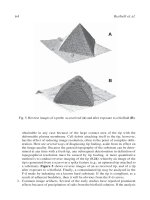
Fig. 9 Three-dimensional surface plot of a stimulating glass fiber (right side) attached to a V-shaped OHC
hair bundle (white cones). The overlaid sawtooth-like pattern of the pipette tip is caused by the sinusoidal
movement of the driving piezo-electric actuator. The scan size is 3.46 ×4.3 μm. This plot clearly demonstrates
the capability of AFM to control and detect the relative position between fiber and stereociliary bundle at high
spatial resolution.
could not be measured. Therefore, the maximum force detected at position 0 varies from
41 to 102 pN. We can conclude that forces transmitted by lateral links are much less than
those measured for a directly stimulated stereocilium, which is mediated by the elasticity
of the stereocilium itself. This result supports the hypothesis of a weak interaction
between stereocilia by lateral links. The background amplitude level of about 20% in
Fig. 8 makes it difficult to distinguish a weak coupling by lateral links from a lack of
coupling. Besides the transmitted force at 357 Hz, AFM curves contain information about
the stereocilia stiffness. We used this information for detecting the mechanical effect of
the touching glass fiber on stiffness of adjacent stereocilia. If lateral links contribute to the
stiffness obtained at individual stereocilia we would expect to see an increase in stiffness
for stereocilia adjacent to those touched with a fiber. Stiffness data were calculated
according to Eq. [3] only for the excitatory direction, where the AFM tip displaces
the stereocilia toward the fiber tip (Fig. 10). Not only the stiffness (filled circles) of the
160 Langer and Koitschev
Fig. 10 Elastic effect of a fiber attached to a single stereocilium on stiffness of adjacent stereocilia. Here,
we successively measured the stiffness of individual stereocilia within a hair bundle while one of these
stereocilia was attached to a glass fiber. Depending on force transmission by side links, stiffness measured
at a single stereocilium should also represent more or less the stiffness of adjacent stereocilia. Open squares
mark the stiffness of stereocilia investigated without a supporting glass fiber; Filled circles mark the stiffness
of stereocilia investigated with a glass fiber attached to a single stereocilium (mean values ±SD). Stiffness of
stereocilia investigated without a fiber shows no significant dependence on position. In contrast, measurements
with a fiber attached to a stereocilium show an increased stiffness not only for the directly supported but also
for adjacent stereocilia (1 to 6), indicating a weak effect of side links on total stiffness. Reproduced from
Langer, M. G., Fink, S., Koitscher, A., Rexhausen, U., H¨orber, J. K. H., and Ruppersberg, J. P. (2001). Lateral
mechanical coupling of stereocilia in cochlear hair bundles. Biophys. J. 80, 2608–2621, with permission from
the Biophysical Society.
directly touched stereocilium at position 0 was found to be increased but also the stiffness
of stereocilia 1 to 4. Mean stiffness in excitatory direction was (4.8 ±1.8) × 10
−3
N/m.
This is about 1.9 times higher compared to the mean stiffness of stereocilia not touched
with a fiber. For better comparison, stiffness data of stereocilia examined without a
fiber were plotted as open symbols into the same graph. For position 7, the stereocilium
with and the stereocilium without a touching glass fiber show approximately identical
stiffness. This demonstrates that lateral links do not tightly couple individual stereocilia
of outer hair cells and small independent movements of single stereocilia are possible.
Side links between the stereocilia may transmit a force when sufficiently stretched and
coupling of stereocilia has a similar stiffness as a single displaced stereocilium. We can
conclude that, depending on elongation of side links, it is in principle possible to deflect
single stereocilia by AFM at least for the preparation of postnatal rats used here.
7. Sensory Cells of Inner Ear Examined by AFM 161
Fig. 11 Principle of simultaneous AFM and patch-clamp recording. This image displays an arrangement
of three separate SEM images with identical scale. The center represents a part of the organ of Corti with
V-shaped hair bundles on top. The upper right side displays a small part of the cantilever beam with the
pyramidal-shaped tip scanning the top of a V-shaped hair bundle of an OHC. The arrow indicates the scan
direction. The lower left side shows a glass pipette in contact with the lipid membrane of an OHC allowing
the transmembrane currents to be recorded as the transduction current. (See Color Plate.)
B. Gating of the Transduction Channel in OHC
In the experiments described previously, we used AFM only for examination of the
elastic properties of hair cells. But how can we benefit from using patch clamp simultane-
ously with AFM? Many micromechanical measurements have already been performed
at entire stereociliary bundles of sensory hair cells using thin glass fibers directly at-
tached to the bundle or fluid jets. The receptor potential or transduction current was
measured in response to the displacement of stereocilia. The possibility to study the
kinetics of a single transduction channel over the whole range of its open probability
requires a technique allowing the stimulation of a single stereocilium. As shown in the
162 Langer and Koitschev
previous section, AFM offers the opportunity of exerting a force very locally to an in-
dividual stereocilium. After supporting cells were removed using a cleaning pipette,
a patch pipette filled with intracellular solution (millimolar concentrations: KCl, 135;
MgCl
2
, 3.5; CaCl
2
, 0.1; EGTA, 5; Hepes, 5; Na
2
ATP, 2.5; pH 7.4) was attached to the
lateral wall of an OHC of the outermost row of OHC. Thereby, the glass microelectrode
forming a seal on the plasma membrane of an intact OHC (Fig. 11) isolates a small
patch. This so-called “cell-attached” configuration is the precursor of the whole-cell
configuration where the microelectrode is in direct electrical contact with the inside of
the cell. For low-noise measurements of single-ion channels the seal resistance should
be typically in the range >1G. A pulse of suction applied to the pipette breaks the
patch creating a hole in the plasma membrane and provides access to the cell interior.
During recording, the electrical resistance between the inside of the pipette and the hair
cell should be very small. Many voltage-activated K
+
-ion channels are embedded in
the lipid membrane of outer hair cells. Opening and closing of these channels increase
the background noise level during transduction current measurements. The current re-
sponse of outward rectifying K
+
-ion channels was controlled by applying 10-mV steps
across the cell membrane (progressively increased from −100 to +40 mV) as shown
in Fig. 12. The outward currents mainly correspond to K
+
currents of voltage-gated
K
+
channels. During transduction current measurements, the holding potential of the
Fig. 12 Activation of voltage-dependent K
+
channels in OHC. This graph displays the electrical response
of voltage-dependent K+ channels located in the cell membrane of OHC. Outward currents were activated,
applying small voltage steps across the cell membrane. Starting at a holding potential of −80 mV the intracel-
lular potential was changed in 10-mV steps from −100 to +40 mV. At voltages below the holding potential
(−80 mV) only small inward currents (less than 50 pA) were detected. Starting from −40 mV to a more
positive potential, outward-rectifying K
+
currents were detectable. These measurements qualitatively allow
testing the leakage and the electrical contact to the intracellular space of examined hair cells.
7. Sensory Cells of Inner Ear Examined by AFM 163
Fig. 13 Simultaneous AFM/patch clamp measurements. The AFM tip scans in the same line while ap-
proaching the organ of Corti to the tip. Transduction currents were measured in the whole cell-recording mode
keeping the holding potential at −80 mV. (A) Current and vertical force measured before interaction between
AFM tip and stereocilium. (B) First contact between the AFM tip and a stereocilium of an OHC. The specimen
is continuously moved toward the AFM tip at from 0 to 470 ms. While scanning in excitatory direction the
specimen does not yet touch the AFM tip. At about 390 ms the hair bundle is near enough to the tip resulting
in a vertical deflection of the AFM cantilever. The cantilever is modulated at about 98 Hz, thereby stimulating
the transduction channel several times. (C) The approach was stopped. Now, the stereocilium is displaced
in both excitatory and inhibitory directions. At about 130 ms an inward current of 19 pA is detected while
displacement in an inhibitory direction does not result in the opening of transduction channels. (D) A second
example demonstrating that a transduction current is detectable only for the excitatory but not for the inhibitory
direction.
164 Langer and Koitschev
Fig. 14 Examination of the transduction current amplitude. Recorded current and force traces are separately
displayed for the excitatory and inhibitory directions of stimulation. (A) These graphs correspond to the force
applied to a single stereocilium in excitatory and inhibitory directions. (B) Successive stimulation of the
identical stereocilium by AFM resulted in the reproducible opening of transduction channels at between 89
and 131 ms. In contrast, force application in an inhibitory direction did not result in the opening of transduction
channels. (C) For detailed analysis, currents recorded during stimulation by AFM (from 89 to 131 ms and from
366 to 407 ms) were pooled and separately displayed for excitatory an inhibitory directions in histograms. The
distribution around 0 pA represents the current for the closed state of the channel while the second smaller
distribution at around19 pA represents the current for the open state of transduction channels. Data demonstrate
that AFM allows stimulation of only a few transduction channels rather than of all channels of the entire hair
bundle.
7. Sensory Cells of Inner Ear Examined by AFM 165
hair cell was set to −80 mV corresponding to the reversal potential of K
+
-ion channels.
Currents are expected to be in the range of a few nanoamperes rather than in the range
of hundreds of picoamperes. After forming a seal, the AFM tip was moved to the top
of the corresponding hair bundle under light microscopic control. The AFM tip succes-
sively displaced each stereocilium within a hair bundle as described in Section IV,A.
In contrast to force transmission measurements, a sinusoidal voltage was added to the
normal AFM scan signal modulating the AFM tip in a horizontal direction with 190 nm
at 98 Hz. Thus, the hair bundle was slightly displaced several times while interacting with
the lateral face of the AFM tip (Fig. 13). The AFM tip repeatedly scanned in the same
line while approaching the hair bundle towards the AFM tip (Fig. 13). As expected,
an inward current was not detected until displacement of a stereocilium in excitatory
direction (Fig. 13C). In the previous section we demonstrated a weak transmission of
force from the directly stimulated stereocilium to adjacent stereocilia implying that
only few channels are opened. Are these results of elasticity measurement confirmed
by electrophysiological findings? A set of transduction current measurements is dis-
played in Fig. 14. The tip of an AFM cantilever repeatedly scanned across the same
stereocilium of an OHC (from medial turn of a postnatal rat, day 3). Applied horizontal
forces ranged up to 0.8 nN (Fig. 14A) resulting in stereocilia displacements of about
350 nm in an excitatory direction and of 250 nm in an inhibitory direction. The AFM tip
displaces the stereocilium in the excitatory direction between 89 and 131 ms, thereby
opening transduction channels (Fig. 14B). The current amplitude was determined for
the period of interaction with the AFM tip plotting histograms of the current (Fig. 14C).
The distribution around 0 pA corresponds to the closed state of the channels, while the
distribution around 19.1 ± 7.6 pA corresponds to the maximal activation. This current
amplitude is twice that expected for a single transduction channel (9.7 ± 1.2 pA;
G´el´eoc et al., 1997). What might underlie this discrepancy? For the detection of sin-
gle channel currents we have to improve the electrical shielding to reduce the noise
level, currently 12.6 pA. Additional high-frequency recordings might be necessary to
distinguish the current of a single channel from the total current of several smaller chan-
nels. The cut-off frequency of our patch-clamp amplifier was currently limited to 3 kHz
allowing the recording of currents at low noise level, but preventing the detection of fast
events. However, if we compare the current amplitude of 19.1 ± 7.6 pA in Fig. 14 to
the maximum transduction current of about 462 pA measured for entire hair bundles of
postnatal mice (G´el´eoc et al., 1997), the results confirm that AFM is well suited for local
stimulation of individual stereocilia.
V. Discussion
In the inner ear the tallest stereocilia of OHC directly contact the tectorial membrane.
The process of opening and closing of transduction channels therefore depends on the
mechanism of force transmission from the top of stereocilia to the transduction channel.
Three structural components are important for understanding the gating process: (i) the
complex mechanics of the cytoskeleton of individual stereocilia, (ii) the cross-linkage


