Báo cáo sinh học: " Hide and seek: the secret identity of the phosphatidylserine receptor" pps
Bạn đang xem bản rút gọn của tài liệu. Xem và tải ngay bản đầy đủ của tài liệu tại đây (73.96 KB, 4 trang )
Minireview
Hide and seek: the secret identity of the phosphatidylserine
receptor
Patrick Williamson* and Robert A Schlegel
†
Addresses: *Department of Biology, Amherst College, Amherst, MA 01002, USA.
†
Department of Biochemistry and Molecular Biology, Penn
State University, University Park, PA 16802, USA.
Correspondence: Patrick Williamson. E-mail:
The genome project and sequence databases have generally
illuminated the molecular basis of cellular functions, but
sometimes this illumination can be an electronic will-o’-the-
wisp. Over the past few months, a series of papers culminat-
ing in the description of a knockout mouse by Lengeling and
co-workers in Journal of Biology [1] has strongly suggested
that a gene sequence thought to illuminate the molecular
basis of apoptotic cell clearance has in fact led us astray, and
that the path to it will have to be retraced.
The problem
Apoptosis, or programmed cell death, is a normal physio-
logic process for orderly removal of effete cells. As a process,
apoptosis fell below the notice of cell biologists for quite
some time, in part because cells dying an apoptotic death in
vivo vanish almost immediately from view. They vanish
because they are promptly engulfed, either by a neighbor or
by a professional phagocytic macrophage; within the
confines of the resulting phagosome, the dying cell digests
itself from the inside out while the engulfing cell digests it
from the outside in. To orchestrate its disappearance, the sui-
cidal cell must make its intentions known to its neighbors,
triggering signaling pathways that activate the engulfment
machinery of the phagocyte. Investigation of the molecules
involved in apoptotic cell recognition is a growing and
industrious scientific subfield of the larger apoptosis research
enterprise, with a host of specific proteins identified and
cloned. One salient feature of the proteins identified is that
most are receptors or enzymes residing on the surface of the
phagocytic cell, along with a burgeoning number of bridging
molecules from the extracellular fluid (Figure 1). Almost
none of the molecules identified is a feature of the apoptotic
cell surface. One molecule that universally distinguishes
apoptotic cells is known, however, although it has the disad-
vantageous property that it cannot be cloned: it is the phos-
pholipid phosphatidylserine (PS), which newly appears on
the surface of cells undergoing apoptosis.
Abstract
Phosphatidylserine on the dying cell surface helps identify apoptotic cells to phagocytes, which
then engulf them. A candidate phagocyte receptor for phosphatidylserine was identified using
phage display, but the phenotypes of knockout mice lacking this presumptive receptor, as well
as the location of the protein within cells, cast doubt on the assignment of this protein as the
phosphatidylserine receptor.
BioMed Central
Journal
of Biology
Journal of Biology 2004, 3:14
Published: 23 September 2004
Journal of Biology 2004, 3:14
The electronic version of this article is the complete one and can be
found online at />© 2004 BioMed Central Ltd
Although the mechanistic details of PS appearance on the
apoptotic cell surface have not been completely resolved,
the basic outlines are clear (Figure 1). PS is normally
sequestered in the inner leaflet of the plasma membrane
bilayer by an active transporter, the aminophospholipid
translocase (APLT). In apoptotic cells this enzyme is down-
regulated, and an enzyme activity called scramblase is up-
regulated; scramblase acts to randomize the lipids between
the two leaflets of the plasma membrane, bringing PS to the
surface [2]. This rearrangement is universal, occurring in vivo
in a wide variety of cell types [3,4] and in both vertebrate
and invertebrate organisms [5]. The ready conclusion is that
PS has something to do with apoptotic cells identifying
themselves to phagocytes, a conclusion borne out by the
finding that masking PS on the apoptotic cell surface with
the PS-binding protein annexin V blocks phagocytosis [6].
Moreover, many of the known bridging and receptor mol-
ecules bind to PS with varying degrees of specificity (Figure
1), including serum-derived protein S [7], milk fat globule
protein MFG-E8 [8], and the scavenger receptors such as
SR-BI [9]. Nevertheless, because the inhibition of engulf-
ment of apoptotic cells by vesicles of PS is stereospecific
[10], in the absence of bridging molecules there must be a
receptor on phagocytes that directly and specifically recog-
nizes PS itself.
Identifying the phosphatidylserine receptor
The first experimental evidence for the existence of a PS
receptor came from Fadok and co-workers [11]. Their
approach began with the production of monoclonal anti-
bodies against ‘stimulated’ macrophages whose recognition
of apoptotic cells is inhibited by PS vesicles in a stereospe-
cific fashion [10], in contrast to ‘unstimulated’ macrophages,
whose uptake of PS-expressing target cells is insensitive to
PS vesicles (even though it is sensitive to annexin V) [6].
One monoclonal antibody, mAb 217, was selected because
it bound preferentially to unfixed stimulated macrophages,
and this binding was inhibited by PS vesicles. The antigenic
target of mAb 217 would thus appear to have the hallmarks
of a PS receptor: it is on the cell surface, it recognizes PS,
and, as the authors went on to show, mAb 217 blocks
engulfment of apoptotic cells [11].
What exactly might this receptor do? This question was
examined in more detail in a later paper from the same lab-
oratory [12], where an experimental system was established
that allowed the binding step of the uptake of an apoptotic
cell to be distinguished from the engulfment step. Bound
cells or particles not expressing PS were engulfed only upon
addition of PS vesicles, but coating target cells with PS vesi-
cles did not result in significant binding to macrophages.
The authors concluded that the PS receptor did not bind PS-
expressing targets tightly (tethering), but that low-affinity
binding could nonetheless stimulate engulfment. Curiously,
it was reported that addition of mAb 217 would itself
induce uptake of previously bound bystander cells, in con-
trast to the earlier observation that pre-incubation with the
antibody prevented uptake of subsequently added apoptotic
cells [11], suggesting perhaps that signal transduction
induced by occupancy of the PS receptor is transient.
The next step was to identify the molecule corresponding to
the PS receptor. On western blots, mAb 217 reacted with a
protein of an apparent molecular weight of 70 kDa. Treat-
ment of cellular extracts with a mixture of four O-glyco-
sidases reduced the size of the protein to roughly 50 kDa,
suggesting that the target of the antibody is glycosylated and
is thus presumably a cell-surface protein. When a phage
display library expressing proteins from mouse
macrophages was panned with mAb 217, half of the one
dozen phages sequenced contained identical sequences,
from a protein in the sequence databases with a theoretical
14.2 Journal of Biology 2004, Volume 3, Article 14 Williamson and Schlegel />Journal of Biology 2004, 3:14
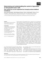
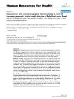
Figure 1
Phosphatidylserine (PS) is a central player in the recognition and
engulfment of apoptotic cells. PS may be recognized by a variety of
tethering receptors (shown as a single entity in green) and bridging
molecules (shown as a single entity in pink) that help tether the
apoptotic target to the phagocyte. The PS receptor signals to a pathway
that leads to engulfment, for example by rearranging elements of the
cytoskeleton (shown as cross-hatching). The proteins that correspond
to the PS receptor, the aminophospholipid translocase (APLT), and the
scramblase are unknown, as are the functions of ABCA1 and the PS
exposed on the surface of the phagocyte. PSRp denotes the protein
encoded by the psr gene, which is found within the nucleus.
PSRp
PS
PS
PS
PS
PS
PS
PS
PS
PSPS
PS
PS
PS
PS
PS
PS Receptor
Tethering
PS
APLT Scramblase
PS
Bridging
ABCA1
ABCA1
PS
PS
PS
PS
PS
?
?
?
PS
PS
PS
PS
PS
PS
PS
?
?
?
Phagocyte
Apoptotic target
PS
PS
PS
molecular weight of 47 kDa [11]. Information from the
databases suggested that the gene encoding the protein was
highly expressed in heart, skeletal muscle, and kidney, with
lower levels of expression in many tissues. Sequence analy-
sis suggested that the protein contained one potential trans-
membrane domain, although this hydrophobic region was
interrupted by an aspartic acid residue in mammals and two
glutamic acid residues in the Caenorhabditis elegans (nema-
tode) homolog. Two pieces of evidence linked this gene to
the engulfment of apoptotic cells. One was that expression
of the mammalian gene, denoted psr, or its nematode
homolog PSR-1 [13], in lymphoid cells conferred an ineffi-
cient capability to bind apoptotic targets, and perhaps to
engulf them as well, notable in view of the conclusion
(mentioned above) that the PS receptor does not contribute
to the tethering step of engulfment [12]. The second piece
of evidence was that transfecting cells with small-interfering
RNA corresponding to this gene, so as to decrease expres-
sion, reduced both binding of mAb 217 to transfected cells
and phagocytosis of apoptotic targets by transfected cells;
whether binding of apoptotic targets by transfected cells was
affected was not reported.
PS receptor knockouts
What happens when the psr gene is deleted? Wang and col-
leagues [13] examined this question in C. elegans, and
found that, in their words, “in a time course analysis of cell
corpses during development, in almost all embryonic
stages, more cell corpses were observed in psr-1 embryos
than in wild type embryos… On average, cell corpses of
psr-1 embryos persisted for 55% longer than those of wild
type animals.” In these studies, expression of the protein
recognized by mAb 217 was not compared in wild-type and
knockout animals.
The first report of the effects of deletion of the psr gene in
mammals came from the laboratory of Richard Flavell [14].
These investigators concluded that the protein encoded by
the gene is required for clearance of apoptotic cells in the
mouse. They observed lung developmental abnormalities
and occasional brain hyperplasia, which were “associated
with increased numbers of nonphagocytosed apoptotic
cells”. They also adoptively transferred fetal liver cells from
knockout mice into lethally irradiated hosts and found that
fewer of the stimulated macrophages recovered from these
animals were able to engulf apoptotic cells compared with
wild-type controls. Reactivity with mAb 217 was not com-
pared between cells from knockout and wild-type animals.
The second report of a knockout of this gene in mice
appeared earlier this year [15], and this study noted severe
developmental defects in definitive erythropoietic and
T-lymphopoietic lineages. Reduced numbers of macrophages
and apoptotic cells were observed in fetal livers of knockout
versus wild-type animals; the fraction of apoptotic cells that
were not phagocytosed was higher in the knockouts. Once
again, reactivity with mAb 217 was not compared between
cells from knockout and wild-type animals.
In contrast to these studies, in the third report of a knockout
of this gene by Lengeling and colleagues [1], which appears
in these pages, reactivity with mAb 217 was compared
between cells from knockout and wild-type animals, and no
difference was observed; in each case, staining was restricted
to the plasma membrane of fetal-liver-derived
macrophages. This finding was foreshadowed by recent
reports that the product of the psr gene is actually a nuclear
protein, as judged from localization of green fluorescent
protein (GFP) constructs as well as immunolocalization
with antibodies directed specifically against the protein
encoded by the psr gene [16,17]. Sequence analysis also
indicates the presence of nuclear localization sequences in
the encoded protein. Western blot analysis by Lengeling and
colleagues, using a commercial antibody generated against a
peptide present in the protein encoded by the psr gene, indi-
cated that this protein does disappear from the knockout
mouse. Lengeling and colleagues also document that dele-
tion of this gene causes “perinatal lethality, growth retard-
ation and a delay in the terminal differentiation of kidney,
intestine, liver, and lungs during embryogenesis.” On the
other hand, in a variety of assays, no defect was observed in
vivo or in vitro in the clearance of apoptotic cells in the
knockout mouse.
Where do we stand?
The simplest interpretation of these studies is that the psr
gene does not encode a PS receptor; rather, the gene appears
to encode a nuclear protein that plays a role in development
and differentiation, perhaps as a regulatory protein related
to the iron-oxidase family of proteins [16]. The experimen-
tal link between the PS receptor and the presumptive psr
gene is mAb 217 binding to an epitope in phage display.
This methodology has the potential to identify weak cross-
reacting epitopes, and the Lengeling study has shown that
mAb 217 does weakly cross-react with a peptide within the
protein encoded by the psr gene. This cross-reactivity could
explain the reactivity of mAb 217 with recombinant PSR
protein expressed in bacteria [13], although it is not clear
whether it can also explain the results using RNA interfer-
ence (RNAi) [12]. If the cloned gene does indeed encode a
critical regulator of hematopoietic differentiation, it is
perhaps not surprising that defects were observed in
macrophage function in worms and mammals. If this inter-
pretation is correct, this gene should no longer be the
Journal of Biology 2004, Volume 3, Article 14 Williamson and Schlegel 14.3
Journal of Biology 2004, 3:14
subject of concentrated attention from those who study the
clearance of apoptotic cells. More importantly, mAb 217
remains as an important foundation for renewed attempts
to identify the genuine PS receptor.
In the meantime, the product of the psr gene will live on in
the databases identified as encoding a PS receptor. In doing
so, it joins a rogue’s gallery of functions without molecules
and molecules without functions that are linked to PS
(Figure 1). In a very similar story, there is a protein identi-
fied in the databases as the phospholipid scramblase that is
not that protein [18], and another identified as an
aminophospholipid translocase [19] that is probably not
the one responsible for transport and sequestration of PS at
the plasma membrane. At the same time, there is a protein,
known as ced-7 in nematodes [20] or ABCA1 in mammals
[21], that is required for engulfment of apoptotic cells and
that seems to be involved in phospholipid movements [22]
but whose function is unclear. Finally, PS itself poses some
puzzles, since engulfment seems to require its exposure not
only on the apoptotic target, but at lower levels on the
phagocyte surface as well [23]. Why this should be the case
is a mystery. All in all, PS seems to have an involved secret
life whose molecular outlines are frustratingly well hidden.
Acknowledgements
We thank Margaret Halleck for her spirited assistance in the generation
of Figure 1.
References
1. Böse J, Gruber AD, Helming L, Schiebe S, Wegener I, Hafner M,
Beales M, Köntgen F, Lengeling A: The phosphatidylserine
receptor has essential functions during embryogenesis but
not in apoptotic cell removal. J Biol 2004, 3:15.
2. Verhoven B, Schlegel RA, Williamson P: Mechanisms of phos-
phatidylserine exposure, a phagocyte recognition signal,
on apoptotic T lymphocytes. J Exp Med 1995, 182:1597-1601.
3. Van den Eijnde SM, Boshart L, Reutelingsperger CPM, De Zeeuw CI,
Vermeij-Keers C: Phosphatidylserine is exposed by apoptotic
cells in mouse embryos in vivo. Eur J Morphol 1997, 35:54-55.
4. Van den Eijnde SM, Boshart L, Reutelingsperger CPM, De Zeeuw
CI, Vermeij-Keers C: Phosphatidylserine plasma membrane
asymmetry in vivo: a pancellular phenomenon which
alters during apoptosis. Cell Death Differ 1997, 4:311-316.
5. Van den Eijnde SM, Boshart L, Baehrecke EH, De Zeeuw CI,
Reutelingsperger CPM, Vermeij-Keers C: Cell surface exposure
of phosphatidylserine during apoptosis is phylogenetically
conserved. Apoptosis 1998, 3:9-16.
6. Krahling S, Callahan MK, Williamson P, Schlegel RA: Exposure of
phosphatidylserine is a general feature in the phagocytosis
of apoptotic lymphocytes by macrophages. Cell Death Differ
1999, 6:183-189.
7. Anderson HA, Maylock CA, Williams JA, Paweletz CP, Shu HJ,
Shacter E: Serum-derived protein S binds to phos-
phatidylserine and stimulates the phagocytosis of apop-
totic cells. Nature Immunol 2003, 4:87-91.
8. Hanayama R, Tanaka M, Miwa K, Shinohara A, Iwamatsu A, Nagata
S: Identification of a factor that links apoptotic cells to
phagocytes. Nature 2002, 417:182-187.
9. Shiratsuchi A, Kawasaki Y, Ikemoto M, Arai H, Nakanishi Y: Role
of class B scavenger receptor type I in phagocytosis of
apoptotic rat spermatogenic cells by Sertoli cells. J Biol
Chem 1999, 274:5901-5908.
10. Fadok VA, Savill JS, Haslett C, Bratton DL, Doherty DE, Campbell
PA, Henson PM: Different populations of macrophages use
either the vitronectin receptor or the phosphatidylserine
receptor to recognize and remove apoptotic cells.
J Immunol 1992, 149:4029-4035.
11. Fadok VA, Bratton DL, Rose DM, Pearson A, Ezekewitz RA,
Henson PM: A receptor for phosphatidylserine-specific
clearance of apoptotic cells. Nature 2000, 405:85-90.
12. Hoffmann PR, deCathelineau AM, Ogden CA, Leverrier Y, Bratton
DL, Daleke DL, Ridley AJ, Fadok VA, Henson PM: Phos-
phatidylserine (PS) induces PS receptor-mediated
macropinocytosis and promotes clearance of apoptotic
cells. J Cell Biol 2001, 155:649-659.
13. Wang X, Wu YC, Fadok VA, Lee MC, Gengyo-Ando K, Cheng LC,
Ledwich D, Hsu PK, Chen JY, Chou BK, et al.: Cell corpse
engulfment mediated by C. elegans phosphatidylserine
receptor through CED-5 and CED-12. Science 2003,
302:1563-1566.
14. Li MO, Sarkisian MR, Mehal WZ, Rakic P, Flavell RA: Phos-
phatidylserine receptor is required for clearance of apop-
totic cells. Science 2003, 302:1560-1563.
15. Kunisaki Y, Masuko S, Noda M, Inayoshi A, Sanui T, Harada M,
Sasazuki T, Fukui Y: Defective fetal liver erythropoiesis and
T lymphopoiesis in mice lacking the phosphatidylserine
receptor. Blood 2004, 103:3362-3364.
16. Cikala M, Alexandrova O, David CN, Proschel M, Stiening B,
Cramer P, Bottger A: The phosphatidylserine receptor from
Hydra is a nuclear protein with potential Fe(II)-dependent
oxygenase activity. BMC Cell Biol 2004, 5:26.
17. Cui P, Qin B, Liu N, Pan G, Pei D: Nuclear localization of the
phosphatidylserine receptor protein via multiple nuclear
localization signals. Exp Cell Res 2004, 293:154-163.
18. Zhou QS, Zhao J, Wiedmer T, Sims PJ: Normal hemostasis but
defective hematopoietic response to growth factors in
mice deficient in phospholipid scramblase 1. Blood 2002,
99:4030-4038.
19. Tang X, Halleck MS, Schlegel RA, Williamson P: A subfamily of
P-type ATPases with aminophospholipid transporting
activity. Science 1996, 272:1495-1497.
20. Wu YC, Horvitz HR: The C. elegans cell corpse engulfment
gene ced-7 encodes a protein similar to ABC trans-
porters. Cell 1998, 93:951-960.
21. Luciani MF, Chimini G: The ATP binding cassette trans-
porter ABC1, is required for the engulfment of corpses
generated by apoptotic cell death. EMBO J 1996, 15:226-235.
22. Fielding PE, Nagao K, Hakamata H, Chimini G, Fielding CJ: A two-
step mechanism for free cholesterol and phospholipid
efflux from human vascular cells to apolipoprotein A-1.
Biochemistry 2000, 39:14113-14120.
23. Callahan MK, Williamson P, Schlegel RA: Surface expression of
phosphatidylserine on macrophages is required for
phagocytosis of apoptotic thymocytes. Cell Death Differ
2000, 7:645-653.
14.4 Journal of Biology 2004, Volume 3, Article 14 Williamson and Schlegel />Journal of Biology 2004, 3:14