Báo cáo sinh học: "X-chromosome inactivation: the molecular basis of silencing" ppt
Bạn đang xem bản rút gọn của tài liệu. Xem và tải ngay bản đầy đủ của tài liệu tại đây (179.33 KB, 4 trang )
Minireview
XX cchhrroommoossoommee iinnaaccttiivvaattiioonn:: tthhee mmoolleeccuullaarr bbaassiiss ooff ssiilleenncciinngg
Barbara Panning
Address: Department of Biochemistry and Biophysics, University of California, San Francisco, CA 94158, USA.
Email:
X-chromosome inactivation is the transcriptional silencing
of one X chromosome in female mammalian cells that
equalizes dosage of gene products from the X chromosome
between XX females and XY males [1-3]. X-chromosome
inactivation in the embryo proper occurs early in develop-
ment. The two X chromosomes have an equal probability of
being silenced [4]. Silencing, once established, is stable: the
same X chromosome remains inactivated in all subsequent
cell generations. As a result, each female is a mosaic of cells
in which either the maternally inherited or the paternally
inherited X is silenced. Nesterova and colleagues in the first
issue of Epigenetics and Chromatin shed new light on how
this process is regulated [5].
An antisense pair of non-coding RNAs, encoded by Xist and
Tsix (Figure 1), is important in the regulation of the random
inactivation of mouse X chromosomes. Before the signal
that initiates random X-chromosome inactivation is received,
Xist and Tsix are transcribed from all active X chromosomes
in each male and female cell [6]. Once inactivation is
initiated, Xist and Tsix are differentially regulated on the X
that will become the active X chromosome (X
A
) and the one
that will become the inactive X chromosome (X
I
). On the X
chromosome that will become the X
I
, Xist transcripts spread
in cis from their site of synthesis to coat the entire X
chromosome and establish transcriptional silencing.
Concomitant with Xist RNA coating, Tsix is silenced on the
X
I
. The expression of Xist and Tsix persists on the X
A
for a
brief period after silencing of the X
I
is complete, and is
eventually extinguished. Xist RNA continues to coat the X
I
throughout all subsequent cell divisions, where it contri-
butes to the maintenance of silencing. These patterns of Xist
and Tsix expression are also seen in mouse female embry-
onic stem (ES) cells, which have two X
A
s and which
undergo X-chromosome inactivation when they are induced
to differentiate in vitro. Thus, ES cells provide a useful model
system to study X-chromosome inactivation.
MMuuttaattiioonnss iinn
XXiisstt
oorr
TTssiixx
ccaann ccaauussee nnoonn rraannddoomm XX
iinnaaccttiivvaattiioonn
Heterozygous mutation of Xist or Tsix causes non-random
X-chromosome inactivation in female cells. When Xist
expression is increased from one X chromosome in pre-X-
chromosome-inactivation cells, that X chromosome always
becomes the X
I
and the wild-type X always becomes the X
A
[7]. In female ES cells or embryos in which Xist is disrupted
on one X chromosome, the mutant X chromosome always
becomes the X
A
and the wild-type X chromosome always
becomes the X
I
[8-10]. Disruption of Tsix has the opposite
AAbbssttrraacctt
X-chromosome inactivation occurs randomly for one of the two X chromosomes in female
cells during development. Inactivation occurs when RNA transcribed from the
Xist
gene on
the X chromosome from which it is expressed spreads to coat the whole X chromosome. In
the first issue of
Epigenetics and Chromatin
, Nesterova and colleagues investigate the role of
the RNA interference pathway enzyme Dicer in DNA methylation of the
Xist
promoter.
Journal of Biology
2008,
77::
30
Published: 27 October 2008
Journal of Biology
2008,
77::
30 (doi:10.1186/jbiol95)
The electronic version of this article is the complete one and can be
found online at />© 2008 BioMed Central Ltd
effect: the mutant X chromosome becomes the X
I
and the
wild-type X chromosome is always the X
A
[11-13]. This is
known as primary non-random X-chromosome inactivation
because the X chromosomes are chosen as the X
A
and X
I
before silencing is initiated. A second cause of non-random
X-chromosome inactivation is the selective death of cells
that inactivate the incorrect number of X chromosomes:
because the fates of the X chromosomes are not determined
before silencing, this is known as secondary non-random X-
chromosome inactivation [14]. Because Xist and Tsix muta-
tions cause primary non-random X-chromosome inactiva-
tion, it is likely that these non-coding RNAs function in the
choice of the X
A
and X
I
before silencing is initiated.
Understanding how Xist and Tsix are regulated in pre-X-
chromosome-inactivation cells is central to understanding
how one X chromosome is randomly selected as the X
A
and
the other as the X
I
in each cell.
In addition to having opposing roles in random choice, Xist
and Tsix also negatively regulate each other in ES cells. Xist
and Tsix are transcribed from overlapping regions on
opposite strands of the X-chromosome DNA (Figure 1).
Deletion of Tsix promoter sequences or a mutation that
blocks Tsix transcription before it reaches Xist RNA coding
sequences abolishes Tsix transcription and causes a roughly
ten-fold increase in Xist RNA levels from the mutant X
chromosome [11-13]. Thus, transcription of Tsix across Xist
is necessary for Tsix to negatively regulate Xist. In the Tsix
truncation mutant the Tsix promoter has histone modifica-
tion patterns that are generally associated with transcriptional
silencing [15]. These epigenetic marks also characterize the X
I
,
and their recruitment to the X
I
requires transcription of Xist
[16-18]. Together, these results suggest that the increase in
Xist RNA that occurs on Tsix mutant chromosomes represses
Tsix. Consistent with the possibility that Xist negatively
regulates Tsix, Tsix RNA levels are increased from Xist
mutant X chromosomes [10,19]. Insights into the nature of
factors that are involved in the mutual regulation of Xist and
Tsix in pre-X-chromosome-inactivation cells are likely to be
important in developing an understanding of how these
non-coding RNAs ensure that the two X chromosomes have
an equal probability of being silenced in each cell.
TThhee rroollee ooff DDNNAA mmeetthhyyllaattiioonn
The mechanisms underlying the mutual regulation of Xist
and Tsix in pre-X-chromosome-inactivation cells are not
well characterized. An interesting new study by Nesterova
and colleagues suggests that DNA methylation may be
involved in this mutual negative regulation [5]. Nesterova et
al. demonstrate a correlation between Xist promoter DNA
methylation and Xist expression in ES cells. In XY ES cells
(in which the single X chromosome remains active), two
regions flanking the Xist transcription start site show high
levels of DNA methylation. Two XY ES cell lines bearing Xist
promoter mutations that result in increased Xist expression
showed DNA hypomethylation at these sites. In addition, a
mutation that truncates Tsix transcription before it traverses
Xist also resulted in increased Xist expression and DNA
hypomethylation at these sites. These results establish a
clear correlation between the levels of DNA methylation at
Xist and expression of Xist in ES cells. It remains to be
established whether the increase in Xist expression triggers
demethylation or vice versa. In addition, Xist and Tsix
negatively regulate each other, raising the possibility that
Tsix also has a role in regulation of Xist DNA methylation.
Tsix has also been implicated in the direct regulation of
DNA methylation. The de novo DNA methyltransferase
Dnmt3a can be immunoprecipitated with Tsix RNA using
an RNA-chromatin immunoprecipitation procedure [20].
Furthermore, Dnmt3a can de novo methylate Xist [21,22].
Together, these data suggest a model in which Tsix RNA
directs Dnmt3a to Xist in ES cells (Figure 2a). Thus, the
hypomethylation of Xist DNA in the Tsix truncation line
may occur because Dnmt3a cannot act on Xist when Tsix
RNA is not present to recruit it there.
This model explains the hypomethylation of Xist DNA in
the Tsix truncation line, but how does it account for the
hypomethylation in the Xist promoter mutation lines? As in
the Tsix truncation line, the Xist promoter mutation lines
show increased Xist expression. In contrast to the truncation
line, which does not produce Tsix RNA, the Xist promoter
mutation lines continue to express Tsix RNA. However, Tsix
RNA levels have not been quantitated in these cell lines, so
it is not possible to establish a correlation between Tsix
expression levels and Xist DNA methylation. One possibility
is that the increase in Xist expression causes a decrease in
Tsix RNA levels and a corresponding decrease in Dnmt3a
activity at Xist DNA. There is also an alternative possibility:
it may be that Xist RNA (or an epigenetic modification
induced by Xist RNA) interferes with the activity of Dnmt3a
30.2
Journal of Biology
2008, Volume 7, Article 30 Panning />Journal of Biology
2008,
77::
30
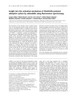
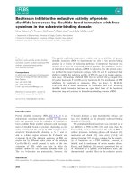
FFiigguurree 11
Transcription of
Xist
and
Tsix
on the X chromosome. The coding
sequences of
Xist
and
Tsix
overlap on opposite strands of the
X-chromosome DNA.
Xist
Tsix
or other de novo methyltransferases (Figure 2b). Indeed, the
Xist RNA-coated X
I
shows overall lower levels of DNA
methylation than the X
A
, consistent with Xist RNA inter-
fering with DNA methylation [23]. Because Xist RNA
accumulates only locally in ES cells, this activity would be
restricted to the Xist locus and perhaps nearby genes.
Analysis of Xist DNA methylation in Xist and combined Xist +
Tsix mutant ES cells will be required to distinguish between
these possibilities.
XX iinnaaccttiivvaattiioonn aanndd DDiicceerr ddeeffiicciieennccyy
Nesterova and colleagues have further investigated the role
of de novo methyltransferases in regulation of Xist expression
in an analysis of Dicer mutant male ES cells. Dicer is an
RNAse III enzyme that is central to the RNA interference
(RNAi) pathway. RNAi regulates many aspects of gene
expression and involves the production of antisense RNA
complementary to sequences in the mRNA of the gene that
is being regulated [24]. The formation of sense-antisense
double-stranded RNA can trigger transcriptional or post-
transcriptional gene silencing. Given that Tsix RNA contains
sequences complementary to Xist RNA, an obvious question
is whether the RNAi pathway has a role in X-chromosome
inactivation. Nesterova et al. show that several indepen-
dently derived Dicer-deficient male ES cell lines show Xist
DNA hypomethylation and upregulation of Xist expression.
They also find that the two imprinted loci H19 and Igf2rAir
show hypomethylation in Dicer-deficient cells. Hypomethy-
lation of Xist, H19 and Igf2rAir seems to be the consequence
of changes in the levels of the de novo methyltransferases
Dnmt3a, Dnmt3b and DnmtL, all of which were down-
regulated upon deletion of Dicer. This decrease in de novo
methyltransferase activity in Dicer-deficient cells was also
seen in two other studies of independently derived Dicer
mutant ES cell lines [25,26]. In these studies Dicer mutant
ES cells show hypomethylation of subtelomeric repeats or
of Oct4, Tsp50 and Sox30 promoters, which are normally
methylated. The downregulation of the de novo methyl-
transferases could be attributed to an increase in levels of
the repressor Rbl2, which is negatively regulated by the
miR-290 microRNA cluster [25,26]. Together, these results
provide a compelling argument that the change in Xist DNA
methylation seen in Dicer mutant ES cells is an indirect
consequence of the loss of de novo methyltransferase activity
(Figure 2c).
Does the change in Xist DNA methylation in pre-X-
chromosome-inactivation cells affect the fate of the X
chromosomes after inactivation is initiated? To answer this
question Nesterova et al. analyzed Dicer mutant embryos.
Dicer mutants die shortly after implantation, between
embryonic day (E)7.5 and E8.5. X-chromosome inactivation
is initiated at approximately E5.5, providing a brief window
in which X-chromosome inactivation can be assayed in Dicer
mutants. The cells of male and female Dicer-deficient E6.5
embryos and their wild-type littermates did not show any
appreciable difference in either Xist or Tsix expression. These
results indicate that one X chromosome can be selected as
the inactive X and Xist RNA can coat that X chromosome in
Dicer mutant embryos. Thus, X-chromosome inactivation
seems unaffected by Dicer deficiency in vivo.
The results of Nesterova et al. contrast with those from
another study of the role of Dicer in X-chromosome
inactivation. Ogawa et al. [27] examined X-chromosome
inactivation in
Dicer mutant female ES cells and found that
Xist RNA could not coat and silence an X chromosome on
/>Journal of Biology
2008, Volume 7, Article 30 Panning 30.3
Journal of Biology
2008,
77::
30
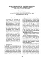
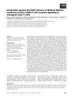
FFiigguurree 22
Models for the coordinate regulation of
Xist
DNA methylation and
expression by
Tsix
,
de novo
DNA methyltransferases and Dicer.
De novo
DNA methyltransferases (Dnmt) promote methylation of
Xist
DNA.
Increased
Xist
expression, as is seen in the
Xist
promoter mutants, could
trigger
Xist
DNA hypomethylation
((aa))
indirectly by affecting
Tsix
RNA
levels, if
Tsix
is necessary to direct
de novo
DNA methyltransferases to
the
Xist
gene, or
((bb))
directly, if
Xist
RNA can interfere with
de novo
DNA methyltransferase activity locally.
((cc))
Because Dicer deficiency
causes a global decrease in levels of
de novo
DNA methyltransferases,
Dicer must lie directly upstream of the
de novo
DNA methyltransferases
and need not function through either
Xist
or
Tsix
to regulate
Xist
DNA
methylation. (The DNA is shown as methylated in a, b and c (bottom),
although in a and b if the inhibitory interactions between
Xist
and
Tsix
RNA (a) or Dmt (b) prevail, the DNA will be hypomethylated.)
Xist DNA
methylated Xist DNA
Xist RNA
Tsix RNA
(a)
Xist DNA
methylated Xist DNA
Xist RNA
(b)
recruits
Dnmt
inhibits
Dnmt
Xist DNA
methylated Xist DNA
(c)
Dicer
upregulates
Dnmt
Dnmt
Dnmt
Dnmt
methyl
group
methyl
group
methyl
group
differentiation. These results indicate that Dicer is necessary
for X-chromosome inactivation in vitro. Why do female ES
cells and embryos differ in their requirements for Dicer
during X-chromosome inactivation? One possibility is that
maternal stores of Dicer persist long enough to promote
X-chromosome inactivation in female Dicer mutant embryos.
However, the homozygous Dicer mutant female ES cells
used by Ogawa et al. contained a Dicer transgene that was
expressed at less than 5% of wild-type levels (this was
deployed to overcome the block to differentiation in Dicer
mutants that would have otherwise interfered with analysis
of X-chromosome inactivation), suggesting that small
amounts of Dicer are not sufficient to promote random
inactivation. A second possibility is that Dicer mutant female
embryos fail to reverse imprinted X-chromosome inactiva-
tion in their embryonic compartment. In mice, the extra-
embryonic tissues undergo imprinted X-chromosome
inactivation, in which there is exclusive silencing of the
paternal X chromosome [28]. Imprinted X-chromosome
inactivation is initiated in pre-implantation development
and seems to occur in all cells of the early embryo.
Imprinted X-chromosome inactivation is reversed in the cells
that will go on to form the embryo proper, and these cells
subsequently undergo random X-chromosome inactivation
after implantation [29,30]. Determining whether Dicer
mutant female embryos show random or imprinted X-
chromosome inactivation will establish whether Dicer is
important to erase imprinted X-chromosome inactivation.
Clearly much work remains to be done to determine how
Dicer regulates Xist expression during development.
RReeffeerreenncceess
1. Heard E, Chaumeil J, Masui O, Okamoto I:
MMaammmmaalliiaann XX cchhrroommoo
ssoommee iinnaaccttiivvaattiioonn:: aann eeppiiggeenneettiiccss ppaarraaddiiggmm
Cold Spring Harb Symp
Quant Biol
2004,
6699::
89-102.
2. Boumil RM, Lee JT:
FFoorrttyy yyeeaarrss ooff ddeeccooddiinngg tthhee ssiilleennccee iinn XX cchhrroo
mmoossoommee iinnaaccttiivvaattiioonn
Hum Mol Genet
2001,
1100::
2225-2232.
3. Lyon MF:
GGeennee aaccttiioonn iinn tthhee XX cchhrroommoossoommee ooff tthhee mmoouussee ((
MMuuss
mmuussccuulluuss
LL ))
Nature
1961,
119900::
372-373.
4. Wutz A, Gribnau J:
XX iinnaaccttiivvaattiioonn XXppllaaiinneedd
Curr Opin Genet Dev
2007,
1177::
387-393.
5. Nesterova TB, Popova BC, Cobb BS, Norton S, Senner C, Tang YA,
Spruce T, Rodriguez TA, Sado T, Merkenschlager M, Brockdorff N:
DDiicceerr rreegguullaatteess
XXiisstt
pprroommootteerr mmeetthhyyllaattiioonn iinn EESS cceellllss iinnddiirreeccttllyy
tthhrroouugghh ttrraannssccrriippttiioonnaall ccoonnttrrooll ooff DDnnmmtt33aa
Epigenetics Chromatin
2008,
11::
2.
6. Mlynarczyk SK, Panning B:
XX iinnaaccttiivvaattiioonn:: TTssiixx aanndd XXiisstt aass yyiinn aanndd
yyaanngg
Curr Biol
2000,
1100::
R899-R903.
7. Nesterova TB, Johnston CM, Appanah R, Newall AE, Godwin J,
Alexiou M, Brockdorff N:
SSkkeewwiinngg XX cchhrroommoossoommee cchhooiiccee bbyy
mmoodduullaattiinngg sseennssee ttrraannssccrriippttiioonn aaccrroossss tthhee XXiisstt llooccuuss
Genes Dev
2003,
1177::
2177-2190.
8. Marahrens Y, Loring J, Jaenisch R:
RRoollee ooff tthhee XXiisstt ggeennee iinn XX
cchhrroommoossoommee cchhoooossiinngg
Cell
1998,
9922::
657-664.
9. Gribnau J, Luikenhuis S, Hochedlinger K, Monkhorst K, Jaenisch R:
XX cchhrroommoossoommee cchhooiiccee ooccccuurrss iinnddeeppeennddeennttllyy ooff aassyynncchhrroonnoouuss
rreepplliiccaattiioonn ttiimmiinngg
J Cell Biol
2005,
116688::
365-373.
10. Sado T, Hoki Y, Sasaki H:
TTssiixx ddeeffeeccttiivvee iinn sspplliicciinngg iiss ccoommppeetteenntt ttoo
eessttaabblliisshh XXiisstt ssiilleenncciinngg
Development
2006,
113333::
4925-4931.
11. Lee JT, Lu N:
TTaarrggeetteedd mmuuttaaggeenneessiiss ooff TTssiixx lleeaaddss ttoo nnoonnrraannddoomm XX
iinnaaccttiivvaattiioonn
Cell
1999,
9999::
47-57.
12. Luikenhuis S, Wutz A, Jaenisch R:
AAnnttiisseennssee ttrraannssccrriippttiioonn tthhrroouugghh
tthhee XXiisstt llooccuuss mmeeddiiaatteess TTssiixx ffuunnccttiioonn iinn eemmbbrryyoonniicc sstteemm cceellllss
Mol
Cell Biol
2001,
2211::
8512-8520.
13. Sado T, Wang Z, Sasaki H, Li E:
RReegguullaattiioonn ooff iimmpprriinntteedd XX cchhrroommoo
ssoommee iinnaaccttiivvaattiioonn iinn mmiiccee bbyy TTssiixx
Development
2001,
112288::
1275-1286.
14. McMahon A, Monk M:
XX cchhrroommoossoommee aaccttiivviittyy iinn ffeemmaallee mmoouussee
eemmbbrryyooss hheetteerroozzyyggoouuss ffoorr PPggkk 11 aanndd SSeeaarrllee’’ss ttrraannssllooccaattiioonn,, TT((XX;;
1166)) 1166HH
Genet Res
1983,
4411::
69-83.
15. Navarro P, Pichard S, Ciaudo C, Avner P, Rougeulle C:
TTssiixx ttrraannssccrriipp
ttiioonn aaccrroossss tthhee XXiisstt ggeennee aalltteerrss cchhrroommaattiinn ccoonnffoorrmmaattiioonn wwiitthhoouutt
aaffffeeccttiinngg XXiisstt ttrraannssccrriippttiioonn:: iimmpplliiccaattiioonnss ffoorr XX cchhrroommoossoommee iinnaaccttiivvaa
ttiioonn
Genes Dev
2005,
1199::
1474-1484.
16. Silva J, Mak W, Zvetkova I, Appanah R, Nesterova TB, Webster Z,
Peters AH, Jenuwein T, Otte AP, Brockdorff N:
EEssttaabblliisshhmmeenntt ooff
hhiissttoonnee hh33 mmeetthhyyllaattiioonn oonn tthhee iinnaaccttiivvee XX cchhrroommoossoommee rreeqquuiirreess
ttrraannssiieenntt rreeccrruuiittmmeenntt ooff EEeedd EEnnxx11 PPoollyyccoommbb ggrroouupp ccoommpplleexxeess
Dev Cell
2003,
44::
481-495.
17. Plath K, Fang J, Mlynarczyk-Evans SK, Cao R, Worringer KA,
Wang H, de la Cruz CC, Otte AP, Panning B, Zhang Y:
RRoollee ooff
hhiissttoonnee HH33 llyyssiinnee 2277 mmeetthhyyllaattiioonn iinn XX iinnaaccttiivvaattiioonn
Science
2003,
330000::
131-135.
18. Kohlmaier A, Savarese F, Lachner M, Martens J, Jenuwein T, Wutz A:
AA cchhrroommoossoommaall mmeemmoorryy ttrriiggggeerreedd bbyy XXiisstt rreegguullaatteess hhiissttoonnee
mmeetthhyyllaattiioonn iinn XX iinnaaccttiivvaattiioonn
PLoS Biol
2004,
22::
E171.
19. Sado T, Hoki Y, Sasaki H:
TTssiixx ssiilleenncceess XXiisstt tthhrroouugghh mmooddiiffiiccaattiioonn
ooff cchhrroommaattiinn ssttrruuccttuurree
Dev Cell
2005,
99::
159-165.
20. Sun BK, Deaton AM, Lee JT:
AA ttrraannssiieenntt hheetteerroocchhrroommaattiicc ssttaattee iinn
XXiisstt pprreeeemmppttss XX iinnaaccttiivvaattiioonn cchhooiiccee wwiitthhoouutt RRNNAA ssttaabbiilliizzaattiioonn
Mol Cell
2006,
2211::
617-628.
21. Sado T, Okano M, Li E, Sasaki H:
DDee nnoovvoo DDNNAA mmeetthhyyllaattiioonn iiss
ddiissppeennssaabbllee ffoorr tthhee iinniittiiaattiioonn aanndd pprrooppaaggaattiioonn ooff XX cchhrroommoossoommee
iinnaaccttiivvaattiioonn
Development
2004,
113311::
975-982.
22. Okano M, Bell DW, Haber DA, Li E:
DDNNAA mmeetthhyyllttrraannssffeerraasseess
DDnnmmtt33aa aanndd DDnnmmtt33bb aarree eesssseennttiiaall ffoorr ddee nnoovvoo mmeetthhyyllaattiioonn aanndd
mmaammmmaalliiaann ddeevveellooppmmeenntt
Cell
1999,
9999::
247-257.
23. Hellman A, Chess A:
GGeennee bbooddyy ssppeecciiffiicc mmeetthhyyllaattiioonn oonn tthhee aaccttiivvee
XX cchhrroommoossoommee
Science
2007,
331155::
1141-1143.
24. Campbell TN, Choy FY:
RRNNAA iinntteerrffeerreennccee:: ppaasstt,, pprreesseenntt aanndd
ffuuttuurree
Curr Issues Mol Biol
2005,
77::
1-6.
25. Sinkkonen L, Hugenschmidt T, Berninger P, Gaidatzis D, Mohn F,
Artus-Revel CG, Zavolan M, Svoboda P, Filipowicz W:
MMiiccrrooRRNNAAss
ccoonnttrrooll ddee nnoovvoo DDNNAA mmeetthhyyllaattiioonn tthhrroouugghh rreegguullaattiioonn ooff ttrraannssccrriipp
ttiioonnaall rreepprreessssoorrss iinn mmoouussee eemmbbrryyoonniicc sstteemm cceellllss
Nat Struct Mol
Biol
2008,
1155::
259-267.
26. Benetti R, Gonzalo S, Jaco I, Muñoz P, Gonzalez S, Schoeftner S,
Murchison E, Andl T, Chen T, Klatt P, Li E, Serrano M, Millar S,
Hannon G, Blasco MA:
AA mmaammmmaalliiaann mmiiccrrooRRNNAA cclluusstteerr ccoonnttrroollss
DDNNAA mmeetthhyyllaattiioonn aanndd tteelloommeerree rreeccoommbbiinnaattiioonn vviiaa RRbbll22 ddeeppeennddeenntt
rreegguullaattiioonn ooff DDNNAA mmeetthhyyllttrraannssffeerraasseess
Nat Struct Mol Biol
2008,
1155::
268-279.
27. Ogawa Y, Sun BK, Lee JT:
IInntteerrsseeccttiioonn ooff tthhee RRNNAA iinntteerrffeerreennccee
aanndd XX iinnaaccttiivvaattiioonn ppaatthhwwaayyss
Science
2008,
332200::
1336-1341.
28. Lyon MF:
TThhee XX iinnaaccttiivvaattiioonn cceennttrree aanndd XX cchhrroommoossoommee iimmpprriinnttiinngg
Eur J Hum Genet
1994,
22::
255-261.
29. Okamoto I, Otte AP, Allis CD, Reinberg D, Heard E:
EEppiiggeenneettiicc
ddyynnaammiiccss ooff iimmpprriinntteedd XX iinnaaccttiivvaattiioonn dduurriinngg eeaarrllyy mmoouussee ddeevveelloopp
mmeenntt
Science
2004,
330033::
644-649.
30. Mak W, Nesterova TB, de Napoles M, Appanah R, Yamanaka S,
Otte AP, Brockdorff N:
RReeaaccttiivvaattiioonn ooff tthhee ppaatteerrnnaall XX cchhrroommoo
ssoommee iinn eeaarrllyy mmoouussee eemmbbrryyooss
Science
2004,
330033::
666-669.
30.4
Journal of Biology
2008, Volume 7, Article 30 Panning />Journal of Biology
2008,
77::
30