Báo cáo sinh học: " Fishing for the signals that pattern the face" doc
Bạn đang xem bản rút gọn của tài liệu. Xem và tải ngay bản đầy đủ của tài liệu tại đây (716.77 KB, 4 trang )
Schilling and Le Pabic: Journal of Biology 2009, 8:101
Abstract
Zebrafish are a powerful system for studying the early
embryonic events that form the skull and face, as a model for
human craniofacial birth defects such as cleft palate. Signaling
pathways that pattern the pharyngeal arches (which contain
skeletal precursors of the palate, as well as jaws and gills) are
discussed in light of a recent paper in BMC Developmental
Biology on requirements for Hedgehog signaling in craniofacial
development.
See research article />It has been over a century since the discovery that
migratory cells of the neural crest give rise to the cranio-
facial skeleton, unlike the vertebral and limb skeletons,
which are derived from embryonic mesoderm [1]. Despite
the extraordinarily high frequency of cleft palate and other
craniofacial problems in human birth defects, the genetic
control of this part of the skeleton remains mysterious.
However, recent identification of the genetic basis for
many craniofacial syndromes, combined with functional
studies in animal models, are beginning to illuminate the
cellular and molecular mechanisms underlying skull
develop ment and its origins in the neural crest. Studies
over the past few years, including one by Schwend and
Ahlgren [2] published recently in BMC Developmental
Biology, have taken advantage of the availability of
zebrafish mutations that disrupt the skull to uncover genes
that control its patterning in the early embryo. Moreover,
advances in the ability to create transgenic zebrafish have
opened up new avenues for following neural crest cells in
living embryos and thereby elucidating how genes control
skull morphogenesis.
Building the ventral skull: anterior-posterior
patterning
A fundamental feature of the head is its modular
organization - the embryonic hindbrain and the pharynx
become partitioned into a series of segments. Within 24
hours postfertilization the zebrafish hindbrain has become
segmented into seven rhombomeres and the tissues
surrounding the pharynx subdivide into seven so-called
arches (Figure 1a). These include the mandibular arch
(arch 1), which forms the jaw, the hyoid arch (arch 2),
which forms the jaw support, and five gill arches (arches
3-7). All have a similar structure, including skeletal, neural
and glial components derived from neural crest, muscles
and blood vessels derived from mesoderm, and an outer
sheath of surface ectoderm and endoderm.
These similarities led early comparative anatomists to
argue that jaws, which form from the prominent mandi-
bular arch, evolved from an ancestral gill arch (reviewed in
[3]). Consistent with this idea, early vertebrates had
pharyngeal arches but were jawless - arches are one of the
most ancient vertebrate traits, found even in non-
vertebrate chordates such as amphioxus. Evidence to
support the gill-arch origin for jaws has come with the
discovery of similar patterns of gene expression in every
arch in the series. For example, recent studies in a
chondrichthyan fish, a skate, suggest that the network of
secreted growth factors found in the mandibular arch,
which includes Sonic Hedgehog (Shh; see below),
fibroblast growth factor and retinoic acid, is conserved in
the gill arches, and inhibitors of Shh signaling disrupt
skeletal patterning similarly in every arch [4].
Surprisingly, however, recent genetic evidence argues
against the model of a gill-arch ground plan and instead
suggests that the default state for an arch is more
mandibular in character. Combinatorial expression of Hox
genes underlies arch-specific morphologies, with the
exception of the mandibular arch, which is devoid of Hox
activity. This ‘Hox code’ applies to all non-mandibular
arches (there are four arches in mammals but up to nine in
other vertebrates) and in each case posterior arches acquire
more anterior characteristics in the absence of Hox gene
function. For instance genetic knockout of Hoxa2 in mice,
or combined loss of hoxa2 and hoxb2 in zebrafish, leads to a
replacement of second-arch-derived structures by those of
the first arch [5,6]. Strikingly, Minoux et al. [7] recently
showed that all posterior arches take on a mandibular
identity following the conditional loss of all Hoxa cluster
genes in mouse cranial neural crest cells. Thus, posterior
arch identity must override a default ‘mandibular’ program
that dominates in the absence of Hox expression.
Minireview
Fishing for the signals that pattern the face
Thomas F Schilling and Pierre Le Pabic
Address: Department of Developmental and Cell Biology, 4462 Natural Sciences II, University of California, Irvine, CA 92697-2300, USA.
Correspondence: Thomas F Schilling. Email:
101.2
Schilling and Le Pabic: Journal of Biology 2009, 8:101
Epithelial-mesenchymal interactions pattern
the arches
Once head segmentation is established, the next critical
step is differential growth and patterning within each arch.
In most vertebrates the mandibular arch is much larger
than the others. Gill arches are prominent structures in
aquatic vertebrates such as fish, but are only transiently
present in mammals and later become incorporated into
the throat and larynx. How does the identity acquired
through the Hox code result in such large differences in
arch morphogenesis?
The answer appears to lie in how neural crest cells within
the arch respond to signals from surrounding epithelia.
Early grafting studies in avian embryos demonstrated that
mandibular neural crest transplanted posteriorly gave rise
to an ectopic beak and other mandibular structures [8],
suggesting that similar signals exist in both anterior and
posterior arches, and that segmental identity is intrinsic to
neural crest cells. More recent variations on these types of
grafts suggest that this is only part of the story [9,10].
Nevertheless, further evidence for an intrinsic Hox code in
the neural crest has come from studies of zebrafish moz
mutants, which disrupt an essential activator of Hox
paralog group 2 gene expression and exhibit trans-
formations of the hyoid (arch 2) to a mandibular (arch 1)
identity, similar to Hoxa2/b2 double morpholino-injected
(morphants) [6,11]. Mosaic studies combining mutant and
wild-type cells suggest that Moz and Hox paralog group 2
proteins act cell-autonomously in the second arch skeleto-
genic neural crest cells - without them skeletal elements
acquire the size and shape of their counterparts in the
mandibular arch [12]. Thus, Hox genes appear to instruct
neural crest cells to interpret arch signals appropriately.
These signals largely come from the endodermal and/or
ectodermal epithelia that ensheath each arch. Here again,
mutational studies and transgenics in zebrafish have been
very informative. Time-lapse movies of fluorescent trans-
genes that mark skeletogenic neural crest cells reveal that
early cartilage formation occurs in close proximity to the
endodermal ‘pouches’ that separate adjacent arches - these
are highly conserved from fish to humans. Zebrafish
mutants that lack endoderm fail to form cartilage, and
restoring endoderm rescues skeletogenesis [13]. Mutants
that disrupt individual pouches disrupt adjacent cartilages,
presumably because of the loss of chondrogenic signals
emitted by the pharyngeal endoderm [14,15]. One secreted
signaling molecule expressed by this endoderm and
required for craniofacial development is Shh, a vertebrate
relative of the product of the Drosophila segment polarity
gene hedgehog.
Requirements for Hedgehog signaling in the
face
Hh signaling influences multiple aspects of craniofacial
development. In humans, disruption of SHH, as well as
several other components of the Hh signal transduction
pathway, causes holoprosencephaly (HPE), which is among
the most common craniofacial birth defects, characterized
by cyclopia, midfacial clefting and arch defects [16].
Conversely, ectopic expression of Shh in chick embryos
causes complete duplications of the mandible [17]. Hh
signaling impinges on multiple cell types, including
pharyngeal endoderm, surface ectoderm, ventral brain and
neural crest. Much of what is known centers around the
midline - fusion or loss of midfacial bones are typical of
HPE patients, as well as of Shh-deficient mice or zebrafish
(Figure 1b,c). New insights came with the knockout of the
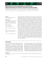
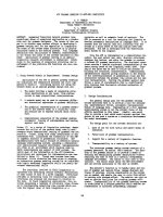
Figure 1
Pharyngeal arch and neurocranial cartilage patterns in wild-type and Hh-deficient zebrafish larvae. Alcian-stained cartilages were dissected
and flat mounted; dorsal views are shown, anterior to the left. (a) The wild-type (WT) pharyngeal arches at 4.5 days postfertilization include:
the mandibular arch (arch 1), containing Meckel’s cartilage (m) and the palatoquadrate (pq); the hyoid arch (arch 2), containing the
ceratohyal (ch) and hyosymplectic (hs); and five more posterior, branchial arches (3-7), each containing a ceratobranchial cartilage. (b) The
wild-type neurocranium at 4.5 days postfertilization includes paired trabeculae (t) and an ethmoid plate (e). (c) Neurocranial defects in sonic
hedgehog (shh) mutants. Trabecular cartilages fuse (arrows) at the midline. e, ethmoid; n, notochord; t, trabecula. Panels (b) and (c) taken
from [19].
WT shh
-/-
e n n
1 2
ch
m
pq
hs
5 6 7
3 4
t
t
(a) (b) (c)
101.3
Schilling and Le Pabic: Journal of Biology 2009, 8:101
essential Hh co-receptor, Smoothened (Smo), which when
removed specifically from neural crest cells causes similar
craniofacial defects in mice as the knockout [18]. Many
outstanding questions remain, however, including the
location of the sources of Shh and its relatives in the face,
the precise timing of their actions, and how defects in Hh
signaling cause such variable defects (for example, midline
fusions versus cleft palate).
Here again, zebrafish offer the advantage of a forward
genetic approach and a large collection of mutants in the
Hh signaling pathway. Fate-mapping studies in both Shh
and Smo mutants, using transgenics that label migrating
cranial neural crest cells, have revealed that Hh signaling
plays two distinct roles in patterning the cartilages of the
primary palate: one in neural crest cells in the maxillary
region of the first arch, and the second in a more anterior
population of neural crest cells that forms the ethmoid
plate in the midline [19]. Disruption of Hh signaling leads
to defects in one or both populations, resulting in cyclopia
(when maxillary regions fuse; Figure 1c) or mid-facial
clefting (when the midline ethmoid is not induced), and
treatment with the alkaloid cyclopamine, which chemically
blocks Smo function, only disrupt one or the other of these
populations, depending on the stage of treatment. Shh
expressed in the ventral forebrain and oral ectoderm
appears to be the critical source in the midline [19,20].
These results exemplify the complex spatial and temporal
nature of Hh signaling in the face, and help explain why
human HPE manifests itself in so many different ways.
Hh signaling also appears to have distinct roles in the
mandibular and more posterior arches. Schwend and
Ahlgren [2] show that chameleon (con) mutants, which
lack a fully functional Dispatched (Disp1) protein crucial
for Hh secretion, develop maxillary fusions, loss of jaw
joints and a complete lack of cartilage in arches 3-7. disp1
co-localizes with Shh in pharyngeal epithelia shortly after
neural crest migration into the arches. Defects in the
mandibular arch are not surprising, though the results in
this paper hint at a previously unappreciated role for Hh
signaling in jaw joint development. What is more
surprising is the selective loss of expression of some genes
involved in skeletogenesis (sox9a, dlx2a) and not others
(for example, sox9b, hand2) in the gill arches in con/disp1
mutants. In zebrafish, defects in the gill arches are often
secondary consequences of developmental delay or heart
defects. However, this is clearly not the case in con/disp1
mutants and instead, fibrous connective tissue appears to
replace cartilage. Thus, Hh signaling is required for
cartilage differentiation in the posterior arches, and several
lines of evidence suggest that this is distinct from its roles
in the palate or mandible. Such specific regulation of gene
expression and skeletogenesis by Hh may offer insights
into the nature of defects in other zebrafish mutants that
affect the posterior arches [21].
Deciphering the secrets of the throat
Given that Shh and other signals are expressed in similar
patterns in different arches, one big open question is how
the responses to these signals are modulated in a segment-
specific manner. Why is the mandible in a fish so much
larger than its gills? We still know relatively little about the
targets of Hox genes within different segments, or how the
combinatorial Hox code is interpreted to give different
morphologies. What seems clear is that not all cells within
a segment necessarily interpret the code in the same
fashion, nor do the signals impinging upon them have a
single effect on cell behavior. For example, Shh appears to
have at least two roles in the face, one in establishing the
patterns of palate or joint precursors and one in cartilage
differentiation, both in the palate and in the gills. Whether
this is truly a dual role for Shh or reflects distinct roles for
its relatives, such as Indian hedgehog (Ihh), remains
unclear.
Zebrafish provide the opportunity to address these
questions more directly than is possible in a chick or
mouse, as we can watch neural crest and arch development
in real time using transgenics, taking advantage of the
transparency of the live fish embryos. The added ability to
screen for craniofacial mutants in zebrafish is proving
fruitful for discovering novel factors involved in arch
development. These are candidates for genes mutated in as
yet unidentified human craniofacial syndromes. The
importance of this approach is apparent when considering
that craniofacial defects are so common (for example, 1 in
700 live births have cleft lip or palate), yet the genetic basis
for most is unknown. Most of the genes known to be
involved have roles in embryonic development, arguing
that as new genetic causes are revealed, their functions can
be rapidly evaluated in the context of the known pathways
that pattern the arches. Craniofacial research is now
undergoing a rapid expansion, with the accelerated
identification of human disease genes and new model
systems for functional analysis, and the zebrafish promises
to be a central player.
Acknowledgements
Craniofacial research in the Schilling lab is funded by the NIH - R01
DE13828.
References
1. Platt J: Ectodermic origin of the cartilages of the head. Anat
Anz 1893, 8:506-509.
2. Schwend T, Ahlgren SC: Zebrafish con/disp1 reveals multi-
ple spatiotemporal requirements for Hedgehog-signaling
in craniofacial development. BMC Dev Biol 2009, 9:59.
3. Northcutt RG: Historical hypotheses regarding segmenta-
tion of the vertebrate head. Integr Comp Biol 2008, 48:611-
619.
4. Gillis JA, Dahn RD, Shubin NH: Shared developmental
mechanisms pattern the vertebrate gill arch and paired fin
skeletons. Proc Natl Acad Sci USA 2009, 106:5720-5724.
5. Rijli FM, Mark M, Lakkaraju S, Dierich A, Dolle P, Chambon P:
A homeotic transformation is generated in the rostral
101.4
Schilling and Le Pabic: Journal of Biology 2009, 8:101
branchial region of the head by disruption of HoxA-2,
which acts as a selector gene. Cell 1993, 75:1333-1349.
6. Hunter MP, Prince VE: Zebrafish Hox paralogue group 2
genes function redundantly as selector genes to pattern
the second pharyngeal arch. Dev Biol 2002, 247:367-389.
7. Minoux M, Antonarakis GS, Kmita M, Duboule D, Rijli FM:
Rostral and caudal pharyngeal arches share a common
neural crest ground pattern. Development 2009, 136:637-
645.
8. Noden DM: The embryonic origins of avian cephalic and
cervical muscles and associated connective tissues. Am J
Anat 1983, 168:257-276.
9. Trainor P, Krumlauf R: Plasticity in mouse neural crest cells
reveals a new patterning role for cranial mesoderm. Nat
Cell Biol 2000, 2:96-102.
10. Couly G, Creuzet S, Bennaceur S, Vincent C, Le Douarin NM:
Interactions between Hox-negative cephalic neural crest
cells and the foregut endoderm in patterning the facial
skeleton in the vertebrate head. Development 2002, 129:
1061-1073.
11. Miller CT, Maves L, Kimmel CB: moz regulates Hox expres-
sion and pharyngeal segmental identity in zebrafish.
Development 2004, 131:2443-2461.
12. Crump JG, Swartz ME, Eberhart JK, Kimmel CB: Moz-
dependent Hox expression controls segment-specific fate
maps of skeletal precursors in the face. Development 2006,
133: 2661-2669.
13. David NB, Saint-Etienne L, Tsang M, Schilling TF, Rosa FM:
Requirement for endoderm and FGF3 in ventral head skel-
eton formation. Development 2002, 129:4457-4468.
14. Crump JG, Maves L, Lawson ND, Weinstein BM, Kimmel CB:
An essential role for Fgfs in endodermal pouch formation
influences later craniofacial skeletal patterning.
Development 2004, 131:5703-5716.
15. Crump JG, Swartz ME, Kimmel CB: An integrin-dependent
role of pouch endoderm in hyoid cartilage development.
PLoS Biol 2004, 2:1432-1445.
16. Roessler E, Belloni E, Gaudenz K, Jay P, Berta P, Scherer SW,
Tsui LC, Muenke M: Mutations in the human Sonic Hedge-
hog gene cause holoprosencephaly. Nat Genet 1996, 14:
357-360.
17. Brito JM, Teillet MA, Le Douarin NM: Induction of mirror-
image supernumerary jaws in chicken mandibular mesen-
chyme by Sonic Hedgehog-producing cells. Development
2008, 135:2311-2319.
18. Jeong J, Mao J, Tenzen T, Kottman AM. McMahon AP:
Hedgehog signaling in the neural crest cells regulates the
patterning and growth of facial primordia. Genes Dev 2004,
18: 937-951.
19. Wada N, Javidan Y, Nelson S, Carney TJ, Kelsh RN, Schilling
TF: Hedgehog signaling is required for cranial neural crest
morphogenesis and chondrogenesis at the midline in the
zebrafish skull. Development 2005, 132:3977-3988.
20. Eberhart JK, Swartz ME, Crump JG, Kimmel CB: Early
Hedgehog signaling from neural to oral epithelium organ-
izes anterior craniofacial development. Development 2005,
133: 1069-1077.
21. Schilling TF, Piotrowski T, Grandel H, Brand M, Heisenberg CP,
Jiang YJ, Beuchle D, Hammerschmidt M, Kane DA, Mullins
MC, van Eeden FJ, Kelsh RN, Furutani-Seiki M, Granato M,
Haffter P, Odenthal J, Warga RM, Trowe T, Nüsslein-Volhard C:
Jaw and branchial arch mutants in zebrafish I: branchial
arches. Development 1996, 123:329-344.
Published: 22 December 2009
doi:10.1186/jbiol205
© 2009 BioMed Central Ltd