Báo cáo lâm nghiệp: "Changes in phenolic acids and stilbenes induced in embryogenic cell cultures of Norway spruce by two fractions of Sirococcus strobilinus mycelia" pot
Bạn đang xem bản rút gọn của tài liệu. Xem và tải ngay bản đầy đủ của tài liệu tại đây (766.27 KB, 7 trang )
J. FOR. SCI., 57, 2011 (1): 1–7 1
JOURNAL OF FOREST SCIENCE, 57, 2011 (1): 1–7
Changes in phenolic acids and stilbenes induced
in embryogenic cell cultures of Norway spruce
by two fractions of Sirococcus strobilinus mycelia
J. M
1
, M. H
2
, P. M
1
, H. C
1
,
O. M
2
, M. C
2
1
Forestry and Game Management Research Institute, Jíloviště, Czech Republic
2
Institute of Experimental Botany, Academy of Sciences of the Czech Republic,
Prague, Czech Republic
ABSTRACT: We examined defence responses in embryogenic cell suspension cultures of Norway spruce (Picea abies
[L.] Karst) elicited by intracellular protein and cell wall fractions (PF and WF, respectively) prepared from mycelia of
the fungus Sirococcus strobilinus Preuss focusing on changes in (soluble and cell wall-bound) phenolic and stilbene
concentrations. Treatment with both preparations induced an increase in the total contents of phenolic acids in Norway
spruce cells and variations in the levels of stilbene glycosides. More rapid and intense induction of defence response
was observed in cells after WF application. The contents of soluble phenolic acids (especially benzoic acid derivatives)
and cell wall-bound phenolic acids (especially ferulic acid) started to increase (relative to controls) within 4 h after the
addition of the WF preparation and remained high in elicited cells for 8–12 h. A moderate increase in phenolic acids
in cells exposed to the PF preparation was observed within 8 h after application. However, after 24h of WF treatment
a decline of total phenolics was observed, while in PF elicited Norway spruce cells the phenolic content continued to
increase. Significantly decreased concentrations of stilbene glycosides, isorhapontin, astringin and piceid, were de-
termined in PF and WF treated Norway spruce cell cultures. The total content of stilbene glycosides decreased within
8 h after WF application to 68% of the amount determined in the control and within 12 h to 73% of the control in
PF-treated cells. These results demonstrate that both PF and WF prepared from the Sirococcus strobilinus mycelium
elicit changes in the metabolism of phenylpropanoids, which are involved in the defence responses of plants to pathogens.
Keywords: defence response; Norway spruce; phenylpropanoids; stilbenoids
Supported by the Ministry of Agriculture of the Czech Republic, Projects No. QH82303 and No. MZE 0002070203.
e decline of the forest tree population caused
by fungal diseases is a long-term factor influencing
the stability of forest ecosystem. e recent wide-
spread dieback of Norway spruces in the Orlické
hory Mountains, Czech Republic, was caused by
the combined effects of air pollution, climatic con-
ditions and attacks by the potentially pathogenic
fungi Sirococcus strobilinus, Phoma spp., and As-
cocalyx abietina. Plants respond to a pathogen
challenge by activating the range of defence mech-
anisms that can be local or result in systemic ac-
quired resistance (B, B 2003). A
universal feature of plant responses to pathogens or
other elicitors is the activation of phenylpropanoid
synthesis. An important manifestation of defence
is the accumulation of polyphenols in the cell walls,
which is accompanied by an increase in lignifica-
tion and suberization (D A, D
2003). e induction of phenylpropanoid biosyn-
thesis and consequent increase in bark polyphenols
in Norway spruce trees following wounding or fun-
gal infection were documented histo- and immuno-
chemically (F et al. 1998). An increase
in cell wall phenolics in the bark of Norway spruce
2 J. FOR. SCI., 57, 2011 (1): 1–7
branches infected by Ascocalyx abietina was also
reported in our previous paper (C et al.
2006). Stilbenoids are an important group of phe-
nolics, specifically linked with resistance to fungal
attack. Stilbenes occur as glycosides in the healthy
phloem of Norway spruce (B et al. 1995).
e main constitutive stilbene glycosides in Picea
species are astringin and isorhapontin (L
et al. 1992). Rapid accumulation of stilbene agly-
cones in response to injury or fungal infection is
considered to be an active defence response of
Norway spruce (N, H
1992).
e use of tissue cultures facilitates detailed
studies of early response to challenge with patho-
gen or elicitor preparations (G, B 2000).
e aim of the study was to characterize changes in
(soluble and cell wall-bound) phenolic and stilbene
concentrations during defence response in Nor-
way spruce embryogenic cell suspension cultures
induced by intracellular protein and wall prepara-
tions from Sirococcus strobilinus mycelia.
MATERIAL AND METHODS
Plant material
e embryogenic tissue derived from zygotic em-
bryos of mature seeds of Norway spruce was initi-
ated on modified AE medium (A, E
1979). Embryogenic cultures were grown on gelrite-
solidified medium (2 g·l
–1
) supplemented with 6-ben-
zylaminopurine and 6-furfurylaminopurine (both
0.5mg·l
–1
), 2,4-dichlorophenoxyacetic acid (1 mg·l
–1
),
glutamine (400 mg·l
–1
), casein hydrolyzate (400 mg·l
–1
),
FeSO
4
·7 H
2
O (27.8 mg·l
–1
) and sucrose (20 mg·l
–1
), pH
of the media was adjusted to 5.8. Cultures were culti-
vated under controlled conditions (24°C) in the dark
and subcultured every 3 weeks (M 1991). For
establishment of suspension cultures, approximately
3 g fresh weight of embryogenic tissue were inoculat-
ed to 100 ml of liquid medium of the same composi-
tion as mentioned above in 250 ml Erlenmeyer flasks
and grown at 24°C in the dark on an orbital incuba-
tor (IOC.400.XX2.C SANYO-Gallenkamp, Leicester,
UK) at 110 rpm. Five-day-old cell suspension cultures
were used for the experiments.
Pathogen culture
Stock culture of the non-lyophilized mycelium of
Sirococcus strobilinus was obtained from Dr. A. L
(METLA, Finnish Forest Research Institute, Vantaa
Research Unit, FI-01301 Vantaa, Finland). Pieces of
the stock fungus were plated onto the Malt Extract
Agar (MA: 12 g·l
–1
Difco Maltose Extract, 12g·l
–1
Difco Agar, Detroit, Michigan, USA) and incubated
at 24°C (L et al. 2005). After multiplication, the
mycelium was transferred into 100 ml Erlenmeyer
flasks containing 50 ml of 12 g·l
–1
Difco Maltose
Extract and incubated at 24°C in an orbital incuba-
tor (IOC.400.XX2.C SANYO-Gallenkamp, Leices-
ter, UK) at 120 rpm. Approximately 5 g of fresh
mycelium was transferred into 50 ml of fresh liquid
medium (Difco Maltose extract 12 g·l
–1
) and cul-
tured under the above mentioned conditions for
4weeks.
Preparation of mycelial intracellular
protein fraction
e Sirococcus strobilinus mycelium was washed
three times with distilled water, harvested by filtra-
tion through Whatman No. 1 filter paper and the
mycelial mass was then ground in liquid nitrogen
and homogenized with 0.1 M Tris-HCl buffer pH 7.2
containing 2mM β-mercaptoethanol, 500 µg·ml
–1
amoxicillinum, and 100 µg·ml
–1
acidum clavu-
lanicum (Augmentin 600, SmithKlineBeechcham
Pharmaceuticals, Worthing, UK). e resulting ho-
mogenate was centrifuged at 14,000 g for 20 min at
4°C to obtain a supernatant containing intracellu-
lar proteins – 12.20 mg protein g
–1
mycelium fresh
weight, according to assays following the method of
B (1976) using bovine serum albumin as a
standard. e final protein content of the intracel-
lular fraction in 100 ml of liquid medium was 4 mg.
Preparation of mycelial wall fraction
e mycelial cell wall fraction (WF) was prepared
according to the method described by M
et al. (2004), with slight modifications, as follows.
Cultures of Sirococcus strobilinus were filtered
through Whatman No. 1 filter and washed with
distilled H
2
O. e resulting mycelial mass was
ground in liquid nitrogen and homogenized with
0.1M Tris-HCl buffer (pH 7.2) containing 2mM
β-mercaptoethanol. e cell walls were separated
by centrifugation at 3,000 g for 10 min and the pel-
let was repeatedly washed with distilled water. To
determine the amount of ionically bound protein in
the mycelial walls a part of the mycelial wall prep-
aration was resuspended in 0.1M Tris-HCl buffer
J. FOR. SCI., 57, 2011 (1): 1–7 3
(pH 7.2) containing 0.1M KCl and stirred for 1 h
at 20°C. e extract was centrifuged (3,000 g for
10 min) and the protein content of the supernatant
was determined (B 1976) using bovine se-
rum albumin as a standard. e results indicated
that the mycelial cell walls contained 19.40 mg of
ionically bound protein per gram of the cell wall
preparation (fresh weight) and 77.6 mg per gram
of the pellet material dried at 40°C for 24 h. e
dried mycelial wall powder was suspended in dis-
tilled water (pH 5.2) and autoclaved for 5 min. e
concentration of the WF used in the experiment
was 30 mg of mycelium powder per 100 ml of liq-
uid medium.
Extraction and analysis of phenolic acids
Phenolic acids were extracted as described by
C et al. (1991). Briefly, free, ester-bound
(released after alkaline hydrolysis) and glycoside-
bound (released after acid hydrolysis) phenolic ac-
ids were obtained from a methanol extract of the
tissue ground in liquid nitrogen. e fraction of cell
wall-bound phenolic acids was obtained after alka-
line hydrolysis of the residual material following the
methanol extraction. 2,6-Di-tert-butyl β-cresol was
used as an antioxidant to minimize the oxidation of
phenolic acids during alkaline hydrolysis and nitro-
gen was immediately bubbled through the sample
after NaOH addition. Phenolic acids were analysed
by means of HPLC using a Dionex liquid chroma-
tograph (P660-HPLC pump, ASI-100 automated
sample injector, TCC-100 thermostated column
compartment, PDA-100 photodiode array detector,
Chromeleon software 6.5) with C18 Spherisorb 5
ODS column (250 × 4.6 mm). Acetonitrile and
acetic acid gradient was used for elution. Phenolic
acids were detected at their absorption maximum.
λ
max
was detected from the authentic compounds
(Sigma-Aldrich, Prague, Czech Republic) that were
used as references for quantitative analyses.
Extraction and analysis of stilbenes
For the extraction of stilbenes the procedure
described by V et al. (2001) was followed with
slight modifications. Briefly, samples of cell culture
(0.5 g fresh weight) were frozen in liquid nitrogen,
homogenized with 5.0 ml of 80% (v/v) methanol in
mortar, stirred on an orbital shaker for 60 min at
room temperature and then centrifuged (5,000×g
for 20 min). e supernatant was evaporated in
the vacuum to dryness. Aliquots of methanol-
soluble material were analyzed by means of HPLC
using a Dionex liquid chromatograph (P660-
HPLC pump, ASI-100 automated sample injector,
TCC-100 thermostated column compartment,
PDA-100 photodiode array detector, Chromeleon
software 6.5) with C18 Spherisorb 5 ODS 2 column
(250×4.6mm). Acetonitrile and acetic acid gradi-
ent was used for elution. Stilbenes were detected at
303 nm. Authentic samples of stilbenes (Polyphe-
nol Laboratories AS, Sandnes, Norway) were used
for qualitative and quantitative determinations.
RESULTS
Phenolic acid contents
Variations in the total contents of phenolic acids
(represented by the sum of free, methanol soluble
conjugated forms, i.e. ester- and glycoside-bound
phenolics, and methanol-insoluble cell wall-bound
phenolic esters) induced by both elicitor prepara-
tions are presented in Fig. 1. In the control Norway
spruce cells the soluble glycoside-bound forms of
phenolic acids (SG) accounted for most of the to-
tal content (about 85%), followed by the methanol-
insoluble cell wall-bound phenolic esters (CWE;
7–8%). e amounts of methanol soluble esters
(SE) and free phenolic acids (F) were low in control
cells, accounting for ca 2 and 4–5% of total pheno-
lic contents, respectively. Responses to challenges
with both elicitors were manifested most clearly by
marked increases (compared with controls) in SG
contents. During the WF treatment, SG levels sig-
nificantly increased after 4 h and almost doubled
after 8 h. In addition, increases in CWE contents
by 50% and doubled content of F after 12 h were
observed in WF-elicited cells (Fig. 1). In PF-elicited
cells, the level of SG increased by about 40% after
8 h. After 12h of treatment with PF the amount
of SG was still at its 8-h level, but levels of the
other forms of phenolic acids increased; levels of
F by 70% and levels of CWE by 25% (Fig. 1). After
24 h of WF treatment a decline in the contents of
total phenolics was observed, while in PF-elicited
cells a further significant rise in SG and CWE was
determined.
e HPLC analyses indicated the presence of
two cinnamic acid derivatives, p-coumaric and
ferulic acids and of five benzoic acid derivatives
(anisic, p-hydroxybenzoic, vanillic and syringic
acids) in the Norway spruce cells, and there were
no qualitative differences in the phenolic acid com-
4 J. FOR. SCI., 57, 2011 (1): 1–7
plements between the control and elicited cells.
e enhancement of phenolic contents in treated
cells was mainly due to increases in SG forms of
p-hydroxybenzoic and vanillic acids. We focused
predominately on changes in the contents of
p-hydroxybenzoic acid. Marked increases in p-hy-
droxybenzoic acid glycosides were detected in WF-
elicited cells after 8 h and remained high after 12h.
e level of glycosides of the above-mentioned
phenolic acid was maximal after 24 h in PF-treated
cells (Fig. 2). Both elicitor treatments induced in-
creases in CWE forms of p-hydroxybenzoic acid
and both cinnamic acid derivatives, p-coumaric
and ferulic acids.
Contents of stilbenes
Significantly decreased concentrations of stil-
bene glycosides, isorhapontin, astringin and piceid,
were determined in WF and PF treated Norway
spruce cell cultures (Fig. 3). Within 8 h after WF
application the total content of stilbene glycosides
decreased to 68% of the amount determined in
the control. e level of isorhapontin, the stilbene
Fig. 1. Changes in the contents of free (F), soluble ester-bound
(SE), cell wall ester-bound (SWE) and soluble glycoside-bound
(SG) phenolic acids in control cells of embryogenic cultures of
Norway spruce (C) and in the cells elicited by mycelium protein
fraction (PF) and mycelium wall fraction (WF) in the course of
24 h. Means ± Standard Error of two independent experiments
with two replicates. Different letters above the bars indicate
significant differences in SG contents from the controls (P < 0.05)
Fig. 2. Changes in the contents of soluble glycoside-bound
p-hydroxybenzoic (p-HBA) acid in control cells of embryo-
genic cultures of Norway spruce (C) and in the cells elicited
by mycelium protein fraction (PF) and mycelium wall frac-
tion (WF) in the course of 24 h. Means ± Standard Error of
two independent experiments with two replicates. Different
letters above the bars indicate significant differences from
the controls (P < 0.05)
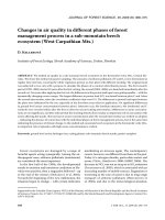
Fig. 3. Contents of stilbene glycosides, isorhapontin (iRHAP),
astringin (ASTR) and piceid (PIC), determined in Norway
spruce cell cultures treated with 5% and 20% A. abietina
culture filtrate and from the control cells (C). Means of two
independent experiments with two replicates. Bars represent
the sum of SE of isorhaportin, astringin and piceid. Different
letters above the bars indicate significant differences from
the controls (P < 0.05)
Stilbenes (nmol·g
–1
FW)
C PF WF C PF WF C PF WF C PF WF
4h 8h
12 h 24 h
60
40
20
0
4h
8h
12 h
24 h
200
150
100
50
0
p-HBA - glycosides (nmol·g
–1
FW)
Phenolic acids (nmol·g
–1
FW)
C PF WF C PF WF C PF WF C PF WF
4h 8h
12 h 24 h
250
200
150
100
50
0
J. FOR. SCI., 57, 2011 (1): 1–7 5
which occurred in the highest concentration in em-
bryogenic cell cultures, decreased to less than 63%
(compared with the control) within 8 h after WF
application. e content of piceid, which was pre-
sent in the lowest amount in cells, did not change
markedly. e stilbene glycoside levels in WF-elic-
ited cultures remained more or less stable till the
end of 24 h treatment. During 12 h of treatment
PF evoked the decline of total stilbene glycosides
to 73% (compared with the control). e decline in
the levels of isorhapontin and astringin continued
24 h after PF application representing less than 68%
and 50%, respectively, of the contents determined
in the control cells (Fig. 3).
DISCUSSION
We showed that the Norway spruce cells re-
sponded more rapidly to the mycelial WF prepa-
ration than to the mycelial PF preparation. e
contents of soluble (especially benzoic acid deriva-
tives) and cell wall-bound phenolic acids (especial-
ly ferulic acid) started to increase within 4h after
the addition of the WF preparation. e response
of cells to the PF fraction was slower; a significant
increase was first detected after 8 h (Fig. 1). It is in
agreement with findings reported by P et al.
(2003, 2005) in winter oilseed rape calli. ese au-
thors found out that pectinase (PF is a rich source
of soluble, hydrolytic enzymes) activated the phe-
nylpropanoid pathway in the calli less strongly than
chitosan (major polysaccharide components of fun-
gal cell walls). In a study examining possible corre-
lations between the synthesis of hydroxycinnamic
amides and the formation of wall-bound phenolic
polymers it was also shown that the phenylpropa-
noid pathway could be induced by pectinase and
pronase in tobacco cell suspension cultures (N-
, J 1995).
e fungal cell wall has a highly complex struc-
ture. It forms a network of polysaccharides in which
various proteins are embedded (S et al. 2006).
It could be supposed that the defence response of
Norway spruce cells was induced by mycelial wall
polysaccharides. is is in agreement with the ac-
cepted knowledge that major polysaccharide com-
ponents of fungal cell walls, glucans and chitin act
as general elicitors of defence responses (Y-
et al. 2000). Similarly, induction of phenylpro-
panoid biosynthesis and accumulation of phenolics
were observed in soybean leaves following the ex-
posure to chitin and chitosan (K et al. 2003).
An increase in the levels of soluble glycosides of
p-hydroxybenzoic acid culminated in WF-elicit-
ed cells after 12 h, while in PF-treated cells their
levels were maximal at the end of 24-h treatment
(Fig. 2). It corresponds to results obtained in callus
cultures of Pinus sylvestris treated with mycelial
extracts of Fusarium nivale which were reported
by S et al. (2003), who concluded that the ac-
cumulation of p-hydroxybenzoic acid plays an im-
portant role in the protection of conifer cells by
acting as a fungicidal agent when fungi penetrate
into the cytosol. Furthermore, plant glycosides are
often hydrolysed by vacuolar glycosidases follow-
ing the pathogen invasion, releasing aglycones that
may be quite toxic to the invader (K 1999).
Stilbenes are generally described as phytoalexin-
like compounds or phytoanticipins in conifers as
they are often present in certain tissues consti-
tutively rather than appearing de novo following
the infection (M 2000). Because of their
strong antimicrobial properties in vitro they are
implicated in the defence of conifers against patho-
gens (L et al. 1992; C et al. 2001).
e present Norway spruce cell cultures responded
to treatment with both elicitor preparations by a
decrease in concentrations of stilbene glycosides.
e decrease in isorhapontin (occurring in the
highest concentrations in embryogenic cell cul-
tures), astringin and piceid levels was observed
in WF-elicited cells after 12 h, while in PF-treat-
ed cells at the end of the 24 h treatment (Fig.3).
Our results agree with those of L et al.
(1992), who concluded that the bark of Norway
spruce contains more isorhapontin than astringin
(Fig. 3). e rapid decline of the levels of glycosides
was described in in vitro maintained excised bark
discs of Sitka spruce following the fungal challenge
(W, P 1988).
It is known that β-glycosidase enzymes are able
to metabolize stilbene glycosides to the respective
aglycones (W, P 1988). Since the
β-glycosidase activities were not measured in this
experiment, we can only speculate that the signifi-
cant decrease in isorhapontin, astringin and piceid
contents in Norway spruce cells after treatment
with both WF and PF preparations might result
from the activities of β-glycosidase enzymes. e
decrease in stilbene levels in treated cells might
also be partly explained by their incorporation into
the cell walls (L et al. 1994).
us, our results show that although the com-
ponents of the pathogen cell walls and intracellu-
lar protein preparations of Sirococcus strobilinus
mycelium differed substantially, the responses of
treated cells to them (characterized by variations in
6 J. FOR. SCI., 57, 2011 (1): 1–7
contents of phenolics and stilbenes) were similar,
although there were differences in the kinetics of
these responses.
References
A S., E T. (1979): Induction of adventitious
buds on buds of Norway spruce (Picea abies) grown in
vitro. Physiologia Plantarum, 45: 29–34.
B P., B J.T. (2003): Pinus nigra–Sphaeropsis
sapinea as a model pathosystem to investigate local and
systemic effects of fungal infection of pines. Physiological
and Molecular Plant Pathology, 63: 249–261.
B M.M. (1976): A rapid and sensitive method for
quantification of microquantities of protein utilizing the
principle of protein dye binding. Analytical Biochemistry,
72: 248–254.
B F., L B., L F., S D.,
D A., C A.C., Y A., B A.A.,
C E. (1995): Induced responses in phenolic
metabolism in two Norway spruce clones after wound-
ing and inoculations with Ophiostoma polonicum, a bark
beetle-associated fungus. Plant Physiology, 109: 821–827.
C C.C., S D.R., Y R.A., S G.R.
(2001): In vitro inhibition of Sphaeropsis sapinea by natural
stilbenes. Phytochemistry, 56: 161–165.
C M., M L., M I., E J.
(1991): Phenylalanine ammonia-lyase, phenolic acids and
ethylene in alfalfa (Medicago sativa L.) cell cultures in
relation to their embryogenic ability. Plant Cell Reports,
10: 251–255.
C M., M J., H M., E J. (2006):
Soluble and cell wall-bound phenolics and lignin in As-
cocalyx abietina infected Norway spruces. Plant Science,
170: 563–570.
D A A.R.F.D.C., D I.A. (2003): Soluble and
wall-bound phenolics and phenolic polymers in Musa
acuminate roots exposed to elicitors from Fusarium ox-
ysporum f.sp. cubense. Phytochemistry, 63: 679–686.
F V.R., K T., B A.A., C-
E. (1998): Specialized phloem parenchyma cells in
Norway spruce (Pinaceae) bark are an important site of de-
fense reactions. American Journal of Botany, 85: 601–615.
G K., B W. 2000): Elicitor-induced defense re-
actions in cell suspension cultures of soybean cultivars.
Zeitschrift für Naturforschung C-A, 55: 718–730.
K N.T. (1999): Plant disease resistance: Progress in basic
understanding and practical application. Advances in
Botanical Research, 30: 291–328.
K W., P B., S D.L. 2003): Chitosan and
chitin oligomers increase phenylalanine ammonia-lyase
and tyrosine ammonia-lyase activities in soybean leaves.
Journal of Plant Physiology, 160: 859–863.
L B.M., T M., K W., L C.,
S H. (1994): Elicitor-induced formation of free
and cell-wall-bound stilbenes in cell-suspension cultures
of Scots pine (Pinus sylvestris L.). Planta, 194: 143–148.
L A. P ., ., ., .
2005: Cultural and PCR-based indetification of the two
most common fungi from cankers on container-grown
Norway spruce seedlings. Canadian Journal of Forest
Research, 35: 432–439.
L M., L L., G R., J M.
(1992): Stilbenes and resin acids in relation to the penetra-
tion of Heterobasidion annosum through the bark of Picea
abies. European Journal of Forest Pathology, 22: 95–106.
M J. 1991: Organogenesis and somatic embryogenesis in
Spruce (Picea abies (L.) Karst.). Communicationes Instituti
Forestalis Cechoslovakiae, 17: 59–72.
M J.W. (2000): Antimicrobial compounds and re-
sistance: the role of phytoalexins and phytoanticipins. In:
S A.J., F R.S.S., L L.C. (eds):
Mechanisms of Resistance to Plant Diseases. Dodrecht,
Kluwer Academic Publishers: 325–370.
M M., L R., H T.W., R E.A.,
M C., P M., G G.M., F J.F., H-
R.B., R K.A. (2004): e Aspergillus fumigatus
cell wall is organized in domains that are remodeled during
polarity establishment. Microbiology, 150: 3261–3268.
N J., J F. (1995): Induction of phenylpropanoid
and tyramine metabolism in pectinase- or pronase-elicited
cell suspension cultures of tobacco (Nicotiana tabacum).
Physiologia Plantarum, 95: 569–574.
N R.L., H R. 1992): Phenolic
compounds and their role in disease resistance. Annual
Review of Phytopathology, 30: 369–38.
P A., H K., Z I. (2003): Reaction of winter oilseed
rape callus to different concentrations of elicitors: pectinase
or chitosan. Acta Physiologiae Plantarum, 25: 83–89.
P A., H K., Z I. (2005): Influence of chitosan,
pectinase and fungal metabolites on activation of phenyl-
propanoid pathway and antioxidant activity in oilseed rape
callus. Acta Physiologiae Plantarum, 27: 95–102.
S R., Y M., S B.P., G D.K., S T.,
A D.K. (2006): Induction of resistance in chicpea by
cell wall protein of Fusarium oxysporum sp. ciceri and Ma-
crophomina phaseolina. Current Science, 91: 1543–1546.
S I.V., A O.N., P G.G., Z
G.K. (2003): Effect of pine callus elicitation by the Fusarium
strains of various pathogenicity on the content of phenolic
compounds. Russian Journal of Plant Physiology, 50: 634–639.
V H., A E., K V., N P. (2001): In-
duced responses in stilbenes and terpenes in fertilized
Norway spruce after inoculation with blue-stain fungus,
Ceratocystis polonica. Trees, 15: 112–122.
W S., P R.B. (1988): e role of stilbenes in
resistance of Sitka spruce [Picea sitchensis (Bong.) Carr.]
J. FOR. SCI., 57, 2011 (1): 1–7 7
to entry of fungal pathogens. Physiological and Molecular
Plant Pathology, 33: 127–149.
Y T., Y A., H N., O T., I
T., S N. (2000): Differences in the recognition of
glucan elicitor signals between rice and soybean: β-glucan
fragments from the rice blast disease fungus Pyricularia
Corresponding author:
RNDr. J M, CS., Forestry and Game Management Research Institute,
Strnady 136, 252 02 Jíloviště, Czech Republic
e-mail:
oryzae that elicit phytoalexin biosynthesis in suspension-
cultured rice cells. Plant Cell, 12: 817–826.
Recieved for publication June 1, 2010
Accepted after corrections July 26, 2010