Báo cáo y học: "Delayed resolution of acute inflammation during zymosan-induced arthritis in leptin-deficient mice" pptx
Bạn đang xem bản rút gọn của tài liệu. Xem và tải ngay bản đầy đủ của tài liệu tại đây (265.6 KB, 8 trang )
Open Access
Available online />R256
Vol 6 No 3
Research article
Delayed resolution of acute inflammation during
zymosan-induced arthritis in leptin-deficient mice
Eiva Bernotiene
1,2
, Gaby Palmer
1
, Dominique Talabot-Ayer
1
, Ildiko Szalay-Quinodoz
3
,
Michel L Aubert
4
and Cem Gabay
1
1
Division of Rheumatology, University Hospital and Department of Pathology, University of Geneva School of Medicine, Geneva, Switzerland
2
Department of Immunology, Institute of Experimental and Clinical Medicine at Vilnius University, Vilnius, Lithuania
3
Division of Clinical Pathology, University Hospital, Geneva, Switzerland
4
Division of Development and Growth, Department of Pediatrics, University of Geneva School of Medicine, Geneva, Switzerland
Corresponding author: Cem Gabay,
Received: 09 Jul 2003 Revisions requested: 11 Aug 2003 Revisions received: 3 Mar 2004 Accepted: 15 Mar 2004 Published: 26 Apr 2004
Arthritis Res Ther 2004, 6:R256-R263 (DOI 10.1186/ar1174)
http://arthr itis-research.com/conte nt/6/3/R256
© 2004 Bernotiene et al.; licensee BioMed Central Ltd. This is an Open Access article: verbatim copying and redistribution of this article are permitted
in all media for any purpose, provided this notice is preserved along with the article's original URL.
Abstract
The severity of antigen-induced arthritis (AIA) is decreased in
leptin-deficient ob/ob mice. However, joint inflammation in AIA
depends on the immune response, which is impaired in ob/ob
mice. In the present study we investigated the effects of leptin
deficiency on zymosan-induced arthritis (ZIA), which is
independent of adaptive immunity. Arthritis was induced by
injection of zymosan into the knee joint. Joint swelling was similar
after 6 and 24 hours in ob/ob and control mice. However, it
remained elevated in ob/ob animals on day 3 whereas values
normalized in controls. Histology revealed similar articular
lesions in all animals on day 3, but on days 14 and 21 arthritis
tended to be more severe in ob/ob mice. The acute phase
response, reflected by circulating levels of IL-6 and serum
amyloid A, was also more pronounced in ob/ob mice, although
corticosterone was significantly elevated in these animals.
Similar results were obtained in leptin receptor-deficient db/db
mice. Thus, in contrast to AIA, ZIA is not impaired in leptin-
deficient animals. On the contrary, resolution of acute
inflammation appears to be delayed in the absence of leptin or
leptin signalling, suggesting that chronic leptin deficiency
interferes with adequate control of the inflammatory response in
ZIA.
Keywords: acute phase response, arthritis, inflammation, interleukin-6, leptin
Introduction
Leptin is a peptide hormone that plays an important role in
the regulation of body weight by inhibiting food intake and
stimulating energy expenditure. Moreover, leptin exhibits a
variety of other effects, including regulation of endocrine
function, reproduction and immunity. Consistently, leptin-
deficient (ob/ob) mice and leptin-receptor deficient (db/db)
mice are not only obese, but also exhibit important hormo-
nal imbalances, infertility, abnormalities in thermoregula-
tion, and evidence of immune and haematopoietic defects
[1-4].
The role of leptin in the modulation of immune response
and inflammation has recently become increasingly evident.
The increase in leptin production that occurs during infec-
tion and inflammation strongly suggests that leptin is a part
of the cytokine cascade, which orchestrates the innate
immune response and host defence mechanisms [5]. How-
ever, both proinflammatory and anti-inflammatory effects
have been described for leptin, depending on the experi-
mental model investigated [5]. Leptin plays an important
role in inflammatory processes involving T cells, and has
been reported to promote T-helper (Th)1 polarization of the
cellular immune response [6-9]. Several studies have impli-
cated leptin in the pathogenesis of autoimmune inflamma-
tory conditions, such as experimental autoimmune
encephalomyelitis and type 1 diabetes [10-13].
Furthermore, in leptin-deficient ob/ob mice we recently
demonstrated reduced severity of antigen-induced arthritis
(AIA), which is a model of immune-mediated joint inflamma-
tion [14]. Leptin appeared to contribute to the mechanisms
AIA = antigen-induced arthritis; GAPDH = glyceraldehyde-3-phosphate dehydrogenase; IL = interleukin; LPS = lipopolysaccharide; MIF = macro-
phage migration inhibitory factor; OB-Ra = leptin receptor, long isoform; SAA = serum amyloid A; Th = T-helper (cell); TNF = tumour necrosis factor;
ZIA = zymosan-induced arthritis.
Arthritis Research & Therapy Vol 6 No 3 Bernotiene et al.
R257
of joint inflammation in AIA by regulating both humoral and
cell-mediated immune responses. Essentially identical
results were obtained in db/db mice, which lack expression
of the functional leptin receptor, long isoform (OB-Rb).
However, joint inflammation in AIA depends on the adaptive
immune response, which is known to be impaired in ob/ob
and db/db mice. We conducted the present study to inves-
tigate the effect of leptin and leptin receptor deficiency on
inflammatory events in the joint, independent of their effects
on T-cell and B-cell responses. Therefore, we explored the
effect of leptin deficiency in zymosan-induced arthritis
(ZIA), a model of proliferative arthritis, which is restricted to
the joint injected with zymosan A and is not dependent on
the adaptive immune response. Indeed, zymosan A, which
is a ligand for toll-like receptor 2 as well as an activator of
the alternate complement pathway, triggers local activation
of the innate immune system, causing inflammation in the
injected joint [15,16].
We followed the development of ZIA in ob/ob C57BL/6
mice and in their control +/? (i.e. +/+ or ob/+) lean litterma-
tes. In addition, in order to evaluate the role of OB-Rb in
ZIA, we also used db/db and control db/+ C57BL/KS
mice. The results of the experiments show that, in contrast
to AIA, ZIA is not impaired in ob/ob and db/db mice. Fur-
thermore, resolution of acute inflammation during ZIA
appears to be delayed in the absence of leptin.
Materials and methods
Animals
Eight-week-old male, leptin deficient C57BL/6 ob/ob mice
and their control +/+ or ob/+ (i.e. +/?) littermates, as well
as OB-Rb leptin receptor deficient C57BL/KS db/db mice
and their control db/+ littermates, were purchased from
Elevage Janvier (Le Genest-St-Isle, France). Animals were
housed under conventional conditions in the animal facility
of the Geneva University School of Medicine. Water and
standard laboratory chow were provided ad libitum. The
experimental protocol received the approval from the Ani-
mal Ethics Committee of the Geneva University School of
Medicine and of the Geneva Veterinarian Office.
Induction of arthritis
Arthritis was induced by injection of 180 µg zymosan A via
a small skin cut along the suprapatellar ligament directly
into the knee joint cavity, as described previously [17].
Zymosan A (30 mg) from Saccharomyces cerevisiae
(Sigma-Aldrich, Buchs, Switzerland) was suspended in 1
ml endotoxin-free saline (Laboratory Dr G Bichsel AG,
Interlaken, Switzerland) by boiling and sonification. Mice
were injected with 6 µl of this suspension into the right
knee joint, under inhalation anaesthesia with 5% isofluran
(Forene
®
; Abbott AG, Baar, Switzerland). The left knee
joint was simultaneously injected with an equal amount (6
µl) of saline and served as the control.
Study design
Ob/ob and +/? mice were killed at different time points
after injection of zymosan A (i.e. on days 1 and 3 for evalu-
ation of the acute phase of arthritis, and on days 14 or 21
for evaluation of the chronic phase). The knee joints were
either processed for histology (days 3, 14 and 21) or used
for RNA extraction (days 1 and 14). One experiment was
performed using db/db and control db/+ mice to evaluate
the effect of OB-Rb deficiency on ZIA. In this experiment,
mice were killed on day 14 after injection of zymosan A and
the knee joints were processed for histology. All animals
were killed by exsanguination (cardiac puncture) followed
by cervical dislocation, under intraperitoneal anaesthesia
with 0.01 ml/g saline solution containing 12 mg/ml ketasol
(Dr E Graub AG, Bern, Switzerland) and 0.16% rompun
(Bayer, Provet AG, Lyssach, Switzerland).
Isotopic quantification of joint swelling
Joint swelling was quantified at various time points after
injection of zymosan A by measuring uptake of circulating
99m
Tc-pertechnetate in the knee joint, as previously
described [14,17]. Animals were injected subcutaneously
in the neck region with 10 µCi of
99m
Tc-pertechnetate in
0.2 ml saline. After 30 min mice were sedated by inhalation
anaesthesia with 5% isofluran, and accumulation of the iso-
tope due to increased blood flow and oedema in the knee
was determined in duplicate by external gamma counting.
The ratio between
99m
Tc-pertechnetate uptake in the
inflamed and that in the contralateral knee joint was calcu-
lated. A ratio greater than 1.1 was taken to indicate joint
swelling.
Histological studies
Knee joints were fixed in 10% formalin for a minimum of 24
hours and decalcified using 'd-calcifier' (Lerner Laborato-
ries, Pittsburg, PA, USA), containing 14% HCl, over 6–7
hours. They were embedded in paraffin and serial sections
of 4 µm were cut for histological analysis. Sections were
stained with either haematoxylin–eosin or toluidine blue.
Histological assessment was performed in a blinded man-
ner, using an established scoring system for synovial hyper-
plasia (from 0 = no hyperplasia to 3 = most severe
hyperplasia) and inflammatory cell infiltration in the syn-
ovium (from 0 = no inflammation to 3 = most severe inflam-
mation). Cartilage damage was determined by toluidine
blue staining (from 0 = no change and fully stained carti-
lage to 3 = total loss of toluidine blue staining and erosions
in cartilage). These three scores were added together to
obtain a total histological score, as previously described
[18]. Only processes taking place inside the joint cavity
were taken into account.
Available online />R258
Measurement of IL-6, serum amyloid A and
corticosterone levels
Blood samples (100 µl) were taken at baseline and differ-
ent time points after injection of zymosan A from the tail vein
and at the end of experiment by cardiac punction. Serum
levels of IL-6 were measured using a commercial DuoSet
ELISA Development System (R&D Systems, Abington,
UK). Detection limit for this test is 39 pg/ml. Serum levels
of serum amyloid A (SAA) were determined using a direct
enzyme-linked immunosorbent assay, as previously
described [19]. The detection limit for this test is 13 µg/ml.
Serum corticosterone levels were determined by using a
radioimmunoassay (Diagnostic Systems Laboratories,
Webster, TX, USA), as previously described [20].
RNase protection assay
On days 1 and 14 after injection of zymosan A, knee joints
from five ob/ob and five +/? mice were used for RNA isola-
tion. Total RNA was prepared using the TRIzol reagent
(Gibco – Life Technologies AG, Basel, Switzerland)
according to the manufacturer's instructions. Expression
levels of IL-12, IL-10, IL-1α, IL-1β, IL-1 receptor antagonist,
IL-18, IL-6, interferon-γ, macrophage migration inhibitory
factor (MIF), L32 ribosomal protein and glyceraldehyde-3-
phosphate dehydrogenase (GAPDH) mRNA were ana-
lyzed by RiboQuant™ RNase protection assay, using the
mCK-2b multiprobe template set from BD Biosciences
(Heidelberg, Germany). Briefly, riboprobes were
32
P-
labelled and hybridized overnight in solution with 10 µg
total RNA. The hybridized RNA was digested with RNases
A and T1, and the remaining RNase-protected probes were
purified, resolved on denaturing polyacrylamide gels, and
imaged by autoradiography according to the RiboQuant
protocol. The protected bands representing cytokine
mRNA expression were quantified by phosphor-imaging
using a Cyclone Storage Phosphor System (PerkinElmer
Life Sciences, Zaventem, Belgium) and normalized for
GAPDH expression. Data represent mean ± SEM (n = 5)
of values obtained for all detectable cytokines.
Statistical analysis
All data were analyzed using one-way analysis of variance.
Data are expressed as means ± SEM. P < 0.05 was con-
sidered statistically significant.
Results
Effect of leptin and leptin receptor deficiency on joint
swelling in zymosan-induced arthritis
Arthritis was induced in ob/ob and control +/? C57BL/6
mice by injecting zymosan A into the right knee joint. To
assess whether leptin deficiency had an effect on the
development of ZIA, we quantified increased blood flow
and oedema (which reflect the severity of acute arthritis) by
measuring
99m
Tc-pertechnetate uptake at various time
points after zymosan A injection (Fig. 1a). The ratio
between
99m
Tc-pertechnetate uptake in the arthritic joint
and that in the control knee increased within 6 hours after
injection of zymosan A. They were maximal after 24 hours
and declined thereafter. Joint swelling was similar at 6 and
24 hours after injection of zymosan A in ob/ob mice and in
their +/? controls. However, on day 3
99m
Tc-pertechnetate
uptake ratios remained elevated in ob/ob animals whereas
they normalized in +/? controls. Resolution of the acute
phase of ZIA thus appeared to be delayed in leptin deficient
mice. Joint swelling was similar between groups at 7 days
(Fig. 1a). In order to investigate whether the observed dif-
ferences were mediated via OB-Rb, we investigated joint
swelling in db/db mice and control db/+ C57BL/KS mice.
As for ob/ob animals, acute arthritis was similar in db/db
mice and in db/+ controls at 6 and 24 hours, but its reso-
lution was delayed and swelling remained detectable on
day 3 in db/db mice while subsiding in db/+ animals (Fig.
1b).
Effect of leptin and leptin receptor deficiency on the
severity of arthritis
We then investigated the histological features of knee
joints from ob/ob and +/? mice. In both groups overt signs
of synovitis were observed in zymosan A injected joints at
early (3 days) and late (14 and 21 days) time points after
injection (Fig. 2a,2b). In the left control knees (i.e. saline
injected) focal accumulation of some synovial cells was
observed on day 3 in some of the mice, which was consid-
ered to be due to the mechanical injury during intra-articular
injection, and no alterations were detected at later stages.
On day 3 similar histological changes were observed in
zymosan A injected knees of ob/ob and +/? mice. Both
groups exhibited a moderate inflammatory cell infiltration
associated with discrete synovial hyperplasia (Fig. 2a,2c).
At this early time point cartilage was generally well pre-
served, as indicated by homogenous toluidine blue staining
(data not shown). On day 3 histological scoring repeatedly
revealed similar severity of articular lesions in ob/ob mice
and controls (Fig. 2c). However, in later stages synovial
hyperplasia, inflammatory infiltration and cartilage damage
were generally greater in ob/ob mice than in controls, sug-
gesting a delayed resolution of arthritis in leptin-deficient
animals (Fig. 2b,2c). In fact, according to the total histolog-
ical scores, arthritis always tended to be more severe in ob/
ob than in +/? mice on days 14 and 21, although the differ-
ences did not achieve statistical significance (Fig. 2c). This
was mostly due to large variation from animal to animal,
which was particularly marked in the ob/ob group. It is
important to note that, at later time points, some very severe
cases of arthritis were observed exclusively in the groups of
obese animals (Fig. 2b, right panel).
Similar histological features were observed on day 14 in
db/db mice. Total histological scores tended to be some-
what higher in db/db than in db/+ animals, although again
Arthritis Research & Therapy Vol 6 No 3 Bernotiene et al.
R259
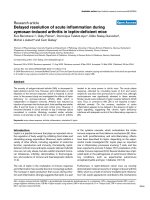
Figure 1
Knee joint
99m
Tc-pertechnetate uptake during zymosan-induced arthritisKnee joint
99m
Tc-pertechnetate (Tc) uptake during zymosan-induced
arthritis (ZIA). Tc uptake was assessed at different time points after
injection of zymosan A in (a) ob/ob (black symbols) and +/? (white
symbols) mice (6 hours, 24 hours, day 3: n = 9–10 per group; day 7: n
= 5 per group), and in (b) db/db (black symbols) and db/+ (white sym-
bols) mice (n = 6 per group). Results are expressed as the ratio of Tc
uptake in the inflamed to that in the control knee joints. A ratio greater
than 1.1 indicates inflammation. In panel (a) data shown for each time
point are means ± SEM in one representative experiment out of five; in
panel (b) data shown are means ± SEM in one experiment. Joint swell-
ing was significantly greater on days 3 and 7 in ob/ob as compared
with +/? mice, and in db/db as compared to db/+ mice (*P < 0.01).
(a)
(b)
0
0.5
1.0
1.5
2.0
2.5
time after Zy injection
6h 24h day 3 day 7
*
*
Tc ratio
Tc ratio
0
0.5
1.0
1.5
2.0
6h 24h
day 3
day 7
*
*
time after Zy injection
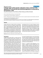
Figure 2
Histological changes in the knee joints of ob/ob and +/? mice during zymosan-induced arthritisHistological changes in the knee joints of ob/ob and +/? mice during
zymosan-induced arthritis (ZIA). (a) Histological changes were exam-
ined in zymosan A injected knee joints at an early (day 3) stage of ZIA.
Representative sections stained with haematoxylin–eosin are shown for
+/? (left panel; × 100) and ob/ob (right panel; × 100) mice. On day 3
we observed similar articular lesions in both +/? and ob/ob mice, which
exhibited a moderate inflammatory cell infiltration associated with dis-
crete synovial hyperplasia. (b) Histological changes were also exam-
ined in zymosan A injected knee joints at a late (day 21) stage of
arthritis. A representative section stained with haematoxylin–eosin is
shown for +/? mice in the left panel (× 100). On day 21 the joints of
+/? mice exhibited only little inflammation, a discrete reactive synovial
hyperplasia and a smooth cartilage surface. At this late stage some very
severe cases of arthritis, with pronounced inflammatory cell infiltration
and synovial hyperplasia, were observed exclusively in ob/ob mice, as
illustrated in the right panel (haematoxylin–eosin; × 100). In this exam-
ple, the articular cartilage has been largely destroyed and overgrown by
a hyperplasic and inflammatory synovium. (c) Histological sections
were scored for synovial hyperplasia, inflammatory cell infiltration and
cartilage destruction. Cumulative total scores are shown for ob/ob
(black columns) and +/? (white columns) mice at early (day 3: n = 9 per
group) and late (day 14: n = 9–10 per group; day 21: n = 5 per group)
time points after injection of zymosan A, representing histological
changes during the early and chronic phases of ZIA, respectively. Data
shown for each time point represent means ± SEM.
Available online />R260
the differences were not statistically significant (total histo-
logical severity scores: 5.58 ± 0.80 for db/+ mice [n = 6]
and 6.58 ± 1.11 for db/db mice [n = 6]). Leptin and OB-
Rb deficiency thus appeared to similarly affect the course
of ZIA.
Increased acute phase response in ob/ob and db/db
mice
The acute phase response was examined in ob/ob and
control mice after injection of zymosan A by measuring cir-
culating levels of IL-6 and of the acute phase protein SAA.
In the sera of naïve mice from both groups, IL-6 was unde-
tectable. Following intra-articular injection of zymosan A,
serum IL-6 increased within 6 hours in all animals but IL-6
levels were significantly higher in ob/ob mice than in con-
trols (Table 1). At 24 hours IL-6 levels were considerably
reduced in both groups, and the differences were no longer
significant. In db/db mice and their lean littermates a similar
transient increase in IL-6 was observed, but differences in
IL-6 levels between the two groups were not significant
(data not shown).
Circulating levels of SAA increased in all animals during the
first 3 days after intra-articular zymosan A injection (Table
1). Early after zymosan A injection the increase in circulat-
ing SAA was delayed in the ob/ob group as compared with
+/? mice, but later, on days 1 and 3, SAA levels were sig-
nificantly higher in ob/ob animals than in controls. Similar
results were obtained in db/db and db/+ mice (data not
shown).
Elevated corticosterone levels in leptin and leptin
receptor deficient mice
Corticosterone secretion is known to be elevated in all
forms of leptin deficiency and in leptin insensitivity [21,22].
During ZIA corticosterone levels increased transiently in
both +/? and ob/ob animals 6 hours after zymosan A injec-
tion; however, in the latter group they remained significantly
higher than in +/? controls throughout the experiment
(Table 1).
Cytokine mRNA expression in zymosan A injected joints
Expression of mRNAs encoding various cytokines was
investigated in knee joints of ob/ob and +/? animals on
days 1 and 14 after zymosan A injection. We observed
increased expression of MIF, IL-1α, IL-1β, IL-1 receptor
antagonist and IL-6 mRNA in zymosan A injected knees
(Fig. 3). Furthermore, low levels of IL-18 were also detected
on day 14 (data not shown). Expression levels of all of these
cytokines were significantly lower or undetectable in the
left, saline-injected knee joints in both groups (data not
shown). IL-12, IL-10 and interferon-γ were undetectable in
both zymosan A and saline injected joints. In contrast to the
observed delayed resolution of arthritis and acute phase
response in ob/ob animals, we could not detect any
differences in the local expression of cytokine mRNA in
knees injected with zymosan A between ob/ob animals and
+/? controls.
Discussion
The results of the present study indicate that resolution of
joint swelling in ZIA was delayed in leptin deficient mice.
Accordingly, the acute phase response, as assessed by cir-
culating levels of IL-6 and SAA, remained elevated for a
longer period of time in ob/ob mice than in control, lean lit-
termates. Furthermore, at late time points histological fea-
tures of arthritis tended to be more severe in ob/ob mice.
Similar results were obtained in db/db mice, suggesting
that the observed changes in the course of ZIA were medi-
ated by a lack of interaction of leptin with OB-Rb.
In contrast to these findings we previously observed a
milder form of AIA in ob/ob and db/db mice as compared
with their controls, with decreased synovial levels of IL-1β
and tumour necrosis factor (TNF)-α, and a switch toward
production of Th2 cytokines [14]. These contrasting
Table 1
Serum IL-6, serum amyloid A and corticosterone during zymosan-induced arthritis in ob/ob and +/? mice
Time IL-6 (pg/ml) SAA (µg/ml) Corticosterone (ng/ml)
+/?ob/ob +/?ob/ob +/?ob/ob
Baseline <39.0 <39.0 21.6 ± 2.6 25.3 ± 2.5 65.6 ± 15.0 140.6 ± 12.5
a
6 hours 518.0 ± 52.8 1034.1 ± 114.8
a
331.9 ± 14.8 191.8 ± 18.7
b
164.5 ± 15.9 216.3 ± 15.8
a
24 hours 85.7 ± 25.6 154.1 ± 59.2 456± 5 ± 23.4 810.6 ± 48.3
c
56.9 ± 8.5 149.4 ± 31.9
a
72 hours <39.0 <39.0 30.9 ± 4.6 387.5 ± 48.8
c
70.3 ± 8.4 120.6 ± 19.3
a
7 days ND ND 36.4 ± 2.2 184.8 ± 56.5
a
ND ND
14 days ND ND 131.8 ± 68.2 116.0 ± 30.1 ND ND
Data shown represent means ± SEM of five to six mice per group. The results are representative of five independent experiments. Differences
between groups were analyzed using one-way analysis of variance.
a
P < 0.05;
b
P < 0.0002;
c
P < 0.0001 for ob/ob versus +/? mice. ND, not
done; SAA, serum amyloid A.
Arthritis Research & Therapy Vol 6 No 3 Bernotiene et al.
R261
observations in AIA and ZIA further suggest a greater sen-
sitivity to agents stimulating the innate immune responses
in leptin or leptin signalling deficient animals, as opposed to
attenuated inflammation in models involving T-cell
responses, in particular Th1-mediated diseases [4].
Indeed, both T and B lymphocytes participate in the mech-
anisms that lead to articular inflammation in AIA, whereas
ZIA exclusively involves the innate immune response.
Leptin was previously reported to play an important role in
T-cell-mediated immune responses. Evidence of defective
cell-mediated immunity and lymphoid atrophy, analogous to
those observed in chronic under-nutrition in humans, are
detected in ob/ob and db/db mice [23-25]. Leptin
stimulates the proliferation of CD4
+
T cells and promotes
Th1 responses [6]. Congenital leptin deficiency in humans
is associated with a decreased number of circulating CD4
+
T cells, impaired T-cell proliferation and cytokine release, all
of which could be reversed by the administration of recom-
binant leptin [26]. In addition, the OB-Rb receptor is also
expressed on B cells and may participate in the develop-
ment of humoral responses [14]. Consistent with these
findings, leptin deficient mice are protected from inflamma-
tion mediated by T and B cells in different disease models,
including AIA, experimental autoimmune encephalomyelitis,
type 1 diabetes and experimental colitis [11,12,14,27].
Our results in ZIA suggest that chronic leptin deficiency
interferes with adequate control of the inflammatory reac-
tion. Protective effects of leptin were previously observed in
studies of other experimental models conducted to explore
innate immune responses. Ob/ob mice are significantly
more susceptible to lipopolysaccharide (LPS)-induced
death, and this feature can partly be reversed by
administration of leptin [28]. OB receptor deficient fa/fa
rats also exhibit enhanced LPS-induced hepatotoxicity
[29]. Similarly, ob/ob and db/db mice are more likely to
succumb after administration of TNF-α. The protective role
of leptin against TNF-α induced toxicity was further sup-
ported by the deleterious effect of neutralizing anti-leptin
antibodies administered to TNF-α injected mice [30]. The
mechanisms underlying these protective effects of leptin
are still unclear. Although thymic and circulating lym-
phocytes are reduced, a fourfold increase in the number of
circulating monocytes was observed in leptin deficient
mice, suggesting enhanced responses to monocyte activa-
tors [7]. Furthermore, an imbalance between proinflamma-
tory and anti-inflammatory monokines has been observed in
ob/ob mice injected with LPS, with plasma levels of the
anti-inflammatory cytokines IL-10 and IL-1 receptor antago-
nist being lower in leptin deficient than in normal mice [28].
However, LPS or TNF-α mediated systemic inflammation is
a complex syndrome, and susceptibility to these systemic
stimuli might also be influenced by the effects of leptin on
nervous, endocrine, or other responses, independent of the
production of inflammatory mediators.
Intravenous injection of Staphylococcus aureus results in a
severe form of septic arthritis in mice, which is associated
with decreased circulating levels of leptin. In this model,
treatment with leptin significantly decreased the severity of
septic arthritis without interfering with staphylococcal load
in the joints [31]. The levels of IL-6 were significantly lower
in mice with septic arthritis after administration of leptin
[31]. Consistent with these findings, our results in ZIA indi-
cate that IL-6 levels were higher in ob/ob mice than in con-
trols. IL-6 plays an important role in turning acute
inflammation into a chronic synovitis, as demonstrated by
limited duration of ZIA in IL-6 deficient mice [32]. In addi-
tion, IL-6 has also been shown to play a major role in other
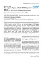
Figure 3
Cytokine mRNA expression in zymosan A injected knee joints of ob/ob and +/? miceCytokine mRNA expression in zymosan A injected knee joints of ob/ob
and +/? mice. Cytokine mRNA expression during acute and chronic
phases of zymosan-induced arthritis was investigated on total RNA iso-
lated from the knee joints of five ob/ob (black columns) and five +/?
(white columns) mice on (a) day 1 and (b) day 14 after injection of
zymosan A. Cytokine mRNA expression levels were assessed by
RNase protection assay (see Materials and methods section), quanti-
fied by phosphor-imaging and normalized for glyceraldehyde-3-phos-
phate dehydrogenase (GAPDH) expression. Data are expressed as a
ratio of cytokine to GAPDH mRNA expression and represent means ±
SEM (n = 5) of values obtained for detectable cytokines in zymosan A
injected knees. Cytokine expression in saline injected control knees
was low or undetectable in all animals.
(a)
(b)
Available online />R262
models of arthritis [33,34]. Thus, control of IL-6 production
may be one of the mechanisms by which leptin is involved
in the control of the inflammatory response during ZIA.
It is noteworthy that anaesthesia, skin cut and intra-articular
injection with saline slightly enhanced serum levels of IL-6
and SAA in ob/ob and +/? mice, although to a lesser extent
than in animals infected with zymosan A. Interestingly, this
small, zymosan A independent inflammatory response was
also greater in ob/ob mice than in lean controls, further sup-
porting the presence of an inappropriate control of inflam-
matory responses in leptin deficiency.
Corticosterone secretion is known to be elevated in all
forms of leptin deficiency and in leptin insensitivity [21,22].
However, despite the presence of elevated levels of gluco-
corticoids, ob/ob mice still exhibited a more pronounced
acute phase response and longer lasting arthritis than did
controls. Thus, it is conceivable that leptin deficiency could
result in an even more severe form of arthritis in the
absence of hypercorticosteronaemia.
The mRNA levels of different cytokines were determined in
arthritic and control joints at two time points. Consistent
with a previous report [17], the levels of IL-1α, IL-1β and IL-
6 mRNA were increased during ZIA. In addition, we also
detected elevated levels of IL-1 receptor antagonist and
MIF, which to the best of our knowledge have not previ-
ously been reported in the joint during ZIA. MIF is a broad-
spectrum proinflammatory cytokine that is implicated both
in animal models of immune-mediated arthritis and in
human rheumatoid arthritis [35]. The levels of mRNAs
encoding these different cytokines, including IL-6, were not
different between ob/ob mice and their lean controls. How-
ever, we cannot exclude variations in post-transcriptional
regulation, which might still result in different levels of
active proteins at the site of inflammation.
Conclusion
Our results indicate that resolution of ZIA is delayed in lep-
tin and leptin receptor deficient animals. Like in other exper-
imental models involving the innate immune response,
leptin deficiency thus appears to cause inadequate control
of the inflammatory response during ZIA, leading to
increased joint swelling, acute phase response and
delayed resolution of acute articular inflammation.
Competing interests
None declared.
Acknowledgements
We thank Nathalie Busso and Véronique Chobaz-Peclat for precious
advice and discussions. We are very grateful to Joan Stalder and Ber-
nard Folliat for their expert technical assistance. EB was the recipient of
an exchange scholarship from the Swiss Government. This work was
supported by the Swiss National Science Foundation (grants 3200-
054955.98 and 3231-05454.98 to CG).
References
1. Ahima RS, Flier JS: Leptin. Annu Rev Physiol 2000, 62:413-437.
2. Trayhurn P: Thermoregulation in the diabetic-obese (db/db)
mouse. The role of non-shivering thermogenesis in energy
balance. Pflugers Arch 1979, 380:227-232.
3. Mantzoros CS: Role of leptin in reproduction. Ann N Y Acad Sci
2000, 900:174-183.
4. Fantuzzi G, Faggioni R: Leptin in the regulation of immunity,
inflammation, and hematopoiesis. J Leukoc Biol 2000,
68:437-446.
5. Faggioni R, Feingold KR, Grunfeld C: Leptin regulation of the
immune response and the immunodeficiency of malnutrition.
FASEB J 2001, 15:2565-2571.
6. Lord GM, Matarese G, Howard JK, Baker RJ, Bloom SR, Lechler
RI: Leptin modulates the T-cell immune response and
reverses starvation-induced immunosuppression. Nature
1998, 394:897-901.
7. Faggioni R, Jones-Carson J, Reed DA, Dinarello CA, Feingold KR,
Grunfeld C, Fantuzzi G: Leptin-deficient (ob/ob) mice are pro-
tected from T cell-mediated hepatotoxicity: role of tumor
necrosis factor alpha and IL-18. Proc Natl Acad Sci USA 2000,
97:2367-2372.
8. Martin-Romero C, Santos-Alvarez J, Goberna R, Sanchez-Margalet
V: Human leptin enhances activation and proliferation of
human circulating T lymphocytes. Cell Immunol 2000,
199:15-24.
9. Lord GM, Matarese G, Howard JK, Bloom SR, Lechler RI: Leptin
inhibits the anti-CD3-driven proliferation of peripheral blood T
cells but enhances the production of proinflammatory
cytokines. J Leukoc Biol 2002, 72:330-338.
10. Matarese G, Sanna V, Di Giacomo A, Lord GM, Howard JK, Bloom
SR, Lechler RI, Fontana S, Zappacosta S: Leptin potentiates
experimental autoimmune encephalomyelitis in SJL female
mice and confers susceptibility to males. Eur J Immunol 2001,
31:1324-1332.
11. Matarese G, Di Giacomo A, Sanna V, Lord GM, Howard JK, Di
Tuoro A, Bloom SR, Lechler RI, Zappacosta S, Fontana S:
Requirement for leptin in the induction and progression of
autoimmune encephalomyelitis. J Immunol 2001,
166:5909-5916.
12. Matarese G, Sanna V, Lechler RI, Sarvetnick N, Fontana S, Zappa-
costa S, La Cava A: Leptin accelerates autoimmune diabetes in
female NOD mice. Diabetes 2002, 51:1356-1361.
13. Sanna V, Di Giacomo A, La Cava A, Lechler RI, Fontana S, Zappa-
costa S, Matarese G: Leptin surge precedes onset of autoim-
mune encephalomyelitis and correlates with development of
pathogenic T cell responses. J Clin Invest 2003, 111:241-250.
14. Busso N, So A, Chobaz-Peclat V, Morard C, Martinez-Soria E, Tal-
abot-Ayer D, Gabay C: Leptin signaling deficiency impairs
humoral and cellular immune responses and attenuates
experimental arthritis. J Immunol 2002, 168:875-882.
15. Keystone EC, Schorlemmer HU, Pope C, Allison AC: Zymosan-
induced arthritis: a model of chronic proliferative arthritis fol-
lowing activation of the alternative pathway of complement.
Arthritis Rheum 1977, 20:1396-1401.
16. Takeuchi O, Akira S: Toll-like receptors; their physiological role
and signal transduction system. Int Immunopharmacol 2001,
1:625-635.
17. van de Loo FA, Kuiper S, van Enckevort FH, Arntz OJ, van den Berg
WB: Interleukin-6 reduces cartilage destruction during exper-
imental arthritis. A study in interleukin-6-deficient mice. Am J
Pathol 1997, 151:177-191.
18. Palmer G, Talabot-Ayer D, Szalay-Quinodoz I, Maret M, Arend WP,
Gabay C: Mice transgenic for intracellular interleukin-1 recep-
tor antagonist type 1 are protected from collagen-induced
arthritis. Eur J immunol 2003, 33:434-440.
19. Sipe JD, Gonnerman WA, Loose LD, Knapschaefer G, Xie WJ,
Franzblau C: Direct binding enzyme-linked immunosorbent
assay (ELISA) for serum amyloid A (SAA). J Immunol Methods
1989, 125:125-135.
20. Walker CD, Sizonenko PC, Aubert ML: Modulation of the neona-
tal pituitary and adrenocortical responses to stress by thyroid
Arthritis Research & Therapy Vol 6 No 3 Bernotiene et al.
R263
hormones in the rat: effects of hypothyroidism and
hyperthyroidism. Neuroendocrinology 1989, 50:265-273.
21. Coleman DL, Burkart DL: Plasma corticosterone concentrations
in diabetic (db) mice. Diabetologia 1977, 13:25-26.
22. Guillaume-Gentil C, Rohner-Jeanrenaud F, Abramo F, Bestetti GE,
Rossi GL, Jeanrenaud B: Abnormal regulation of the hypotha-
lamo-pituitary-adrenal axis in the genetically obese fa/fa rat.
Endocrinology 1990, 126:1873-1879.
23. Chandra RK: Cell-mediated immunity in genetically obese
C57BL/6J ob/ob) mice. Am J Clin Nutr 1980, 33:13-16.
24. Fernandes G, Handwerger BS, Yunis EJ, Brown DM: Immune
response in the mutant diabetic C57BL/Ks-dt+ mouse. Dis-
crepancies between in vitro and in vivo immunological assays.
J Clin Invest 1978, 61:243-250.
25. Howard JK, Lord GM, Matarese G, Vendetti S, Ghatei MA, Ritter
MA, Lechler RI, Bloom SR: Leptin protects mice from starvation-
induced lymphoid atrophy and increases thymic cellularity in
ob/ob mice. J Clin Invest 1999, 104:1051-1059.
26. Farooqi IS, Matarese G, Lord GM, Keogh JM, Lawrence E, Agwu
C, Sanna V, Jebb SA, Perna F, Fontana S, Lechler RI, DePaoli AM,
O'Rahilly S: Beneficial effects of leptin on obesity, T cell
hyporesponsiveness, and neuroendocrine/metabolic dys-
function of human congenital leptin deficiency. J Clin Invest
2002, 110:1093-1103.
27. Siegmund B, Lehr HA, Fantuzzi G: Leptin: A pivotal mediator of
intestinal inflammation in mice. Gastroenterology 2002,
122:2011-2025.
28. Faggioni R, Fantuzzi G, Gabay C, Moser A, Dinarello CA, Feingold
KR, Grunfeld C: Leptin deficiency enhances sensitivity to endo-
toxin-induced lethality. Am J Physiol 1999, 276:R136-142.
29. Yang SQ, Lin HZ, Lane MD, Clemens M, Diehl AM: Obesity
increases sensitivity to endotoxin liver injury: implications for
the pathogenesis of steatohepatitis. Proc Natl Acad Sci USA
1997, 94:2557-2562.
30. Takahashi N, Waelput W, Guisez Y: Leptin is an endogenous
protective protein against the toxicity exerted by tumor necro-
sis factor. J Exp Med 1999, 189:207-212.
31. Hultgren OH, Tarkowski A: Leptin in septic arthritis: decreased
levels during infection and amelioration of disease activity
upon its administration. Arthritis Res 2001, 3:389-394.
32. de Hooge AS, van De Loo FA, Arntz OJ, van Den Berg WB:
Involvement of IL-6, apart from its role in immunity, in mediat-
ing a chronic response during experimental arthritis. Am J
Pathol 2000, 157:2081-2091.
33. Ohshima S, Saeki Y, Mima T, Sasai M, Nishioka K, Nomura S, Kopf
M, Katada Y, Tanaka T, Suemura M, Kishimoto T: Interleukin 6
plays a key role in the development of antigen-induced
arthritis. Proc Natl Acad Sci USA 1998, 95:8222-8226.
34. Alonzi T, Fattori E, Lazzaro D, Costa P, Probert L, Kollias G, De
Benedetti F, Poli V, Ciliberto G: Interleukin 6 is required for the
development of collagen-induced arthritis. J Exp Med 1998,
187:461-468.
35. Morand EF, Leech M, Weedon H, Metz C, Bucala R, Smith MD:
Macrophage migration inhibitory factor in rheumatoid arthritis:
clinical correlations. Rheumatology (Oxford) 2002, 41:558-562.