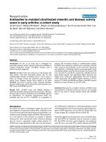
Báo cáo y học: "Strong inhibition of TNF-α production and inhibition of IL-8 and COX-2 mRNA expression in monocyte-derived macrophages by RWJ 67657, a p38 mitogen-activated protein kinase (MAPK) inhibitor" ppt
Bạn đang xem bản rút gọn của tài liệu. Xem và tải ngay bản đầy đủ của tài liệu tại đây (326.81 KB, 9 trang )
Open Access
Available online />R384
Vol 6 No 4
Research article
Strong inhibition of TNF-α production and inhibition of IL-8 and
COX-2 mRNA expression in monocyte-derived macrophages by
RWJ 67657, a p38 mitogen-activated protein kinase (MAPK)
inhibitor
Johanna Westra
1
, Berber Doornbos-van der Meer
1
, Peter de Boer
2
, Miek A van Leeuwen
1
,
Martin H van Rijswijk
1
and Pieter C Limburg
1,3
1
Department of Rheumatology, University Hospital Groningen, Groningen, The Netherlands
2
Pharmaceutical Research and Development, Johnson and Johnson, High Wycombe, UK
3
Department of Pathology and Laboratory Medicine, University Hospital Groningen, Groningen, The Netherlands
Corresponding author: Johanna Westra,
Received: 18 Mar 2004 Revisions requested: 8 Apr 2004 Revisions received: 19 Apr 2004 Accepted: 7 May 2004 Published: 21 Jun 2004
Arthritis Res Ther 2004, 6:R384-R392 (DOI 10.1186/ar1204)
http://arthr itis-research.com/conte nt/6/4/R384
© 2004 Westra et al.; licensee BioMed Central Ltd. This is an Open Access article: verbatim copying and redistribution of this article are permitted
in all media for any purpose, provided this notice is preserved along with the article's original URL.
Abstract
In inflammatory processes, the p38 mitogen-activated protein
kinase (MAPK) signal transduction route regulates production
and expression of cytokines and other inflammatory mediators.
Tumor necrosis factor α (TNF-α) is a pivotal cytokine in
rheumatoid arthritis and its production in macrophages is under
control of the p38 MAPK route. Inhibition of the p38 MAPK
route may inhibit production not only of TNF-α, but also of other
inflammatory mediators produced by macrophages, and
indirectly of inflammatory mediators by other cells induced by
TNF-α stimulation. Here we investigate the effects of RWJ
67657, a p38 MAPK inhibitor, on mRNA expression and protein
production of TNF-α and other inflammatory mediators, in
monocyte-derived macrophages. A strong inhibition of TNF-α
was seen at pharmacologically relevant concentrations of RWJ
67657, but also inhibition of mRNA expression of IL-1β, IL-8,
and cyclooxygenase-2 was shown. Furthermore, it was shown
that monocyte-derived macrophages have a high constitutive
production of matrix metalloproteinase 9, which is not affected
by p38 MAPK inhibition. The results presented here may have
important implications for the treatment of rheumatoid arthritis.
Keywords: COX-2, matrix metalloproteinase, monocyte-derived macrophage, p38 MAPK inhibitor, TNF-α
Introduction
Rheumatoid arthritis (RA) is characterized by chronic
inflammation of synovial tissue and destruction of cartilage
and bone in the joints [1]. Macrophages play an important
role in RA, as the rheumatoid synovium is intensively infil-
trated by macrophages and their numbers correlate with
clinical scores [2] and articular destruction in RA [3]. RA
patients with active disease display a faster generation of
CD14
+
myelomonocytic cells from the bone marrow and
faster differentiation into HLA-DR
+
cells than control indi-
viduals do [4]. Activation of the monocytic lineage in inflam-
matory disease is not restricted to synovial macrophages,
but extends to circulating monocytes and other cells of the
mononuclear phagocyte system [5]. The activation state of
monocytes/macrophages is characterized by increased
expression and transcription of interleukin (IL)-1β and
tumor necrosis factor α (TNF-α), but also of other proin-
flammatory and regulatory cytokines and growth factors [6].
Highly specific therapeutics have been developed to target
these cytokines, such as monoclonal antibodies, soluble
receptors, binding proteins, and receptor antagonists.
COX-2 = cyclooxygenase-2; DMSO = dimethyl sulfoxide; ELISA = enzyme-linked immunosorbent assay; ERK = extracellular signal-regulated kinase;
FCS = fetal calf serum; GAPDH = glyceraldehyde-3-phosphate dehydrogenase; IC
50
= median inhibitory concentration; IL = interleukin; LPS =
lipopolysaccharide; MAPK = mitogen-activated protein kinase; MAPKAPK-2 = MAPK-activating protein kinase-2; M-CSF = macrophage-colony-stim-
ulating factor; MDM = monocyte-derived macrophage; MMP = matrix metalloproteinase; NF-κB = nuclear factor κB; RA = rheumatoid arthritis; RPMI
= Roswell Park Memorial Institute [medium]; RT-PCR = reverse transcriptase polymerase chain reaction; SEM = standard error of the mean; TIMP =
tissue inhibitor of metalloproteinases; TNF = tumor necrosis factor.
Arthritis Research & Therapy Vol 6 No 4 Westra et al.
R385
TNF-α blockade has been the major breakthrough in the
therapy of RA during the past 10 years. However, more
than half of patients do not achieve adequate responses,
remissions are rare, and these drugs do have side effects
[7,8].
The importance of mitogen-activated protein kinases
(MAPKs) in cell biology has been reported in many studies
concerning different inflammatory diseases. These MAPKs
belong to three families: the extracellular signal-regulated
kinases (ERKs); the c-Jun N-terminal or stress-activated
protein kinases (JNK/SAPKs); and the p38 MAPKs. All
three families have been shown to become activated in
macrophages in response to a variety of stimuli both in pri-
mary cells and in cell lines [9]. In the RA synovium, p38
MAPK is predominantly activated in endothelial cells and in
the lining layer [10]. Inhibition of p38 MAPK therefore could
provide an interesting target for intervention in inflamma-
tion, as it occurs in the synovia in RA.
In vitro stimulation of macrophages with lipopolysaccharide
(LPS) leads to activation of MAPK cascades through the
LPS receptor (CD14) or Toll-like receptors [9]. The ability
of bacterial toxins or super-antigens to induce proinflamma-
tory responses leading to the production of TNF-α and IL-
1 is relevant in view of the possible microorganism etiology
in RA [11]. Stimulation of monocytes with LPS induces a
number of matrix metalloproteinases (MMPs), including two
prominent monocytic MMPs: interstitial collagenase
(MMP1) and gelatinase B (MMP9). These enzymes are
involved in the connective-tissue loss associated with
chronic inflammatory diseases. In vivo, a significant part of
macrophage effector responses occurs through cell-con-
tact-dependent signalling with several inflammatory cells,
mainly T cells and fibroblasts. A few soluble stimuli, such as
IL-15 and IL-17, are known to have a stimulatory effect on
macrophages. It has also been reported that IL-17 induces
the production of MMP9 and cyclooxygenase-2 (COX-2),
which is the rate-limiting enzyme in prostaglandin and leu-
kotriene synthesis in monocytes/macrophages [12].
The p38 MAPK inhibitor RWJ 67657 (4-[4-(4-fluorophe-
nyl)-1-(3-phenylpropyl)-5-(4-pyridinyl)-1H-imidazol-2-yl]-3-
butyn-1-ol) has been shown to inhibit the release of TNF-α
from LPS-treated human peripheral blood mononuclear
cells, with a median inhibitory concentration (IC
50
) of 3 nM
[13]. Moreover, this compound effectively inhibited endo-
toxin-induced clinical effects and cytokine release in normal
healthy volunteers [14]. Furthermore a report was pub-
lished in which pharmacokinetics and pharmacodynamics
of RWJ 67657 were presented, showing that the com-
pound has acceptable safety to warrant further investiga-
tion [15]. Our group recently showed that RWJ 67657
significantly inhibited IL-6, IL-8, MMP3, and COX-2 mRNA
expressed by IL-1β and/or TNF-α stimulated rheumatoid
synovial fibroblasts [16].
It has been shown that differences occur in signalling path-
ways in myeloid cells in relation to the maturation stage of
macrophages [17]. Strong inhibition of TNF-α in mono-
cytes has been reported due to p38 MAPK inhibition [13],
but the effects in matured macrophages are not fully
known. Therefore in this study we investigated the effects
of the p38 MAPK inhibitor RWJ 67657 on the mRNA
expression and production of inflammatory cytokines and
MMPs by stimulated monocyte-derived macrophages
(MDMs). We investigated macrophages, differentiated in
medium with pooled serum or by the method described by
Plesner and co-workers [18], and compared MDMs from
healthy controls and RA patients. Strong inhibition of
mRNA expression and production of TNF-α by RWJ 67657
was found, as well as inhibition of IL-1β, IL-8, and COX-2
mRNA expression.
Materials and methods
Reagents
RWJ 67657 was provided by Johnson and Johnson (RW
Johnson Pharmaceutical Research Institute, Raritan, NJ,
USA). Antibodies for flow cytometry were obtained from
IQProducts, Groningen, the Netherlands. LPS was pur-
chased from Sigma-Aldrich (Zwijndrecht, the Netherlands).
Recombinant human macrophage-colony-stimulating fac-
tor (M-CSF) and ELISA antibodies were from R&D Sys-
tems (Minneapolis, MN, USA). Fetal calf serum (FCS) and
RPMI 1640 culture medium were obtained from Biowhit-
taker (Verviers, Belgium). All reagents for RNA isolation and
reverse transcriptase reaction were purchased from Invitro-
gen, Life Technologies (Gaithersburg, MD, USA). Rea-
gents for real-time RT-PCR were obtained from Applied
Biosystems (Foster City, CA, USA). Specific antibodies to
p38 MAPK, phospho-p38 MAPK, and phospho-MAP-
KAPK-2 were purchased from Cell Signalling Technologies
(Beverly, MA, USA) and detecting antibody peroxidase-
swine-anti-rabbit was from DAKO (Glostrup, Denmark).
Macrophage culture
Blood was obtained from RA patients who had given their
informed consent and from healthy laboratory workers.
Peripheral mononuclear cells were isolated by Lymphoprep
density-gradient centrifugation from citrated blood. Cells
were suspended in RPMI with gentamicin at 10
6
cells/ml
and seeded in 6-well plates (Costar, Badhoevedorp, the
Netherlands) in 3 ml or 0.5 ml into 24-well plates and cul-
tured at 37°C in a 5% CO
2
atmosphere. After 2 hours, non-
adherent cells were discarded and adherent monocytes
were allowed to differentiate into macrophages in RPMI
containing gentamicin, 50 ng/ml M-CSF + 1% FCS [18];
medium was refreshed at day 2. To compare methods,
monocytes were also differentiated in RPMI with gen-
Available online />R386
tamicin + 2% pooled human serum; on days 2 and 5, fresh
medium was added to the wells. Cells were cultured for 5
or 7 days. We found no marked differences between
differentiation methods and decided to perform further
experiments with macrophages differentiated with M-CSF
and FCS.
Flow cytometric analysis
Expression of surface markers CD14 (LPS receptor), HLA-
DR, CD18 (β
2
integrin subunit), CD36 (GPIIIb, GPIV), and
CD83 (dendritic cell marker) on monocytes in the periph-
eral blood mononuclear cell fraction and on differentiated
macrophages was detected by flow cytometric analysis in
an Epics-Elite flow cytometer (Coulter Electronics,
Mijdrecht, the Netherlands).
Phosphorylation studies
Phosphorylation of p38 MAPK in MDMs was analyzed by
western blotting. Cells were cultured in RPMI containing
gentamicin, 50 ng/ml M-CSF + 1% FCS for 5 days and
stimulated with 50 ng/ml LPS for various periods of time.
Cell extracts were prepared by lysing the cells with 1 ×
SDS sample buffer (containing 2% SDS, 10% glycerol, 50
mM dithiothreitol, 62.5 mM Tris–HCl [pH 6.8], and 0.01%
bromphenol blue). Cells were scraped off the wells and the
lysates were subsequently sonicated for 5 to 10 seconds
and boiled for 5 minutes. After centrifugation, the samples
were loaded onto a 10% SDS–PAGE gel and resolved by
running at a constant 200 V and 15 W. Semidry blotting
was performed onto nitrocellulose membrane and immuno-
detection was with anti-phospho-p38 MAPK and peroxi-
dase-anti-rabbit. Enhanced chemiluminescence detection
was performed in accordance with the manufacturer's
guidelines (Lumi-Light
plus
, Roche Diagnostics, Mannheim,
Germany).
To determine the effect of RWJ 67657 on phosphorylation
of p38 MAPK and its downstream substrate MAPK-activat-
ing protein kinase-2 (MAPKAPK-2), a concentration range
of the p38 MAPK inhibitor (0, 0.01, 0.1, 1, and 10 µM) was
added 1 hour before stimulation with 50 ng/ml LPS for 30
minutes. Blotting experiments were performed with specific
antibodies to p38 MAPK, phospho-p38 MAPK, and
phospho-MAPKAPK-2.
Determination of TNF-α, IL-1β, IL-6, IL-8, MMP1, MMP9,
and TIMP-1 levels in cell culture supernatants
MDMs from eight healthy controls and nine RA patients,
who were not treated with steroids, were cultured in 24-
well plates in RPMI containing gentamicin, 50 ng/ml M-
CSF + 1% FCS for 5 days. The cells were subsequently
pretreated with increasing concentrations of RWJ 67657
(stock solution 10 mM in dimethyl sulfoxide [DMSO]), from
0.01 µM to 10 µM for 1 hour prior to stimulation with 50 ng/
ml LPS for 24 hours. Levels of cytokines (TNF-α, IL-1β, IL-
6, and IL-8), MMP1, MMP9, and tissue inhibitor of metallo-
proteinases (TIMP)-1 were measured in cell supernatants
by ELISA, using matched antibody pairs for ELISA and
recombinant proteins as standards from R&D Systems.
Detection limits for all cytokine ELISAs was 20 pg/ml. For
optimal determination of MMP1, MMP9, and TIMP-1, 96-
well plates (Greiner M129A) were precoated with F(ab)
2
fragments of goat-anti-mouse IgG-Fc (Jackson, West
Grove, PA, USA) in 0.1 M carbonate buffer (pH 9.6) for at
least 48 hours before coating of the capture antibody. After
sample incubation and binding of the biotinylated detection
antibodies, the color reaction was performed with strepta-
vidin-poly-horseradish peroxidase (Sanquin, Amsterdam,
The Netherlands) and tetramethylbenzidine (TMB; Roth,
Karlsruhe, Germany). The detection limit for MMP1 and
TIMP-1 was 1 ng/ml and for MMP9, 0.1 ng/ml.
RNA isolation and real-time RT-PCR
MDMs from healthy controls were cultured in 6-well plates
in RPMI containing gentamicin, 50 ng/ml M-CSF + 1%
FCS for 5 days. First, macrophages were stimulated with
50 ng/ml LPS for various periods of time to determine opti-
mal mRNA expression of TNF-α, IL-1β, IL-6, IL-8, MMP9,
and COX-2 genes. At the optimal time point, the effect of
p38 MAPK inhibition on LPS-stimulated MDMs was deter-
mined by pretreatment of the cells with increasing concen-
trations of RWJ 67657 for 1 hour. Total RNA was isolated
from the cells with TRIZOL reagent in accordance with the
manufacturer's instructions (Life Technologies). After
DNAse treatment (Ambion DNA-free, Austin, TX, USA)
cDNA was synthesized from 1.0 µg of total RNA using M-
MLV Reverse Transcriptase and oligo (dT)
24
(Life
Technologies).
For detection of mRNA expression, a fluorescence-based
real-time RT-PCR was performed, which allows relative
quantification of steady-state mRNA. The amount of emit-
ted fluorescence is proportional to the amount of PCR
product and enables the monitoring of the PCR reaction.
For the measurement of IL-1β, TNF-α, IL-6, IL-8, MMP9,
COX-2, and glyceraldehyde-3-phosphate dehydrogenase
(GAPDH), 1 µl of cDNA in triplicate was used for amplifica-
tion by the real-time quantitative PCR system (ABI Prism
7900HT Sequence Detection System, Applied Biosys-
tems) with specific Taqman primers/probes. The Assay-on-
Demand numbers for the genes were as follows: IL-1β,
Hs00174097_m1; TNF-α, Hs00174128_m1; IL-6,
Hs00174131_m1; IL-8, Hs00174103_m1; MMP9,
Hs00234579_m1; COX-2, Hs00153133_m1 and
GAPDH, Hs99999905_m1.
The amount of target, normalized to an endogenous refer-
ence (GAPDH) and relative to a control sample, is given by
2
-∆∆CT
, in which C
T
is the threshold cycle. The results are
expressed as fold induction relative to untreated samples.
Arthritis Research & Therapy Vol 6 No 4 Westra et al.
R387
Statistics
Paired t-tests were performed using GraphPad Prism ver-
sion 3.00 for Windows, GraphPad Software (San Diego,
CA, USA).
Results
Macrophage differentiation
Expression of surface markers on monocytes and macro-
phages was measured by flow cytometry. In Fig. 1, the
mean fluorescence intensity (MFI) of CD14 and HLA-DR
on monocytes (left panel) and macrophages differentiated
in 50 ng/ml M-CSF and 1% FCS after 5 days (right panel)
is shown, versus the number of cells measured in the flow
cytometer. Macrophages differentiated in this way showed
lower expression of CD14 than monocytes, relative to the
isotype control, whereas the HLA-DR expression did not
differ between the two cell types. Expression of CD18
increased with differentiation, while expression of CD36
decreased (data not shown). Expression of CD83 was not
seen on matured macrophages, excluding differentiation
into dendritic cells. For further experiments, monocytes dif-
ferentiated in medium with 50 ng/ml M-CSF and 1% FCS
for 5 days were used.
Effect of RWJ 67657 on phosphorylation of p38 MAPK
and MAPKAPK-2
In Fig. 2a, a representative example is shown of phosphor-
ylation of p38 MAPK in MDMs after stimulation with 50 ng/
ml LPS. Phosphorylation occurs after 15 minutes, is maxi-
mal at 30–60 minutes, and diminishes after 2 hours. p38
MAPK is constitutively expressed in the cells, as is demon-
strated in the control blot. The effect of RWJ 67657 on
phosphorylation of p38 MAPK and its direct downstream
substrate, MAPKAPK-2, measured after 30 minutes of
stimulation with 50 ng/ml LPS, is shown in Fig. 2b. RWJ
67657 does not inhibit phosphorylation of p38 MAPK but
does inhibit its activity, as can be seen from the strong inhi-
bition at 0.01 µM and the complete inhibition of MAPKAPK-
2 phosphorylation at 0.1 µM RWJ 67657. The solvent,
0.1% DMSO, did not affect phosphorylation of either
kinase.
Effect of RWJ 67657 on cytokine and MMP production
Stimulation of MDMs with 50 ng/ml LPS for 24 hours
resulted in increased production of TNF-α, IL-6, IL-8, and
MMP9 both in control macrophages and in RA macro-
phages (Fig. 3a). Production of IL-1β, MMP1, and TIMP-1
was too low for detection. Before stimulation, TNF-α pro-
duction was below the detection limit, while after stimula-
Figure 1
Expression of CD14 (bold grey line) and HLA-DR (bold black line) in monocytes (left panel) and macrophages, differentiated with 50 ng/ml macro-phage-colony-stimulating factor and 1% FCS after 5 days (right panel), measured by flow cytometry and expressed in mean fluorescence intensity (MFI)Expression of CD14 (bold grey line) and HLA-DR (bold black line) in monocytes (left panel) and macrophages, differentiated with 50 ng/ml macro-
phage-colony-stimulating factor and 1% FCS after 5 days (right panel), measured by flow cytometry and expressed in mean fluorescence intensity
(MFI). The isotype control is an IgG2a antibody (dashed line); the blank is in solid fill. The MFI is shown on the x-axis, while on the y-axis the number
of cells measured in the flow cytometer is expressed. PE = phycoerythrin.
Figure 2
Effect of RWJ 67657 on phosphorylation of p38 MAPK and MAPKAPK-2Effect of RWJ 67657 on phosphorylation of p38 MAPK and MAP-
KAPK-2. (a) Representative presentation of phosphorylation of p38
mitogen-activated protein kinase (MAPK) in monocyte-derived macro-
phages after stimulation with lipopolysaccharide (LPS) at various time
points. Phosphorylation was measured by western blotting using spe-
cific antibodies to p38 MAPK and phospho-p38 MAPK. (b) Effect of
RWJ 67657 on phosphorylation of the direct substrate of p38 MAPK,
MAPKAPK-2 (MAPK-activating protein kinase-2), measured after 30
minutes of stimulation. DMSO = dimethyl sulfoxide.
Available online />R388
tion, the mean production in control MDMs was 2.204 (±
1.993) ng/ml and 2.150 (± 1.816) ng/ml in RA MDMs.
Pretreatment of the macrophages with increasing concen-
trations of RWJ 67657 showed a dose-dependent
decrease of protein production for TNF-α and IL-8. Inhibi-
tion of TNF-α production was seen, with an IC
50
of 0.015
µM for control cells and 0.03 µM for RA cells (Fig. 3b). For
IL-8 production, the IC
50
was 0.3 µM for control cells and
1.2 µM for RA cells.
IL-6 and MMP9 production was inhibited only at concentra-
tions between 1 and 10 µM RWJ 67657. Pretreatment of
MDMs with 0.1% DMSO had no significant effect on pro-
tein production (data not shown).
Effects of RWJ 67657 on mRNA expression
To determine optimal stimulation time points for the genes
involved in this study, MDMs of two healthy control
subjects were stimulated for 0.5, 2, 4, 8, 12, and 24 hours
with 50 ng/ml LPS. mRNA expression of TNF-α, IL-1β, IL-
Figure 3
Protein production of tumor necrosis factor (TNF)-α, IL-6, IL-8, and matrix metalloproteinase 9 (MMP9) by monocyte-derived macrophages from healthy controls (n = 8, open squares) and rheumatoid arthritis patients (n = 9, filled squares)Protein production of tumor necrosis factor (TNF)-α, IL-6, IL-8, and matrix metalloproteinase 9 (MMP9) by monocyte-derived macrophages from
healthy controls (n = 8, open squares) and rheumatoid arthritis patients (n = 9, filled squares). Cells were stimulated with LPS for 24 hours and pre-
treated 1 hour beforehand with RWJ 67657 at various concentrations. Protein production was measured in supernatants by ELISA and is expressed
in ng/ml (a). Inhibition was calculated against the stimulated control (b). Bars show mean and SEM. *P < 0.05; **P < 0.001, paired t-test, calculated
versus the stimulated control. unst, unstimulated.
Arthritis Research & Therapy Vol 6 No 4 Westra et al.
R389
6, IL-8, and COX-2 was measured with real-time RT-PCR,
as depicted in Fig. 4. A significant increase in mRNA
expression of all but MMP9 was found after stimulation with
LPS. After 4 and 8 hours of stimulation, most genes were
highly expressed. IL-8 mRNA expression was still increased
after 12 and 24 hours. For measurement of the effects of
the p38 MAPK inhibitor, MDMs were stimulated for 4
hours.
As can be seen in Fig. 5, there is a dose-dependent
decrease in mRNA expression with increasing RWJ 67657
concentration. TNF-α mRNA expression is inhibited by
48.4% at 0.01 µM and 65.8% at 0.1 µM p38 MAPK inhib-
itor. IL-1β, IL-8, and COX-2 mRNA expression was reduced
by 40.2%, 56.6%, and 65.0%, respectively, at 1 µM.
MMP9 mRNA expression is not induced and not inhibited
and proved to be constitutively expressed at a high level.
Control incubations with 0.1% DMSO had no significant
effect on mRNA expression, as can been seen in Fig. 5.
Discussion
In this study, we have shown the significant inhibition of
TNF-α production in MDMs by the p38 MAPK inhibitor
RWJ 67657. The strong inhibition was seen at the level of
both mRNA expression and protein production, so inhibi-
tion is already apparent at the level of transcription.
p38 MAPK activity in RA is found predominantly in the syn-
ovial lining layer and in endothelial cells in the synovium
[10]. The lining layer consists mainly of fibroblasts and
macrophages, both important players in the process of
inflammation by the production of cytokines and degrading
enzymes. Recently our group demonstrated strong inhibi-
tion of IL-6, IL-8, COX-2, and MMP3 expression in rheuma-
toid synovial fibroblasts by the p38 MAPK inhibitor RWJ
67657 [16]. Studying macrophages in vitro raises some
difficulties, because isolation and culture of macrophages
from synovial tissue is disturbed by the overgrowth of
fibroblasts. Therefore, macrophages differentiated from
peripheral blood monocytes are widely used for in vitro
studies. By using M-CSF and low FCS concentrations, we
generated macrophages with high HLA-DR expression that
were not activated [18].
Current treatment strategies in RA, including TNF-α- and
IL-1-blocking agents, alone or in combination with, for
example, methotrexate, still have limited efficacy in a sub-
stantial proportion of patients. Recently, Redlich and co-
workers reviewed the multiple pathogenesis pathways
involved in RA, and the possible targets for therapies, and
stressed the importance of aiming at interference both with
the pathways leading to inflammation and with those ulti-
mately leading to destruction [8]. Inhibition of signal trans-
duction cascades may fit in this concept, because they are
involved in the activation of proinflammatory cytokines as
well as of MMP genes.
The p38 MAPK inhibitor RWJ 67657 has been reported to
be specific for p38 α and β, and has no activity for other
kinases [13]. The first study investigating the pharmacoki-
netics and pharmacodynamics of RWJ 67657 to be done
in humans showed that a single oral intake of 0.25 ranging
up to 30 mg/kg resulted in plasma levels of 0.01 µM to 6
µM [15]. Furthermore, the study showed that at the doses
tested there were no significant adverse effects.
p38 MAPK acts mainly through phosphorylation of its
downstream substrate, MAPKAPK-2, and a variety of tran-
scription factors [19]. Mice that lack MAPKAPK-2 show
increased stress resistance and survive LPS-induced
endotoxic shock, due to a 90% reduction in the production
of TNF-α [20]. The level and stability of TNF-α mRNA was
not reduced in these mice, so the inhibition was at the post-
transcriptional level. With western blotting, we demon-
strated complete inhibition of phosphorylation of
MAPKAPK-2 at 0.1 µM RWJ 67657, and strong inhibition
at 0.01 µM. In our study, TNF-α production was signifi-
cantly inhibited at nanomolar concentrations of RWJ
67657, and mRNA expression was also decreased, by
nearly 50% at 10 nM. Inhibition of p38 MAPK activity leads
to reduced TNF-α mRNA expression and therefore
reduced TNF-α production, while inhibition of MAPKAPK-
Figure 4
Time course of induction of mRNA expression of tumor necrosis factor (TNF)-α, IL-1β, IL-6, IL-8, and cyclooxygenase 2 (COX-2)Time course of induction of mRNA expression of tumor necrosis factor
(TNF)-α, IL-1β, IL-6, IL-8, and cyclooxygenase 2 (COX-2). Monocyte-
derived macrophages from healthy controls (n = 2) were stimulated for
increasing periods of time with lipopolysaccharide (50 ng/ml). mRNA
expression was determined with real-time RT-PCR and results were
calculated as fold induction in comparison with unstimulated cells (fold
induction = 1).
Available online />R390
2, as in MAPKAPK-2 knockout mice, leads to reduced TNF-
α production without affecting the mRNA levels.
IL-6 production and mRNA expression were not inhibited
by p38 MAPK inhibition, but IL-8 production and mRNA
expression were inhibited by more than 50% at 1 µM RWJ
67657. Bhattacharyya and co-workers showed that LPS
from Helicobacter pylori stimulates IL-8 release from cells
of the monocytic lineage through activation of NF-κB and
MAPK cascades [21], and we demonstrated that in MDMs
the p38 MAPK route plays an important role in this process.
In 1992, Herzyk and co-workers published findings show-
ing that macrophage and monocyte IL-1β regulation differs
at multiple sites [22]. Macrophages did not differ from
monocytes in LPS sensitivity but had limitations in IL-1β
release. Our study shows mRNA expression of IL-1β in
MDMs, as well as a reduction of mRNA expression at 1 µM
RWJ 67657, but protein production in MDMs was below
the detection limit of the measurement.
We found a reduction of 65% in COX-2 mRNA expression
after treatment with 1 µM RWJ 67657. It was previously
demonstrated that p38 MAPK plays a role in transcription
and stabilization of COX-2 mRNA [23]. However, Caivano
and Cohen showed that both p38 MAPK and ERK influ-
ence COX-2 mRNA expression through activation of
mitogen- and stress-activated protein kinase 1 (MSK-1)
[24], an activator of important transcription factors such as
activating transcription factor (ATF)-2 and cyclic AMP
response element binding protein (CREB). Our results
indicate an important role for p38 MAPK in COX-2 mRNA
expression in MDMs.
In synovial tissue, the presence of macrophages is often
seen together with the expression of MMPs and TIMP-1
Figure 5
mRNA expression of tumor necrosis factor (TNF)-α, IL-1β, cyclooxygenase 2 (COX-2), IL-6, IL-8, and matrix metalloproteinase 9 (MMP9) in mono-cyte-derived macrophages from healthy controls (n = 5)mRNA expression of tumor necrosis factor (TNF)-α, IL-1β, cyclooxygenase 2 (COX-2), IL-6, IL-8, and matrix metalloproteinase 9 (MMP9) in mono-
cyte-derived macrophages from healthy controls (n = 5). Cells were stimulated with lipopolysaccharide for 4 hours and pretreated with a RWJ
67657 at various concentrations. mRNA expression was determined with real-time RT-PCR and results are expressed as fold induction in compari-
son with unstimulated cells (fold induction = 1). Bars show means and SEM (*P < 0.05, paired t-test, calculated against the stimulated control).
Arthritis Research & Therapy Vol 6 No 4 Westra et al.
R391
[25]. In our study, MMP1 and TIMP-1 could not be
detected in the cell supernatants of MDMs after LPS stim-
ulation, in contrast to high production of MMP9. MMP9 is
associated with macrophages and peripheral blood mono-
nuclear cells and has a broad substrate specificity and may
contribute together with collagenases to the degradation of
fibrillar collagens, basement membrane components, and
stromal extracellular matrix molecules [26]. Stimulation with
LPS induced a twofold induction of MMP9 levels, which
was not inhibited by pretreatment with RWJ 67657 at low
concentrations. Also, mRNA expression of MMP9 could
not be induced by LPS stimulation, and no inhibition by p38
MAPK inhibitor was observed. This latter finding is in
accordance with the study by Lai and co-workers showing
that LPS induction of MMP9 in monocytes is mainly regu-
lated by the ERK1/2 pathway and not the p38 MAPK path-
way [27].
p38 MAPK inhibitors have effects on various cell types,
thereby possibly enhancing the therapeutic effects but also
increasing the risk of side effects. One of the reasons for
undesirable effects might be the cross-reactivity against
other kinases, which is not the case for RWJ 67657 [13].
The preliminary pharmacokinetic data suggest a twice-daily
dosing regimen [15], while our data show significant
effects at low concentrations.
Conclusion
A significant inhibition of TNF-α production and mRNA
expression in LPS-stimulated MDMs was observed after
pretreatment with RWJ 67657, a p38 MAPK inhibitor, at
pharmacologically relevant concentrations. Inhibition of
mRNA expression of IL-1β, IL-8, and COX-2 was also
detected. MMP9 was found to be constitutively produced
at high levels and not inhibited by RWJ 67657. The results
presented here could have important implications for the
treatment of RA, since the drug used in this study has
already proved to be safe in a study in humans [15]. More
research into the effects of p38 MAPK inhibition on other
cell types involved in inflammation should establish its
applicability as a drug in the near future.
Competing interests
Part of this study was financed by an unrestricted grant
from Johnson and Johnson Pharmaceutical Research and
Development, Raritan, NJ, USA. One of the authors (PdB)
is an employee of this company.
Acknowledgements
This work was supported by the Dutch Rheumatology Foundation and
Johnson and Johnson Pharmaceutical Research and Development, Rar-
itan, NJ, USA.
References
1. Choy EH, Panayi GS: Cytokine pathways and joint inflamma-
tion in rheumatoid arthritis. N Engl J Med 2001, 344:907-916.
2. Tak PP, Smeets TJ, Daha MR, Kluin PM, Meijers KA, Brand R,
Meinders AE, Breedveld FC: Analysis of the synovial cell infil-
trate in early rheumatoid synovial tissue in relation to local dis-
ease activity. Arthritis Rheum 1997, 40:217-225.
3. Mulherin D, Fitzgerald O, Bresnihan B: Synovial tissue macro-
phage populations and articular damage in rheumatoid
arthritis. Arthritis Rheum 1996, 39:115-124.
4. Hirohata S, Yanagida T, Itoh K, Nakamura H, Yoshino S, Tomita T,
Ochi T: Accelerated generation of CD14+ monocyte-lineage
cells from the bone marrow of rheumatoid arthritis patients.
Arthritis Rheum 1996, 39:836-843.
5. Burmester GR, Stuhlmuller B, Keyszer G, Kinne RW: Mononu-
clear phagocytes and rheumatoid synovitis. Mastermind or
workhorse in arthritis? Arthritis Rheum 1997, 40:5-18.
6. Kinne RW, Brauer R, Stuhlmuller B, Palombo-Kinne E, Burmester
GR: Macrophages in rheumatoid arthritis. Arthritis Res 2000,
2:189-202.
7. Smolen JS, Steiner G: Therapeutic strategies for rheumatoid
arthritis. Nat Rev Drug Discov 2003, 2:473-488.
8. Redlich K, Schett G, Steiner G, Hayer S, Wagner EF, Smolen JS:
Rheumatoid arthritis therapy after tumor necrosis factor and
interleukin-1 blockade. Arthritis Rheum 2003, 48:3308-3319.
9. Rao KM: MAP kinase activation in macrophages. J Leukoc Biol
2001, 69:3-10.
10. Schett G, Tohidast-Akrad M, Smolen JS, Schmid BJ, Steiner CW,
Bitzan P, Zenz P, Redlich K, Xu Q, Steiner G: Activation, differen-
tial localization, and regulation of the stress-activated protein
kinases, extracellular signal-regulated kinase, c-JUN N-termi-
nal kinase, and p38 mitogen-activated protein kinase, in syno-
vial tissue and cells in rheumatoid arthritis. Arthritis Rheum
2000, 43:2501-2512.
11. Ebringer A, Wilson C: HLA molecules, bacteria and
autoimmunity. J Med Microbiol 2000, 49:305-311.
12. Jovanovic DV, Martel-Pelletier J, Di Battista JA, Mineau F, Jolicoeur
FC, Benderdour M, Pelletier JP: Stimulation of 92-kd gelatinase
(matrix metalloproteinase 9) production by interleukin-17 in
human monocyte/macrophages: a possible role in rheuma-
toid arthritis. Arthritis Rheum 2000, 43:1134-1144.
13. Wadsworth SA, Cavender DE, Beers SA, Lalan P, Schafer PH,
Malloy EA, Wu W, Fahmy B, Olini GC, Davis JE, Pellegrino-Gensey
JL, Wachter MP, Siekierka JJ: RWJ 67657, a potent, orally active
inhibitor of p38 mitogen-activated protein kinase. J Pharmacol
Exp Ther 1999, 291:680-687.
14. Fijen JW, Zijlstra JG, de Boer P, Spanjersberg R, Cohen Tervaert
JW, van der Werf TS, Ligtenberg JJ, Tulleken JE: Suppression of
the clinical and cytokine response to endotoxin by RWJ-67657,
a p38 mitogen-activated protein-kinase inhibitor, in healthy
human volunteers. Clin Exp Immunol 2001, 124:16-20.
15. Parasrampuria DA, de Boer P, Desai-Krieger D, Chow AT, Jones
CR: Single-dose pharmacokinetics and pharmacodynamics of
RWJ 67657, a specific p38 mitogen-activated protein kinase
inhibitor: a first-in-human study. J Clin Pharmacol 2003,
43:406-413.
16. Westra J, Limburg PC, de Boer P, van Rijswijk MH: Effects of RWJ
67657, a p38 mitogen activated protein kinase (MAPK) inhibi-
tor, on the production of inflammatory mediators by rheuma-
toid synovial fibroblasts. Ann Rheum Dis 2004 in press.
17. Lucas DM, Lokuta MA, McDowell MA, Doan JE, Paulnock DM:
Analysis of the IFN-gamma-signaling pathway in macro-
phages at different stages of maturation. J Immunol 1998,
160:4337-4342.
18. Plesner A, Greenbaum CJ, Lernmark A: Low serum conditions
for in vitro generation of human macrophages with macro-
phage colony stimulating factor. J Immunol Methods 2001,
249:53-61.
19. Kumar S, Boehm J, Lee JC: p38 MAP kinases: key signalling
molecules as therapeutic targets for inflammatory diseases.
Nat Rev Drug Discov 2003, 2:717-726.
20. Kotlyarov A, Neininger A, Schubert C, Eckert R, Birchmeier C, Volk
HD, Gaestel M: MAPKAP kinase 2 is essential for LPS-induced
TNF-alpha biosynthesis. Nat Cell Biol 1999, 1:94-97.
21. Bhattacharyya A, Pathak S, Datta S, Chattopadhyay S, Basu J,
Kundu M: Mitogen-activated protein kinases and nuclear fac-
tor-kappaB regulate Helicobacter pylori-mediated interleukin-
8 release from macrophages. Biochem J 2002, 368:121-129.
22. Herzyk DJ, Allen JN, Marsh CB, Wewers MD: Macrophage and
monocyte IL-1 beta regulation differs at multiple sites. Mes-
Available online />R392
senger RNA expression, translation, and post-translational
processing. J Immunol 1992, 149:3052-3058.
23. Dean JL, Brook M, Clark AR, Saklatvala J: p38 mitogen-activated
protein kinase regulates cyclooxygenase-2 mRNA stability
and transcription in lipopolysaccharide-treated human
monocytes. J Biol Chem 1999, 274:264-269.
24. Caivano M, Cohen P: Role of mitogen-activated protein kinase
cascades in mediating lipopolysaccharide-stimulated induc-
tion of cyclooxygenase-2 and IL-1 beta in RAW264
macrophages. J Immunol 2000, 164:3018-3025.
25. Smeets TJ, Barg EC, Kraan MC, Smith MD, Breedveld FC, Tak PP:
Analysis of the cell infiltrate and expression of proinflamma-
tory cytokines and matrix metalloproteinases in arthroscopic
synovial biopsies: comparison with synovial samples from
patients with end stage, destructive rheumatoid arthritis. Ann
Rheum Dis 2003, 62:635-638.
26. Murphy G, Knauper V, Atkinson S, Butler G, English W, Hutton M,
Stracke J, Clark I: Matrix metalloproteinases in arthritic disease.
Arthritis Res 2002, Suppl 3:39-49.
27. Lai WC, Zhou M, Shankavaram U, Peng G, Wahl LM: Differential
regulation of lipopolysaccharide-induced monocyte matrix
metalloproteinase MMP-1 and MMP-9 by p38 and extracellular
signal-regulated kinase 1/2 mitogen-activated protein
kinases. J Immunol 2003, 170:6244-6249.