báo cáo khoa học: "A male case of an undifferentiated carcinoma with osteoclast-like giant cells originating in an indeterminate mucin-producing cystic neoplasm of the pancreas. A case report and review of the literature" docx
Bạn đang xem bản rút gọn của tài liệu. Xem và tải ngay bản đầy đủ của tài liệu tại đây (4.18 MB, 6 trang )
CASE REP O R T Open Access
A male case of an undifferentiated carcinoma
with osteoclast-like giant cells originating in an
indeterminate mucin-producing cystic neoplasm
of the pancreas. A case report and review of the
literature
Takeyuki Wada
1
, Osamu Itano
1*
, Go Oshima
1
, Naokazu Chiba
1
, Hideki Ishikawa
1
, Yasumasa Koyama
1
, Wenlin Du
2
and Yuko Kitagawa
3
Abstract
We report a rare male case of an undifferentiated carcinoma with osteoclast-like giant cells originating in an
indeterminate mucin-producing cystic neoplasm of the pancreas. A 59-year-old Japanese man with diabetes visited
our hospital, complaining of fullness in the upper abdomen. A laboratory analysis revealed anemia (Hemoglobin;
9.7 g/dl) and elevated C-reactive protein (3.01 mg/dl). Carbohydrate antigen 19-9 was 274 U/ml and
Carcinoembryonic antigen was 29.6 ng/ml. A computed tomography scan of the abdomen revealed a 14-cm cystic
mass in the upper left quadrant of the abdomen that appeared to originate from the pancreatic tail. The patient
underwent distal pancreatectomy/splenectomy/total gastrectomy/cholecystectomy. The mass consisted of a
multilocular cystic lesion. Microscopically, the cyst was lined by cuboidal or columnar epithelium, including
mucinous epithelium. Sarcomatous mononuclear cells and multinucleated osteoclast-like giant cells were found in
the stroma. Ovarian-type stroma was not seen. We made a diagnosis of osteoclast-like giant cell tumor originating
in an indeterminate mucin-producing cystic neoplasm of the pancreas. All surgical margins were negativ e,
however, two peripancreatic lymph nodes were positive. The patient recovered uneventfully. Two months after the
operation, multiple metastases occurred in the liver. He died 4 months after the operation.
Keywords: undifferentiated carcinoma with osteoclast-like giant cells, Mucin-pr oducing, Mucinous, Cystic neoplasm,
Pancreas
Background
Undifferentiated carcinoma (UC) with osteoclast-like
giant cells (OGCs) is rare neoplasm of the pancreas.
The tumor was first described by Rosai in 1968 [1], an d
similar tumors also have been identified in the skin,
thyroidgland,ovary,breast,kidney, prostate, and soft
tissue. In the pancreas, it was mostly recorded in ductal
adenocarcinomas. Since Posen et al. reported the first
case of an UC with OGCs of the pancreas associated
with a mucus-secreting cystadenocarcinoma in 1981 [2] ,
there have been 11 additional cases of UC with OGCs
of the pancreas originating in mucinous cystic neo-
plasms (MCN) and indeterminate mucin-producing cys-
tic neoplasm reported in the English language literat ure
[2-12]. Among these cases, only one male ca se has been
reported [8]. In this report, we describe a new male case
of UC with OGCs that originated in an indeterminate
mucin-producing cystic neoplasm of the pancreas, and
discuss the clinicopathological features as well as pre-
sent a review of the pertinent literature.
Case report
A 59 year-old man p resented at our hospital with a
complaint of fullness in the upper abdomen. A physical
* Correspondence:
1
Department of Surgery, Eiju General Hospital 2-23-16 Higashiueno Taitouku
Tokyo 110-8645 Japan
Full list of author information is available at the end of the article
Wada et al. World Journal of Surgical Oncology 2011, 9:100
/>WORLD JOURNAL OF
SURGICAL ONCOLOGY
© 2011 Wada et al; lice nsee BioMed Central Ltd. This is an Open Access article distributed under the terms of the Crea tive Commons
Attribution Lic ense (http://creativecom mons .org/licenses/by/2.0), which permits unrestricted use, distribution, and reproduction in
any medium, provided the original work is prope rly cited.
examination showed a palpable mass in the upper left
abdomen. Labo ratory tests showed anemia and inflam-
matory reactivity, hemoglobin (Hgb) was 9.7 g/dl and C-
reactive protein (CRP) was 3.01 mg/dl. Carbohydrate
antigen 19-9 (CA19-9) was 274 U/ml and carcinoem-
bryonic antigen (CEA) was 29.6 ng/ml. A computed
tomography scan revealed a la rge cystic mass in the
upper left quadrant of the abdomen that appeared to
originate from the pancreatic tail (Figure 1). In magnetic
resonance images, the cystic component showed variable
signal intensities, and nodular components were seen in
the cystic wall. Magnetic resonance cholangio-pancrea-
tography showed narrowing and irregular ity of the main
pancreatic duct. Although i t was a male case, we con-
cluded tentatively that tumor might be a MCN of pan-
creas ba sed on its characteristic appearance resembling
the shape of an orange. An operation was performed. At
laparotomy, a large cystic mass was found in t he pan-
creas tail. The tumor invaded to the stomach, but dis-
tant metastasis was not discovered. The patient
underwent distal pancreatect omy with splenectomy,
total gastrectomy and cholecystectomy. Histological ana-
lysis revealed a multilocular cystic tumor that was 20
cm wide at its largest diameter and located in the cauda
pancreatis (Figure 2-A). The cystic cavities, which were
separated by thin, transparent septations, were filled
with fluid of a low viscosity (Figure 2-B). In some parts
the lining was dotted, occasionally p resenting papillary
projections. A 3-cm solid part was observed consisting
of yellow to brown material. The cystic spaces were
lined by a columnar mucinous epithelium that presented
with papillary folding (Figure 3-A). The epithelium pre-
sented severe dysplasia, reaching the degree of a carci-
noma in s itu. The walls of the cysts did not display an
ovarian-type stroma. There were a small number of
stromal invasive features in the bottom of the solid part
of this cystic tumor (Figure 3-B). Close to the carcinoma
in situ, the OGCs were distributed diffusely in the
stroma of the cyst wall, with more than 10 nuclei per
cell and lacking features of atypia. In Figure 2-B we pre-
sent views of the cut surface of the cystic tumor deli-
neating the pathological mapping of carcinoma in situ,
stromal invasion and gastric invasion. In the stroma of
the cyst wall, some pleomorphic large cells (PLCs) were
also observed. The PLC was a large cell with irregular,
pleomorphic or bizarre nuclei and frequently demon-
strating atypical mitosis (Figure 3-C). The tumor showed
invasion to the stomach across the serosal layer (Figure
3-D). The epithelium of the cyst wall showed mucus
production, as demonstrated by positive reactions with
Periodic acid-Schiff stain (PAS), alcian blue and Muc-2
(Figure 4-A, B, C). The papillary epithelium was positive
for the epithelial marker cytokeratin AE1/AE3, but the
stroma associated with OGCs and PLCs was negative
for cytokeratin AE1/AE3 (Figure 5-A). OGCs expressed
the histiocytic marker CD68 (Figure 5-B). Almost all of
the PLCs were positive for p53 (Figure 5-C) and nega-
tive for CD68. The Ki-67 positivity of the stroma asso-
ciated with OGCs and PLCs was about 30% (Fi gure 5-
D). This tumor was not diagnosed as a MCN, because it
did not display an ovarian-type stroma. However, it
seemed inappropriate to diagnose this tumor as an
intra-ductal papillary mucinous neoplasm (IPMN), con-
sidering invasive featu res to stroma and stomach and
lymph nodes metastases of this tumor. Therefore, we
diagnosed our case as an indeterminate mucin-produ-
cing cystic neoplasm, according to the international con-
sensus guidelines for management of intraductal
papillary mucinous neoplasms and mucinous cystic neo-
plasms of the pancreas, in whic h an ovarian-type stroma
is a histological requirement for the diagnosis of a MCN
[13]. Based on these findings, this case was diagnosed as
an UC with O GCs originating in an indeterminate
mucin-producing cystic neoplasm of the pancreas. The
patient recovered uneventfully and was d ischarged from
the hospital on the 23rd post-operative day. Multiple
liver metastases were detected 2 months after the opera-
tion, and the patient died 4 months after the operation.
Discussion
Since Posen et al. reported the first case of an UC
with OGCs of the pancreas associated with a mucus-
secreting cystadenocarcinoma in 1981 [2], there have
been 11 additional cases reported in the English lan-
guage literature of UC with OGCs of the pancreas ori-
ginating in MCN and indeterminate mucin-producing
cystic neoplasm [2-12]. Only one male case was
reported in addition to our case. We searched the
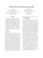
Figure 1 Abdominal CT showing the lar ge cystic tumor in the
upper left quadrant of the abdomen. A computed tomography
scan of the abdomen revealed a large cystic mass appeared to
originate from the pancreatic tail.
Wada et al. World Journal of Surgical Oncology 2011, 9:100
/>Page 2 of 6
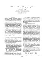
A
B
carcinoma in situ
stromal invasion
gastric invasion
Figure 2 Macroscopic findings showing a multilocular cystic tumor. (A) A multilocular cystic tumor that was 20 cm wide at its largest
diameter was located in the cauda pancreatis. (B) The cystic cavities, which were separated by thin, transparent septations, were filled with fluid
of low viscosity. The pathological mapping shows carcinoma in situ, stromal invasion and gastric invasion.
C
A
D
B
Figure 3 HE staining image of the tumor tissue. (A) The cystic spaces were lined by a columnar mucinous epithelium that presented
papillary folding. Higher power view of columnar mucinous epithelium is displayed on the bottom-right corner. (B) There was a small number
of stromal invasive features in the bottom of the solid part of this cystic tumor. (C) Near the carcinoma in situ, OGCs were distributed diffusely in
the stroma of the cyst wall. (D) The tumor showed the invasion to the stomach across the serosal layer.
Wada et al. World Journal of Surgical Oncology 2011, 9:100
/>Page 3 of 6
literature by the PubMed database. The characteristics
of our case and the previously reported cases are sum-
marized in Table 1.
The reports described 2 men and 10 women ranging
in age from 25 to 77 years with a median age of 47
years, suggesting that this type of tumor tends to
develop in middle age and predominantly in females.
That spectrum was compatible with that of o rdinary
MCN. Patients showed s ymptoms such as abdominal
pain or discomfort, anemia, and weight loss. The tumor
aros e from the head of the pancreas in 2, body in 1, tail
in 6, and body and tail in 3 patients. The lesions were
A
B
C
Figure 4 Histological findings showing mucus production of cyst wall. The epithelium of the cyst wall showed mucus production, as
demonstrated by the positive reactions with PAS, alcian blue and Muc-2.
Cytokeratin CD68
p53 MIB-1
A
B
CD
Figure 5 Immunohistochemical examination of OGC and PLC. (A) The stroma associated with OGCs and PLCs was negative for cytokerati n
AE1/AE3. (B) OGCs expressed the histiocytic marker CD68. (C) Almost all of the PLCs were positive for p53. (D) The Ki-67 positivity of the stroma
associated with OGCs and PLCs was about 30%.
Wada et al. World Journal of Surgical Oncology 2011, 9:100
/>Page 4 of 6
resected in all of the patients. T he average tumor size
was12.5cmatthelargestdiameter,rangingfrom5to
20 cm. Lymph node meta stasis was seen in two cases.
Invasion to another organ was seen only in our case, in
which the tumor invaded to the stomach. With the
exception of the two male cases, the patients had experi-
enced favorable courses of their disease and were alive
when papers were pub lished. An o varian-type stroma
was seen in 6 cases, and 5 cases did not mention it. Our
case did not display an ovarian-type stroma.
Although some authors have stated that UC with
OGCs of the pancreas is a pt to present as a large mass
with a slow metastatic spread and a much better prog-
nosis than ordinary carcinoma [14,15], the prognosis o f
UC with OGCs of the pancreas originating in a MCN
and indeterminate mucin-producing cy stic neoplasm
remains unclear due to the small number of reported
cases and short follow-up periods.
Zamboni et al. reported that 14% of MCNs of the
pancreas did not demonstrate an ovarian-type stroma
and that these tumors had a high tendency to invade
compared to the tumors with ovarian-type stroma [16].
Furthermore, some h ave suggested that MCN may lose
its ovarian-type stroma with malignant transformation
[17,18]. Our case did not display an ovarian-type
stroma, and demonstrated gastric invasion and lymph
nodes metastasis consisted of ductal adenocarcinoma
component. And, similar to our case, another male
casereportedbyNaietal.[8]alsodiedfromliver
metastasis 1 year after the operation. These authors
did not state whether or not an ovarian-ty pe stroma
was present. An UC with OGCs originating in an inde-
terminate mucin-producing cystic neoplasm of the
pancreas may also have a poor prognosis compared to
an UC with OGCs originating in a MCN with ovarian-
type stro ma.
UC with OGCs is a rare neoplasm of the pancreas.
In most cases, UCs with OGCs originate in ductal ade-
nocarcinoma, classified as a subtype of undifferentiated
carcinoma in the WHO classification [19], and are
only rarely combined with MCNs. Since the first
description of UC with OGCs by Rosai, the origin of
the tumor has been controversial. In our case, OGCs
were positive for the histiocytic marker CD68 and
negative for p53. On the other hand, almost all of the
PLCs were positive for p53 and negative for CD68. In
this type of tumor, PLC may have a neoplastic poten-
tial and produce chemotactic and growth factors that
stimulate the proliferation of circulating precursor cells
to OGCs.
Table 1 Clinicopathological findings of UC with OGCs of the pancreas originating in mucinous cystic neoplasms (MCN)
and indeterminate mucin-producing cystic neoplasm
Case Author Year Age
(years)
Sex Location Size
(cm)
Symptom Lymph
node
metastasis
Invasion to another
organ
Ovarian-
type
stroma
Survival
1 Posen et al.
[2]
1981 45 F Body 14 Abdominal pain - - ND ND
2 Aoki et al. [3] 1989 44 F Tail 15 Palpable tumor
in the abdomen
- - ND NR at 3 years
3 Bergman et
al. [4]
1995 77 F Head 5 Nausea,
weight loss
- - + Lost to
follow up
4 Suda et al. [6] 2001 35 F Tail 11 ND + - + NR at 14
years
5 Leighton et
al. [5]
2001 40 F Body&Tail 15 Back pain,
nausea
- - ND NR at 10
months
6 Sarnaik et al.
[7]
2003 25 F Tail 17 Abdominal pain - - ND NR at 22
months
7 Sedivy et al.
[9]
2005 44 F Tail 12 Anemia - - + NR at 48
months
8 Nai et al. [8] 2005 69 M Head 5 Weight loss,
jaundice
- - ND Died at 1
year
9 Pan et al. [10] 2007 70 F Body&Tail 14 Anemia,
weight loss,
appetite loss
+NRat4
months
10 Hirano et al.
[11]
2008 26 F Body&Tail 11 Abdominal pain - - + NR at 8
months
11 Burkadze et
al. [12]
2009 34 F Tail 11 Abdominal pain - - + NR at 4 years
12 Our case 2010 59 M Tail 20 Fullness in the lower
abdomen
+ + - Died at 4
months
ND, not desc ribed; NR, no recurrence
Wada et al. World Journal of Surgical Oncology 2011, 9:100
/>Page 5 of 6
Conclusions
In conclusion , we have reported a male case of UC with
OGCs originating in an indeterminat e mucin- producing
cystic neoplasm of the pancreas. Because the number of
cases is too small to arrive at definitive conclusions,
more studies are needed to establish the treatment strat-
egy for this tumor.
Consent
Written informed consent was obtained from the patient
for publication of this case report and a ny accompany-
ing images. A copy o f the written consent is available
for review by the Editor-in-Chief of this journal.
List of abbreviations used
UC: undifferentiated carcinoma; OGC: Osteoclast-like giant cell; MCN:
Mucinous cystic neoplasms; Hgb: Hemoglob in; CRP: C-reactive protein;
CA19-9: Carcinoembryonic antigen; PLC: Pleomorphic large cell; PAS: Periodic
acid-Schiff stain; IPMN: Intra- ductal papillary-mucinous neoplasms.
Author details
1
Department of Surgery, Eiju General Hospital 2-23-16 Higashiueno Taitouku
Tokyo 110-8645 Japan.
2
Department of Pathology, Keio University, School of
Medicine, 35 Shinanomachi, Shinjuku-ku, Tokyo 160-8582, Japan.
3
Department of Surgery, Keio University, School of Medicine, 35
Shinanomachi, Shinjuku-ku, Tokyo 160-8582, Japan.
Authors’ contributions
TW and OI wrote the manuscript. OI have operated this case. TW, GO, NC, HI
and YK did the assistant of the operation. WD diagnosed the pathology of
this case. YK reviewed the manuscript. All authors read and approved the
final manuscript.
Conflict of interest s statement
The authors declare that they have no competing interests.
Received: 26 December 2010 Accepted: 8 September 2011
Published: 8 September 2011
References
1. Rosai J: Carcinoma of pancreas simulating giant cell tumor of bone.
Electron-microscopic evidence of its acinar cell origin. Cancer 1968,
22:333-344.
2. Posen JA: Giant cell tumor of the pancreas of the osteoclastic type
associated with a mucous secreting cystadenocarcinoma. Hum Pathol
1981, 12:944-947.
3. Aoki Y, Tanimura H, Mori K, Kodama E, Uesaka K, Kawaguchi T, Sugimoto Y,
Sakamoto Y, Uchiyama K, Sasaki M, et al: Osteoclast-like giant cell tumor
of the pancreas associated with cystadenocarcinoma. Nippon Geka Hokan
1989, 58:452-460.
4. Bergman S, Medeiros LJ, Radr T, Mangham DC, Lewandrowski KB: Giant cell
tumor of the pancreas arising in the ovarian-like stroma of a mucinous
cystadenocarcinoma. Int J Pancreatol 1995, 18:71-75.
5. Leighton CC, Shum DT: Osteoclastic giant cell tumor of the pancreas:
case report and literature review. Am J Clin Oncol 2001, 24:77-80.
6. Suda K, Takase M, Oyama T, Mitsui T, Horike S: An osteoclast-like giant cell
tumor pattern in a mucinous cystadenocarcinoma of the pancreas with
lymph node metastasis in a patient surviving over 10 years. Virchows
Arch 2001, 438:519-520.
7. Sarnaik AA, Saad AG, Mutema GK, Martin SP, Attar A, Lowy AM: Osteoclast-
like giant cell tumor of the pancreas associated with a mucinous
cystadenocarcinoma. Surgery 2003, 133:700-701.
8. Nai GA, Amico E, Gimenez VR, Guilmar M: Osteoclast-like giant cell tumor
of the pancreas associated with mucus-secreting adenocarcinoma. Case
report and discussion of the histogenesis. Pancreatology 2005, 5:279-284.
9. Sedivy R, Kalipciyan M, Mazal PR, Wolf B, Wrba F, Karner-Hanusch J,
Muhlbacher F, Mader RM: Osteoclast-like giant cell tumor in mucinous
cystadenocarcinoma of the pancreas: an immunohistochemical and
molecular analysis. Cancer Detect Prev 2005, 29:8-14.
10. Pan ZG, Wang B: Anaplastic carcinoma of the pancreas associated with a
mucinous cystic adenocarcinoma. A case report and review of the
literature. JOP 2007, 8:775-782.
11. Hirano H, Morita K, Tachibana S, Okimura A, Fujisawa T, Ouchi S, Nakasho K,
Ueyama S, Nishigami T, Terada N: Undifferentiated carcinoma with
osteoclast-like giant cells arising in a mucinous cystic neoplasm of the
pancreas. Pathol Int 2008, 58:383-389.
12. Burkadze G, Turashvili G: A case of osteoclast-like giant cell tumor of the
pancreas associated with borderline mucinous cystic neoplasm. Pathol
Oncol Res 2009, 15:129-131.
13. Tanaka M, Chari S, Adsay V, Fernandez-del Castillo C, Falconi M, Shimizu M,
Yamaguchi K, Yamao K, Matsuno S: International consensus guidelines for
management of intraductal papillary mucinous neoplasms and
mucinous cystic neoplasms of the pancreas. Pancreatology 2006, 6
:17-32.
14. Jeffrey I, Crow J, Ellis BW: Osteoclast-type giant cell tumour of the
pancreas. J Clin Pathol 1983, 36:1165-1170.
15. Baniel J, Konichezky M, Wolloch Y: Osteoclast-type giant cell tumor of the
pancreas. Case report. Acta Chir Scand 1987, 153:67-69.
16. Zamboni G, Scarpa A, Bogina G, Iacono C, Bassi C, Talamini G, Sessa F,
Capella C, Solcia E, Rickaert F, et al: Mucinous cystic tumors of the
pancreas: clinicopathological features, prognosis, and relationship to
other mucinous cystic tumors. Am J Surg Pathol 1999, 23:410-422.
17. Shimizu Y, Yasui K, Yamao K, Ohhashi K, Kato T, Yamamura Y, Hirai T,
Kodera Y, Kanemitsu Y, Ito S, Yanagisawa A: Possible oncogenesis of
mucinous cystic tumors of the pancreas lacking ovarian-like stroma.
Pancreatology 2002, 2:413-420.
18. Sugiyama M, Atomi Y: Recent topics in mucinous cystic tumor and
intraductal papillary mucinous tumor of the pancreas. J Hepatobiliary
Pancreat Surg 2003, 10:123-124.
19. Stanley R, Hamilton LAA: Pathology and genetics ofTumours of the
Digestive System. Lyon: IARCPress; 2000.
doi:10.1186/1477-7819-9-100
Cite this article as: Wada et al.: A male case of an undifferentiated
carcinoma with osteoclast-like giant cells originating in an
indeterminate mucin-producing cystic neoplasm of the pancreas. A
case report and review of the literature. World Journal of Surgical
Oncology 2011 9:100.
Submit your next manuscript to BioMed Central
and take full advantage of:
• Convenient online submission
• Thorough peer review
• No space constraints or color figure charges
• Immediate publication on acceptance
• Inclusion in PubMed, CAS, Scopus and Google Scholar
• Research which is freely available for redistribution
Submit your manuscript at
www.biomedcentral.com/submit
Wada et al. World Journal of Surgical Oncology 2011, 9:100
/>Page 6 of 6