Báo cáo y học: "Transcriptional regulation of collagenase (MMP-1, MMP-13) genes in arthritis: integration of complex signaling pathways for the recruitment of gene-specific transcription factor" ppt
Bạn đang xem bản rút gọn của tài liệu. Xem và tải ngay bản đầy đủ của tài liệu tại đây (588.39 KB, 8 trang )
AP-1 = activating protein-1; bp = base pairs; CDDO = 2-cyano-3,12-dioxoolean-1,9,dien-28-oic acid; ERK = extracellular signal-regulated kinase;
Ets = erythroblastosis twenty-six; GR = glucocorticoid receptor; IκB = inhibitor of κB; IKK = inhibitor of κB kinase; IL = interleukin; JNK = c-Jun N-
terminal kinase; MAPK = mitogen-activated protein kinase; MAPKK = MAPK kinase; MAPKKK = MAPKK kinase; MMP = matrix metalloproteinase;
NF-κB = nuclear factor κB; NIK = NF-κB-inducing kinase; OA = osteoarthritis; PPAR-γ = peroxisome proliferator-activated receptor-γ; RA =
rheumatoid arthritis; Runx-2 = Runt domain factor-2; TNF-α = tumor necrosis factor-α.
Available online />Introduction
The matrix metalloproteinase (MMP) family members are
the major enzymes that degrade the components of the
extracellular matrix [1,2]. At the time of writing this article,
20 members of this family have been identified [3]. All are
active at neutral pH, require Ca
2+
for activity and contain a
central zinc atom as part of their structure. Most MMPs
are secreted into the extracellular space in a latent pro-
form, and require proteolytic cleavage for enzymatic activ-
ity. A few MMPs, however, are activated intracellularly by a
furin-like mechanism and therefore, these enzymes are
fully active when they reach the extracellular space [2].
Most cells in the body express MMPs, even though some
enzymes are often associated with a particular cell type.
For example, the principle substrate of MMP-2 (also
known as gelatinase A) and MMP-9 (also known as gelati-
nase B) is the type IV collagen in basement membrane
and thus, these enzymes are usually expressed by
endothelial cells, although other cells (e.g. stromal fibrob-
lasts, macrophages, tumor cells) also express them [1,4].
MMP-3 (also known as stromelysin) activates MMP-1
(also known as collagenase-1) and cleaves a broad range
of matrix proteins [5]; MMP-1, which is an interstitial colla-
Review
Transcriptional regulation of collagenase (MMP-1, MMP-13)
genes in arthritis: integration of complex signaling pathways for
the recruitment of gene-specific transcription factors
Matthew P Vincenti
1
and Constance E Brinckerhoff
1,2
1
Department of Medicine, Dartmouth Medical School, Hanover, New Hampshire, USA
2
Department of Biochemistry, Dartmouth Medical School, Hanover, New Hampshire, USA
Correspondence: Matthew P Vincenti, Department of Medicine, Dartmouth Medical School, Hanover, New Hampshire 03755, USA.
Tel: +1 603 650 1607; fax: +1 603 650 1128; e-mail:
Abstract
Matrix metalloproteinase (MMP)-1, MMP-8 and MMP-13 are interstitial collagenases that degrade type
II collagen in cartilage; this is a committed step in the progression of rheumatoid arthritis and
osteoarthritis. Of these enzymes, the expression of MMP-1 and MMP-13 is substantially increased in
response to IL-1 and tumor necrosis factor-α, and elevated levels of these collagenases are observed
in arthritic tissues. Therefore, cytokine-mediated MMP-1 and MMP-13 gene regulation is an important
issue in arthritis research. In this review, we discuss current models of MMP-1 and MMP-13
transcriptional regulation, with a focus on signaling intermediates and transcription factors that may be
future targets for the development of new arthritis drugs.
Keywords: arthritis, matrix metalloproteinases, mitogen-activated protein kinases, nuclear factor κB, transcription.
Received: 31 August 2001
Revisions requested: 5 October 2001
Revisions received: 2 November 2001
Accepted: 9 November 2001
Published: 23 November 2001
Arthritis Res 2002, 4:157-164
This article may contain supplementary data which can only be found
online at />© 2002 BioMed Central Ltd
(
Print ISSN 1465-9905; Online ISSN
1465-9913)
Arthritis Research Vol 4 No 3 Vincenti and Brinckerhoff
genase, and MMP-3 are among the most ubiquitously
expressed MMPs. In contrast, MMP-13 (also known as
collagenase-3) has a more restricted pattern of expression
within connective tissue, and is usually produced only by
cartilage and bone during development, and by chondro-
cytes in osteoarthritis (OA) [6–8].
Expression of MMPs is low in normal cells, and these low
levels allow for healthy connective tissue remodeling. In
pathologic conditions, however, the level of MMP expres-
sion increases considerably, resulting in aberrant connec-
tive tissue destruction. Excess MMP production is
associated with the pathology of many diseases, including
periodontitis, atherosclerosis, tumor invasion/metastasis
and arthritic disease [1,4,9]. In rheumatoid arthritis (RA)
and OA, connective tissue destruction is mediated primar-
ily by chondrocytes, by synovial fibroblasts and on occa-
sion, by osteoclasts [7,10–12].
The interstitial collagens (types I, II and III), are the princi-
ple targets of destruction, and the secreted collagenases
(MMP-1 and MMP-13) have the major role in this process.
These MMPs are induced in response to the cytokines
and growth factors usually found in arthritic joints. MMP-9
is also an inducible MMP, but its role in connective tissue
destruction in arthritis appears to be secondary, since it
contributes to the degradation of collagen only after the
chains of the triple helix have been cleaved by the intersti-
tial collagenases [2]. In contrast, MMP-2 and MMP-14
(membrane type 1-MMP), are constitutively expressed,
with minimal regulation, and they may have a relatively
minor role in the pathophysiology of arthritis. Thus, the col-
lagenases (MMP-1, MMP-8 [also known as neutrophil col-
lagenase] and MMP-13) have the unique ability to cleave
the triple helix of collagen, thereby allowing the chains to
unwind; the chains then become susceptible to further
degradation by other MMPs. Recently, MMP-8 (tradition-
ally termed neutrophil collagenase) has been found in
arthritic lesions, even in the absence of neutrophils, indi-
cating that chondrocytes, and perhaps synovial cells, can
produce this enzyme [13,14]. MMP-13 may have a partic-
ular role in cartilage degradation because it is expressed
by chondrocytes, and because it hydrolyzes type-II collagen
more efficiently than the other collagenases [15]. However,
MMP-1 is more abundant and it also degrades interstitial
collagens effectively [1,2]. We will, therefore, focus this dis-
cussion on the mechanisms controlling transcription of
MMP-1 and MMP-13 in arthritic disease, although the con-
cepts may be applicable to other members of this gene
family and to other pathologic conditions.
Signal transduction of transcription factor
activation
The etiologies of RA and OA are quite different, in that RA
is caused by immune dysfunction and chronic inflamma-
tion, while OA is the consequence of years of mechanical
stress on the articular cartilage. A common feature,
however, of these two diseases is the proteolytic degrada-
tion and ultimate destruction of articular cartilage, which
results in loss of joint function. In RA, inflammatory
cytokines such as IL-1 and tumor necrosis factor-α (TNF-
α) are produced by activated macrophages in the syn-
ovium. These cytokines stimulate connective tissue cells
such as synovial fibroblasts and articular chondrocytes to
produce MMP-1 and MMP-13 [15–18]. In OA, the
mechanical insult causes cytokine expression by articular
chondrocytes, with subsequent autocrine MMP expres-
sion [14]. Upon ligand binding, IL-1 and TNF-α receptors
each recruit a unique set of receptor-associated proteins
that transduce the stimulus into the cell. Beyond the par-
ticularities of their receptors, however, IL-1 and TNF-α
elicit a series of shared phosphorylation events within the
cells that facilitate transcriptional induction of MMPs.
One group of proteins that mediate some of these phos-
phorylation events is the mitogen-activated protein kinase
(MAPK) group [19]. The MAPK family of serine/threonine
kinases consists of the c-Jun N-terminal kinases (JNKs),
the extracellular signal-regulated kinases (ERKs) and the
p38 kinases. The JNKs and p38 kinases are activated in
response to inflammatory cytokines, osmotic stress and
apoptotic signals [20], while the ERKs respond to
cytokines, growth factors and phorbol esters [19,21]
(Fig. 1). These stimuli first activate a group of protein
kinases (MAPK kinase kinases [MAPKKKs]) that phospho-
rylate other kinases (MAPK kinases [MAPKKs]), which in
turn are responsible for phosphorylation and activation of
MAPK. Upon activation by MAPKKs, MAPKs translocate
to the nucleus to phosphorylate and activate various tran-
scription factors. Of particular relevance to MMP tran-
scription, JNKs and ERKs phosphorylate and activate the
activating protein-1 (AP-1) family member c-Jun [22,23],
which dimerizes with c-Fos to drive transcription of multi-
ple MMP genes. In addition to c-jun, the ERK pathway
regulates the activity of erythroblastosis twenty-six (Ets)
transcription factors [24,25], which cooperate with AP-1
proteins in multiple MMP promoters. To date, there are no
known targets of p38 that directly regulate MMP promot-
ers. However, p38 phosphorylates activating transcription
factor-2, which then drives both the c-jun promoter and
the ternary complex factor Elk-1, which activates the c-fos
promoter [20]. Thus, by promoting expression of AP-1
genes, p38 may indirectly contribute to MMP transcrip-
tion.
Another major cytokine-induced signaling pathway
involves translocation of nuclear factor-κB (NF-κB) family
members from the cytoplasm to the nucleus (Fig. 2). Upon
binding of IL-1 to its cognate receptor, transforming-
growth-factor-β-activated kinase becomes active, leading
to the activation of the NF-κB-inducing kinase (NIK) [26].
In turn, NIK is responsible for the phosphorylation and acti-
vation of the inhibitor of κB (IκB) kinases (IKKs), which
then phosphorylate IκB [27]. In resting cells, IκB binds to,
and sequesters, dimers of the NF-κB1/p50 and c-rel-
related factor A (RelA)/p65 NF-κB subunits in the cyto-
plasm. Upon phosphorylation, however, IκB becomes
ubiquitinated and is targeted for proteosome-mediated
degradation. Loss of IκB leaves the p50/p65 dimers free
to translocate to the nucleus and transactivate several
genes including those for some MMPs. Indeed, when NF-
κB is maintained in the cytoplasm by constitutive levels of
IκB, reduced expression of MMP-1, MMP-3 and MMP-13
is observed in cytokine-stimulated cells [7,28–30].
In their latent forms, some NF-κB family members function
as IκB proteins. NF-κB1 is a 105 kDa protein that has its
carboxyl terminus cleaved to yield the 50 kDa NF-κB
subunit (p50). NF-κB2 is a 100 kDa protein that is cleaved
similarly to yield the 52 kDa NF-κB subunit (p52) [27]. In
their latent states, both NF-κB1/p105 and NF-κB2/p100
can sequester p50 and p52 in the cytoplasm. Recent evi-
dence suggests that the NIK and IKK activation leads to
phosphorylation, ubiquitination and degradation of NF-κB1
and NF-κB2 [31–33]. This process results in the release of
p50 and p52, so that they can translocate to the nucleus.
Heissmeyer et al. reported, however, that IKK-dependent
degradation of NF-κB1 is independent of NF-κB1 process-
ing [32], so that changes in the total amount of p50 and
p52 may be controlled by a different mechanism. The func-
tional consequence of this alternative pathway is not com-
pletely understood, since liberation of p50 from p105 leads
to the association of p50 homodimers in the nucleus [34],
and p50 homodimers can repress NF-κB-dependent tran-
scription by p50/p65 heterodimers [35].
Transcriptional regulation by dimers of NF-κB containing
p50 and/or p52 appears to require an IκB-related protein,
Bcl-3. Following degradation of p105, Bcl-3 promotes
p50 homodimer formation by creating a stable p50/p50/
Bcl-3 trimeric complex [34]. Bcl-3 can then act as a coac-
tivator molecule for p50 and directly contribute to tran-
scriptional activation by p50 homodimers. Alternatively,
Bcl-3 can inhibit the binding of p50 homodimers to certain
promoter elements, and this frees these sites for transacti-
vation by p50/p65 heterodimers [36].
The MAPK and NF-κB pathways are coordinately acti-
vated by IL-1 and TNF-α, and are central pathways in RA
and OA pathogenesis. While these kinase cascades lead
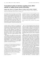
Available online />Figure 1
Activation of mitogen-activated protein kinase (MAPK; shown in yellow) pathways by IL-1. Stimulation by IL-1 activates the MAPK kinase kinases
(MAPKKKs; shown in blue), transforming-growth-factor-β-activated kinase-1 (TAK 1) and Raf, which then phosphorylate and activate several MAPK
kinases (MAPKKs; shown in green): MKK6, MKK4, MKK7, MEK. These MAPKKs in turn phosphorylate and activate the MAPKs, p38, c-Jun N-
terminal kinase (JNK) and extracellular signal-regulated kinase (ERK), which translocate to the nucleus. There, these MAPKs phosphorylate and
activate the transcription factors (activating transcription factor 2 [ATF2], c-Jun, Elk-1 and erythroblastosis twenty-six [Ets]-1; shown in red) that
contribute to matrix metalloproteinase (MMP) transcription. IL-1R, IL-1 receptor; IL-1RAcP, IL-1 receptor-associated protein; IRAK, IL-1 receptor
activated kinase; SRF, serum response factor; TAB, Tak-binding protein; TRAF, TNF receptor-associated factor.
to the transcription of an array of inflammatory genes, their
direct regulation of MMP transcription is just beginning to
be elucidated. In the remainder of this review, we address
how these pathway-specific signals lead to the recruit-
ment of a cohort of transcription factors that cooperate to
initiate MMP-1 and MMP-13 transcription.
Regulation of transcription
The promoters of MMP-1 and MMP-13 (and most other
MMPs) contain a TATA box, the core transcriptional unit,
at approximately –30 bp, and an AP-1 site at approxi-
mately –70 bp [37]. The AP-1 site (5′-TGAG/CTCA-3′)
binds dimers of the Fos and Jun families. Several addi-
tional AP-1 sites are present throughout the MMP promot-
ers, and may contribute to gene expression. One site
(5′-TTAATCA-3′) is found at –186 bp in the rabbit and
human MMP-1 promoters [38]. In contrast to the proximal
AP-1 site at –70 bp, this upstream site has only a modest
role in basal transcription, but it increases transcription in
response to phorbol esters [38]. A third AP-1 site has
been identified in the human MMP-1 promoter that coop-
erates with an adjacent Ets site. Thus, there may be dis-
tinct roles for various AP-1 elements, and these functions
may depend, at least in part, on the particular AP-1 family
members that bind to each site [38,39].
Although initial studies demonstrated the pivotal role of
the AP-1 site in MMP transcription in many cells, later
studies have clearly shown that it must cooperate with a
variety of cis-acting sequences found in the upstream
regions of the MMP promoters. For example, induction of
MMP-1 by IL-1 in rabbit fibroblasts requires interaction
between the AP-1 site at –77 bp and a NF-κB-like
element located upstream at –3030 bp [29,30]. Interest-
ingly, while both IL-1 and TNF-α activate NF-κB in primary
rabbit synovial fibroblasts, only IL-1 is capable of inducing
MMP-1 transcription [29]. This is due, in part, to the inabil-
ity of TNF to activate the ERK pathways in these cells,
both of which contribute to IL-1 induction. Recently,
Firestein and colleagues have demonstrated that the JNK
pathway, but not the p38 pathway is required for cytokine
induction of MMP-1 in human rheumatoid synovial fibrob-
lasts [40], and for MMP-13 induction in murine inflamma-
tory arthritis [11]. In contrast to synovial fibroblasts,
Arthritis Research Vol 4 No 3 Vincenti and Brinckerhoff
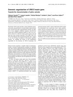
Figure 2
Activation of the NF-κB pathway by IL-1. IL-1 binds to its receptor (IL-1R1) and receptor-associated protein (IL-1RAcP), causing conformational
changes in multiple receptor-bound proteins (MyD88, IRAK, TRAF6, TAB2; see Figure 1 for definition). This results in recruitment and activation of
transforming-growth-factor-κ-activated kinase-1 (TAK 1), which phosphorylates and activates NF-κB-inducing kinase (NIK). In turn, NIK activates
the inhibitor of κB kinase (IKK) complex, which is responsible for phosphorylation of inhibitor of κB (IκB) and NF-κB1 (a 105 kDa protein, p105).
Upon phosphorylation, IκB and p105 become polyubiquitinated (U), which targets these proteins for degradation by the proteosome. Degradation
of these cellular inhibitors allows translocation of NF-κB subunits to translocate to the nucleus and transactivate matrix metalloproteinase (MMP)
promoters. The IκB-like protein, Bcl-3, promotes dimerization of the 50 kDa NF-κB subunit (p50) and regulates the transcriptional activity of p50.
chondrocytes rely on the p38, JNK and NF-κB pathways
to activate MMP-13 [7]. A working model [7,29] that
explains these observations is that MAPKs, either directly
or indirectly, activate AP-1 family members, which cooper-
ate with NF-κB to activate MMP-1 and MMP-13 transcrip-
tion. Additional transcription factors, however, such as Ets
family members, may also be nuclear targets of MAPKs in
this complex transcriptional program.
The AP-1 site at –1602 bp is of interest because it cooper-
ates with an Ets site created by a single nucleotide polymor-
phism at –1607 bp. This single nucleotide polymorphism is
represented by the insertion of an extra guanine base (G),
which creates a core binding site for the Ets family of tran-
scription factors (5′-GGA-3′). At least one copy of this ‘2G
allele’ is present in about 75% of the human population,
where it has the potential to enhance MMP-1 transcription
in both normal fibroblasts and some tumor cells [41–43].
Thus, this Ets site may contribute substantially to MMP-1
gene expression, at least under certain conditions. Ets sites
have critical roles in MMP transcription and they often inter-
act with AP-1 sites. In addition, multiple Ets sites are
present in all the MMP promoters, except MMP-2. The
number of these sites and their location within a given pro-
moter vary among the MMP family members, and these vari-
ations influence the regulation of these genes [37].
The interaction of AP-1 with other transcription factors
may also regulate tissue-specific expression of MMPs. The
transcription factor Runt domain factor-2 (Runx-2)
appears to be expressed almost exclusively in developing
cartilage and bone [44–47], and these are precisely the
cells that normally express MMP-13. Among the MMPs, a
Runx-2 binding site is unique to the MMP-13 promoter,
and it cooperates with the AP-1 site to mediate MMP-13
transcription [48–50]. Important future studies will define
the role of MAPK in Runx-2 activation, and how NF-κB
interacts with the AP-1/Runx-2 complex.
Repression of MMP transcription
Since joint destruction in arthritic diseases is not
reversible, the inhibition of the proteases responsible for
cartilage degradation is an important part of therapy.
While potent inhibitors of MMP enzymatic activity have
been developed, their use has been limited due to safety
issues [51]. Inhibition of MMP gene expression at the tran-
scriptional level may be a viable alternative option. With
the exception of the glucocorticoid hormones, however,
none of the several compounds now available to reduce
MMP transcription are in clinical use, although some have
been used successfully in animal models of arthritis.
The glucocorticoids bind to their receptors (GRs), which
then usually interact with glucocorticoid response ele-
ments present in the promoters of many genes [52].
Because the MMP promoters do not contain glucocorti-
coid response elements, inhibition of transcription occurs
through an indirect mechanism. This ‘transrepression’
involves binding of the activated receptor to Fos and Jun
proteins present at the proximal AP-1 site, with a subse-
quent change in their conformation and a reduction in
transcription [53]. Activated GRs can also potently
repress NF-κB-dependent transcription by two separate
mechanisms. First, GRs physically interact with RelA/p65,
resulting in inhibition of NF-κB-dependent transcription
[54,55]. This interaction is specific for p65, and is distinct
from the domain involved in AP-1 transrepression [56].
Second, glucocorticoids enhance IκBα synthesis, result-
ing in sequestration of NF-κB in the cytoplasm.
The vitamin A analogues, retinoids, also block MMP tran-
scription through the AP-1 site [57–60]. Ligand activated
receptors (e.g. the retinoic acid receptors α, β and γ, and
the retinoid x receptors α, β and γ) reduce MMP transcrip-
tion by binding to Fos and Jun proteins at the AP-1 site,
sequestering these proteins away from the promoter
and/or reducing the level of Fos and Jun mRNAs [61].
Although retinoids have reduced joint destruction in
animal models of arthritis [62], they have not been used in
patients. In addition, both glucocorticoids and retinoids
affect a broad number of genes, and this lack of specificity
may contribute to the side effects associated with these
compounds.
There is one novel compound that may block the expres-
sion of specific MMPs in arthritis. This is a synthetic triter-
penoid, 2-cyano-3,12-dioxoolean-1,9,dien-28-oic acid
(CDDO) [63,64]. At nanomolar concentrations, CDDO
selectively inhibits the induction of MMP-1 and MMP-13
by inflammatory cytokines in IL-1-stimulated chondrosar-
coma cells, without affecting basal expression [63]. In
addition, the expression of other MMPs is not affected,
and thus the low constitutive levels of MMPs required for
normal physiology may remain untouched. CDDO inhibits
MMP-1 and MMP-13 gene expression, at least in part, by
reducing IL-1-induced transcription [63]. While this mech-
anism is not fully understood, this drug is known to be a
ligand for peroxisome proliferator-activated receptor-γ
(PPAR-γ) [65]; other PPAR-γ agonists such as 15-deoxy-
prostaglandin J2 can also inhibit MMP-13 synthesis [66].
Since PPAR-γ can physically interact with c-Jun, it is
tempting to speculate that CDDO treatment induces an
AP-1/PPAR-γ association that is transcriptionally repres-
sive. Additional work is required to determine if CDDO
represses in an AP-1-dependent manner, or if it works
through a novel mechanism to repress MMPs.
Finally, increased knowledge of the specific signal/trans-
duction pathways driving MMP-1 and MMP-13 expression
in arthritic chondrocytes and synovial cells has led to the
search for agents that can inhibit these pathways. Block-
ing MAPK pathways inhibits gene expression of MMPs in
Available online />tissue culture experiments, and prevents progression of
arthritis in animal models. For example, the p38 MAPK
inhibitor, SB203580, blocked MMP-13 gene expression in
cultured chondrocytes [7] and inhibited IL-1 mediated col-
lagen degradation in cartilage explants [67]. In the colla-
gen-induced arthritis model of rheumatoid arthritis,
SB203580 significantly inhibited TNF-α and IL-6 produc-
tion, reduced paw inflammation, and inhibited the formation
of joint lesions [68]. In addition, orally active p38 inhibitors
were also effective in animal models of inflammatory arthri-
tis [69,70] presumably by blocking MMP synthesis. Inhibi-
tion of JNK by the novel inhibitor SP600125 inhibited bone
destruction in adjuvant-induced arthritis, suggesting a role
for this MAPK in disease pathogenesis [11].
Since NF-κB activation is required for the expression of
MMP-1 and MMP-13, as well as inflammatory stimuli such
as IL-1, IL-6 and TNF-α [7,29,71,72], this pathway is
another potential therapeutic target. This is supported by
the work of Bondeson et al. [73], in which over-expression
of IκBα reduced expression of inflammatory cytokines and
MMPs, but did not reduce anti-inflammatory cytokines or
tissue inhibitor of metalloproteinases. Furthermore, mice
deficient in the p50 subunit are refractory to collagen-
induced arthritis [74], indicating that this factor has a
prominent role in arthritic disease. Indeed, p50 was the
only subunit found to bind to an IL-1-responsive element
of the MMP-1 promoter [30]. Thus, direct blockade of the
NF-κB pathway, at least in joint cells, may be a viable
therapy to reduce MMP transcription in arthritis.
Conclusion
MMP-1 and MMP-13 play dominant roles in the progression
of RA and OA, making them prime targets for arthritis thera-
pies. Since the genes for both of these enzymes are tran-
scriptionally activated by inflammatory cytokines, an
understanding of the molecular pathways involved is crucial.
As our knowledge of these molecular mechanisms
increases, our ability to use this knowledge to develop novel
and effective therapies will also increase. In the twenty-first
century, the era of molecular medicine will surely include
strategies targeted at the control MMP gene transcription.
Acknowledgements
The authors would like to acknowledge the National Institute of Arthritis
and Musculoskeletal and Skin Diseases (AR-46977 and AR-02024 to
MPV; AR-26599 to CEB), the National Cancer Institute (CA-77267 to
CEB) the RGK Foundation, Austin Texas (to CEB), the Susan B
Komen Foundation (to CEB) and the Department of Defense
(BC991121 to CEB) for funding of this research.
References
1. Vincenti MP: The matrix metalloproteinase (MMP) and tissue
inhibitor of metalloproteinase (TIMP) genes. Transcriptional
and posttranscriptional regulation, signal transduction and
cell-type-specific expression. Methods Mol Biol 2001, 151:
121-148.
2. Nagase H, Woessner JF Jr: Matrix metalloproteinases. J Biol
Chem 1999, 274:21491-21494.
3. Stetler-Stevenson WG, Yu AE: Proteases in invasion: matrix
metalloproteinases. Semin Cancer Biol 2001, 11:143-152.
4. Borden P, Heller RA: Transcriptional control of matrix metallo-
proteinases and the tissue inhibitors of matrix metallopro-
teinases. Crit Rev Eukaryotic Gene Expr 1997, 7:159-178.
5. Vincenti MP, White LA, Schroen DJ, Benbow U, Brinckerhoff CE:
Regulating expression of the gene for matrix metallopro-
teinase-1 (collagenase): mechanisms that control enzyme
activity, transcription, and mRNA stability. Crit Rev Eukaryotic
Gene Expr 1996, 6:391-411.
6. Borden P, Solymar D, Sucharczuk A, Lindman B, Cannon P,
Heller RA: Cytokine control of interstitial collagenase and col-
lagenase-3 gene expression in human chondrocytes. J Biol
Chem 1996, 271:23577-23581.
7. Mengshol JA, Vincenti MP, Coon CI, Barchowsky A, Brinckerhoff
CE: Interleukin-1 induction of collagenase 3 (matrix metallo-
proteinase 13) gene expression in chondrocytes requires p38,
c-Jun N-terminal kinase, and nuclear factor kappaB: differen-
tial regulation of collagenase 1 and collagenase 3. Arthritis
Rheum 2000, 43:801-811.
8. Vincenti MP, Coon CI, Mengshol JA, Yocum S, Mitchell P, Brinck-
erhoff CE: Cloning of the gene for interstitial collagenase-3
(matrix metalloproteinase-13) from rabbit synovial fibroblasts:
differential expression with collagenase-1 (matrix metallopro-
teinase-1). Biochem J 1998, 331:341-346.
9. Brinckerhoff CE, Rutter JL, Benbow U: Interstitial collagenases
as markers of tumor progression. Clin Cancer Res 2000, 6:
4823-4830.
10. Goldring MB: The role of the chondrocyte in osteoarthritis.
Arthritis Rheum 2000, 43:1916-1926.
11. Han Z, Boyle DL, Chang L, Bennett B, Karin M, Yang L, Manning
AM, Firestein GS: c-Jun N-terminal kinase is required for met-
alloproteinase expression and joint destruction in inflamma-
tory arthritis. J Clin Invest 2001, 108:73-81.
12. Vincenti MP, Brinckerhoff CE: The potential of signal transduc-
tion inhibitors for the treatment of arthritis: Is it all just JNK? J
Clin Invest 2001, 108:181-183.
13. Tetlow LC, Adlam DJ, Woolley DE: Matrix metalloproteinase
and proinflammatory cytokine production by chondrocytes of
human osteoarthritic cartilage: associations with degenera-
tive changes. Arthritis Rheum 2001, 44:585-594.
14. Shlopov BV, Lie WR, Mainardi CL, Cole AA, Chubinskaya S,
Hasty KA: Osteoarthritic lesions: involvement of three differ-
ent collagenases. Arthritis Rheum 1997, 40:2065-2074.
15. Mitchell PG, Magna HA, Reeves LM, Lopresti-Morrow LL, Yocum
SA, Rosner PJ, Geoghegan KF, Hambor JE: Cloning, expression,
and type II collagenolytic activity of matrix metalloproteinase-
13 from human osteoarthritic cartilage. J Clin Invest 1996, 97:
761-768.
16. Firestein GS, Paine MM, Littman BH: Gene expression (collage-
nase, tissue inhibitor of metalloproteinases, complement, and
HLA-DR) in rheumatoid arthritis and osteoarthritis synovium.
Quantitative analysis and effect of intraarticular corticos-
teroids. Arthritis Rheum 1991, 34:1094-1105.
17. Clark IM, Powell LK, Ramsey S, Hazleman BL, Cawston TE: The
measurement of collagenase, tissue inhibitor of metallopro-
teinases (TIMP), and collagenase-TIMP complex in synovial
fluids from patients with osteoarthritis and rheumatoid arthri-
tis. Arthritis Rheum 1993, 36:372-379.
18. Westhoff CS, Freudiger D, Petrow P, Seyfert C, Zacher J, Kriegs-
mann J, Pap T, Gay S, Stiehl P, Gromnica-Ihle E, Wernicke D:
Characterization of collagenase 3 (matrix metalloproteinase
13) messenger RNA expression in the synovial membrane
and synovial fibroblasts of patients with rheumatoid arthritis.
Arthritis Rheum 1999, 42:1517-1527.
19. Garrington TP, Johnson GL: Organization and regulation of
mitogen-activated protein kinase signaling pathways. Curr
Opin Cell Biol 1999, 11:211-218.
20. Davis RJ: Signal transduction by the JNK group of MAP
kinases. Cell 2000, 103:239-252.
21. Kolch W: Meaningful relationships: the regulation of the
Ras/Raf/MEK/ERK pathway by protein interactions. Biochem
J 2000, 351:289-305.
22. Karin M: The regulation of AP-1 activity by mitogen-activated
protein kinases. J Biol Chem 1995, 270:16483-16486.
23. Leppa S, Saffrich R, Ansorge W, Bohmann D: Differential regu-
lation of c-Jun by ERK and JNK during PC12 cell differentia-
tion. Embo J 1998, 17:4404-4413.
Arthritis Research Vol 4 No 3 Vincenti and Brinckerhoff
24. Janknecht R, Ernst WH, Nordheim A: SAP1a is a nuclear target
of signaling cascades involving ERKs. Oncogene 1995, 10:
1209-1216.
25. O’Hagan RC, Tozer RG, Symons M, McCormick F, Hassell JA:
The activity of the Ets transcription factor PEA3 is regulated
by two distinct MAPK cascades. Oncogene 1996, 13:1323-
1333.
26. Ninomiya-Tsuji J, Kishimoto K, Hiyama A, Inoue J, Cao Z, Mat-
sumoto K: The kinase TAK1 can activate the NIK-I kappaB as
well as the MAP kinase cascade in the IL-1 signalling
pathway. Nature 1999, 398:252-256.
27. Karin M, Ben-Neriah Y: Phosphorylation meets ubiquitination:
the control of NF-[kappa]B activity. Annu Rev Immunol 2000,
18:621-663.
28. Bondeson J, Brennan F, Foxwell B, Feldmann M: Effective aden-
oviral transfer of IkappaBalpha into human fibroblasts and
chondrosarcoma cells reveals that the induction of matrix
metalloproteinases and proinflammatory cytokines is nuclear
factor- kappaB dependent. J Rheumatol 2000, 27:2078-2089.
29. Barchowsky A, Frleta D, Vincenti MP: Integration of the NF-
kappaB and mitogen-activated protein kinase/AP-1 pathways
at the collagenase-1 promoter: divergence of IL-1 and TNF-
dependent signal transduction in rabbit primary synovial
fibroblasts. Cytokine 2000, 12:1469-1479.
30. Vincenti MP, Coon CI, Brinckerhoff CE: Nuclear factor
kappaB/p50 activates an element in the distal matrix metallo-
proteinase 1 promoter in interleukin-1beta-stimulated syn-
ovial fibroblasts. Arthritis Rheum 1998, 41:1987-1994.
31. Xiao G, Harhaj EW, Sun SC: NF-kappaB-inducing kinase regu-
lates the processing of NF-kappaB2 p100. Mol Cell 2001, 7:
401-409.
32. Heissmeyer V, Krappmann D, Hatada EN, Scheidereit C: Shared
pathways of IkappaB kinase-induced SCF(betaTrCP)-medi-
ated ubiquitination and degradation for the NF-kappaB pre-
cursor p105 and IkappaBalpha. Mol Cell Biol 2001, 21:
1024-1035.
33. Heusch M, Lin L, Geleziunas R, Greene WC: The generation of
nfkb2 p52: mechanism and efficiency. Oncogene 1999, 18:
6201-6208.
34. Heissmeyer V, Krappmann D, Wulczyn FG, Scheidereit C: NF-
kappaB p105 is a target of IkappaB kinases and controls
signal induction of Bcl-3-p50 complexes. Embo J 1999, 18:
4766-4778.
35. Ziegler-Heitbrock HW, Wedel A, Schraut W, Strobel M, Wendel-
gass P, Sternsdorf T, Bauerle PA, Haas JG, Riethmuller G: Toler-
ance to lipopolysaccharide involves mobilization of nuclear
factor kappa B with predominance of p50 homodimers. J Biol
Chem 1994, 269:17001-17004.
36. Franzoso G, Bours V, Azarenko V, Park S, Tomita-Yamaguchi M,
Kanno T, Brown K, Siebenlist U: The oncoprotein Bcl-3 can
facilitate NF-kappa B-mediated transactivation by removing
inhibiting p50 homodimers from select kappa B sites [pub-
lished erratum appears in EMBO J 1997, 16:440]. Embo J
1993, 12:3893-3901.
37. Benbow U, Brinckerhoff CE: The AP-1 site and MMP gene reg-
ulation: what is all the fuss about? Matrix Biol 1997, 15:519-
526.
38. Chamberlain SH, Hemmer RM, Brinckerhoff CE: Novel phorbol
ester response region in the collagenase promoter binds Fos
and Jun. J Cell Biochem 1993, 52:337-351.
39. White LA, Brinckerhoff CE: Two activator protein-1 elements in
the matrix metalloproteinase-1 promoter have different
effects on transcription and bind Jun D, c-Fos, and Fra-2.
Matrix Biol 1995, 14:715-725.
40. Han Z, Boyle DL, Aupperle KR, Bennett B, Manning AM, Firestein
GS: Jun N-terminal kinase in rheumatoid arthritis. J Pharmacol
Exp Ther 1999, 291:124-130.
41. Rutter JL, Mitchell TI, Buttice G, Meyers J, Gusella JF, Ozelius LJ,
Brinckerhoff CE: A single nucleotide polymorphism in the
matrix metalloproteinase-1 promoter creates an Ets binding
site and augments transcription. Cancer Res 1998, 58:5321-
5325.
42. Kanamori Y, Matsushima M, Minaguchi T, Kobayashi K, Sagae S,
Kudo R, Terakawa N, Nakamura Y: Correlation between expres-
sion of the matrix metalloproteinase-1 gene in ovarian
cancers and an insertion/deletion polymorphism in its pro-
moter region. Cancer Res 1999, 59:4225-4227.
43. Nishioka Y, Kobayashi K, Sagae S, Ishioka S, Nishikawa A, Mat-
sushima M, Kanamori Y, Minaguchi T, Nakamura Y, Tokino T,
Kudo R: A single nucleotide polymorphism in the matrix met-
alloproteinase-1 promoter in endometrial carcinomas. Jpn J
Cancer Res 2000, 91:612-615.
44. Enomoto H, Enomoto-Iwamoto M, Iwamoto M, Nomura S, Himeno
M, Kitamura Y, Kishimoto T, Komori T: Cbfa1 is a positive regu-
latory factor in chondrocyte maturation. J Biol Chem 2000,
275:8695-8702.
45. Inada M, Yasui T, Nomura S, Miyake S, Deguchi K, Himeno M,
Sato M, Yamagiwa H, Kimura T, Yasui N, Ochi T, Endo N, Kita-
mura Y, Kishimoto T, Komori T: Maturational disturbance of
chondrocytes in Cbfa1-deficient mice. Dev Dyn 1999, 214:
279-290.
46. Komori T, Yagi H, Nomura S, Yamaguchi A, Sasaki K, Deguchi K,
Shimizu Y, Bronson RT, Gao YH, Inada M, Sato M, Okamoto R,
Kitamura Y, Yoshiki S, Kishimoto T: Targeted disruption of
Cbfa1 results in a complete lack of bone formation owing to
maturational arrest of osteoblasts. Cell 1997, 89:755-764.
47. Ducy P, Zhang R, Geoffroy V, Ridall AL, Karsenty G: Osf2/Cbfa1:
a transcriptional activator of osteoblast differentiation. Cell
1997, 89:747-754.
48. Hess J, Porte D, Munz C and Angel P: AP-1 and Cbfa/runt phys-
ically interact and regulate parathyroid hormone- dependent
MMP13 expression in osteoblasts through a new osteoblast-
specific element 2/AP-1 composite element. J Biol Chem
2001, 276:20029-20038.
49. Selvamurugan N, Chou WY, Pearman AT, Pulumati MR, Partridge
NC: Parathyroid hormone regulates the rat collagenase-3
promoter in osteoblastic cells through the cooperative inter-
action of the activator protein-1 site and the runt domain
binding sequence. J Biol Chem 1998, 273:10647-10657.
50. Porte D, Tuckermann J, Becker M, Baumann B, Teurich S, Higgins
T, Owen MJ, Schorpp-Kistner M, Angel P: Both AP-1 and Cbfa1-
like factors are required for the induction of interstitial colla-
genase by parathyroid hormone. Oncogene 1999, 18:667-678.
51. Brown PD: Ongoing trials with matrix metalloproteinase
inhibitors. Expert Opin Invest Drugs 2000, 9:2167-2177.
52. Funder JW: Glucocorticoid and mineralocorticoid receptors:
biology and clinical relevance. Annu Rev Med 1997, 48:231-
240.
53. Diamond MI, Miner JN, Yoshinaga SK, Yamamoto KR: Transcrip-
tion factor interactions: selectors of positive or negative regu-
lation from a single DNA element. Science 1990, 249:
1266-1272.
54. Scheinman RI, Gualberto A, Jewell CM, Cidlowski JA, Baldwin AS
Jr: Characterization of mechanisms involved in transrepres-
sion of NF-kappa B by activated glucocorticoid receptors. Mol
Cell Biol 1995, 15:943-953.
55. Ray A, Prefontaine KE: Physical association and functional
antagonism between the p65 subunit of transcription factor
NF-kappa B and the glucocorticoid receptor. Proc Natl Acad
Sci USA 1994, 91:752-756.
56. Tao Y, Williams-Skipp C, Scheinman RI: Mapping of glucocorti-
coid receptor DNA binding domain surfaces contributing to
transrepression of NF-kappa B and induction of apoptosis. J
Biol Chem 2001, 276:2329-2332.
57. Pan L, Eckhoff C, Brinckerhoff CE: Suppression of collagenase
gene expression by all-trans and 9-cis retinoic acid is ligand
dependent and requires both RARs and RXRs. J Cell Biochem
1995, 57:575-589.
58. Schroen DJ, Brinckerhoff CE: Inhibition of rabbit collagenase
(matrix metalloproteinase-1; MMP-1) transcription by retinoid
receptors: evidence for binding of RARs/RXRs to the -77 AP-1
site through interactions with c-Jun. J Cell Physiol 1996, 169:
320-332.
59. Yang-Yen HF, Zhang XK, Graupner G, Tzukerman M, Sakamoto
B, Karin M, Pfahl M: Antagonism between retinoic acid recep-
tors and AP-1: implications for tumor promotion and inflam-
mation. New Biol 1991, 3:1206-1219.
60. Schule R, Rangarajan P, Yang N, Kliewer S, Ransone LJ, Bolado
J, Verma IM, Evans RM: Retinoic acid is a negative regulator of
AP-1-responsive genes. Proc Natl Acad Sci USA 1991, 88:
6092-6096.
61. Schroen DJ, Brinckerhoff CE: Nuclear hormone receptors
inhibit matrix metalloproteinase (MMP) gene expression
through diverse mechanisms. Gene Expr 1996, 6:197-207.
Available online />62. Kuwabara K, Shudo K, Hori Y: Novel synthetic retinoic acid
inhibits rat collagen arthritis and differentially affects serum
immunoglobulin subclass levels. FEBS Lett 1996, 378:153-
156.
63. Mix KS, Mengshol JA, Benbow U, Vincenti MP, Sporn MB, Brinck-
erhoff CE: A synthetic triterpenoid selectively inhibits the
induction of matrix metalloproteinases 1 and 13 by inflamma-
tory cytokines. Arthritis Rheum 2001, 44:1096-1104.
64. Suh N, Wang Y, Honda T, Gribble GW, Dmitrovsky E, Hickey WF,
Maue RA, Place AE, Porter DM, Spinella MJ, Williams CR, Wu G,
Dannenberg AJ, Flanders KC, Letterio JJ, Mangelsdorf DJ, Nathan
CF, Nguyen L, Porter WW, Ren RF, Roberts AB, Roche NS, Sub-
baramaiah K, Sporn MB: A novel synthetic oleanane triter-
penoid, 2-cyano-3,12-dioxoolean-1,9- dien-28-oic acid, with
potent differentiating, antiproliferative, and anti-inflammatory
activity. Cancer Res 1999, 59:336-341.
65. Wang Y, Porter WW, Suh N, Honda T, Gribble GW, Leesnitzer
LM, Plunket KD, Mangelsdorf DJ, Blanchard SG, Willson TM,
Sporn MB: A synthetic triterpenoid, 2-cyano-3,12-dioxooleana-
1,9-dien-28-oic acid (CDDO), is a ligand for the peroxisome
proliferator-activated receptor gamma. Mol Endocrinol 2000,
14:1550-1556.
66. Fahmi H, Di Battista JA, Pelletier JP, Mineau F, Ranger P, Martel-
Pelletier J: Peroxisome proliferator—activated receptor gamma
activators inhibit interleukin-1beta-induced nitric oxide and
matrix metalloproteinase 13 production in human chondro-
cytes. Arthritis Rheum 2001, 44:595-607.
67. Ridley SH, Sarsfield SJ, Lee JC, Bigg HF, Cawston TE, Taylor DJ,
DeWitt DL, Saklatvala J: Actions of IL-1 are selectively con-
trolled by p38 mitogen-activated protein kinase: regulation of
prostaglandin H synthase-2, metalloproteinases, and IL-6 at
different levels. J Immunol 1997, 158:3165-3173.
68. Badger AM, Bradbeer JN, Votta B, Lee JC, Adams JL, Griswold
DE: Pharmacological profile of SB 203580, a selective
inhibitor of cytokine suppressive binding protein/p38 kinase,
in animal models of arthritis, bone resorption, endotoxin
shock and immune function. J Pharmacol Exp Ther 1996, 279:
1453-1461.
69. Jackson JR, Bolognese B, Hillegass L, Kassis S, Adams J, Gris-
wold DE, Winkler JD: Pharmacological effects of SB 220025, a
selective inhibitor of P38 mitogen-activated protein kinase, in
angiogenesis and chronic inflammatory disease models. J
Pharmacol Exp Ther 1998, 284:687-692.
70. Badger AM, Griswold DE, Kapadia R, Blake S, Swift BA, Hoffman
SJ, Stroup GB, Webb E, Rieman DJ, Gowen M, Boehm JC,
Adams JL, Lee JC: Disease-modifying activity of SB 242235, a
selective inhibitor of p38 mitogen-activated protein kinase, in
rat adjuvant-induced arthritis. Arthritis Rheum 2000, 43:175-
183.
71. Firestein GS, Manning AM: Signal transduction and transcrip-
tion factors in rheumatic disease. Arthritis Rheum 1999, 42:
609-621.
72. Tak PP, Firestein GS: NF-kappaB: a key role in inflammatory
diseases. J Clin Invest 2001, 107:7-11.
73. Bondeson J, Foxwell B, Brennan F, Feldmann M: Defining thera-
peutic targets by using adenovirus: blocking NF-kappaB
inhibits both inflammatory and destructive mechanisms in
rheumatoid synovium but spares anti-inflammatory media-
tors. Proc Natl Acad Sci USA 1999, 96:5668-5673.
74. Campbell IK, Gerondakis S, O’Donnell K, Wicks IP: Distinct roles
for the NF-kappaB1 (p50) and c-Rel transcription factors in
inflammatory arthritis. J Clin Invest 2000, 105:1799-1806.
Arthritis Research Vol 4 No 3 Vincenti and Brinckerhoff