Báo cáo khoa học: "Low early ototoxicity rates for pediatric medulloblastoma patients treated with proton radiotherapy" pptx
Bạn đang xem bản rút gọn của tài liệu. Xem và tải ngay bản đầy đủ của tài liệu tại đây (1.69 MB, 7 trang )
RESEARCH Open Access
Low early ototoxicity rates for pediatric
medulloblastoma patients treated with proton
radiotherapy
Benjamin J Moeller
1
, Murali Chintagumpala
2
, Jimmy J Philip
1
, David R Grosshans
1
, Mary F McAleer
1
, Shiao Y Woo
3
,
Paul W Gidley
4
, Tribhawan S Vats
5
and Anita Mahajan
1*
Abstract
Background: Hearing loss is common following chemoradiotherapy for children with medulloblastoma. Compared
to photons, proton radiotherapy reduces radiation dose to the cochlea for these patients. Here we examine
whether this dosimetric advantage leads to a clinical benefit in audiometric outcomes.
Methods: From 2006-2009, 23 children treated with proton radiotherapy for medulloblastoma were enrolled on a
prospective observational study, through which they underwent pre- and 1 year post-radiotherapy pure-tone
audiometric testing. Ears with moderate to severe hearin g loss prior to therapy were censored, leaving 35 ears in
19 patients available for analysis.
Results: The predicted mean cochlear radiation dose was 30
60
Co-Gy Equivalents (range 19-43), and the mean
cumulative cisplatin dose was 303 mg/m
2
(range 298-330). Hearing sensitivity significantly declined following
radiotherapy across all frequencies analyzed (P < 0.05). There was partial sparing of mean post-radiation hearing
thresholds at low-to-midrange frequencies and, consequently, the rate of high-grade (grade 3 or 4) ototoxicity at 1
year was favorable (5%). Ototoxicity did not correlate with predicted dose to the auditory apparatus for proton-
treated patients, potentially reflecting a lower-limit threshold for radiation effect on the cochlea.
Conclusions: Rates of high-grade early post-radiation ototoxicity following proton radiotherapy for pediatric
medulloblastoma are low. Preservation of hearing in the audible speech range, as observed here, may improve
both quality of life and cognitive functioning for these patients.
Keywords: Proton, radiotherapy, pediatric, medulloblastoma, ototoxicity
Background
Hearing loss is an important consequence of therapy for
children with intracranial malignancies, including medul-
loblastoma [1,2]. It can have a profound impact on a
child’s quality of life, affecting not only communication
skills but also social and cognitive development [3-5].
Chemotherapy and radiotherapy are major causes of
ototoxicity for children with medulloblastoma [6,7].
Efforts to mitigate treatment-related ototoxicity for
these patients tumors have included the use of confor-
mal radiotherapy techniques to minimize radiation dose
to the auditory apparatus. Compared to conventional
photon-based radiotherapy techniques, IMRT reduces
cochlear radiation doses and improves both earl y and
late audiometric outcomes [8-10]. Dosimetric studies
have suggested that proton techniques can further
reduce radiation dose to the auditory apparatus [11-13].
However, whether this translates into a clinical benefit
is as yet unknown.
Although ototoxicity is typically considered to be a
late effect of radiotherapy,withalatencyofapproxi-
mately four years [14,15], radiation also potentiates early
cisplatin-induced ototoxicity when the two ar e delivered
concomitantly [6,7], an effect typically peaking within a
year of treatment [8]. The objective of this study is to
* Correspondence:
1
Department of Radiation Oncology, University of Texas M.D. Anderson
Cancer Center, Houston, TX, USA
Full list of author information is available at the end of the article
Moeller et al. Radiation Oncology 2011, 6:58
/>© 2011 Moelle r et al; licensee BioMed Central Ltd. This is an Open Access article distributed under the terms of the Creative Commons
Attribution License ( which permits unrestricted use, distr ibution, and reproduction in
any medium, provided the original work is prope rly cited.
determine whether proton radiotherapy technique spares
this early ototoxicity for children with medulloblastoma.
Methods
Patients
Between 2006 and 2009, twenty-three consecutive chil-
dren with resected and histologically-confirmed medul-
loblastoma were enrolled on a prospective IRB-ap proved
institutional observational study investigating the effects
of proton radiotherapy on normal tissues. Relevant base-
line clinicopathologic a nd demographic features are
listed in Table 1.
Treatment
All patients received proton-based adjuvant radiother-
apy. Patients were positioned supine, and anesthesia was
used when necessary to optimize immo bilization, at the
discretion of the treating physician. CT simulation was
performed for each patient (LightSpeed RT16, GE
Healthcare). Treatment planning was performed using
comm ercial software (Eclipse, version 8, Varian Medical
Systems). Clinical target volumes were defined by the
treating physician, and planning margins were calculated
as previously described [16-18]. Standard-risk patients
(n = 17) received craniospinal irradiation (CSI) to a dose
of 23.4
60
Co-Gy Equivalents (CGE); high-risk patients (n
= 6) received CSI to 36 CGE. The tumor bed, plus a
clinical target volume expansion, was boosted to a total
dose of between 54 and 55.8 CGE. Relevant details
regarding radiation targets a nd doses are included in
Table 1. All patients received platinum-based che-
motherapy, with a median cumulative cisplatin dose of
303 mg/m
2
(range 298-330 mg/m
2
). Al l but five patients
rec eived adjuvant chem otherapy following radiotherapy;
the remainder received it beforehand principally to delay
cranial i rradiation. The mean total duration of all che-
motherapy and radiotherapy was approximately 28
weeks. A chart review confirmed that no other ototoxic
drugswereinusebyanypatientatthetimetheywere
simulated for radiotherapy.
Audiometry
Pure-tone audiometry was perf ormed for each patient at
baseline and at 1 year post-radiotherapy. Age-appropri-
ate audiometric techniques were used, at the discre tion
of the testing audiometrist. Each patient was confirmed
free of middle ear disease by tympanometry, in both
ears and at both time points. Each audiogram reported
hearing threshold, in decibels (dB), for each ear at 0.5,
1, 2, 4, 6, and 8 kHz. Ears with moderate-to-severe hear-
ing loss prior to any therapy were censor ed. For the
remaining patients, Brock ototoxicity rates (Table 2)
were determined for each patient from the raw post-
radiation audiometric data [19, 20]. Ototoxicity rates
were calculated per patient and, in the uncommon cases
where threshold loss was asymmetric following radia-
tion, toxicity grading reflected the worse of the two ears
tested.
Radiation Dosimetry
Both cochleae were contoured for each case, and the
treatment planning software (Eclipse, version 8, Varian
Medical Systems) was used to estimate the mean and
maximum delivered organ doses.
Statistics
Changes in raw audiometric thresholds following radio-
therapy were tested for significance by one-way
ANOVA (SPSS, version 16). Associations between clini-
cal, demographic, treatment, and audiometric variables
were estimated using Spearman’s correlations and uni-
variate linear modeling (SPSS, version 16).
Results
Of the twenty-three patients enrolled, baseline audiome-
try showed that four had bilateral and three had unilat-
eral severe hearing loss before starting radiotherapy
(ot otoxicity grad es 3 or 4). Of those with bilateral base-
line severe hearing loss, two had prior chemotherapy
and two had hearing loss attributed to unrelated genetic
Table 1 Clinical and treatment characteristics
PROTON COHORT
(n = 19)
Age 6 (3-16)
Time to Audiogram (months) 11 (8-16)
Gender Male Female 14 (74) 5 (26)
Risk Grouping Standard High 16 (84) 3 (16)
Cisplatin Dose (mg/m
2
) 303 (298-330)
CSI Dose (CGE or Gy) SR HR 23.4 36.0
Total Dose (CGE or Gy) 54.0 or 55.8
Cochear Dose (CGE or Gy) 30 (19-43)
Mean values are shown, with data ranges or percentages of total in
parentheses. “Time to Audiogram” refers to the interval, in months, between
the end of radio therapy and audiometry. SR = standard-risk, HR = high-risk,
CSI = craniospinal irradiation.
Table 2 Brock ototoxicity grading scale
FREQUENCY (kHz) GRADE
-0
8 1
4 2
2 3
1 4
On this scale, ototoxicity is graded by the lowest frequency level at which a
hearing threshold loss of at least 40 dB occurs [19]. If no threshold loss of this
magnitude is detected at or below 8 kHz, the toxicity grade is zero.
Moeller et al. Radiation Oncology 2011, 6:58
/>Page 2 of 7
syndromes. All three patients with unilateral severe
hearing loss developed the deficit either before or imme-
diately following surgery, with no prior exposure to che-
motherapy or radi otherapy. These ears were censored
from analysis, leaving 35 ears in 19 patients available for
further study. Baseline demographics were similar to
those of most children with medulloblastoma treated at
the authors’ institution (Table 1).
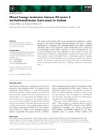
A pair of posterior oblique proton beams was used for
the cranial portion of each patient’s treatment in order
to spare the lenses of the eye while adequ atel y covering
the cribriform plate. The auditory apparatus was not
typically included as a target volume during the cra-
niospinal portion of treatment. The tumor bed b oost
portion of treatment was typically carried out using a
cone-down postero-lateral beam pair (Figure 1). Consis-
tent with prior reports, proton technique resulted in a
favorably low me an cochlear radiation (30
60
Co-Gy
Equivalents [range 19-43]).
Compared to baseline testing, post-radiation audiome-
try showed a clinically and statistically significant wor-
sening of hearing thresho ld acros s all frequencies tested
(P < 0.05, Figure 2). However, we noted a relatively
modest threshold change in the audible speech range
(0.5-6 kHz). The preservation of hearing in the audible
speech range is of critical functional importance for
patients, and this is reflected in t he heavy weighting of
threshold loss in this range on ototoxicity grading scales.
Accordingly, overall ototoxicity grade was found to be
low following proton-based treatment (Figure 3), and
the rate of high-grade ototoxicity was favorable at 5%.
In keeping with the low rates of high-grade ototoxicity
for this cohort, hearing amplification was re commended
for only a relatively small number of patients (3 of 19)
following radiotherapy.
Prior published data s uggest that the risk of ototoxi-
city is linearly related to cochlear radiation dose, with
an apparent lower-limit threshold at approximately 36
Gy [15]. If this is the case, then reducing cochlear
radiation dose to 36 Gy should minimize ototoxicity,
but further reduction of dose below 36 Gy should have
little additional impact on hearing loss. Our data sup-
port this hypothesis. The audiometric benefits
described above for this cohort likely reflect the fact
that 84% (16 of 19) of these patients received cochlear
doses below 36 CGE; however, we found no evidence
Figure 1 Proton radiotherapy dosimetry. A representative plan is
shown depicting the sparing of dose to the auditory apparatus (red
arrows) in a child with medulloblastoma treated with proton
technique. Colored isodose curves are shown depicting the
absolute radiation dose in CGE. The clinical tumor bed boost target
volume is outlined (blue).
Figure 2 Audiometr ic outcomes. (A) Mean pure-tone audiometry
for the proton cohort at baseline (blue) and following radiotherapy
(red) are shown. Note the sparing of threshold loss following proton
radiotherapy in the audible speech range (0.5-4 kHz). (B) Box and
whisker plots of the same data are shown, representing the 2
nd
/3
rd
quartile data range (boxes), the mean values (horizontal line), and
the total data range (whiskers).
Figure 3 Ototoxicity rates. Brock ototoxicity rates, p er patient,
were favorable following proton radiotherapy (High-Grade = Grades
3 or 4, Low-Grade = Grades 1 or 2, None = Grade 0).
Moeller et al. Radiation Oncology 2011, 6:58
/>Page 3 of 7
that further reducing the cochlear radiation dose below
36 CGE offered any additional benefit to these
patients. Although there wa s a weakly positive correla-
tion between the two (Spearman’ s r = 0.33), radiation
dose to the cochlea across the observed range (16-43
CGE) ultimately failed to predict ototoxicity on uni-
variate analysis for these patients. Similarly, scatter
plots of cochlear radiation dose versus ototoxicity
revealed no obvious correlation between the two (Fig-
ure 4). This supports the concept of there being a
threshold effect for radiation dose to the cochlea near
36 Gy, and suggests that further reduction in dose
below this threshold is unlikely to achieve additional
clinical benefit. Of note, cisplatin dose also failed to
predict ototoxicity for this cohort, though this is not
surprising given the small range of cumulative doses
delivered (298-330 mg/m
2
).
Discussion
The above data support our hypothesis that children
with m edulloblast oma treated with proton radiotherapy
have low rates of ototoxicity at one year after treatment.
These data validate the many pre-existing dosimetry stu-
dies suggesting that proton technique spares radiation
dose to the auditory apparatus, and establish a relation-
ship between this dosimetric advantage and improved
clinical outcomes.
To date, published data on audiometric outcomes fol-
lowing proton-based radiotherapy for pediatri c medullo-
blastoma are lacking. Physicians f rom the Francis H.
Burr Proton Center at the Massachusetts General Hos-
pital recently presented in abstract form their early
audiometric results in 31 children with medulloblastoma
treated with proton radiotherapy [21]. Predicted mean
cochlear doses were identical to those for our cohort
Figure 4 Dose-response analysis. Shown are scatter plots of mean predicted cochlear radiation dose versus ototoxicity grade (A), as well as
post-proton radiotherapy hearing threshold at 4 kHz (B), 6 kHz (C), and 8 kHz (D). Correlations are weak for all metrics, suggesting a lack of
influence of cochlear radiation dose on ototoxicity rates over the range of doses seen in this cohort.
Moeller et al. Radiation Oncology 2011, 6:58
/>Page 4 of 7
(30 CGE). At a mea n follow-up of 2.5 years, the authors
reported high-grade ototoxicity rates of 8% (when cor-
recting for baseline rates). Although this rate is slightly
higher than that reported here, the difference is likely
related to a higher cumulative cisplatin dose for this
cohort (395 versus 303 mg/m
2
) as well as longer follow-
up (2.5 versus 1 year). These results corroborate our
findings and further support our conclusions that early
audiometric outcomes following chemoradiotherapy for
children with medulloblastoma are favorable with pro-
ton technique.
An unanswered question raised by these results is
whether ototoxicity rates following proton therapy a re
better than those seen following photon therapy. Given
the m any proposed benefits of proton radiotherapy for
pediatric cancer patients, it is unlikely that randomized
trials of proton versus photon radiation techniques will
ever be pursued in this population. This limits our capa-
city to make definitive judgments on outcomes between
the two techniques. In the absence of higher-quality
data, we are left to contrast results across series of
patients treated with proton versus photon techniques.
We acknowledge that such comparisons are susceptible
to many sources of bias and error, and should be inter-
preted accordingly.
One useful series for comparison is that published by
Huang et al [9], which reported early audiometr ic out-
comes after IMRT for children with medulloblastoma.
These data demonstrate a higher rate of grade 3-4 toxi-
cityfollowingIMRT(18%)comparedtothatseenfol-
lowing proton radiotherapy on o ur study (5%). When
comparing the mean post-radiation audiometric data
between the two cohorts, there appears to be a sparing
of threshold loss following radiation of approximately 10
dB in the audible speech frequency range (1-4 kHz), but
little no ticeable difference in outcomes between the two
modalities at higher or lower frequency ranges.
A potential flaw in this comparison, however, is that
the definition of target volume is discrepant between the
cohorts. There has been increasing interest re cently in
reducingthetargetvolumefortheboostportionof
radiotherapy for children with medulloblastoma to the
surgical cavity, alone, without boosting the entire poster-
ior fossa. Accordin gly, the entire posterior fossa was tar-
geted only during the craniospinal portion of treatment
for the proton patients described above; i n the series
published by Huang et al [9], the entire posterior fossa
was treated to 36 Gy prior to a cone-down boost to the
surgical cavity. It is possible that this difference in plan-
ning approach, alone, might explain the improvement in
audiometric outcomes between the cohorts.
A more robust comparator, then, may be the cohort of
IMRT-treated children with medulloblastoma recently
reported by Polkinghorn et al [10]. The target volumes
and doses for the majo rity of the pa tients treated in this
cohort were identical to those in our own (23.4 Gy CSI,
55.8 Gy boost). At a median follow-up of 19 months,
the reported rate o f grade 3-4 hearing loss was similar
to that for our cohort (6%). However, whereas more
than half of the pr oton-treated patients on our c ohort
had no measurable otot oxicity (i.e. grade 0), this was
achieved in less than a quarter of the IMRT-treated
patients in the comparator cohort . Since low-g rade oto-
toxicity can have an impact on a child’s communication
skills, learning, and quality of life, this might represent a
clinically meaningful benefit to proton radiotherapy for
these patients.
Again, however, one must be cautious when drawing
conclusions from these comparisons. The delivered
doses of cisplatin were not reported in the series pub-
lished by Polkinghorn et al, so it is not possible to deter-
mine whether the cohorts were similar in this regard.
Also, though the target volumes for this cohort of
IMRT-treated patients were smaller than those for the
IMRT-treated patients published by Huang et al, the
reported delivered doses to the audit ory apparatus were
similar (38 Gy versus 37 Gy ). Therefore, there may be
radiation -unrelated differences between these two IMRT
cohorts that account for their divergent audi ometric
outcomes.
As discussed above, compared to the mean audio-
metric data available for patients treated w ith IMRT
techni que [9], our data show a selective sparing of hear-
ing threshold loss in the audible speech frequency range
with proton therapy. This outcome is of particular
importance for young radiotherapy patients who are cri-
tically reliant on the proper recognition and processing
of speech for cognitive and social development. The fre-
quency range quoted for audible speech varies some-
what in the literature, but i s most commonly defined as
0.5 to 2 kHz. Recent work has shown that somewhat
higher frequency ranges, including 4 kHz, are also quite
important for the p roper recognition o f certain nuances
in spoken lang uage, suc h as fricative sounds, suggesting
that our defined range for audible speech ought to be
expanded to include these frequencies [5,22]. The
observed reduction in the rate of clinically significant
threshold loss (i.e. beyond 20 dB) in this range may
eventually translate into an improved quality of life for
these patients; further follow-up is required to explore
this hypothesis.
The established literature shows that radiation dose to
the cochlea is clearly an important variable in demon-
strating ototoxicity [15]. Though we were unable to
demonstrate a dose-response relationship for ototoxicity
on our study, this is not surprising given that the vast
majority of the patients i n our cohort received predicted
cochlear doses below the 36 G y threshold proposed by
Moeller et al. Radiation Oncology 2011, 6:58
/>Page 5 of 7
the e xisting literature. Indeed, the fact that overall oto-
toxicity rates were so low on this study supports the
validity of dose constraints for the cochlea at or around
36 Gy. Proton radiotherapy effectively allows this con-
straint to be met in the majority of c ases, adding to the
overall rationale for its use in this patient population.
However, our results also highlight the importance of
variables apart from radiation and chemotherapy dose in
determining ototoxicity rates for these patients. Within
the relatively narrow range of cisplatin and cochlear
radiation doses delivered here, ototoxicity varied widely.
High-grade ototoxicity was observed follo wing cochlear
radiation doses as low as 28 CGE, much lower than the
putative threshold dose. These facts point to the impor-
tance of ototoxic variables unrelated to radiation in
these patients. As added proof of this concept, the num-
ber of enrolled cases censored in this study for having
pre-therapy high-grade ototoxicity was higher than the
number of analyzed cases with post-radiation high-grade
ototoxicity. Further study is needed to better understand
the patient and non-therapy related variables that lead
to high-grade ototoxicity in some of these children. One
such variable may be increased intracranial pressure or,
its surrogate, the use of cerebrospinal fluid shunting
[14]; however, this factor was not predictive in our
cohort. The lack of clear correlations b etween ototoxi-
city and these clinical variables may speak to the impor-
tance of unidentified biologic factors that may inf luence
individual pati ents’ intrinsic sensitivity to ra diation and/
or cisplatin effects on the cochlea; such issues warrant
further investigation.
Continued follow-up of this cohort is needed for sev-
eral reasons. First, it will be critical to determine
whether t he measured clinical gain seen here translates
into a benefit in the quality of life for the patients. As
the absolute number of patients spared high-grade toxi-
city was relatively small, answering this question may
eventually require a larger sample size. It will also be
important to d etermine whether proton radiotherapy
spares late ototoxicity, as this may be a more critical
determina nt of long-term functional outcome s for these
patients than is early toxicit y. It seems logical to predic t
that it will. First, though the data are inherently limited,
updates of prior studies haveconfirmedthestabilityof
early audiometric outcomes with long follow-up for chil-
dren with medulloblastoma [8,9], and there is no reason
to expect that our cohort will behave differently. In fact,
it may be that the advantages in proton-treated children
will become more pronounced with time, owing to the
smaller dose per fra ction delivered to the auditory appa-
ratus with proton radiotherapy.
There are some strengths and limitations of this study
that should be highlighted. The prospective collection of
pre- and post-radiation audiometry on an institutional
protocol makes the quality of this dataset favorable in
comparison to many of the retrospective reports cur-
rently available on this topic. The relative homogeneity
of treatment between patients also improves the quality
of the data analysis. Also, this is the first series reporting
audiometric outcomes for children with m edulloblas-
toma treated with proton radio therapy and, theref ore, it
represents a unique contribution to the literature.
Weaknesses of this study include the lack of long-term
follow-up and the relatively small sample size of the
patient population. For the various reasons outlined in
the Introduction, we believe an analysis of audiometric
data at one year after radiotherapy is valid. The small
sample size is, of course, an inherent obstacle when
studying a rare disease treated with a limited resource.
Continued accrual onto prospective studies of normal
tissue toxicity is critical to further evalua te the proposed
benefits of proton radiotherapy for children with medul-
loblastoma and other malignancies.
It could be argued that omitting the hi gh-risk patients
from each cohort would have i mproved the homogene-
ity of the populations compared. While this may be
true, this approach also would have carried with it the
drawbacks of d ecreasing the cohort size, reducing the
range of the radiation dos e dataset, and decreasing the
scope of the study. Indeed, repeating the major analyses
described above wh ile including only the standard-risk
patients had no noticeable impact on the data, other
than by decreasing the mean radiation dose delivered to
the cochlea (not shown). Therefore, inclusion of these
patients in the above analyses appears to be appropriate.
Conclusions
Proton radiotherapy results in low early high-grade oto-
toxicity rates for children with medulloblastoma. The
sparing of auditory threshold in the audible speech
range with proton radiotherapy may eventually translate
into improved communication skills, quality of life,
social development, and cognitive development fo r these
patients. Further follow-up is needed to address these
questions, and to determine the degree to which proton
technique may prevent late ototoxicity.
Author details
1
Department of Radiation Oncology, University of Texas M.D. Anderson
Cancer Center, Houston, TX, USA.
2
Texas Children’s Cancer Center, Baylor
College of Medicine, Houston, TX, USA.
3
Department of Radiation Oncology,
University of Louisville, Louisville, KY, USA.
4
Department of Head and Neck
Surgery, University of Texas M.D. Anderson Cancer Center, Houston, TX, USA.
5
Department of Pediatrics, University of Texas M.D. Anderson Cancer Center,
Houston, TX, USA.
Authors’ contributions
BJM designed the study, analyzed the data and prepared the manu script.
JJP collected and helped to analyze the data. MC, DRG, MFM, SYW, PWG,
TSV, and AM all participated in the treatment of the patient cohort
Moeller et al. Radiation Oncology 2011, 6:58
/>Page 6 of 7
described, designed the study, helped analyze the data and assisted with
preparation of the manuscript. All authors read and approved the final
manuscript.
Competing interests
The authors declare that they have no competing interests.
Received: 1 April 2011 Accepted: 2 June 2011 Published: 2 June 2011
References
1. Knight KR, Kraemer DF, Neuwelt EA: Ototoxicity in children receiving
platinum chemotherapy: underestimating a commonly occurring toxicity
that may influence academic and social development. J Clin Oncol 2005,
23:8588-8596.
2. Kolinsky DC, Hayashi SS, Karzon R, Mao J, Hayashi RJ: Late onset hearing
loss: a significant complication of cancer survivors treated with Cisplatin
containing chemotherapy regimens. J Pediatr Hematol Oncol 2010,
32:119-123.
3. Moeller MP: Current state of knowledge: psychosocial development in
children with hearing impairment. Ear Hear 2007, 28:729-739.
4. Moeller MP, Tomblin JB, Yoshinaga-Itano C, Connor CM, Jerger S: Current
state of knowledge: language and literacy of children with hearing
impairment. Ear Hear 2007, 28:740-753.
5. Stelmachowicz PG, Pittman AL, Hoover BM, Lewis DE, Moeller MP: The
importance of high-frequency audibility in the speech and language
development of children with hearing loss. Arch Otolaryngol Head Neck
Surg 2004, 130:556-562.
6. Walker DA, Pillow J, Waters KD, Keir E: Enhanced cis-platinum ototoxicity
in children with brain tumours who have received simultaneous or prior
cranial irradiation. Med Pediatr Oncol 1989, 17:48-52.
7. Weatherly RA, Owens JJ, Catlin FI, Mahoney DH: Enhanced cis-platinum
ototoxicity in children. Laryngoscope 1991, 101:917-924.
8. Paulino AC, Lobo M, Teh BS, Okcu MF, South M, Butler EB, Su J,
Chintagumpala M: Ototoxicity After Intensity-Modulated Radiation
Therapy and Cisplatin-Based Chemotherapy in Children with
Medulloblastoma. Int J Radiat Oncol Biol Phys 2010.
9. Huang E, Teh BS, Strother DR, Davis QG, Chiu JK, Lu HH, Carpenter LS,
Mai WY, Chintagumpala MM, South M, et al: Intensity-modulated radiation
therapy for pediatric medulloblastoma: early report on the reduction of
ototoxicity. Int J Radiat Oncol Biol Phys 2002, 52:599-605.
10. Polkinghorn WR, Dunkel IJ, Souweidane MM, Khakoo Y, Lyden DC,
Gilheeney SW, Becher OJ, Budnick AS, Wolden SL: Disease Control and
Ototoxicity Using Intensity-Modulated Radiation Therapy Tumor-Bed
Boost for Medulloblastoma. Int J Radiat Oncol Biol Phys 2011.
11. Merchant TE, Hua CH, Shukla H, Ying X, Nill S, Oelfke U: Proton versus
photon radiotherapy for common pediatric brain tumors: comparison of
models of dose characteristics and their relationship to cognitive
function. Pediatr Blood Cancer 2008, 51:110-117.
12. Lee CT, Bilton SD, Famiglietti RM, Riley BA, Mahajan A, Chang EL, Maor MH,
Woo SY, Cox JD, Smith AR: Treatment planning with protons for pediatric
retinoblastoma, medulloblastoma, and pelvic sarcoma: how do protons
compare with other conformal techniques? Int J Radiat Oncol Biol Phys
2005, 63:362-372.
13. St Clair WH, Adams JA, Bues M, Fullerton BC, La Shell S, Kooy HM,
Loeffler JS, Tarbell NJ: Advantage of protons compared to conventional
X-ray or IMRT in the treatment of a pediatric patient with
medulloblastoma. Int J Radiat Oncol Biol Phys 2004, 58:727-734.
14. Merchant TE, Gould CJ, Xiong X, Robbins N, Zhu J, Pritchard DL, Khan R,
Heideman RL, Krasin MJ, Kun LE: Early neuro-otologic effects of three-
dimensional irradiation in children with primary brain tumors. Int J
Radiat Oncol Biol Phys 2004, 58:1194-1207.
15. Hua C, Bass JK, Khan R, Kun LE, Merchant TE: Hearing loss after
radiotherapy for pediatric brain tumors: effect of cochlear dose. Int J
Radiat Oncol Biol Phys 2008, 72:892-899.
16. Moyers MF, Miller DW: Range, range modulation, and field radius
requirements for proton therapy of prostate cancer. Technol Cancer Res
Treat 2003, 2:445-447.
17. Moyers MF, Miller DW, Bush DA, Slater JD: Methodologies and tools for
proton beam design for lung tumors. Int J Radiat Oncol Biol Phys 2001,
49:1429-1438.
18. Torres MA, Chang EL, Mahajan A, Lege DG, Riley BA, Zhang X, Lii M,
Kornguth DG, Pelloski CE, Woo SY: Optimal treatment planning for skull
base chordoma: photons, protons, or a combination of both? Int J Radiat
Oncol Biol Phys 2009, 74:1033-1039.
19. Brock P, Pritchard J, Bellman S, Pinkerton CR: Ototoxicity of high-dose cis-
platinum in children. Med Pediatr Oncol 1988, 16:368-369.
20. Neuwelt EA, Brock P: Critical need for international consensus on
ototoxicity assessment criteria. J Clin Oncol 2010, 28:1630-1632.
21. Yock T, Yeap B, Ebb D, MacDonald S, Pulsifer M, Marcus K, Tarbell N: A
phase II trial of proton radiotherapy for medulloblastoma: Preliminary
results. J Clin Oncol 2010, 28 :18s(abstr CRA9507).
22. Pittman AL, Stelmachowicz PG: Hearing loss in children and adults:
audiometric configuration, asymmetry, and progression. Ear Hear 2003,
24:198-205.
doi:10.1186/1748-717X-6-58
Cite this article as: Moeller et al.: Low early ototoxicity rates for
pediatric medulloblastoma patients treated with proton radiotherapy.
Radiation Oncology 2011 6:58.
Submit your next manuscript to BioMed Central
and take full advantage of:
• Convenient online submission
• Thorough peer review
• No space constraints or color figure charges
• Immediate publication on acceptance
• Inclusion in PubMed, CAS, Scopus and Google Scholar
• Research which is freely available for redistribution
Submit your manuscript at
www.biomedcentral.com/submit
Moeller et al. Radiation Oncology 2011, 6:58
/>Page 7 of 7