Báo cáo y học: "Seroprevalence of parvovirus B19 IgG in children affected by juvenile idiopathic arthritis" pdf
Bạn đang xem bản rút gọn của tài liệu. Xem và tải ngay bản đầy đủ của tài liệu tại đây (202.11 KB, 6 trang )
Open Access
Available online />Page 1 of 6
(page number not for citation purposes)
Vol 9 No 4
Research article
Seroprevalence of parvovirus B19 IgG in children affected by
juvenile idiopathic arthritis
Benedikt Weissbrich
1
, Yvonne Süß-Fröhlich
1,2
and Hermann J Girschick
2
1
Institute of Virology and Immunobiology, University of Würzburg, Versbacher Str 7, 97078 Würzburg, Germany
2
Section of Paediatric Rheumatology, Immunology and Infectious diseases, Children's Hospital, University of Würzburg, Josef-Schneider-Str 2,
97080 Würzburg, Germany
Corresponding author: Benedikt Weissbrich,
Received: 25 May 2007 Revisions requested: 19 Jul 2007 Revisions received: 29 Jul 2007 Accepted: 30 Aug 2007 Published: 30 Aug 2007
Arthritis Research & Therapy 2007, 9:R82 (doi:10.1186/ar2281)
This article is online at: />© 2007 Weissbrich et al.; licensee BioMed Central Ltd.
This is an open access article distributed under the terms of the Creative Commons Attribution License ( />),
which permits unrestricted use, distribution, and reproduction in any medium, provided the original work is properly cited.
Abstract
Parvovirus (PV) B19 is the causative agent of the childhood
disease erythema infectiosum. An association of PV B19 with
chronic arthropathies, sometimes resembling rheumatoid
arthritis or juvenile idiopathic arthritis (JIA), has repeatedly been
described. Other studies, however, have failed to identify any
such relationship. In order to study further whether there is a link
between PV B19 and JIA, we determined the prevalence of PV
B19 specific IgG antibodies in serum samples from children
with rheumatoid diseases and compared it with the prevalence
in unaffected children We reasoned that if there is an
association between PV B19 and JIA, then the prevalence of PV
B19 IgG in the children with JIA should be higher than in the
control group. PV B19 IgG status was tested in 406 children
with JIA and related diseases, and in 146 children constituting a
control group. The percentage of PV B19 IgG positive children
was not significantly elevated in the disease subgroups
compared with age-matched control groups. In conclusion, our
findings do not support the hypothesis that human parvovirus
B19 is involved in the pathogenesis of JIA.
Background
Parvovirus (PV) B19, the causative agent of the childhood dis-
ease erythema infectiosum (fifth disease), was identified in
1975. Since then a large spectrum of diseases caused by or
associated with PV B19 has been recognized (for review
[1,2]). In addition to erythema infectiosum, nonspecific febrile
illnesses and asymptomatic courses are common. Further-
more, the clinical spectrum of PV B19 includes haematologi-
cal, neurological and cardiovascular manifestations. Infection
during pregnancy may result in hydrops fetalis. Arthralgias and
acute arthritis are well known complications of acute PV B19
infection in children and in adults [3].
An association of PV B19 with chronic arthropathies, some-
times resembling rheumatoid arthritis or juvenile idiopathic
arthritis (JIA), has also been described, but other studies have
been unable to corroborate these findings. It therefore remains
unclear whether there is an aetiological or pathogenic link
between PV B19 and rheumatoid arthritis or JIA (for review
[4]).
The term JIA encapsulates a heterogeneous group of rheu-
matic diseases, which – for most subtypes – is distinct from
adult rheumatoid arthritis [5]. Manifesting as early as in the first
year of life, childhood arthritis can be a serious disease that
affects not only motor neurone but also psychosocial develop-
ment. Autoimmune features have been noted especially in two
subgroups, namely enthesitis related arthritis and early onset
pauciarticular arthritis (EOPA). Infectious diseases have long
been suspected as being trigger factors for the initial manifes-
tation of arthritis and subsequent flare ups, and various bacte-
ria and viruses have been implicated in this regard [6-10]. In
particular, Still's disease in childhood has been associated
with PV B19; in fact, in some children affected by Still's dis-
ease the erythematous rash resembles that of erythema infec-
tiosum [11,12].
In order to evaluate further the possible link between PV B19
and JIA, we determined the prevalence of PV B19 specific IgG
antibodies in serum samples from children with rheumatic dis-
eases and compared it with the prevalence in unaffected
EOPA, early onset pauciarticular arthritis; JIA, juvenile idiopathic arthritis; JRA, juvenile rheumatoid arthritis; NS, nonstructural protein; PV, parvovirus.
Arthritis Research & Therapy Vol 9 No 4 Weissbrich et al.
Page 2 of 6
(page number not for citation purposes)
children. Whereas PV B19 IgM is detectable only for about 1
to 3 months after an acute infection, PV B19 IgG persists
throughout life. Thus, the presence of PV B19 IgG is a marker
of previous exposure to PV B19. We hypothesized that if there
is an association between PV B19 and JIA, then the preva-
lence of PV B19 IgG in children with JIA should be higher than
in the control group.
Materials and methods
Patients
The study population consisted of children who were referred
to the Section of Paediatric Rheumatology at the University of
Würzburg, Germany, between 1988 and 2001. Serum sam-
ples for routine laboratory studies were obtained from the
patients at the initial visit. No selection of patients was per-
formed. Unused serum was kept frozen at -20°C. Demo-
graphic data, clinical diagnosis, and information on PV B19
status were extracted from the records. Stored serum samples
of children with unknown PV B19 status were retrospectively
tested for PV B19 IgG.
In addition, serum samples of 146 children were analyzed as
controls. These samples were collected from children referred
to the Endocrinological Section of the Children's Hospital and
from children referred to the Section of Paediatric Rheumatol-
ogy who presented with complaints not associated with
arthropathy or infections.
All patients and control children were of white Caucasian
descent. The study was conducted in compliance with the
Helsinki Declaration and was approved by the ethics commit-
tee at the University of Würzburg. Informed consent was
obtained from the parents or legal guardians of the children in
the arthritis group for standard of care diagnostic procedures,
which included serology on infectious agents associated with
arthritis. For the control group, the use of anonymized residual
serum samples obtained for routine diagnostic procedures
was approved by the ethics committee.
Parvovirus B19 serology
PV B19 IgG antibodies were determined using an indirect
immunofluorescence assay. Briefly, SF9 insect cells infected
with baculovirus recombinant for VP1 of PV B19 were spotted
on glass slides, air dried, fixed with cold acetone and stored at
-20°C until use. For IgG determination, the fixed cells were
incubated for 90 min with a fourfold dilution series of each
plasma sample, starting at 1:10. Subsequently, the slides
were washed with phosphate-buffered saline and incubated
with fluoresceine-conjugated goat-anti-human-IgG (Medac,
Hamburg, Germany) and Evans blue for 30 min. After another
washing step, coverslips were mounted for immunofluores-
cence microscopy. The IgG slides were independently read by
two experienced persons. If the results did not match, a third
person read the slides and the median value was used for fur-
ther analysis. Samples with a PV B19 IgG titre above 1:10
were counted as positive.
Statistical analysis
Fisher's exact test was used to compare the seroprevalence
proportions of PV B19 between different groups, and the
Mann-Whitney test was used to compare PV B19 IgG titers.
P ≤ 0.05 was considered statistically significant.
Results
Patients and control children
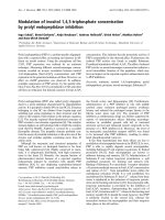
Between 1988 and 2001, 658 children were referred to the
section for paediatric rheumatology. JIA or related rheumatic
diseases were diagnosed in 574 children. In 84 of the chil-
dren, no rheumatic disease was diagnosed. The PV B19 IgG
status had already been determined as part of the routine
check up in 304 of the children. Of the remaining 270 children
in whom information on PV B19 serostatus was not available,
stored serum samples were available for 102. Both the chil-
dren with documented PV B19 status from the initial diagnosis
and children in whom PV B19 status was determined retro-
spectively were included in the analysis (Figure 1). Children
were classified into subgroups of JIA or related rheumatic dis-
eases based on the Durban Criteria of the International
League of Associations for Rheumatology [13]. A minor mod-
ification was used. Because EOPA is considered to be a sep-
arate disease entity, we differentiated this subgroup from other
oligoarthritides. The subgroups of children, along with demo-
graphic data, are summarized in Table 1. Twenty-four children
were excluded from the study because retrospective classifi-
cation ruled out rheumatic or associated diseases. In order to
allow age-matched comparison of PV B19 IgG status, the
control group was randomly divided into three subgroups with
mean ages and age ranges comparable to those in the patient
subgroups (Table 1).
Results of PV B19 serology
The results of PV B19 IgG determination are shown in Table
1. The percentage of PV B19 IgG positive children was not
significantly elevated in the disease subgroups compared with
the age-matched control groups. In fact, for the EOPA-JIA sub-
group, the percentage of PV B19 IgG positive children was
significantly lower than in the control group.
Individual virus specific IgG titres are influenced by various
factors, which prevents definition of normal values. Therefore,
comparison of IgG titres between different groups is usually of
only very limited value. For completeness, IgG titres of the PV
B19 IgG positive children were compared between the major
JIA subgroups (enthesitis associated arthritis, EOPA-JIA and
polyarthritis). No significant differences were identified (data
not shown).
Available online />Page 3 of 6
(page number not for citation purposes)
Discussion
Comparing the seroprevalence of PV B19 IgG between chil-
dren with JIA and a control group, we found no significant dif-
ference in PV B19 seropositivity. Our study was based on the
assumption that if there is an association between PV B19 and
a substantial proportion of JIA cases, then the prevalence of
PV B19 IgG in the children with JIA should be higher than that
in the control group, irrespective of time of infection. However,
our findings do not support an association between PV B19
and JIA.
In general, there are two basic approaches to diagnosing viral
infections: application of serological tests for antibody
detection and application of direct viral detection methods (in
particular polymerase chain reaction). Both approaches have
been used to study the potential link between B19 and JIA, but
thus far no unequivocal evidence favouring or refuting such an
association has been presented.
Following a description of a case of juvenile chronic arthritis
following acute infection with PV B19 [14], an observational
study conducted in 22 children with joint complaints in con-
junction with a recent PV B19 infection [12] identified six chil-
dren who developed chronic arthritis for 2 to 13 months.
Although the arthritis was attributed to the PV B19 infection,
these children would also have fulfilled the diagnostic criteria
for juvenile rheumatoid arthritis (JRA) [15].
In a study conducted in Japan [16], adult as well as juvenile
patients with rheumatoid arthritis were tested for B19 IgG.
Out of four patients with Still's disease, none was positive for
PV B19 IgG. However, five out of 11 patients (mean age 19.1
years, range 12 to 27 years) with polyarticular JRA (according
to the criteria of the American Rheumatism Association of
1976 [15]) were positive for B19 IgG, as compared with five
out of 60 age-matched control patients. The difference was
statistically significant (P = 0.006, by Fisher's exact test), but
the authors expressed concern in their discussion that statisti-
cal bias from case sampling might have occurred. Further-
more, the number of patients with JRA included in the study
was low. In addition, the mean age was high, suggesting blood
sampling later in the course of disease.
In a study conducted in India [17], 69 children and adolescent
persons with JRA (according to the criteria of the American
Rheumatism Association [18]), 26 adults with rheumatoid
Table 1
Demographic data and PV B19 serostatus of the disease subgroups and the corresponding age-related control subgroups
Classification Disease subgroups
a
Age-matched control groups
n Female
(%)
Mean age
(years)
b
Age range
(years)
b
B19 IgG
+
(n [%])
P
c
Subgroup n Female
(%)
Mean age
(years)
b
Age range
(years)
b
B19 IgG
+
(n [%])
EOPA 67 76.1 4.5 1.3–15.0 21 (31.3) <0.05 1 36 38.9 5.5 0.8–9.3 17 (47.2)
Systemic arthritis
(Still's disease)
8 100 6.7 4.8–11.1 5 (62.5) NS
Eye disease,
rheumatoid
(iridocyclitis)
6 83.3 7.3 3.8–11.1 2 (33.3) NS
Reactive arthritis 38 31.6 7.5 1.8–16.2 15 (39.5) <0.05 2 30 50.0 8.9 1.5–15.8 19 (63.3)
Other arthritis
(unclassified)
28 50 8.8 1.0–15.1 16 (57.1) NS
Arthralgias 85 48.2 9.3 2.3–16.5 53 (62.4) NS
Polyarthritis (RF
+
and RF
-
)
19 78.9 9.7 2.6–15.7 11 (57.9) NS
Lyme arthritis 37 43.2 10.2 2.8–15.5 25 (67.6) NS 3 80 41.3 11.2 1.9–18.3 52 (65.0)
Other
oligoarthritis
(nonHEOPA JIA)
13 38.5 10.6 4.8–18.8 7 (53.9) NS
Psoriatic arthritis 11 63.6 10.7 1.3–15.0 7 (63.6) NS
CRMO 12 66.7 11.5 6.8–15.3 6 (50.0) NS
EAA 54 61.1 11.6 1.9–18.3 39 (72.2) NS
SLE 4 100 13.0 9.7–15.9 4 (100) NS
a
Subgroups ordered by mean age.
b
Age when serum sample used for parvovirus (PV) B19 testing was drawn.
c
Comparison of disease subgroup
with age-matched control group. CRMO, Chronic recurrent multifocal osteomyelitis; EAA, Enthesitis associated arthritis; EOPA, Early-onset
pauciarticular arthritis; JIA, juvenile idiopathic arthritis; NS, not significant; RF, rheumatoid factor; SLE, System ic lupus erythematosus.
Arthritis Research & Therapy Vol 9 No 4 Weissbrich et al.
Page 4 of 6
(page number not for citation purposes)
arthritis (disease control individuals) and 12 healthy children
were tested for PV B19 IgG and IgM. Although the proportion
of PV B19 between patients (35/69 [50.7%]) and control indi-
viduals (19/38 [50.0%]) was almost identical, PV B19 IgM
was found significantly more often in the JRA group. Although
detailed information on the distribution of possible constella-
tions of PV B19 IgG and IgM antibodies was not provided in
the results section of the report, it can be deduced from the
abstract that ten JRA patients (14.5%) were positive for PV
B19 IgM but negative for PV B19 IgG. Because none of the
patients had clinical symptoms suggestive of acute PV B19
infection, this finding is highly unlikely. Thus, the specificity of
the PV B19 IgM test used in this study must be questioned.
Oguz and coworkers [19] used a different serological
approach to study the potential association between PV B19
infections and JRA. They determined PV B19 IgM in 75 chil-
dren with acute arthropathy. Sixteen were found to be positive
for PV B19 IgM. Children with persisting joint complaints were
followed up for at least 6 months. Three of the PV B19 IgM
positive patients but only one of the PV B19 IgM negative chil-
dren were diagnosed with JRA (P = 0.03). Prospective studies
such as this one are important in determining whether there is
a potential risk for developing JRA after acute PV B19 infec-
tion. Unfortunately, acute PV B19 infections in this study were
diagnosed solely using a peptide-based PV B19 IgM enzyme
immunoassay. Similar peptide-based assays have been found
to yield a considerable number of false-positive results com-
pared with assays based on recombinant antigens expressed
in the baculovirus system, especially in the presence of
autoantibodies and rheumatoid factor [20-22]. Confirmation of
the findings presented by Oguz and coworkers would there-
fore be desirable.
Figure 1
Enrollment, JIA diagnosis and PV B19 testingEnrollment, JIA diagnosis and PV B19 testing. JIA, juvenile idiopathic arthritis; PV, parvovirus.
Available online />Page 5 of 6
(page number not for citation purposes)
In an Italian study, Angelini and colleagues [23] compared the
prevalence of PV B19 IgG in a group of 35 children fulfilling
the 1987 criteria of the American College of Rheumatology for
rheumatoid arthritis [24] with that in a control group of 93 chil-
dren. The difference between the two groups was significant
(PV B19 IgG positive: 45.7% in the arthritis group versus
24.7% in the control group). The number of cases was consid-
erably smaller than that in the present study. Furthermore, the
patient and control populations cannot be compared with
those included in our study. Whereas we applied the Durban
criteria of juvenile idiopathic arthritis of 1997 to select our
patients [13], Angelini and colleagues used the criteria of the
American College of Rheumatology to diagnose JRA. The con-
trol population in their paper is not described in sufficient
detail to allow a comparison.
In another study of PV B19 and juvenile arthritis, Lehmann and
coworkers [25] examined PV B19 DNA in serum of 48 chil-
dren with joint complaints, which were selected on the basis
of positive IgG antibodies against the PV B19 nonstructural
protein (NS)1. Fifteen patients were positive for PV B19 DNA,
as compared with nine out of 124 healthy control children.
However, only 27 of the control children were positive for NS1
IgG, and three of these were positive for PV B19 DNA. Assum-
ing that the NS1 IgG positive children constitute the appropri-
ate control group, the proportions of PV B19 DNA positive
children were not significantly different between patients and
control children. In a follow-up study with the same control
group but including consecutively enrolled hospitalized chil-
dren with rheumatic diseases [11], the proportions of PV B19
IgG positive children were compared between patients and
control children. In agreement with our findings, there was no
significant difference. However, both NS1 IgG and PV B19
DNA were found more frequently in serum samples of children
with rheumatoid disease. PV B19 DNA was also detected in
22% of synovial membrane samples of children with JIA. How-
ever, a control group for comparison was not assessed. In
consideration of the fact that the overall frequency of PV B19
infections between patients and control individuals exhibited
no difference, it remains to be determined how the greater fre-
quency of PV B19 DNA in serum of children with JIA could
reflect pathogenicity of PV B19 for rheumatoid diseases. Fur-
ther studies will be necessary to prove that the presence of PV
B19 DNA in serum of children with rheumatoid disease is not
merely an epiphenomenon.
This issue is also pertinent to several other studies, both in
adults and in children, that described detection of PV B19
DNA in synovial fluid, cells, and tissue [26-32]. However,
because PV B19 DNA is also found in control samples with
varying frequency, it is still unclear whether the presence of PV
B19 DNA in synovial material is of pathogenic relevance
[26,27,29,30]. It was recently suggested that PV B19 DNA
may persist in human tissues throughout life [33]. Thus, the
presence of PV B19 in synovium appears insufficient in terms
of proving that it causes arthritis [34].
Because of its retrospective nature, our study has limitations
with respect to case finding and description of clinical mani-
festations. Not all case patients were tested for PV B19 IgG.
However, there is no indication of selection bias caused by the
exclusion of children for whom stored serum samples were not
available. PV B19 polymerase chain reaction and PV B19 IgM
tests were not used in this study because of limitations inher-
ent in the interpretation of positive results obtained with these
methods, and because the hypothesis to be tested in this
study was based on PV B19 IgG status.
Conclusion
In summary, there is no conclusive evidence yet for a patho-
genic role of PV B19 in JIA. Analysis of the seroprevalence of
anti-PV B19 IgG antibodies in European Caucasian children
affected by arthritis did not support the hypothesis that human
PV B19 is involved in the pathogenesis of JIA.
Competing interests
The authors declare that they have no competing interests.
Authors' contributions
YSF collected the patient data from the clinical records, per-
formed the PV B19 IgG testing, and analyzed the data. BW
and HJG designed and coordinated the study. BW contrib-
uted to the PV B19 testing and data analysis, and drafted the
manuscript. HJG cared for the patients, participated in the
analysis of the clinical data and contributed to the manuscript.
All authors read and approved the final version of the
manuscript.
Acknowledgements
The dedicated and skillful assistance of the technicians of the viral diag-
nostic laboratory is gratefully acknowledged. We thank Anne Zechel for
helpful comments on the manuscript.
References
1. Corcoran A, Doyle S: Advances in the biology, diagnosis and
host-pathogen interactions of parvovirus B19. J Med Microbiol
2004, 53:459-475.
2. Heegaard ED, Brown KE: Human parvovirus B19. Clin Microbiol
Rev 2002, 15:485-505.
3. Reid DM, Reid TM, Brown T, Rennie JA, Eastmond CJ: Human
parvovirus-associated arthritis: a clinical and laboratory
description. Lancet 1985, 1:422-425.
4. Kerr JR: Pathogenesis of human parvovirus B19 in rheumatic
disease. Ann Rheum Dis 2000, 59:672-683.
5. Prahalad S, Glass DN: Is juvenile rheumatoid arthritis/juvenile
idiopathic arthritis different from rheumatoid arthritis? Arthri-
tis Research 2002:303-310.
6. Fink CW: Reactive arthritis. Pediatr Infect Dis J 1988, 7:58-65.
7. Huppertz HI, Sandhage K: Salmonella enteritidis in reactive
carditis. Lancet 1993, 342:1488-1489.
8. Miller LC: Infectious causes of arthritis in adolescents. Adolesc
Med 1998, 9:115-126, vi.
9. Petty RE: Viruses and childhood arthritis. Ann Med 1997,
29:149-152.
10. Sieper J, Braun J, Doring E, Wu P, Heesemann J, Treharne J, Kings-
ley G: Aetiological role of bacteria associated with reactive
Arthritis Research & Therapy Vol 9 No 4 Weissbrich et al.
Page 6 of 6
(page number not for citation purposes)
arthritis in pauciarticular juvenile chronic arthritis. Ann Rheum
Dis 1992, 51:1208-1214.
11. Lehmann HW, Knoll A, Kuster RM, Modrow S: Frequent infection
with a viral pathogen, parvovirus B19, in rheumatic diseases of
childhood. Arthritis Rheum 2003, 48:1631-1638.
12. Nocton JJ, Miller LC, Tucker LB, Schaller JG: Human parvovirus
B19-associated arthritis in children. J Pediatr 1993,
122:186-190.
13. Petty RE, Southwood TR, Baum J, Bhettay E, Glass DN, Manners
P, Maldonado-Cocco J, Suarez-Almazor M, Orozco-Alcala J, Prieur
AM: Revision of the proposed classification criteria for juvenile
idiopathic arthritis: Durban, 1997. J Rheumatol 1998,
25:1991-1994.
14. Schwarz TF, Roggendorf M, Suschke H, Deinhardt F: Human par-
vovirus B19 infection and juvenile chronic polyarthritis [letter].
Infection 1987, 15:264-265.
15. Brewer EJ Jr, Bass J, Baum J, Cassidy JT, Fink C, Jacobs J, Hanson
V, Levinson JE, Schaller J, Stillman JS: Current proposed revision
of JRA Criteria. JRA Criteria Subcommittee of the Diagnostic
and Therapeutic Criteria Committee of the American Rheuma-
tism Section of The Arthritis Foundation. Arthritis Rheum 1977,
20(Suppl):195-199.
16. Mimori A, Misaki Y, Hachiya T, Ito K, Kano S: Prevalence of anti-
human parvovirus B19 IgG antibodies in patients with refrac-
tory rheumatoid arthritis and polyarticular juvenile rheumatoid
arthritis. Rheumatol Int 1994, 14:87-90.
17. Kishore J, Misra R, Gupta D, Ayyagari A: Raised IgM antibodies
to parvovirus B19 in juvenile rheumatoid arthritis. Indian J Med
Res 1998, 107:15-18.
18. Cassidy JT, Levinson JE, Bass JC, Baum J, Brewer EJ Jr, Fink CW,
Hanson V, Jacobs JC, Masi AT, Schaller JG, et al.: A study of clas-
sification criteria for a diagnosis of juvenile rheumatoid
arthritis. Arthritis Rheum 1986, 29:274-281.
19. Oguz F, Akdeniz C, Unuvar E, Kucukbasmaci O, Sidal M: Parvovi-
rus B19 in the acute arthropathies and juvenile rheumatoid
arthritis. J Paediatr Child Health 2002, 38:358-362.
20. Bruu AL, Nordbo SA: Evaluation of five commercial tests for
detection of immunoglobulin M antibodies to human parvovi-
rus B19. J Clin Microbiol
1995, 33:1363-1365.
21. Helftenbein E, Kunz H, Hartter P: Laboratory diagnosis of parvo-
virus B 19 infection: comparison of ten commercial IgG- and
IgM-antibody tests [in German]. Immun Infekt 1994,
22:181-186.
22. Sloots T, Devine PL: Evaluation of four commercial enzyme
immunoassays for detection of immunoglobulin M antibodies
to human parvovirus B19. Eur J Clin Microbiol Infect Dis 1996,
15:758-761.
23. Angelini F, Cancrini C, Colavita M, Panei P, Concato C, Romiti ML,
Chini L: Role of parvovirus B19 infection in juvenile chronic
arthritis. Is more investigation needed? Clin Exp Rheumatol
2003, 21:684.
24. Arnett FC, Edworthy SM, Bloch DA, McShane DJ, Fries JF, Cooper
NS, Healey LA, Kaplan SR, Liang MH, Luthra HS, et al.: The Amer-
ican Rheumatism Association 1987 revised criteria for the
classification of rheumatoid arthritis. Arthritis Rheum 1988,
31:315-324.
25. Lehmann HW, Kuhner L, Beckenlehner K, Muller-Godeffroy E,
Heide KG, Kuster RM, Modrow S: Chronic human parvovirus
B19 infection in rheumatic disease of childhood and
adolescence. J Clin Virol 2002, 25:135-143.
26. Cassinotti P, Siegl G, Michel BA, Bruhlmann P: Presence and
significance of human parvovirus B19 DNA in synovial mem-
branes and bone marrow from patients with arthritis of
unknown origin. J Med Virol 1998, 56:199-204.
27. Kerr JR, Cartron JP, Curran MD, Moore JE, Elliott JR, Mollan RA: A
study of the role of parvovirus B19 in rheumatoid arthritis. Br
J Rheumatol 1995, 34:809-813.
28. Nikkari S, Roivainen A, Hannonen P, Mottonen T, Luukkainen R, Yli-
Jama T, Toivanen P: Persistence of parvovirus B19 in synovial
fluid and bone marrow. Ann Rheum Dis 1995, 54:597-600.
29. Peterlana D, Puccetti A, Beri R, Ricci M, Simeoni S, Borgato L, Sci-
langa L, Ceru S, Corrocher R, Lunardi C: The presence of parvo-
virus B19 VP and NS1 genes in the synovium is not correlated
with rheumatoid arthritis. J Rheumatol 2003, 30:1907-1910.
30. Söderlund M, von Essen R, Haapasaari J, Kiistala U, Kiviluoto O,
Hedman K: Persistence of parvovirus B19 DNA in synovial
membranes of young patients with and without chronic
arthropathy [see comments]. Lancet 1997, 349:1063-1065.
31. Stahl HD, Seidl B, Hubner B, Altrichter S, Pfeiffer R, Pustowoit B,
Liebert UG, Hofmann J, von Salis-Soglio G, Emmrich F: High inci-
dence of parvovirus B19 DNA in synovial tissue of patients
with undifferentiated mono- and oligoarthritis. Clin Rheumatol
2000, 19:281-286.
32. Zakrzewska K, Azzi A, De Biasi E, Radossi P, De Santis R, Davoli
PG, Tagariello G: Persistence of parvovirus B19 DNA in syn-
ovium of patients with haemophilic arthritis. J Med Virol 2001,
65:402-407.
33. Norja P, Hokynar K, Aaltonen LM, Chen R, Ranki A, Partio EK,
Kiviluoto O, Davidkin I, Leivo T, Eis-Hubinger AM, et al.: Bioportfo-
lio: lifelong persistence of variant and prototypic erythrovirus
DNA genomes in human tissue. Proc Natl Acad Sci USA 2006,
103:7450-7453.
34. Kingsley G: Microbial DNA in the synovium: a role in aetiology
or a mere bystander? Lancet 1997, 349:1038-1039.