Báo cáo y học: "Detection of bone erosions in rheumatoid arthritis wrist joints with magnetic resonance imaging, computed tomography and radiography" ppt
Bạn đang xem bản rút gọn của tài liệu. Xem và tải ngay bản đầy đủ của tài liệu tại đây (289.79 KB, 8 trang )
Open Access
Available online />Page 1 of 8
(page number not for citation purposes)
Vol 10 No 1
Research article
Detection of bone erosions in rheumatoid arthritis wrist joints
with magnetic resonance imaging, computed tomography and
radiography
Uffe Møller Døhn
1
, Bo J Ejbjerg
1
, Maria Hasselquist
2
, Eva Narvestad
3
, Jakob Møller
2
,
Henrik S Thomsen
2
and Mikkel Østergaard
1,4
1
Department of Rheumatology, Copenhagen University Hospital Hvidovre, Kettegaard Allé 30, 2650 Hvidovre, Denmark
2
Department of Diagnostic Radiology, Copenhagen University Hospital Herlev, Herlev Ringvej 75, 2630 Herlev, Denmark
3
Department of Radiology, Copenhagen University Hospital Rigshospitalet, Blegdamsvej 1, 2100 Copenhagen, Denmark
4
Department of Rheumatology, Copenhagen University Hospital Herlev, Herlev Ringvej 75, 2630 Herlev, Denmark
Corresponding author: Uffe Møller Døhn,
Received: 7 Nov 2007 Revisions requested: 19 Dec 2007 Revisions received: 9 Jan 2008 Published: 28 Feb 2008
Arthritis Research & Therapy 2008, 10:R25 (doi:10.1186/ar2378)
This article is online at: />© 2008 Møller Døhn et al.; licensee BioMed Central Ltd.
This is an open access article distributed under the terms of the Creative Commons Attribution License ( />),
which permits unrestricted use, distribution, and reproduction in any medium, provided the original work is properly cited.
Abstract
Background The objectives of the present study were, with
multidetector computed tomography (CT) as the reference
method, to determine the performance of magnetic resonance
imaging (MRI) and radiography for the detection of bone
erosions in rheumatoid arthritis wrist bones, and to test whether
measuring volumes of erosions on CT and MRI is reproducible
and correlated to semiquantitative assessments (scores) of
erosions on CT, MRI and radiography.
Methods Seventeen patients with rheumatoid arthritis and four
healthy control individuals underwent CT, MRI and radiography
of one wrist, performed on the same day. CT was performed on
a Philips Mx8000IDT unit (voxel size 0.4 mm × 0.4 mm × 1 mm)
and MRI was performed on a Philips Panorama 0.6T unit (voxel
size 0.4 mm × 0.4 mm × 0.4 mm). Images were evaluated
separately for erosions in all wrist bones and were scored
according to the principles of the Outcome Measures in
Rheumatology Rheumatoid Arthritis MRI Scoring System (CT
and MRI) and the Sharp/van der Heijde (radiographs) scoring
methods. Measurements of erosion volumes of all erosions were
performed twice with a 1-week interval.
Results With CT as the reference method, the overall sensitivity,
specificity and accuracy (concordance) of MRI for detecting
erosions were 61%, 93% and 77%, respectively, while the
respective values were 24%, 99% and 63% for radiography.
The intramodality agreements when measuring erosion volumes
were high for both CT and MRI (Spearman correlation
coefficients 0.92 and 0.90 (both P < 0.01), respectively).
Correlations between volumes and scores of individual erosions
were 0.96 for CT and 0.99 for MRI, while they were 0.83 (CT)
and 0.80 (MRI) for persons' total erosion volume and total score
(all P < 0.01).
Conclusion With CT as the reference method, MRI showed
moderate sensitivity and good specificity and accuracy for
detection of erosions in rheumatoid arthritis and healthy wrist
bones, while radiography showed very low sensitivity. The
tested volumetric method was highly reproducible and
correlated to scores of erosions.
Introduction
Radiography, traditionally considered the golden standard for
assessing structural joint damage in patients with rheumatoid
arthritis (RA), is used routinely for diagnosing and monitoring
RA patients, and is used as an endpoint in clinical trials [1,2].
In early undifferentiated arthritis, the presence of bone ero-
sions is a risk factor for developing persisting arthritis [3], and
the presence of erosions when diagnosing RA is related to a
poor long-term functional and radiographic outcome [4-8]. For
these reasons, detection of erosions as early as possible is
desirable. Radiography does not visualise the earliest stages
of erosive changes in RA, however, and other imaging
CT = computed tomography; MRI = magnetic resonance imaging; OMERACT = Outcome Measures in Rheumatology; RA = rheumatoid arthritis;
RAMRIS = Rheumatoid Arthritis MRI Scoring System.
Arthritis Research & Therapy Vol 10 No 1 Døhn et al.
Page 2 of 8
(page number not for citation purposes)
modalities have emerged as methods for more sensitive detec-
tion of early bone erosions [9-12].
Magnetic resonance imaging (MRI) has been demonstrated to
be more sensitive than radiography in detecting erosive bone
changes in RA, especially the subtle changes that occur in
early disease [9-11,13,14]. The Outcome Measures in Rheu-
matology (OMERACT) Rheumatoid Arthritis MRI Scoring Sys-
tem (RAMRIS) has been developed [15,16] with data from
iterative multicenter studies [15,17,18]. The OMERACT RAM-
RIS is a semiquantitative scoring system for assessing synovi-
tis, bone erosions and bone edema on MRI in RA hands and
wrists. Studies on volumetric quantification of bone erosion
volumes with MRI have previously shown it is a reliable and
feasible method [19-21], and it could possibly be beneficial in
documenting progression or regression of structural joint dam-
age in longitudinal studies.
Multidetector computed tomography (CT) is a tomographic
radiographic imaging method offering isotropic high-resolution
and three-dimensional visualisation of calcified tissue. CT
seems to be even more sensitive than MRI for detection of
bone erosions, and can be considered a standard reference
for detection of bone erosions in RA [12,22,23].
An objective of the present cross-sectional methodological
study was, with CT as the reference method, to investigate the
sensitivity, specificity and accuracy (concordance) of MRI and
radiography for detection of bone erosions in RA wrist bones.
A second objective was to determine the intramodality and
intermodality agreement when measuring erosion volumes on
CT and MRI in RA wrist bones, using a semiautomated com-
puterised method. A third objective was to evaluate whether
semiquantitative scoring methods for bone erosions (the
OMERACT erosion score and the Sharp/van der Heijde radi-
ographic erosion score) correlated with erosion volumes
determined with CT and MRI.
Patients and methods
Patients and control individuals
Seventeen RA patients fulfilling the American College of
Rheumatology 1987 criteria [24] – of which 14 were rheuma-
toid factor positive – and four healthy control individuals were
included in the study. Fourteen patients were female and three
were male (median age 51 years (range 33–78 years), median
disease duration 7 years (range 4–22 years)), and three con-
trol individuals were female and one was male (median age 36
years (range 34–57 years)). All individuals underwent CT, MRI
and radiography of one wrist joint on the same day. The study
was approved by the local ethics committee, and written
informed consent was obtained from all participants.
Computed tomography
A Philips Mx8000 IDT multidetector unit (Philips Medical Sys-
tems, Cleveland, OH, USA) was used for all examinations
(parameters: 90 kV, 100 mAs, pitch 0.4 mm, slice spacing 0.4
mm, overlap 50%). Patients were placed in a prone position
with the arm stretched and the palm facing down. Images with
a voxel size of 0.4 mm × 0.4 mm × 1.0 mm were obtained.
Axial and coronal reconstructions with a slice thickness of 1.0
mm were created and used for image evaluation.
Magnetic resonance imaging
A Philips Panorama 0.6 T unit (Philips Medical Systems, Hel-
sinki, Finland) using a receive-only, three-channel, phased
solenoid coil was used for all examinations. Patients were
placed in a supine position with the hand alongside the body
and the palm facing the body. Acquired images included a
coronal T1-weighted three-dimensional fast field echo (repeti-
tion time 20 ms, echo time 8 ms, flip angle 25°, voxel size 0.4
mm × 0.4 mm × 0.4 mm, matrix 216 × 216, number of acqui-
sitions 2, acquisition time 5.23 min). Images in the axial and
coronal planes with a slice thickness of 0.4 mm were created
by multiplanar reconstruction of the T1 three-dimensional fast
field echo sequence, and these were used for image
evaluation.
Conventional radiography
Radiography was performed on a Philips Digital Diagnost unit
(Philips Medical Systems, Hamburg, Germany) (resolution 0.3
mm). Posterior-anterior and semisupine projections were
obtained and were printed on mammography films.
Image evaluation
Images obtained with CT, MRI and radiography were evalu-
ated for erosions by separate investigators, blinded to clinical
and other imaging data, with large experience from previous
imaging studies on RA. Erosions were marked on preformed
scoring sheets, allowing exact positioning in all three planes,
and an erosion score was assigned as described below.
Definitions of MRI erosions were as suggested by OMERACT
RAMRIS; that is, a sharply marginated bone lesion, with cor-
rect juxtaarticular localisation and typical signal characteris-
tics, visible in two planes with a cortical break seen in at least
one plane [15]. MRI bone erosions were scored according to
the OMERACT RAMRIS; that is, all wrist bones were assigned
a score by the percentage of bone volume involved (score 0–
10, by 10% volume increments) [15,25], leading to a total ero-
sion score for one wrist ranging from 0 to 150.
Erosions on CT images were defined as a sharply demarcated
area of focal bone loss seen in two planes, with a cortical
break (loss of cortex) seen in at least one plane. CT bone ero-
sions were scored according to the principles of the OMER-
ACT RAMRIS method described above.
We applied the principles from the Sharp/van der Heijde scor-
ing method in assessing radiographs, assigning an erosion
score ranging from 0 to 5 to all wrist bones [26]. Briefly,
Available online />Page 3 of 8
(page number not for citation purposes)
individual erosions are given a score of 1 when discrete, a
score of 2 if larger and a score of 3 when the erosion extends
over the imaginary middle of the bone. If more than one erosion
is present in a single bone, the sum of the scores (with a max-
imum of 5) of the individual erosions is calculated. With this
modification of the scoring method, the total erosion score of
one wrist ranges from 0 to 75.
Erosion volume measurements
Owing to the severity of bone damage or ankylosis we
excluded two patients from the analysis on erosion volume,
leaving 19 patients and 285 bones for further analysis. The vol-
umes of all erosions in the remaining 19 persons, detected by
CT or MRI in the evaluation described above, were calculated
using OsiriX medical imaging software (a free DICOM viewer
for Apple computers that can be downloaded [27]). To calcu-
late the erosion volume, erosions were manually outlined on
coronal images, on all slices where visible. The outlining of ero-
sion borders was done using an Intous3 A5 pen tablet system
(Wacom Technology Corporation, Vancouver, WA, USA). The
erosion volume is calculated by the software, according to the
formula: Vol
ero
= Σ(Area
ero
x ST), where Vol
ero
is the erosion vol-
ume, Area
ero
the erosion area on one slice and ST is the slice
thickness. All erosion volume measurements were performed
by the same person (UMD) on two occasions with a 1-week
interval between measuring on the same sets of images.
Statistical analysis
The specificity, sensitivity and accuracy of MRI and radiogra-
phy, with CT as the reference method, were calculated for
bone erosions. To determine the reliability of erosion volume
measurements, the absolute and relative differences, Spear-
man's correlation coefficients and the coefficient of variation of
erosion volumes obtained with CT and MRI at the two read-
ings (intramodality agreement) were calculated. Spearman's
correlation coefficients were calculated between the OMER-
ACT erosion scores and the erosion volumes of individual ero-
sions and between the persons' total OMERACT erosion
score and the persons' total erosion volumes (sum of the 15
evaluated joint areas). For erosions that were seen on both CT
and MRI – that is, concordant erosions – the absolute and rel-
ative differences between CT and MRI erosion volumes (inter-
modality agreement) were calculated. Furthermore,
intermodality agreements were assessed by calculation of
Spearman's correlation coefficients and coefficients of varia-
tion. Correlation coefficients between the erosion volume, CT
and MRI erosion scores and the radiographic erosion score
were calculated. For calculation of intermodality agreement,
the mean value of the volumes found at the two readings of CT
respective to MRI was used. SPSS version 12.0 for Windows
(SPSS Inc., Chicago, IL, USA) was used for statistical
calculations.
Results
In total, 315 wrist bones from 21 persons were assessed for
erosions. A total of 166 erosions in 151 bones were detected
with CT, while 119 erosions in 104 bones were detected on
MRI, and 43 erosions in 38 bones were detected with radiog-
raphy. With CT as the reference method for bone erosions, the
overall sensitivity, specificity and accuracy of MRI were 61%,
93% and 77%, respectively. The corresponding values for
radiography were 24%, 99% and 63%, respectively. Of the
119 MRI erosions, 92 (77%) could be confirmed with CT,
whereas 36 (84%) of the 43 radiographic erosions were con-
firmed with CT. If considering only bones without radiographic
erosions (n = 277), the overall sensitivity, specificity and accu-
racy of MRI were 59%, 93% and 79%, respectively. See Table
1 for further details.
Erosion-like changes were registered in two healthy controls
on CT, while one healthy control had three erosion-like
changes on MRI (the same control also had erosion-like
changes on CT) and none were seen on radiography.
Persons had a wide spectrum of joint destructions as judged
on their erosions scores. The total OMERACT erosion score
of one wrist (0–150) in all 21 persons was a mean of 10
(median 5, range 0–108) on MRI, while the mean was 15
(median 8, range 0–103) on CT. The total Sharp/van der Hei-
jde erosion score (modified as mentioned in Materials and
methods) produced a mean of 4 (median 1, range 0–43).
Erosion volume
Results on erosion volume measurements and values on
intramodality agreement of reading A and reading B (CT vs CT
and MRI vs MRI) and intermodality (CT vs MRI) agreements
are presented in Table 2. The intramodality agreements of sin-
gle erosion volume measurements at the two occasions were
very high for both CT (Spearman's ρ = 0.92, P < 0.01) and
MRI (ρ = 0.90, P < 0.01). The intramodality agreements of per-
sons' total erosion volume were also very high for CT (ρ =
0.83) and MRI (ρ = 0.80) (both P < 0.01). Volumes of erosions
seen on both CT and MRI (concordant erosions) were com-
pared. The volumes of the concordant erosions (n = 64) were
correlated (ρ = 0.55, P < 0.01), as were the total volumes of
concordant erosions on CT and MRI in the 15 persons with at
least one concordant erosion (ρ = 0.89, P < 0.01). A signifi-
cant correlation (ρ = 0.82, P < 0.01) between persons' (n =
19) total erosion volume on CT and MRI was also observed if
all erosions – that is, not only concordant erosions – were
included in the analysis.
Erosion volume versus the OMERACT erosion score
The OMERACT erosion scores in the 15 evaluated wrist joint
bones of the 19 examined persons (n = 285) were compared
with the corresponding erosion volumes. The Spearman's cor-
relation coefficients for CT and MRI erosion volumes and the
corresponding OMERACT CT and MRI scores were 0.96 and
Arthritis Research & Therapy Vol 10 No 1 Døhn et al.
Page 4 of 8
(page number not for citation purposes)
0.99 (both P < 0.01), respectively, when considering all 285
areas. When more than one erosion was present in a bone, the
sum of the volumes of erosions in the bone was used for com-
parison with the OMERACT score. The total erosion volume
per person (n = 19) and the total OMERACT erosion score of
the wrist were closely correlated, as Spearman's correlation
coefficients between volumes and scores on CT and MRI
were 0.83 and 0.80, respectively (both P < 0.01). The corre-
lation between the total MRI erosion score and the erosion vol-
ume determined on CT was ρ = 0.70 (P < 0.01).
Erosion volumes and OMERACT erosion scores versus
radiographic erosion scores
The correlation coefficients between the radiographic erosion
score of the individual wrist bones (n = 285), according to the
principles of the Sharp/van der Heijde scoring method, and
the erosion volume in the corresponding bone, as measured
on CT and MRI, were ρ = 0.27 (P < 0.01) and ρ = 0.10 (P =
0.10), respectively. Persons' total Sharp/van der Heijde ero-
sion score of all wrist bones in all persons (n = 19) correlated
with the total erosion volume on CT (ρ = 0.73, P < 0.01) and
MRI (ρ = 0.70, P < 0.01).
The Sharp/van der Heijde erosion score of the individual wrist
bones correlated weakly with the OMERACT erosion score on
CT (ρ = 0.27, P < 0.01) but did not correlate with the MRI
OMERACT erosion score (ρ = 0.10, P = 0.11). The persons'
total Sharp/van der Heijde erosion score, however, correlated
with the total OMERACT erosion score on both CT (ρ = 0.83,
P < 0.01) and MRI (ρ = 0.66, P < 0.01).
Discussion
With CT as the standard reference method for detecting bone
erosions in wrist joints, a moderate sensitivity (61%) and a
high specificity (93%) of MRI was demonstrated. Although
radiography was also highly specific (99%), only a low sensi-
tivity (24%) for erosions was reached when compared with
CT. The very low sensitivity of radiography, as compared with
CT, found in the present study can be explained by the two-
dimensional visualisation of the joint, and is in accordance with
findings from previous comparisons with MRI [9-13,23] and
with CT [12,23].
Since the amount of mobile protons in bone is very low, corti-
cal bone is depicted on MRI as signal voids against signal-
emitting bone marrow and periosseous tissues. MRI has con-
sequently been argued not to be a method well suited for vis-
ualising bone lesions, and the nature of erosions visualised
with MRI but invisible on radiography has been questioned
[22]. In the present study, however, MRI was markedly more
sensitive than radiography and was in good agreement with
CT even in regions without radiographic erosions, supporting
that even radiographically invisible MRI erosions represent a
true loss of calcified tissue.
Table 1
Sensitivities, specificities and accuracies for bone erosions of radiography and magnetic resonance imaging (MRI), with computed
tomography (CT) as reference
Bones with erosions
(number of erosions)
Radiography MRI MRI values in bones without
radiographic erosions (n = 277)
CT Radiography MRI Sensitivity
(%)
Specificity
(%)
Accuracy
(%)
Sensitivity
(%)
Specificity
(%)
Accuracy
(%)
Sensitivity
(%)
Specificity
(%)
Accuracy
(%)
Radius 10 (11) 2 (3) 6 (8) 20 100 62 60 100 81 50 100 79
Ulna 15 (15) 2 (2) 14 (15) 13 100 38 93 100 95 92 100 95
Scaphoid 11 (14) 3 (3) 8 (8) 27 100 62 64 90 76 50 90 72
Lunate 10 (11) 3 (3) 11 (14) 30 100 67 90 82 86 86 82 83
Triquetrum 14 (17) 5 (5) 13 (16) 36 100 57 86 86 86 100 86 94
Pisiforme 8 (8) 4 (5) 1 (1) 38 92 71 13 100 67 20 100 76
Trapezium 8 (8) 3 (5) 3 (3) 25 92 67 38 100 76 33 100 78
Trapezoid 8 (10) 2 (2) 9 (9) 25 100 71 86 85 86 83 85 84
Capitate 14 (14) 1 (1) 12 (16) 7 100 38 71 71 71 69 71 70
Hamate 9 (10) 3 (4) 7 (8) 33 100 71 56 83 71 50 85 71
Metacarpal base 1 8 (9) 3 (3) 5 (5) 38 100 76 63 100 86 40 100 83
Metacarpal base 2 16 (19) 1 (1) 9 (10) 6 100 29 56 100 67 53 100 65
Metacarpal base 3 5 (5) 1 (1) 2 (2) 20 100 81 20 94 76 25 94 80
Metacarpal base 4 8 (8) 3 (3) 2 (2) 38 100 76 25 100 71 20 100 78
Metacarpal base 5 7 (7) 2 (2) 2 (2) 29 100 76 14 93 67 20 93 74
Total 151 (166) 38 (43) 104 (119) 24 99 63 61 93 77 59 93 79
Available online />Page 5 of 8
(page number not for citation purposes)
The even higher agreement (87% vs 77%) between CT and
MRI in a study of nine RA wrist joints by Perry and colleagues
[12] may partly be explained by more advanced joint destruc-
tions in their cohort. In comparison with our previous study on
RA metacarpophalangeal joints [23], the level of agreement
between CT and MRI in the present study was lower. The anat-
Table 2
Intramodality and intermodality agreements of single and total erosion volume, measured on computed tomography (CT) and
magnetic resonance imaging (MRI)
Reading A
(mm
3
)
Reading B
(mm
3
)
Mean of
readings A
and B
(mm
3
)
Spearman ρ Absolute
difference
(mm
3
)
a
Absolute
numerical
difference
(mm
3
)
Relative difference
(%)
b
Relative
numerical
difference
(%)
Coefficient of
variation
Intramodality agreement: CT (reading A) vs CT (reading B) and MRI (reading A) vs MRI (reading B)
Volume per
erosion
CT
(n = 135)
13
(4; 1–245)
14
(4; 1–264)
13
(4; 1–255)
0.92* -1
(0; -28 to 12)
2
(1; 0–28)
-7
(0; -120 to 100)
29
(22; 0–120)
0.15
(0.11; 0–0.60)
MRI
(n = 90)
17
(10; 1–132)
17
(11; 1–138)
17
(11; 1–133)
0.90* 0
(0; -23 to 18)
4
(3; 0–23)
0
(0; -100 to 86)
28
(25; 0–100)
0.14
(0.13 0–0.50)
Volume per
person with
erosions
CT (n = 17) 102
(49; 2–519)
108
(56; 3–535)
105
(55; 3–527)
0.99* -6
(-2; -54 to 19)
12
(7; 1–54)
-10
(-6; -43 to 15)
16
(15; 3–43)
0.08
(0.07; 0.02–0.21)
MRI
(n = 15)
101
(80; 5–409)
100
(78; 5–409)
100
(76; 5–409)
0.95* 1
(0; -23 to 18)
7
(5; 0–23)
2
(0; -22 to 25)
8
(6; 0–25)
0.04
(0.03; 0–0.13)
Intermodality agreement (CT vs MRI)
c
Volume per
erosion of all
concordant
erosions
(n = 64)
CT 21
(5; 1–245)
22
(5; 1–255)
21
(5; 1–255)
0.55* 2
(-5; -55 to 132)
17
(9; 0–131)
-54
(-59; -174 to 167)
90
(92; 0–174)
0.46
(0.45; 0–0.87)
MRI 20
(13; 1–132)
19
(13; 1–138)
19
(13; 1–133)
Total volume
per person of
all
concordant
erosions (n =
15)
CT 88
(18; 1–514)
93
(23; 1–528)
91
(21; 1–521)
0.89* 8
(-7; -56 to 147)
40
(16; 4–147)
-48
(-63; -139 to 64)
72
(64; 6–139)
0.36
(0.32; 0.03–0.70)
MRI 83
(78; 5–374)
83
(62; 5–375)
83
(71; 5–375)
Total volume
per person of
all erosions
(n = 19)
CT 91
(36; 0–519)
100
(49, 0–535)
94
(38; 0–527)
0.82* 15
(2; -60 to 118)
41
(31; 0–118)
13
(6; -129 to 200)
68
(52; 0–200)
0.34
(0.26; 0–1.0)
MRI 79
(67; 0–409)
79
(67; 0–409)
79
(63; 0–409)
Data presented as the mean (median; range). Reading A and reading B, volumes obtained at the first (reading A) and second (reading B) volume measurements, done
by the same observer 1 week apart. The mean value of volumes obtained at reading A and B was used for the comparison of CT and MRI volumes. *P < 0.01.
a
Intramodality agreement, reading A minus reading B; intermodality agreement, CT erosion volume minus MRI erosion volume.
b
Intramodality agreement, positive values
refer to larger erosion volume at reading A than reading B, and vice versa; intermodality agreement, positive values refer to larger erosion volume on CT than MRI, and
vice versa.
c
Values on intermodality agreement are comparisons between CT and MRI erosions volumes.
Arthritis Research & Therapy Vol 10 No 1 Døhn et al.
Page 6 of 8
(page number not for citation purposes)
omy of the wrist is much more complicated than that of the
metacarpophalangeal joints, and many of the small carpal
bones have irregular margins with indentations (for example, at
the attachment of ligaments), making discrimination between
normal anatomy and presence of erosions difficult, and nutri-
tive foramina may also resemble erosions [28]. This may, at
least partly, explain the lower sensitivity and accuracy in this
wrist joint study compared with previous results from metacar-
pophalangeal joints [23]. In the present study, erosion-like
changes were registered in two healthy controls on CT and in
one healthy control on MRI. A low prevalence of erosion-like
changes on MRI in healthy controls has previously been
reported for wrists and metacarpophalangeal joints [29]. A 0.6
T (midfield) MRI unit was used in the present study. We expect
values on sensitivities and specificities on erosions are also
applicable to MRI units using higher field strengths, since pre-
vious studies have showed comparable results when images
obtained on MRI units with higher field strengths were com-
pared with images obtained on low-field MRI units [30-32].
As the wrist joint has proved more sensitive to changes in
bone erosions than other joint areas in RA [33], and bone
changes in the wrist joint have been shown to possess predic-
tive value with respect to further radiographic erosive progres-
sion [14,34,35], we found the wrist an important joint area to
investigate.
The number of erosions detected on CT, as compared with
MRI and radiography, indicate that CT is a very sensitive
method for detecting bone erosions in RA wrist bones, and
possibly even more sensitive than MRI. CT may therefore be of
value for detecting and monitoring bone erosions in RA. The
sensitivity to change is not yet established, however, and CT
is disfavored by using ionising radiation and by the inability to
visualise soft tissue changes.
We recently published data on the reliability of erosion volume
measurements on CT and MRI in RA metacarpophalangeal
joints, showing very high reproducibility when measuring ero-
sion volumes on CT and MRI, and good correlations between
CT and MRIerosion volumes and between erosion volumes
and erosion scores. [21] The present study on RA wrist joints
also showed a very high level of reproducibility when measur-
ing volumes of erosions on CT and MRI. Furthermore, semi-
quantitative scores of bone erosions according to the
OMERACT scoring system were closely correlated with both
CT and MRI volumes, both for individual joint regions and for
the wrist joint as a whole, supporting that the OMERACT ero-
sion score reflects the extent of erosive joint damage.
Although high to very high agreements of volumes were
reached between and within imaging modalities (respectively),
there were individual measurements that differed markedly.
Coincidental differences in outlining erosions at the two time
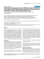
Figure 1
Erosions in the wrist of a rheumatoid arthritis patientErosions in the wrist of a rheumatoid arthritis patient. Wrist of a rheumatoid arthritis patient visualised by (a, b) computed tomography and (c, d) T1-
weighted magnetic resonance imaging in the (a, c) coronal and (b, d) axial planes. A bone erosion at the distal radius is seen on both computed tom-
ography and magnetic resonance images in two planes (white arrows), but not on the corresponding radiograph (e). The erosion was assigned an
OMERACT erosion score of 1 on both computed tomography and magnetic resonance imaging.
Available online />Page 7 of 8
(page number not for citation purposes)
points are potentially a major source of error. Especially, the
peripheral border of erosions can be difficult to define as sig-
nal intensities of erosions and adjacent soft-tissues often are
very similar for both CT and magnetic resonance images. Gen-
erally, the larger and more advanced the erosion, the more dif-
ficult it was to define the exact border of the erosions. The
estimated erosion volumes of concordant erosions were, on
average, larger on MRI than CT, as reflected by the mean rel-
ative difference in erosions' size. As cortical bone appears
black on MRI it may be included in the outlining of erosions,
and may consequently lead to overestimation of erosion size
on MRI compared with CT, where the cortical bone is well
delineated. Furthermore, the majority of erosions in the present
study were small; for small erosions, small absolute differ-
ences will result in large relative differences, with a systematic
bias towards larger volumes on MRI due to a proportionally
large area of cortical bone included in the estimation of erosion
size. The total erosion volume, however, was relatively larger
on CT than MRI due to more erosions being detected with CT.
Using the OMERACT RAMRIS, Haavardsholm and colleagues
have recently shown very good intrareader and good inter-
reader reliability, and a high level of sensitivity to change- dem-
onstrating that the OMERACT RAMRIS system, after proper
training and calibration of readers, appears suitable for use in
monitoring joint inflammation and destruction in RA [36]. The
close correlation with erosion volumes determined by MRI, as
well as CT, provides further important evidence of the OMER-
ACT RAMRIS erosion score being a valid measure of RA bone
destruction.
Conclusion
The present study demonstrated a high specificity of bone ero-
sions detected on MRI and radiography, and showed a mark-
edly higher sensitivity of MRI than radiography when CT was
considered the reference method. Secondly, when measuring
erosion volumes by CT and MRI, a very high intramodality and
a high intermodality agreement was reached, applying both to
individual erosion volume and persons' total erosion volume.
Owing to the high reproducibility, this quantitative method for
assessing bone erosions in RA patients could be a useful tool
in longitudinal studies, including randomised controlled trials,
but further studies, including studies of sensitivity to change,
are needed to clarify this issue. As the OMERACT erosion
scores were closely correlated with erosion volumes deter-
mined on CT and MRI, the present study supports the OMER-
ACT erosion score as a valid measure of RA joint destruction.
Competing interests
The authors declare that they have no competing interests.
Authors' contributions
UMD participated in the study development and recruitment of
patients, performed erosion volume measurements, con-
ducted data evaluation and statistical analysis, and prepared
the manuscript draft. BJE participated in the study
development, performed the evaluation of magnetic reso-
nance images, and was involved in patient recruitment. MH
was involved in the CT scanning protocol. EN performed the
evaluation of radiographs. JM was involved in the MRI scan-
ning protocol and performed all MRI examinations. HST partic-
ipated in the study development and gave substantial input to
data evaluation and manuscript preparation. MØ participated
in the study development, was involved in the CT and MRI
scanning protocol, evaluated CT images, and gave substantial
input to data evaluation and manuscript preparation. All
authors read and approved the final manuscript.
Acknowledgements
The Danish Rheumatism Association and the Copenhagen University
Hospital at Hvidovre are acknowledged for financial support. Photogra-
pher Ms Susanne Østergaard is acknowledged for preparation of the
figure.
References
1. American College of Rheumatology Subcommittee on Rheumatoid
Arthritis: Guidelines for the management of rheumatoid arthri-
tis: 2002 Update. Arthritis Rheum 2002, 46:328-346.
2. Boers M, Felson DT: Clinical measures in rheumatoid arthritis:
which are most useful in assessing patients? J Rheumatol
1994, 21:1773-1774.
3. Visser H, le CS, Vos K, Breedveld FC, Hazes JM: How to diag-
nose rheumatoid arthritis early: a prediction model for persist-
ent (erosive) arthritis. Arthritis Rheum 2002, 46:357-365.
4. Kaarela K: Prognostic factors and diagnostic criteria in early
rheumatoid arthritis. Scand J Rheumatol Suppl 1985, 57:1-54.
5. van der Heijde DM, van Leeuwen MA, van Riel PL, Koster AM, van't
Hof MA, van Rijswijk MH, van de Putte LB: Biannual radiographic
assessments of hands and feet in a three-year prospective fol-
lowup of patients with early rheumatoid arthritis. Arthritis
Rheum 1992, 35:26-34.
6. van der Heijde DM: Joint erosions and patients with early rheu-
matoid arthritis. Br J Rheumatol 1995, 34(Suppl 2):74-78.
7. Nissila M, Isomaki H, Kaarela K, Kiviniemi P, Martio J, Sarna S:
Prognosis of inflammatory joint diseases. A three-year follow-
up study. Scand J Rheumatol 1983, 12:33-38.
8. Mottonen TT: Prediction of erosiveness and rate of develop-
ment of new erosions in early rheumatoid arthritis. Ann Rheum
Dis 1988, 47:648-653.
9. McQueen FM, Stewart N, Crabbe J, Robinson E, Yeoman S, Tan
PL, McLean L: Magnetic resonance imaging of the wrist in early
rheumatoid arthritis reveals a high prevalence of erosions at
four months after symptom onset. Ann Rheum Dis 1998,
57:350-356.
10. Backhaus M, Kamradt T, Sandrock D, Loreck D, Fritz J, Wolf KJ,
Raber H, Hamm B, Burmester GR, Bollow M: Arthritis of the fin-
ger joints: a comprehensive approach comparing conventional
radiography, scintigraphy, ultrasound, and contrast-enhanced
magnetic resonance imaging. Arthritis Rheum 1999,
42:1232-1245.
11. Klarlund M, Østergaard M, Jensen KE, Madsen JL, Skjødt H, Loren-
zen I:
Magnetic resonance imaging, radiography, and scintigra-
phy of the finger joints: one year follow up of patients with
early arthritis. The TIRA Group. Ann Rheum Dis 2000,
59:521-528.
12. Perry D, Stewart N, Benton N, Robinson E, Yeoman S, Crabbe J,
McQueen F: Detection of erosions in the rheumatoid hand; a
comparative study of multidetector computerized tomography
versus magnetic resonance scanning. J Rheumatol 2005,
32:256-267.
13. Conaghan P, O'Connor P, McGonagle D, Astin P, Wakefield RJ,
Gibbon WW, Quinn M, Karim Z, Green MJ, Proudman S, Isaacs J,
Emery P: Elucidation of the relationship between synovitis and
bone damage: a randomized magnetic resonance imaging
Arthritis Research & Therapy Vol 10 No 1 Døhn et al.
Page 8 of 8
(page number not for citation purposes)
study of individual joints in patients with early rheumatoid
arthritis. Arthritis Rheum 2003, 48:64-71.
14. Lindegaard HM, Vallø J, Hørslev-Petersen K, Junker P, Østergaard
M: Low-cost, low-field dedicated extremity magnetic reso-
nance imaging in early rheumatoid arthritis: a 1-year follow-up
study. Ann Rheum Dis 2006, 65:1208-1212.
15. Østergaard M, Peterfy C, Conaghan P, McQueen F, Bird P, Ejbjerg
B, Shnier R, O'Connor P, Klarlund M, Emery P, Genant H, Lassere
M, Edmonds J: OMERACT Rheumatoid Arthritis Magnetic Res-
onance Imaging Studies. Core set of MRI acquisitions, joint
pathology definitions, and the OMERACT RA-MRI scoring
system. J Rheumatol 2003, 30:1385-1386.
16. Lassere M, McQueen F, Østergaard M, Conaghan P, Shnier R,
Peterfy C, Klarlund M, Bird P, O'Connor P, Stewart N, Emery P,
Genant H, Edmonds J: OMERACT Rheumatoid Arthritis Mag-
netic Resonance Imaging Studies. Exercise 3: an international
multicenter reliability study using the RA-MRI score. J
Rheumatol 2003, 30:1366-1375.
17. Østergaard M, Klarlund M, Lassere M, Conaghan P, Peterfy C,
McQueen F, O'Connor P, Shnier R, Stewart N, McGonagle D,
Emery P, Genant H, Edmonds J: Interreader agreement in the
assessment of magnetic resonance images of rheumatoid
arthritis wrist and finger joints – an international multicenter
study. J Rheumatol 2001, 28:1143-1150.
18. Conaghan P, Lassere M, Østergaard M, Peterfy C, McQueen F,
O'Connor P, Bird P, Ejbjerg B, Klarlund M, Shnier R, Genant H,
Emery P, Edmonds J: OMERACT Rheumatoid Arthritis Magnetic
Resonance Imaging Studies. Exercise 4: an international mul-
ticenter longitudinal study using the RA-MRI score. J
Rheumatol 2003, 30:1376-1379.
19. Bird P, Lassere M, Shnier R, Edmonds J: Computerized meas-
urement of magnetic resonance imaging erosion volumes in
patients with rheumatoid arthritis: a comparison with existing
magnetic resonance imaging scoring systems and standard
clinical outcome measures. Arthritis Rheum 2003, 48:614-624.
20. Bird P, Ejbjerg B, McQueen F, Østergaard M, Lassere M,
Edmonds J: OMERACT Rheumatoid Arthritis Magnetic Reso-
nance Imaging Studies. Exercise 5: an international multi-
center reliability study using computerized MRI erosion
volume measurements. J Rheumatol 2003, 30:1380-1384.
21. Møller Døhn U, Ejbjerg BJ, Hasselquist M, Narvestad E, Court-
Payen M, Szkudlarek M, Møller J, Thomsen HS, Østergaard M:
Rheumatoid arthritis bone erosion volumes on CT and MRI:
reliability and correlations with erosion scores on CT, MRI and
radiography. Ann Rheum Dis 2007, 66:
1388-1392.
22. Goldbach-Mansky R, Woodburn J, Yao L, Lipsky PE: Magnetic
resonance imaging in the evaluation of bone damage in rheu-
matoid arthritis: a more precise image or just a more expen-
sive one? Arthritis Rheum 2003, 48:585-589.
23. Møller Døhn U, Ejbjerg BJ, Court-Payen M, Hasselquist M, Narves-
tad E, Szkudlarek M, Møller JM, Thomsen H, Østergaard M: Are
bone erosions detected by magnetic resonance imaging and
ultrasonography true erosions? A comparison with computed
tomography in rheumatoid arthritis metacarpophalangeal
joints. Arthritis Res Therapy 2006, 8:R110.
24. Arnett FC, Edworthy SM, Bloch DA, McShane DJ, Fries JF, Cooper
NS, Healey LA, Kaplan SR, Liang MH, Luthra HS, Medsger TA Jr,
Mitchell DM, Neustadt DH, Pinals RS, Schaller JG, Sharp JT,
Wilder RL, Hunder GG: The American Rheumatism Association
1987 revised criteria for the classification of rheumatoid
arthritis. Arthritis Rheum 1988, 31:315-324.
25. Conaghan P, Bird P, Ejbjerg B, O'Connor P, Peterfy C, McQueen
F, Lassere M, Emery P, Shnier R, Edmonds J, Østergaard M: The
EULAR-OMERACT rheumatoid arthritis MRI reference image
atlas: the metacarpophalangeal joints. Ann Rheum Dis 2005,
64(Suppl 1):i11-i21.
26. van der Heijde DM: How to read radiographs according to the
Sharp/van der Heijde method. J Rheumatol 2000, 27:261-263.
27. OsiriX Imaging Software [
]
28. McQueen F, Østergaard M, Peterfy C, Lassere M, Ejbjerg B, Bird
P, O'Connor P, Genant H, Shnier R, Emery P, Edmonds J, Cona-
ghan P: Pitfalls in scoring MR images of rheumatoid arthritis
wrist and metacarpophalangeal joints. Ann Rheum Dis 2005,
64(Suppl 1):i48-i55.
29. Ejbjerg B, Narvestad E, Rostrup E, Szkudlarek M, Jacobsen S,
Thomsen HS, Østergaard M: Magnetic resonance imaging of
wrist and finger joints in healthy subjects occasionally shows
changes resembling erosions and synovitis as seen in rheu-
matoid arthritis. Arthritis Rheum 2004, 50:1097-1106.
30. Taouli B, Zaim S, Peterfy CG, Lynch JA, Stork A, Guermazi A, Fan
B, Fye KH, Genant HK: Rheumatoid arthritis of the hand and
wrist: comparison of three imaging techniques. Am J
Roentgenol 2004, 182:937-943.
31. Savnik A, Malmskov H, Thomsen HS, Bretlau T, Graff LB, Nielsen
H, Danneskiold-Samsøe B, Boesen J, Bliddal H: MRI of the
arthritic small joints: comparison of extremity MRI (0.2 T) vs
high-field MRI (1.5 T). Eur Radiol 2001, 11:1030-1038.
32. Ejbjerg BJ, Narvestad E, Jacobsen S, Thomsen HS, Østergaard M:
Optimised, low cost, low field dedicated extremity MRI is
highly specific and sensitive for synovitis and bone erosions in
rheumatoid arthritis wrist and finger joints: comparison with
conventional high field MRI and radiography. Ann Rheum Dis
2005, 64:1280-1287.
33. Ejbjerg BJ, Vestergaard A, Jacobsen S, Thomsen HS, Østergaard
M: The smallest detectable difference and sensitivity to
change of magnetic resonance imaging and radiographic
scoring of structural joint damage in rheumatoid arthritis fin-
ger, wrist, and toe joints: a comparison of the OMERACT rheu-
matoid arthritis magnetic resonance imaging score applied to
different joint combinations and the Sharp/van der Heijde
radiographic score. Arthritis Rheum 2005, 52:2300-2306.
34. McQueen FM, Stewart N, Crabbe J, Robinson E, Yeoman S, Tan
PLJ, McLean L: Magnetic resonance imaging of the wrist in
early rheumatoid arthritis reveals progression of erosions
despite clinical improvement. Ann Rheum Dis 1999,
58:156-163.
35. Haavardsholm EA, Boyesen P, Østergaard M, Schildvold A, Kvien
TK: MRI findings in 84 early rheumatoid arthritis patients: bone
marrow edema predicts erosive progression. Ann Rheum Dis
2007 in press.
36. Haavardsholm E, Østergaard M, Ejbjerg B, Kvan N, Uhlig T, Lilleas
F, Kvien T: Reliability and sensitivity to change of the OMER-
ACT rheumatoid arthritis MRI score (RAMRIS) in a multi-
reader longitudinal setting. Arthritis Rheum 2005,
52:3860-3867.