Báo cáo y học: "Immune regulation of bone loss by Th17 cells" doc
Bạn đang xem bản rút gọn của tài liệu. Xem và tải ngay bản đầy đủ của tài liệu tại đây (334.6 KB, 9 trang )
Page 1 of 9
(page number not for citation purposes)
Available online />Abstract
A significant macrophage and T-cell infiltrate commonly occurs in
inflammatory joint conditions such as rheumatoid arthritis that have
significant bone destruction. Cytokines produced by activated
macrophages and T cells are implicated in arthritis pathogenesis
and are involved in osteoclast-mediated bone resorption. The
scope of the present review is to analyze current knowledge and to
provide a better understanding of how macrophage-derived factors
promote the differentiation of a novel T-helper subset (Th17) that
promotes osteoclast formation and activation.
Introduction
Rheumatoid arthritis (RA) is a chronic inflammatory disease
driven by immune dysregulation. The cause remains unknown,
but environmental and genetic factors are believed to contri-
bute to RA development. RA is characterized by joint inflam-
mation, initially resulting in pain and swelling, and in a majority
of the patients there is bone and cartilage erosion. The goal
of current treatment regimens is to control inflammation and
to retard the progression of structural damage of the joint
bone structure as measured by X-ray analysis.
The RA joint synovial fluid and synovium contain a variety of
hematopoeitic cells that are in direct proximity with the
articular cartilage and underlying bone, and that contribute to
the joint-destructive process. The present review will focus
upon the bone-degrading cell, the osteoclast, and on how
different T-cell subsets and their signature cytokines
positively and negatively modulate osteoclast activity in
autoimmune-driven inflammatory bone diseases. A specific T-
cell subset, the Th17 cell, has a significant osteoclastogenic
potential in the arthritic joint and may therefore provide a
suitable target to combat arthritis and loss of joint function.
Osteoclast differentiation
Physiological and pathological bone resorption is carried out
by a specialized cell called the osteoclast. Osteoclasts are
large 20 to 100 μm multinucleated cells containing three to
100 nuclei with many mitochondria, lysosomes, dense
granules, vesicles, and an extensive Golgi network required
for the synthesis and secretion of factors required to degrade
the bone matrix and subsequently phagocytose the resorbed
products [1]. Tartrate-resistant acid phosphatase [2],
cathepsin K [3], calcitonin receptor [4], and α
v
β
3
integrin [5]
are characteristic gene products of the mature osteoclast [6].
The initial event in bone resorption is the attachment of the
mature osteoclast to the bone matrix. Cell surface α
v
β
3
integrins bind to a variety of extracellular matrix proteins,
including vitronectin, osteopontin, and bone sialoprotein. Arg-
Gly-Asp-containing peptides, Arg-Gly-Asp mimetics, and
blocking antibodies to α
v
β
3
integrins inhibit bone resorption
in vitro and in vivo, suggesting that this integrin plays a key
role in osteoclast function [7]. Once attached to bone, the
osteoclast generates an isolated extracellular microenviron-
ment between itself and the bone surface by creating a
sealing zone structure unique to the osteoclast. A ring of
filamentous actin associates with the intracellular proteins
vinculin, talin, α-actinin, and cortactin, and the ring links to
α
v
β
3
integrins that have bound various extracellular proteins.
The ring appears when the osteoclast is immobilized on bone
and during the resorption process. The ring disappears prior
to osteoclast detachment from the eroded site and migration
to another resorption site.
Resorption depends upon acidification of this extracellular
compartment, leading to demineralization of the inorganic
bone component and subsequent organic matrix degradation
by cysteine proteases. Cathepsin K, an acid-activated
cysteine proteinase, plays a critical and necessary role in the
bone resorption process [8]. The expanded membrane within
the filamentous actin ring creates additional surface area for
massive H
+
transport performed by the vacuolar (V-type)
Review
Immune regulation of bone loss by Th17 cells
Iannis E Adamopoulos and Edward P Bowman
Department of Immunology, Schering Plough Biopharma, 901 California Avenue, Palo Alto, CA 94304, USA
Corresponding author: Iannis E Adamopoulos,
Published: 17 October 2008 Arthritis Research & Therapy 2008, 10:225 (doi:10.1186/ar2502)
This article is online at />© 2008 BioMed Central Ltd
CIA = collagen-induced arthritis; CSF = colony-stimulating factor; IFN = interferon; IL = interleukin; NF = nuclear factor; RA = rheumatoid arthritis;
RANK = receptor activator of NFκB; RANKL = receptor activator of NFκB ligand; Th = T-helper cells; TGFβ = transforming growth factor beta; TNF =
tumor necrosis factor; Treg = regulatory T cell.
Page 2 of 9
(page number not for citation purposes)
Arthritis Research & Therapy Vol 10 No 5 Adamopoulos and Bowman
electrogenic H
+
-ATPase [9]. The proton source is carbonic
acid produced by carbonic anhydrase type II [9]. The intra-
cellular pH is balanced by a passive chloride–bicarbonate
exchange in the basolateral membrane. Metalloproteinases
secreted by vesicles into the sealing zone can also degrade
the organic matrix, but their actions are only partly known. The
resorbed material is transcytosed through the osteoclast
[10,11]. Although there are a few reports that macrophages
and fibroblasts degrade or resorb mineralized bone in vitro
[12,13], it is widely accepted that osteoclasts are the only
cells capable of lacunar resorption [6].
The osteoclast lineage is distinct from other mesenchymal cells
of the bone (osteoblasts, chondrocytes, adipocytes, bone-
marrow stromal cells, and fibroblasts). Osteoclasts are
hematopoietic derived [14] and are from the colony-forming
unit granulocyte–macrophage progenitor cells, which give rise
to granulocytes and macrophages [15]. Monocytes are
released from the bone marrow into the blood, where they
home into different tissues and differentiate into tissue-resident
macrophages. Multinucleated osteoclasts are formed under
appropriate stimuli by the fusion of mononuclear precursors
within the monocyte fraction of peripheral blood [16].
PU.1 is a monocyte/macrophage-specific transcription factor
that acts as a master switch in programming hematopoietic cell
commitment and differentiation [17]. PU.1 promotes the cell-
type-specific expression of the myeloid lineage genes CD11b,
CD11c, CD18, the granulocyte colony-stimulating factor (CSF)
receptor, the granulocyte–macrophage CSF receptor, and the
macrophage CSF receptor (c-fms) via binding to these genes’
promoter regions [18]. Mice with homozygous PU.1 deficiency
have osteopetrotic bones due to the lack of osteoclasts that
would form from the myeloid lineage [19].
Osteoclast activation
Macrophage CSF and receptor activator of NFκB ligand
(RANKL) are the most important factors known to date to
drive osteoclast formation and activity [20,21]. Macrophage
CSF is a survival factor for osteoclast precursors due to
upregulating Bcl-X
L
, inhibiting caspase-9 activation [22], and
supporting mature osteoclast survival by preventing
apoptosis [23]. Macrophage CSF stimulation also stimulates
receptor activator of NFκB (RANK) expression in osteoclast
precursor cells, thereby allowing RANKL to drive mature
osteoclast formation [24]. RANKL is a transmembrane
protein expressed by activated osteoblasts, synovial fibro-
blasts, and T cells. It can also be proteolytically cleaved by
TNF convertase (TACE) to generate a soluble molecule that
has osteoclastic activity at distal sites [25,26]. RANKL-
induced osteoclastogenesis is inhibited by osteoprotegerin, a
soluble decoy receptor for RANKL, which is also produced
by a variety of cells including osteoblasts, synovial fibroblasts,
B cells, and T cells [25-29]. Osteoprotegerin-deficient mice
are severely osteoporotic [30], while osteoprotegerin-trans-
genic mice are osteopetrotic [28].
The NFκB pathway is an integral component in the osteoclast
differentiation pathway. RANKL activates NFκB both via the
canonical pathway due to IκBα degradation to release
p50/RelA and p50/c-Rel heterodimers and via the alternative
or noncanonical pathway by promoting p100 processing into
p52 [31]. TNF also stimulates NFκB activation via the
canonical pathway [32], and a synergistic interaction
between RANKL and TNF may account for the pathological
osteoclastogenesis seen in RA [33,34]. Elevated joint TNF
activates TNF receptors expressed by osteoclast precursors
[35] to drive NFκB and Jun N-terminal kinase activation using
the TNF receptor adaptor proteins TRAF1, TRAF2, and
TRAF6 [36-39]. Importantly, TNF induces osteoclastogenesis
in RANK-deficient mice [40] and induces multinucleated cell
formation from osteoclast precursors in the bone marrow
macrophage population [41].
IL-1 receptors are expressed on mature osteoclasts [42], and
exogenous IL-1 induces NFκB activation via TRAF6 [43]. IL-1
signals feed into the tyrosine kinase pathways through a
TRAF6–Src molecular complex, which regulates the cyto-
skeletal reorganization essential for osteoclast activation and
can enhance the ruffled border formation of the mature osteo-
clast and hence its resorbing activity [44].
There is a significant macrophage infiltrate in the arthritic
joint, and the extent of synovial macrophage infiltration corre-
lates strongly with the degree of joint erosion in arthritis [45].
Synovial macrophages isolated from different types of
arthritides differentiate in vitro to fully functional osteoclasts
via RANKL stimulation as well as independently of the
RANK/RANKL signaling pathway, via TNF and IL-1α
signaling [46]. Stimulatory, costimulatory, and/or inhibitory
signals may be provided by adjacent T cells present in the
inflammatory infiltrate.
T-cell subsets and their action on osteoclasts
The seminal observation in 1972 that osteoclast activity was
increased by leukocyte-derived factors [47] gave rise to the
idea that T cells influenced osteoclast differentiation and
activation. Similar to the increased macrophage infiltrate,
activated T cells are also found in inflamed RA synovial tissue
and CD4
+
T-cell infiltration has become a hallmark of RA
pathogenesis. This concept has become central to the under-
standing of RA, and a new field termed osteoimmunology has
recently emerged. How T cells regulate osteoclastogenesis is
only partly known. We provide a brief overview of four T-cell
subsets and the factors that drive their differentiation into
these subsets, and we then concentrate on how the
signature cytokine(s) of these T cells impact inflammation-
driven osteoclastogenesis in arthritis.
T-helper 17 cells
The Th17 lineage has only recently been recognized and the
factors involved in its differentiation are still being identified
and sorted out. If a naïve T cell is activated in the presence of
Page 3 of 9
(page number not for citation purposes)
transforming growth factor beta (TGFβ) plus IL-6 in the
mouse or TGFβ plus an inflammatory stimuli in the human,
then the resulting clonal memory T-cell population will be
instructed to produce the Th17 signature cytokines IL-17A,
IL-17F, IL-22, and IL-26 (there is no mouse IL-26). The inflam-
matory stimuli in the human setting can be IL-1β, IL-6, IL-21,
and/or IL-23 [48-53].
IL-17A is the only Th17 signature cytokine currently known to
influence the biology of osteoclasts. The cytokine’s message
is also present in RAG-deficient mice (that is, T-cell and B-
cell deficient), consistent with IL-17A also being produced
from nonlymphoid sources [54]. IL-17A receptors are single-
pass transmembrane proteins expressed by many cell types,
including synoviocytes [55], chondrocytes, [56] osteoblasts,
[57], and osteoclasts (our data). Receptor activation results
in NFκB activation and the phosphorylation and activation of
the extracellular signal-regulated kinase, Jun N-terminal kinase,
and p38 mitogen-activated protein kinase pathways [56].
Th17 cells express RANKL [58] and TNF [52,59], which
directly act on osteoclast precursors to induce osteoclasto-
genesis. IL-17A induces RANKL expression by synovial fibro-
blasts and osteoblasts to indirectly drive bone erosion [60]
and to activate synovial macrophages to secrete the known
osteoclastogenic factors TNF and IL-1β [61]. Synovial and
synovial fluid macrophages can differentiate to fully functional
bone-resorbing osteoclasts, and Th17-induced synovial
macrophage–osteoclast differentiation may represent an
important cellular mechanism in the bone destruction asso-
ciated with RA [46,62].
Several types of IL-17A antagonists have been used in a
variety of animal arthritis models to address the efficacy of
therapeutic IL-17A neutralization. Polyclonal anti-IL-17A anti-
body treatment after disease induction in the collagen-
induced arthritis (CIA) model decreased clinical scores over
10 days of therapy compared with controls. Ankles and knees
had reduced synovitis, cartilage damage, chondrocyte death,
proteoglycan depletion, and bone erosion (histologically and
radiographically) [63]. Polyclonal anti-IL-17A antibody also
inhibited the antigen-induced arthritis model’s knee swelling,
proteoglycan depletion, and bone erosion in the smoldering
knee following reintroduction of antigen [64]. Mice immunized
with formalin-fixed Borrelia burgdorferi and later challenged
with live B. burgdorferi display transient joint inflammation.
Knee swelling was reduced with either anti-IL-17A or anti-
IL-17RA therapy, and ankles and knees were free of histo-
pathological changes including bone erosions following
either therapy [65]. Lastly, rat adjuvant-induced arthritis
models treated with an IL-17R–Fc fusion protein demon-
strated decreased paw swelling, joint histopathology scores,
and bone radiographic scores [66].
Mechanistically, the bone protection following therapeutic
IL-17A neutralization has been attributed to normalizing
excessive RANKL levels. Anti-IL-17A blockade in CIA and
antigen-induced arthritis models that demonstrated decreased
bone erosion correlated with decreased RANKL message
and RANKL-positive cells in the joints [64,67].
T-helper 1 cells
IL-12 is a master differentiation factor produced by activated
antigen-presenting cells. If a naïve T cell is activated in the
presence of IL-12, then the resulting clonal memory T-cell
population is instructed to produce the Th1 signature
cytokine IFNγ.
IFNγ is also produced by another hematopoeitic cell type, the
natural killer cell, following its activation. Osteoclast
precursors and mature osteoclasts express the IFNγ receptor
and exogenous IFNγ in vitro inhibits murine osteoclasto-
genesis by inducing the rapid degradation of the RANK
adapter protein TRAF6, resulting in strong inhibition of RANKL-
induced activation of NFκB and Jun N-terminal kinase [68].
Activated mouse Th1 cells inhibited osteoclastogenesis when
mixed in cocultures with either RANKL-stimulated mouse bone
marrow macrophages or osteoblasts stimulated with vitamin
D
3
and prostaglandin E
2
[58]. Activated Th1 cells expressed
significant IFNγ quantities, and the inhibitory effects of these
cells on osteoclastogenesis were abrogated when using IFNγ
receptor-deficient osteoclast precursors [58]. Although
exogenous IFNγ inhibits RANKL-induced human osteoclasto-
genesis, it was paradoxical that IFNγ-producing human T cells
induced osteoclast formation in a RANKL-dependent mecha-
nism (discussed below) [69]. The increased osteoclasto-
genesis effect may well be due to a Th1 proinflammatory
cytokine TNF, which is known to synergize with RANKL.
The Th1 master differentiation factor IL-12 has anti-osteo-
clastogenic activities but the mechanism of inhibition remains
unclear. Exogenous IL-12 inhibited RANKL-stimulated osteo-
clast formation from splenocytes or from osteoblast and
splenocyte cocultures. This IL-12-dependent inhibition,
however, was not due to IFNγ [70]. IL-12 was also identified
as the anti-osteoclast factor produced by Toll-like receptor 9
stimulation with CpG oligodeoxynucleotides that opposed
RANKL-induced osteoclast differentiation [71]. CpG oligo-
deoxynucleotides, known to mimic bacterial DNA, modulate
osteoclastogenesis via interactions with osteoclast precursors
and osteoblasts [72]. Mice specifically deficient in IL-12 (that
is, IL-12p35-deficient mice) were not protected from CIA, but
show exacerbated paw swelling responses following collagen
challenge [73]. Similarly, mice lacking IFNγ signaling are not
protected in the CIA model [74]. A Lyme’s disease arthritis
model using a two-step inactivated Borrelia vaccination
followed by live Borrelia challenge shows a greater disease
penetrance and severity when using IFNγ-deficient mice [65].
Collectively, these data do not support that Th1 cells, nor
their signature cytokine IFNγ or their master differentiation
Available online />factor IL-12, are major drivers of inflammation-associated osteo-
clastogenesis. These data instead support that this lineage may
counteract other pro-osteoclast inflammation factors present in
the RA joint, including Th17 products [75,76].
T-helper 2 cells
The source of the Th2 master differentiation factor IL-4 during
the naïve to memory transition is controversial, but mature
Th2 cells, natural killer T cells, basophils, and mast cells
produce IL-4 [77-79]. Th2 cells produce the signature
cytokines IL-4, IL-5, and IL-13 when they encounter a nonself-
antigen (or a cross-reacting self-antigen) being presented by
an antigen-presenting cell. Little is know regarding the role, if
any, that IL-5 plays in osteoclast biology, whereas IL-4 and
IL-13 have been extensively studied. Mouse spleen cells and
bone marrow macrophages express IL-4 and IL-13 receptors,
and the addition of these factors decreased RANKL-
stimulated tartrate-resistant acid phosphatase-positive multi-
nucleated cell formation and cathepsin K message in spleno-
cytes and bnone marrow macrophages [80]. IL-4 was more
potent than IL-13 at inhibiting mouse osteoblast/osteoclast
progenitor cocultures [81]. Mature osteoclasts express the
IL-4 receptor, and exogenous IL-4 decreased tartrate-resistant
acid phosphatase expression, actin ring formation, and bone
resorption by osteoclasts [82]. Since IL-4 inhibits osteoclasto-
genesis by inhibiting NFκB and mitogen-activated protein
kinase signaling [83], the synergistic action of RANKL and TNF
to stimulate osteoclast formation is also inhibited by IL-4 [84].
The Th2 signature cytokines IL-4 and IL-13 also use other
mechanisms to inhibit osteoclastogenesis. IL-4 and IL-13
promote expression of the natural RANKL antagonist, osteo-
protegerin, by endothelial cells and osteoblasts [81,85]. IL-4
and IL-13 decreased RANKL and RANK protein in calvariae
bone. Exogenous IL-4 addition to the CIA model had minimal
impact on the outward signs of inflammation, but showed a
bone-preserving biology including decreased bone erosion,
tartrate-resistant acid phosphatase activity, and RANKL
message [86,87]. In total, these data do not support that Th2
cells or their signature cytokines IL-4 and IL-13 are major
drivers of inflammation-associated osteoclastogenesis, but
instead support that this lineage along with the Th1 lineage
may counteract Th17 products and other pro-osteoclast
inflammation factors present in the RA joint.
Regulatory T-cells
The regulatory T cell (Treg) lineage is composed of over-
lapping T-cell subsets whose role is to dampen the immune
response to minimize tissue destruction. There are a number
of mechanisms by which the Treg dampens the immune
system, including some requiring cell-to-cell contact, but the
present review will focus upon the Treg signature cytokines
TGFβ and IL-10 [88].
There is support for Tregs inhibiting osteoclastogenesis, but
there are discrepancies regarding the key mechanisms by
which it occurs. Zaiss and colleagues concluded that
CD4
+
CD25
+
Foxp3
+
Tregs inhibited osteoclastogenesis in a
cell-contact-dependent manner with a minor contribution by
the signature cytokines IL-4, TGFβ, and IL-10 [89,90]. In
contrast, Kim and colleagues reported that human
CD4
+
CD25
+
Foxp3
+
Tregs inhibit osteoclast differentiation
from peripheral blood mononuclear cells in a cell-contact-
independent manner, but in an IL-4-dependent and TGFβ-
dependent manner [91,92].
Although IL-4 is not considered a Treg signature cytokine, it
does inhibit osteoclastogenesis by inhibiting NFκB and
mitogen-activated protein kinase signaling [83]. The role of
TGFβ in bone turnover is quite complex, with conflicting in
vivo and in vitro data on whether resorption is enhanced or
inhibited [93]. IL-10, however, has a clear inhibitory effect on
osteoclasts by downregulating NFATc1 [94].
Tregs are enriched in inflamed joints of patients with
rheumatic disease [95], but the interaction of Tregs with
activated monocytes in the joint might lead to diminished
suppressive activity of Tregs in vivo – thus contributing to the
chronic inflammation in RA [96]. Other workers have reported
that Tregs have neither an inhibitory effect nor an enhancing
effect on osteoclastogenesis. Collectively, these data do not
support that Tregs nor their signature cytokines TGFβ and
IL-10 are major drivers of inflammation-associated osteo-
clastogenesis, but instead support that this lineage along
with Th1 and Th2 signature cytokines may counteract Th17
and other pro-osteoclast inflammatory factors present in the
RA joint.
Flavors of T-helper 17 cells
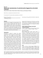
The schematic concept of separate nonoverlapping T-cell
lineages identified by their unique cytokine repertoire, how-
ever, is hampered by reality (Figure 1). A subset of Th17 cells
defined by their expression of the signature cytokine IL-17A
also produce IFNγ, albeit less than that produced by Th1
cells [53]. The IL-17A/IFNγ double-producing human T cells
arise from in vitro naïve to memory T-cell differentiation
cultures and are found in peripheral blood memory T cells
from healthy donors [50,52,53]. It could be envisioned that
factors present in the microenvironment during the naïve to
memory transition could alter either the number of double
producers that arise or could alter the magnitude of IL-17A to
IFNγ protein made by the double producers [49,50,53].
These different flavors of Th17 cells and, even more, the
distinctly different T-cell subsets must be kept in mind when
evaluating the literature precedent regarding T-cell effects on
osteoclasts. The paradoxical observation that IFNγ-producing
T cells promoted osteoclast formation, highlighted above,
provides such an example. IFNγ-producing T cells and IFNγ-
nonproducing T cells were cultured with human monocytes in
the presence of macrophage CSF. To the authors’ surprise,
IFNγ-producing T cells induced osteoclastogenesis in a
Arthritis Research & Therapy Vol 10 No 5 Adamopoulos and Bowman
Page 4 of 9
(page number not for citation purposes)
RANKL-dependent manner. The osteoclastogenesis was
further enhanced when the anti-osteoclast factor IFNγ was
neutralized, implicating other pro-osteoclast factor(s) being
produced by IFNγ-producing T cells [69]. Th17 cells can be
endowed with different cytokine repertoires and/or different
secretion ratios between key cytokines based on master
differentiation factors present in the microenvironment during
initial antigen recognition [49]. All of the various osteoclast-
related factors must be taken into account when hypothesizing
the impact that an activated T cell may have in an inflamed joint.
Mouse models have been key to expanding our knowledge of
various T-cell subsets, the biology of their signature cyto-
kines, and how these cytokines affect osteoclasts. This is also
true for the Th17 lineage; however, there are two keys areas
where mouse studies must be analyzed with caution when
translating to humans.
IL-6 (in combination of TGFβ) is required to differentiate
activated naïve mouse CD4 T cells into memory IL-17A-
producing Th17 cells [52]. Further exposure to IL-23 was
required for full pathogenic activity in vivo (at least in animal
central nervous system inflammation models). IL-6 is just one of
a handful of proinflammatory factors (for example, IL-1β, IL-21,
and IL-23) that can work singly and in combination to drive
human Th17 development [49,50,52,53]. This dichotomy may
overemphasize the role of IL-6 (from mouse studies) in human
Th17-mediated osteoclast formation.
Secondly, Th17 cells from mouse autoimmune models rarely
coexpress IFNγ – whereas this is more the norm when
working with human Th17 cells from healthy donors. Whether
this is due to an inherent mouse/human difference or due to
different disease states remains to be seen. Human Th17
cells may therefore have a more prominent IFNγ anti-osteo-
clastogenesis brake on the human cell’s activity than a
corresponding mouse Th17 cell. This would be consistent
with some rodent arthritis models having an explosive bone
eroding component, in comparison with the much slower
bone erosion seen in RA patients. It is important to note that
these above cautions relate to how a memory Th17 cell is
formed. Once the Th17 cell is formed, however, the
osteoclastogenic factors produced by the cell have similar
biology between the mouse and the human (for example,
RANKL, TNF, IL-17A).
T-helper 17 cells in RA
Multiple lines of evidence point to Th17’s disease association
with RA. IL-17A protein is present in both the synovium and
the synovial fluid of rheumatoid patients [97-101], and a
subset of T-cell lines expanded in vitro from RA synovium
expressed IL-17A and IFNγ following activation [59,102].
Classical IFNγ-only Th1 cells were also present and can be
expanded from RA synovium [102,103]. Exploratory medical
studies, however, not only support IL-17A’s disease asso-
ciation, but implicate IL-17A’s correlation with poor disease
prognosis. IL-17A message in synovial membrane biopsies
Available online />Page 5 of 9
(page number not for citation purposes)
Figure 1
Schematic representation of T cell differentiation and T-cell signature cytokines that inhibit or induce osteoclastogenesis. CFU-GM,
granulocyte–macrophage colony-stimulating factor; RANKL, receptor activator of NFκB ligand; TGFβ, transforming growth factor beta; Treg,
regulatory T cell.
was one factor (including TNF, IL-1β, and IL-10) that was
predictive for subsequent bone erosion and joint damage as
assessed by magnetic resonance imaging and radiography
[104].
Additionally, Raza and colleagues prospectively collected
synovial fluid samples within a few weeks after symptom
onset from patients with early synovitis [105]. The patient’s
outcomes were subsequently determined 18 months after
fluid acquisition to determine what early factors expression
correlated with synovitis progression to RA. Patients were
grouped into those who subsequently progressed to RA,
those who subsequently progressed to non rheumatoid
persistent synovitis, or those whose synovitis resolved.
Importantly, IL-17A was one of a few factors (including IL-2,
IL-4, IL-13, IL-15, basic fibroblast growth factor, and
epidermal growth factor) whose expression in the earliest
stages of disease was associated with subsequent progres-
sion to RA [105]. An IL-17A single nucleotide polymorphism
was significantly associated with radiographic progression
after 2 years [106].
Collectively, these data support that IL-17A is present in the
inflamed synovium and that IL-17A expression levels correlate
with poor prognosis and greater joint destruction.
In summary, Th17 cells are arrayed with factors that can
directly and indirectly drive osteoclastogenesis. It is well
appreciated within the T-cell development literature that
master differentiation factors and signature cytokines of the
Th1, Th2, and Treg lineage inhibit Th17 development. This
same concept holds true from an osteoclastogenesis view-
point since master differentiation factors and signature cyto-
kines of the Th1, Th2, and Treg lineage inhibit osteoclasto-
genesis and Th17 products stimulate osteoclastogenesis.
Conclusion
Th17 cells are a newly discovered T-cell lineage that plays a
role in the adaptive immune response to extracellular patho-
gens. The repertoire of molecules at its disposal to combat
these pathogens is formidable. When these products are
unleashed within an inflamed joint, however, they can directly
drive osteoclast precursors to differentiate into osteoclasts
via RANKL-dependent and RANKL-independent pathways.
Additionally, Th17 factors can act on other cell types to
indirectly increase the osteoclastogenic potential in the
inflamed joint microenvironment. Targeting this T-cell lineage
and/or its osteoclastic factors may provide alternative
strategies to combat inflammatory joint diseases compared
with the gold-standard TNF antagonist therapies.
Competing interests
IEA and EPB are employees of Schering-Plough Corporation
and therefore, receive salary from and/or hold stock in
Schering-Plough.
References
1. Holtrop ME, King GJ: The ultrastructure of the osteoclast and
its functional implications. Clin Orthop 1977, 123:177-196.
2. Minkin C: Bone acid phosphatase: tartrate-resistant acid
phosphatase as a marker of osteoclast function. Calcif Tissue
Int 1982, 34:285-290.
3. Drake FH, Dodds RA, James IE, Connor JR, Debouck C, Richard-
son S, Lee-Rykaczewski E, Coleman L, Rieman D, Barthlow R,
Hastings G, Gowen M: Cathepsin K, but not cathepsins B, L, or
S, is abundantly expressed in human osteoclasts. J Biol Chem
1996, 271:12511-12516.
4. Hattersley G, Chambers TJ: Calcitonin receptors as markers for
osteoclastic differentiation: correlation between generation of
bone-resorptive cells and cells that express calcitonin recep-
tors in mouse bone marrow cultures. Endocrinology 1989,
125:1606-1612.
5. Davies J, Warwick J, Totty N, Philp R, Helfrich M, Horton M: The
osteoclast functional antigen, implicated in the regulation of
bone resorption, is biochemically related to the vitronectin
receptor. J Cell Biol 1989, 109(4 Pt 1):1817-1826.
6. Teitelbaum SL: Bone resorption by osteoclasts. Science (NY)
2000, 289:1504-1508.
7. Horton MA, Taylor ML, Arnett TR, Helfrich MH: Arg-Gly-Asp
(RGD) peptides and the anti-vitronectin receptor antibody
23C6 inhibit dentine resorption and cell spreading by osteo-
clasts. Exp Cell Res 1991, 195:368-375.
8. Zaidi M, Troen B, Moonga BS, Abe E: Cathepsin K, osteoclastic
resorption, and osteoporosis therapy. J Bone Miner Res 2001,
16:1747-1749.
9. Blair HC, Teitelbaum SL, Ghiselli R, Gluck S: Osteoclastic bone
resorption by a polarized vacuolar proton pump. Science (NY)
1989, 245:855-857.
10. Salo J, Lehenkari P, Mulari M, Metsikko K, Vaananen HK:
Removal of osteoclast bone resorption products by transcyto-
sis. Science (NY) 1997, 276:270-273.
11. Nesbitt SA, Horton MA: Trafficking of matrix collagens through
bone-resorbing osteoclasts. Science (NY) 1997, 276:266-269.
12. Eilon G, Mundy GR: Direct resorption of bone by human breast
cancer cells in vitro. Nature 1978, 276:726-728.
13. Pap T, Claus A, Ohtsu S, Hummel KM, Schwartz P, Drynda S,
Pap G, Machner A, Stein B, George M, Gay RE, Neumann W,
Gay S, Aicher WK: Osteoclast-independent bone resorption
by fibroblast-like cells. Arthritis Res Ther 2003, 5:R163-R173.
14. Walker DG: Bone resorption restored in osteopetrotic mice by
transplants of normal bone marrow and spleen cells. Science
1975, 190:784-785.
15. Kurihara N, Chenu C, Miller M, Civin C, Roodman GD: Identifica-
tion of committed mononuclear precursors for osteoclast-like
cells formed in long term human marrow cultures. Endocrinol-
ogy 1990, 126:2733-2741.
16. Fujikawa Y, Quinn JM, Sabokbar A, McGee JO, Athanasou NA:
The human osteoclast precursor circulates in the monocyte
fraction. Endocrinology 1996, 137:4058-4060.
17. Klemsz MJ, McKercher SR, Celada A, Van Beveren C, Maki RA:
The macrophage and B cell-specific transcription factor PU.1
is related to the ets oncogene. Cell 1990, 61:113-124.
18. Zhang DE, Hetherington CJ, Chen HM, Tenen DG: The
macrophage transcription factor PU.1 directs tissue-specific
expression of the macrophage colony-stimulating factor
receptor. Mol Cell Biol 1994, 14:373-381.
19. Tondravi MM, McKercher SR, Anderson K, Erdmann JM, Quiroz
M, Maki R, Teitelbaum SL: Osteopetrosis in mice lacking
haematopoietic transcription factor PU.1. Nature 1997, 386:
81-84.
20. Lacey DL, Timms E, Tan HL, Kelley MJ, Dunstan CR, Burgess T,
Elliott R, Colombero A, Elliott G, Scully S, Hsu H, Sullivan J,
Hawkins N, Davy E, Capparelli C, Eli A, Qian Y X, Kaufman S,
Sarosi I, Shalhoub V, Senaldi G, Guo J, Delaney J, Boyle WJ:
Osteoprotegerin ligand is a cytokine that regulates osteoclast
differentiation and activation. Cell 1998, 93:165-176.
21. Quinn JM, Elliott J, Gillespie MT, Martin TJ: A combination of
osteoclast differentiation factor and macrophage-colony stim-
ulating factor is sufficient for both human and mouse osteo-
clast formation in vitro. Endocrinology 1998, 139:4424-4427.
22. Woo KM, Kim HM, Ko JS: Macrophage colony-stimulating
factor promotes the survival of osteoclast precursors by up-
regulating Bcl-X(L). Exp Mol Med 2002, 34:340-346.
Arthritis Research & Therapy Vol 10 No 5 Adamopoulos and Bowman
Page 6 of 9
(page number not for citation purposes)
23. Fuller K, Owens JM, Jagger CJ, Wilson A, Moss R, Chambers TJ:
Macrophage colony-stimulating factor stimulates survival and
chemotactic behavior in isolated osteoclasts. J Exp Med 1993,
178:1733-1744.
24. Arai F, Miyamoto T, Ohneda O, Inada T, Sudo T, Brasel K, Miyata
T, Anderson DM, Suda T: Commitment and differentiation of
osteoclast precursor cells by the sequential expression of c-
Fms and receptor activator of nuclear factor
κκ
B (RANK)
receptors. J Exp Med 1999, 190:1741-1754.
25. Hofbauer LC, Khosla S, Dunstan CR, Lacey DL, Boyle WJ, Riggs
BL: The roles of osteoprotegerin and osteoprotegerin ligand
in the paracrine regulation of bone resorption. J Bone Miner
Res 2000, 15:2-12.
26. Li Y, Toraldo G, Li A, Yang X, Zhang H, Qian W-P, Weitzmann
MN: B cells and T cells are critical for the preservation of bone
homeostasis and attainment of peak bone mass in vivo. Blood
2007, 109:3839-3848.
27. Takayanagi H, Iizuka H, Juji T, Nakagawa T, Yamamoto A, Miyazaki
T, Koshihara Y, Oda H, Nakamura K, Tanaka S: Involvement of
receptor activator of nuclear factor kappaB ligand/osteoclast
differentiation factor in osteoclastogenesis from synoviocytes
in rheumatoid arthritis. Arthritis Rheum 2000, 43:259-269.
28. Simonet WS, Lacey DL, Dunstan CR, Kelley M, Chang MS, Luthy
R, Nguyen HQ, Wooden S, Bennett L, Boone T, Shimamoto G,
DeRose M, Elliott R, Colombero A, Tan HL, Trail G, Sullivan J,
Davy E, Bucay N, Renshaw-Gegg L, Hughes TM, Hill D, Pattison
W, Campbell P, Sander S, Van G, Tarpley J, Derby P, Lee R,
Boyle WJ: Osteoprotegerin: a novel secreted protein involved
in the regulation of bone density. Cell 1997, 89:309-319.
29. Gillespie MT: Impact of cytokines and T lymphocytes upon
osteoclast differentiation and function. Arthritis Res Ther 2007,
9:103.
30. Bucay N, Sarosi I, Dunstan CR, Morony S, Tarpley J, Capparelli C,
Scully S, Tan HL, Xu W, Lacey DL, Boyle WJ, Simonet WS:
Osteoprotegerin-deficient mice develop early onset osteo-
porosis and arterial calcification. Genes Dev 1998, 12:1260-
1268.
31. Beinke S, Ley SC: Functions of NF-
κκ
B1 and NF-
κκ
B2 in immune
cell biology. Biochem J 2004, 382(Pt 2):393-409.
32. Chaisson ML, Branstetter DG, Derry JM, Armstrong AP, Tometsko
ME, Takeda K, Akira S, Dougall WC: Osteoclast differentiation
is impaired in the absence of inhibitor of kappa B kinase
alpha. J Biol Chem 2004, 279:54841-54848.
33. Zhang G, Ghosh S: Toll-like receptor-mediated NF-kappaB
activation: a phylogenetically conserved paradigm in innate
immunity. J Clin Invest 2001, 107:13-19.
34. Lam J, Takeshita S, Barker JE, Kanagawa O, Ross FP, Teitelbaum
SL: TNF-
αα
induces osteoclastogenesis by direct stimulation
of macrophages exposed to permissive levels of RANK
ligand. J Clin Invest 2000, 106:1481-1488.
35. Abu-Amer Y, Erdmann J, Alexopoulou L, Kollias G, Ross FP, Teit-
elbaum SL: Tumor necrosis factor receptors types 1 and 2 dif-
ferentially regulate osteoclastogenesis. J Biol Chem 2000,
275:27307-27310.
36. Liu ZG, Hsu H, Goeddel DV, Karin M: Dissection of TNF recep-
tor 1 effector functions: JNK activation is not linked to apopto-
sis while NF-
κκ
B activation prevents cell death. Cell 1996, 87:
565-576.
37. Fuller K, Murphy C, Kirstein B, Fox SW, Chambers TJ: TNF
αα
potently activates osteoclasts, through a direct action inde-
pendent of and strongly synergistic with RANKL. Endocrinol-
ogy 2002, 143:1108-1118.
38. Rothe M, Wong SC, Henzel WJ, Goeddel DV: A novel family of
putative signal transducers associated with the cytoplasmic
domain of the 75 kDa tumor necrosis factor receptor. Cell
1994, 78:681-692.
39. Kaji K, Katogi R, Azuma Y, Naito A, Inoue JI, Kudo A: Tumor
necrosis factor alpha-induced osteoclastogenesis requires
tumor necrosis factor receptor-associated factor 6. J Bone
Miner Res 2001, 16:1593-1599.
40. Li J, Sarosi I, Yan X-Q, Morony S, Capparelli C, Tan H-L, McCabe
S, Elliott R, Scully S, Van G, Kaufman S, Juan SC, Sun Yu, Tarpley
J, Martin L, Christensen K, McCabe J, Kostenuik P, Hsu H,
Fletcher F, Dunstan CR, Lacey DL, Boyle WJ: RANK is the intrin-
sic hematopoietic cell surface receptor that controls osteo-
clastogenesis and regulation of bone mass and calcium
metabolism. Proc Natl Acad Sci U S A 2000, 97:1566-1571.
41. Azuma Y, Kaji K, Katogi R, Takeshita S, Kudo A: Tumor necrosis
factor-alpha induces differentiation of and bone resorption by
osteoclasts. J Biol Chem 2000, 275:4858-4864.
42. Xu LX, Kukita T, Nakano Y, Yu H, Hotokebuchi T, Kuratani T, Iijima
T, Koga T: Osteoclasts in normal and adjuvant arthritis bone
tissues express the mRNA for both type I and II interleukin-1
receptors. Lab Invest 1996, 75:677-687.
43. Ye H, Arron JR, Lamothe B, Cirilli M, Kobayashi T, Shevde NK,
Segal D, Dzivenu OK, Vologodskaia M, Yim M,Du K, Singh S, Pike
J, Wesley D, Bryant G, Choi Y, Wu H: Distinct molecular mech-
anism for initiating TRAF6 signalling. Nature 2002, 418:443-
447.
44. Nakamura I, Kadono Y, Takayanagi H, Jimi E, Miyazaki T, Oda H,
Nakamura K, Tanaka S, Rodan GA, Duong LT: IL-1 regulates
cytoskeletal organization in osteoclasts via TNF receptor-
associated factor 6/c-Src complex. J Immunol 2002, 168:
5103-5109.
45. Yanni G, Whelan A, Feighery C, Bresnihan B: Synovial tissue
macrophages and joint erosion in rheumatoid arthritis. Ann
Rheum Dis 1994, 53:39-44.
46. Adamopoulos IE, Sabokbar A, Wordsworth BP, Carr A, Ferguson
DJ, Athanasou NA: Synovial fluid macrophages are capable of
osteoclast formation and resorption. J Pathol 2006, 208:35-43.
47. Horton JE, Raisz LG, Simmons HA, Oppenheim JJ, Mergenhagen
SE: Bone resorbing activity in supernatant fluid from cultured
human peripheral blood leukocytes. Science (NY) 1972, 177:
793-795.
48. Langrish CL, Chen Y, Blumenschein WM, Mattson J, Basham B,
Sedgwick JD, McClanahan T, Kastelein RA, Cua DJ: IL-23 drives
a pathogenic T cell population that induces autoimmune
inflammation. J Exp Med 2005, 201:233-240.
49. Volpe E, Servant N, Zollinger R, Bogiatzi SI, Hupe P, Barillot E,
Soumelis V: A critical function for transforming growth factor-
beta, interleukin 23 and proinflammatory cytokines in driving
and modulating human T(H)-17 responses. Nat Immunol
2008, 9:650-657.
50. Manel N, Unutmaz D, Littman DR: The differentiation of human
T(H)-17 cells requires transforming growth factor-beta and
induction of the nuclear receptor RORgammat. Nat Immunol
2008, 9:641-649.
51. Yang L, Anderson DE, Baecher-Allan C, Hastings WD, Bettelli E,
Oukka M, Kuchroo VK, Hafler DA: IL-21 and TGF-beta are
required for differentiation of human T(H)17 cells. Nature
2008, 454:350-352.
52. Acosta-Rodriguez EV, Napolitani G, Lanzavecchia A, Sallusto F:
Interleukins 1beta and 6 but not transforming growth factor-
beta are essential for the differentiation of interleukin 17-pro-
ducing human T helper cells. Nat Immunol 2007, 8:942-949.
53. Wilson NJ, Boniface K, Chan JR, McKenzie BS, Blumenschein
WM, Mattson JD, Basham B, Smith K, Chen T, Morel F, Lecron
JC, Kastelein RA, Cua DJ, McClanahan TK, Bowman EP, de Waal
Malefyt R: Development, cytokine profile and function of
human interleukin 17-producing helper T cells. Nat Immunol
2007, 8:950-957.
54. Uhlig HH, McKenzie BS, Hue S, Thompson C, Joyce-Shaikh B,
Stepankova R, Robinson N, Buonocore S, Tlaskalova-Hogenova
H, Cua DJ, Powrie F: Differential activity of IL-12 and IL-23 in
mucosal and systemic innate immune pathology. Immunity
2006, 25:309-318.
55. Zrioual S, Toh ML, Tournadre A, Zhou Y, Cazalis MA, Pachot A,
Miossec V, Miossec P: IL-17RA and IL-17RC receptors are
essential for IL-17A-induced ELR
+
CXC chemokine expres-
sion in synoviocytes and are overexpressed in rheumatoid
blood. J Immunol 2008, 180:655-663.
56. Moseley TA, Haudenschild DR, Rose L, Reddi AH: Interleukin-17
family and IL-17 receptors. Cytokine Growth Factor Rev 2003,
14:155-174.
57. Van bezooijen RL, Farih-Sips HC, Papapoulos SE, Lowik CW:
Interleukin-17: a new bone acting cytokine in vitro. J Bone
Miner Res 1999, 14:1513-1521.
58. Sato K, Suematsu A, Okamoto K, Yamaguchi A, Morishita Y,
Kadono Y, Tanaka S, Kodama T, Akira S, Iwakura Y, Cua DJ,
Takayanagi H: Th17 functions as an osteoclastogenic helper T
cell subset that links T cell activation and bone destruction. J
Exp Med 2006, 203:2673-2682.
59. Pene J, Chevalier S, Preisser L, Venereau E, Guilleux MH,
Ghannam S, Moles JP, Danger Y, Ravon E, Lesaux S, Yssel H,
Available online />Page 7 of 9
(page number not for citation purposes)
Gascan H: Chronically inflamed human tissues are infiltrated
by highly differentiated Th17 lymphocytes. J Immunol 2008,
180:7423-7430.
60. Takayanagi H: Osteoimmunology: shared mechanisms and
crosstalk between the immune and bone systems. Nat Rev
2007, 7:292-304.
61. Jovanovic DV, Di Battista JA, Martel-Pelletier J, Jolicoeur FC, He Y,
Zhang M, Mineau F, Pelletier JP: IL-17 stimulates the production
and expression of proinflammatory cytokines, IL-beta and
TNF-alpha, by human macrophages. J Immunol 1998, 160:
3513-3521.
62. Fujikawa Y, Sabokbar A, Neale S, Athanasou NA: Human osteo-
clast formation and bone resorption by monocytes and syn-
ovial macrophages in rheumatoid arthritis. Ann Rheum Dis
1996, 55:816-822.
63. Lubberts E, Koenders MI, Oppers-Walgreen B, van den Bersse-
laar L, Coenen-de Roo CJ, Joosten LA, van den Berg WB: Treat-
ment with a neutralizing anti-murine interleukin-17 antibody
after the onset of collagen-induced arthritis reduces joint
inflammation, cartilage destruction, and bone erosion. Arthritis
Rheum 2004, 50:650-659.
64. Koenders MI, Lubberts E, Oppers-Walgreen B, van den Bersse-
laar L, Helsen MM, Di Padova FE, Boots AM, Gram H, Joosten LA,
van den Berg WB: Blocking of interleukin-17 during reactiva-
tion of experimental arthritis prevents joint inflammation and
bone erosion by decreasing RANKL and interleukin-1. Am J
Pathol 2005, 167:141-149.
65. Burchill MA, Nardelli DT, England DM, DeCoster DJ, Christopher-
son JA, Callister SM, Schell RF: Inhibition of interleukin-17 pre-
vents the development of arthritis in vaccinated mice
challenged with Borrelia burgdorferi. Infect Immun 2003, 71:
3437-3442.
66. Bush KA, Farmer KM, Walker JS, Kirkham BW: Reduction of
joint inflammation and bone erosion in rat adjuvant arthritis
by treatment with interleukin-17 receptor IgG1 Fc fusion
protein. Arthritis Rheum 2002, 46:802-805.
67. Nakae S, Saijo S, Horai R, Sudo K, Mori S, Iwakura Y: IL-17 pro-
duction from activated T cells is required for the spontaneous
development of destructive arthritis in mice deficient in IL-1
receptor antagonist. Proc Natl Acad Sci U S A 2003, 100:
5986-5990.
68. Takayanagi H, Ogasawara K, Hida S, Chiba T, Murata S, Sato K,
Takaoka A, Yokochi T, Oda H, Tanaka K, Nakamura K, Taniguchi
T: T-cell-mediated regulation of osteoclastogenesis by sig-
nalling cross-talk between RANKL and IFN-
γγ
. Nature 2000,
408:600-605.
69. Kotake S, Nanke Y, Mogi M, Kawamoto M, Furuya T, Yago T,
Kobashigawa T, Togari A, Kamatani N: IFN-gamma-producing
human T cells directly induce osteoclastogenesis from
human monocytes via the expression of RANKL. Eur J
Immunol 2005, 35:3353-3363.
70. Horwood NJ, Elliott J, Martin TJ, Gillespie MT: IL-12 Alone and in
synergy with IL-18 inhibits osteoclast formation in vitro. J
Immunol 2001, 166:4915-4921.
71. Amcheslavsky A, Bar-Shavit Z: Interleukin (IL)-12 mediates the
anti-osteoclastogenic activity of CpG-oligodeoxynucleotides.
J Cell Physiol 2006, 207:244-250.
72. Zou W, Amcheslavsky A, Bar-Shavit Z: CpG oligodeoxynu-
cleotides modulate the osteoclastogenic activity of osteo-
blasts via Toll-like receptor 9. J Biol Chem 2003, 278:
16732-16740.
73. Murphy CA, Langrish CL, Chen Y, Blumenschein W, McClanahan
T, Kastelein RA, Sedgwick JD, Cua DJ: Divergent pro- and anti-
inflammatory roles for IL-23 and IL-12 in joint autoimmune
inflammation. J Exp Med 2003, 198:1951-1957.
74. Chu CQ, Song Z, Mayton L, Wu B, Wooley PH: IFN
γγ
deficient
C57BL/6 (H-2b) mice develop collagen induced arthritis with
predominant usage of T cell receptor V
ββ
6 and V
ββ
8 in arthritic
joints. Ann Rheum Dis 2003, 62:983-990.
75. Kotake S, Schumacher HR, Jr, Yarboro CH, Arayssi TK, Pando JA,
Kanik KS, Gourley MF, Klippel JH, Wilder RL: In vivo gene
expression of type 1 and type 2 cytokines in synovial tissues
from patients in early stages of rheumatoid, reactive, and
undifferentiated arthritis. Proc Assoc Am Phys 1997, 109:286-
301.
76. Manoury-Schwartz B, Chiocchia G, Bessis N, Abehsira-Amar O,
Batteux F, Muller S, Huang S, Boissier MC, Fournier C: High sus-
ceptibility to collagen-induced arthritis in mice lacking IFN-
gamma receptors. J Immunol 1997, 158:5501-5506.
77. Boulay JL, Paul WE: The interleukin-4 family of lymphokines.
Curr Opin Immunol 1992, 4:294-298.
78. Romagnani S: Regulation of the T cell response. Clin Exp
Allergy 2006, 36:1357-1366.
79. Chen H, Paul WE: Cultured NK1.1
+
CD4
+
T cells produce large
amounts of IL-4 and IFN-
γγ
upon activation by anti-CD3 or CD1.
J Immunol 1997, 159:2240-2249.
80. Palmqvist P, Lundberg P, Persson E, Johansson A, Lundgren I, Lie
A, Conaway HH, Lerner UH: Inhibition of hormone and
cytokine-stimulated osteoclastogenesis and bone resorption
by interleukin-4 and interleukin-13 is associated with
increased osteoprotegerin and decreased RANKL and RANK
in a STAT6-dependent pathway. J Biol Chem 2006, 281:2414-
2429.
81. Yamada A, Takami M, Kawawa T, Yasuhara R, Zhao B, Mochizuki
A, Miyamoto Y, Eto T, Yasuda H, Nakamichi Y, et al.: Interleukin-
4 inhibition of osteoclast differentiation is stronger than that
of interleukin-13 and they are equivalent for induction of
osteoprotegerin production from osteoblasts. Immunology
2007, 120:573-579.
82. Mangashetti LS, Khapli SM, Wani MR: IL-4 inhibits bone-
resorbing activity of mature osteoclasts by affecting NF-
κκ
B
and Ca
2+
signaling. J Immunol 2005, 175:917-925.
83. Wei S, Wang MWH, Teitelbaum SL, Ross FP: Interleukin-4
reversibly inhibits osteoclastogenesis via inhibition of NF-
κκ
B
and mitogen-activated protein kinase signaling. J Biol Chem
2002, 277:6622-6630.
84. Kitaura H, Nagata N, Fujimura Y, Hotokezaka H, Tatamiya M,
Nakao N, Yoshida N, Nakayama K: Interleukin-4 directly inhibits
tumor necrosis factor-
αα
-mediated osteoclast formation in
mouse bone marrow macrophages. Immunol Lett 2003, 88:
193-198.
85. Stein NC, Kreutzmann C, Zimmermann SP, Niebergall U,
Hellmeyer L, Goettsch C, Schoppet M, Hofbauer LC: Interleukin-
4 and interleukin-13 stimulate the osteoclast inhibitor osteo-
protegerin by human endothelial cells via the STAT6 pathway.
J Bone Miner Res 2008, 23:750-758.
86. Joosten LA, Lubberts E, Durez P, Helsen MM, Jacobs MJ,
Goldman M, van den Berg WB: Role of interleukin-4 and inter-
leukin-10 in murine collagen-induced arthritis. Protective
effect of interleukin-4 and interleukin-10 treatment on carti-
lage destruction. Arthritis Rheum 1997, 40:249-260.
87. Lubberts E, Joosten LA, Chabaud M, van Den Bersselaar L,
Oppers B, Coenen-De Roo CJ, Richards CD, Miossec P, van Den
Berg WB: IL-4 gene therapy for collagen arthritis suppresses
synovial IL-17 and osteoprotegerin ligand and prevents bone
erosion. J Clin Invest 2000, 105:1697-1710.
88. Lohr J, Knoechel B, Abbas AK: Regulatory T cells in the periph-
ery. Immunol Rev 2006, 212:149-162.
89. Zaiss MM, Axmann R, Zwerina J, Polzer K, Guckel E, Skapenko A,
Schulze-Koops H, Horwood N, Cope A, Schett G: Treg cells
suppress osteoclast formation: a new link between the
immune system and bone. Arthritis Rheum 2007, 56:4104-
4112.
90. Zaiss MM, Zwerina J, Polzer K, Gückel E, Skapenko A, Schulze-
Koops H, Horwood N, Cope A, Schett G: Treg cells suppress
osteoclast formation: a new link between the immune system
and bone. Arthritis Rheum 2007, 56:4104-4112.
91. Kim YG, Lee CK, Nah SS, Mun SH, Yoo B, Moon HB: Human
CD4
+
CD25
+
regulatory T cells inhibit the differentiation of
osteoclasts from peripheral blood mononuclear cells.
Biochem Biophys Res Commun 2007, 357:1046-1052.
92. Morgan ME, Sutmuller RP, Witteveen HJ, van Duivenvoorde LM,
Zanelli E, Melief CJ, Snijders A, Offringa R, de Vries RR, Toes RE:
CD25
+
cell depletion hastens the onset of severe disease in
collagen-induced arthritis. Arthritis Rheum 2003, 48:1452-
1460.
93. Lari R, Fleetwood AJ, Kitchener PD, Cook AD, Pavasovic D,
Hertzog PJ, Hamilton JA: Macrophage lineage phenotypes and
osteoclastogenesis – complexity in the control by GM-CSF
and TGF-
ββ
. Bone 2007, 40:323-336.
94. Evans KE, Fox SW: Interleukin-10 inhibits osteoclastogenesis
by reducing NFATc1 expression and preventing its transloca-
tion to the nucleus. BMC Cell Biol 2007, 8:4.
95. van Amelsfort JM, Jacobs KM, Bijlsma JW, Lafeber FP, Taams LS:
Arthritis Research & Therapy Vol 10 No 5 Adamopoulos and Bowman
Page 8 of 9
(page number not for citation purposes)
CD4(+)CD25(+) regulatory T cells in rheumatoid arthritis: dif-
ferences in the presence, phenotype, and function between
peripheral blood and synovial fluid. Arthritis Rheum 2004,
50:2775-2785.
96. van Amelsfort JM, van Roon JA, Noordegraaf M, Jacobs KM,
Bijlsma JW, Lafeber FP, Taams LS: Proinflammatory mediator-
induced reversal of CD4
+
,CD25
+
regulatory T cell-mediated
suppression in rheumatoid arthritis. Arthritis Rheum 2007, 56:
732-742.
97. Chabaud M, Durand JM, Buchs N, Fossiez F, Page G, Frappart L,
Miossec P: Human interleukin-17: A T cell-derived proinflamma-
tory cytokine produced by the rheumatoid synovium. Arthritis
Rheum 1999, 42:963-970.
98. Kotake S, Udagawa N, Takahashi N, Matsuzaki K, Itoh K, Ishiyama
S, Saito S, Inoue K, Kamatani N, Gillespie MT, Martin TJ, Suda T:
IL-17 in synovial fluids from patients with rheumatoid arthritis
is a potent stimulator of osteoclastogenesis. J Clin Invest
1999, 103:1345-1352.
99. Nistala K, Moncrieffe H, Newton KR, Varsani H, Hunter P, Wed-
derburn LR: Interleukin-17-producing T cells are enriched in
the joints of children with arthritis, but have a reciprocal rela-
tionship to regulatory T cell numbers. Arthritis Rheum 2008,
58:875-887.
100. Parsonage G, Filer A, Bik M, Hardie D, Lax S, Howlett K, Church
LD, Raza K, Wong SH, Trebilcock E, Scheel-Toellner D, Salmon
M, Lord JM, Buckley CD: Prolonged, granulocyte-macrophage
colony-stimulating factor-dependent, neutrophil survival fol-
lowing rheumatoid synovial fibroblast activation by IL-17 and
TNF
αα
. Arthritis Res Ther 2008, 10:R47.
101. Hirota K, Hashimoto M, Yoshitomi H, Tanaka S, Nomura T, Yam-
aguchi T, Iwakura Y, Sakaguchi N, Sakaguchi S: T cell self-reac-
tivity forms a cytokine milieu for spontaneous development of
IL-17
+
Th cells that cause autoimmune arthritis. J Exp Med
2007, 204:41-47.
102. Aarvak T, Chabaud M, Miossec P, Natvig JB: IL-17 is produced
by some proinflammatory Th1/Th0 cells but not by Th2 cells.
J Immunol 1999, 162:1246-1251.
103. Yamada H, Nakashima Y, Okazaki K, Mawatari T, Fukushi JI,
Kaibara N, Hori A, Iwamoto Y, Yoshikai Y: Th1 but not Th17 cells
predominate in the joints of patients with rheumatoid arthritis.
Ann Rheum Dis 2007, 67:1299-1304.
104. Kirkham BW, Lassere MN, Edmonds JP, Juhasz KM, Bird PA, Lee
CS, Shnier R, Portek IJ: Synovial membrane cytokine expres-
sion is predictive of joint damage progression in rheumatoid
arthritis: a two-year prospective study (the DAMAGE study
cohort). Arthritis Rheum 2006, 54:1122-1131.
105. Raza K, Falciani F, Curnow SJ, Ross EJ, Lee CY, Akbar AN, Lord
JM, Gordon C, Buckley CD, Salmon M: Early rheumatoid arthri-
tis is characterized by a distinct and transient synovial fluid
cytokine profile of T cell and stromal cell origin. Arthritis Res
Ther 2005, 7:R784-R795.
106. Furuya T, Hakoda M, Ichikawa N, Higami K, Nanke Y, Yago T,
Kamatani N, Kotake S: Associations between HLA-DRB1,
RANK, RANKL, OPG, and IL-17 genotypes and disease sever-
ity phenotypes in Japanese patients with early rheumatoid
arthritis. Clin Rheumatol 2007, 26:2137-2141.
Available online />Page 9 of 9
(page number not for citation purposes)