Báo cáo y học: "Human articular chondrocytes express 15-lipoxygenase-1 and -2: potential role in osteoarthritis" pdf
Bạn đang xem bản rút gọn của tài liệu. Xem và tải ngay bản đầy đủ của tài liệu tại đây (1.42 MB, 12 trang )
Open Access
Available online />Page 1 of 12
(page number not for citation purposes)
Vol 11 No 2
Research article
Human articular chondrocytes express 15-lipoxygenase-1 and -2:
potential role in osteoarthritis
Nadir Chabane
1,2
, Nadia Zayed
1,2
, Mohamed Benderdour
3
, Johanne Martel-Pelletier
1,2
, Jean-
Pierre Pelletier
1,2
, Nicolas Duval
4
and Hassan Fahmi
1,2
1
Osteoarthritis Research Unit, Research Centre of the University of Montreal Hospital Center (CR-CHUM), Notre-Dame Hospital, Sherbrooke Street
East, Montreal, Quebec H2L 4M1, Canada
2
Department of Medicine, University of Montreal, Montreal, Quebec H2L 4M1, Canada
3
Research Centre, Sacré-Coeur Hospital, Gouin Boulevard West, Montreal, Quebec H4J 1C5 Canada
4
Centre de Convalescence, de Charmilles Pavillion, des Laurentides Boulevard, Montreal, Quebec H7M 2Y3 Canada
Corresponding author: Hassan Fahmi,
Received: 12 Nov 2008 Revisions requested: 23 Dec 2008 Revisions received: 4 Mar 2009 Accepted: 18 Mar 2009 Published: 18 Mar 2009
Arthritis Research & Therapy 2009, 11:R44 (doi:10.1186/ar2652)
This article is online at: />© 2009 Chabane et al.; licensee BioMed Central Ltd.
This is an open access article distributed under the terms of the Creative Commons Attribution License ( />),
which permits unrestricted use, distribution, and reproduction in any medium, provided the original work is properly cited.
Abstract
Introduction 15-Lipoxygenases and their metabolites have
been shown to exhibit anti-inflammatory and immunomodulatory
properties, but little is known regarding their expression and
function in chondrocytes. The objective of this study was to
evaluate the expression of 15-lipoxygenase-1 and -2 in human
articular chondrocytes, and to investigate the effects of their
metabolites 13(S)-hydroxy octadecadienoic and 15(S)-
hydroxyeicosatetraenoic acids on IL-1β-induced matrix
metalloproteinase (MMP)-1 and MMP-13 expression.
Methods The expression levels of 15-lipoxygenase-1 and -2
were analyzed by reverse transcription PCR and Western
blotting in chondrocytes, and by immunohistochemistry in
cartilage. Chondrocytes or cartilage explants were stimulated
with IL-1β in the absence or presence of 13(S)-hydroxy
octadecadienoic and 15(S)-hydroxyeicosatetraenoic acids, and
the levels of MMP-1 and MMP-13 protein production and type II
collagen cleavage were evaluated using immunoassays. The
role of peroxisome proliferator-activated receptor (PPAR)γ was
evaluated using transient transfection experiments and the
PPARγ antagonist GW9662.
Results Articular chondrocytes express 15-lipoxygenase-1 and
-2 at the mRNA and protein levels. 13(S)-hydroxy
octadecadienoic and 15(S)-hydroxyeicosatetraenoic acids
dose dependently decreased IL-1β-induced MMP-1 and MMP-
13 protein and mRNA expression as well as type II collagen
cleavage. The effect on MMP-1 and MMP-13 expression does
not require de novo protein synthesis. 13(S)-hydroxy
octadecadienoic and 15(S)-hydroxyeicosatetraenoic acids
activated endogenous PPARγ, and GW9662 prevented their
suppressive effect on MMP-1 and MMP-13 production,
suggesting the involvement of PPARγ in these effects.
Conclusions This study is the first to demonstrate the
expression of 15-lipoxygenase-1 and -2 in articular
chondrocytes. Their respective metabolites, namely 13(S)-
hydroxy octadecadienoic and 15(S)-hydroxyeicosatetraenoic
acids, suppressed IL-1β-induced MMP-1 and MMP-13
expression in a PPARγ-dependent pathway. These data suggest
that 15-lipoxygenases may have chondroprotective properties
by reducing MMP-1 and MMP-13 expression.
Introduction
Osteoarthritis (OA) is the most common form of arthritis,
accounting for a large proportion of disability in adults. The
destruction of articular cartilage is a typical pathological char-
acteristic of the disease [1,2]. and is believed to be largely
mediated by proteases belonging to the matrix metalloprotein-
ase (MMP) family of enzymes [3]. The MMPs can be classified
into at least five main groups, including the collagenases
AP: activator protein; C
T
: threshold cycle; DMEM: Dulbecco's modified Eagle's medium; ELISA: enzyme-linked immunosorbent assay; FCS: fetal calf
serum; GAPDH: glyceraldehyde-3-phosphate dehydrogenase; HETE: hydroxyeicosatetraenoic acid; HODE: hydroxy octadecadienoic acid; IL: inter-
leukin; LOX: lipoxygenase; MMP: matrix metalloproteinase; NF-κB: nuclear factor-κB; OA: osteoarthritis; PBS: phosphate-buffered saline; PCR:
polymerase chain reaction; PPAR: peroxisome proliferator-activated receptor; PPRE: peroxisome proliferator-activated receptor-responsive element;
SD: standard deviation; TNF: tumor necrosis factor; UNG: uracil-N-glycosylase.
Arthritis Research & Therapy Vol 11 No 2 Chabane et al.
Page 2 of 12
(page number not for citation purposes)
(MMP-1, -8, and -13), the gelatinases (MMP-2 and -9), the
stromelysins (MMP-3, -10, and -11), the matrilysins (MMP-7
and -26), and the membrane-bound-type MMPs (MMP-14, -
15, -16, -17, -24, and -25). Among the MMPs, two colla-
genases, namely MMP-1 and MMP-13, are considered key
players in the pathogenesis of OA because they have the
unique ability to cleave most components of cartilage matrix,
including collagen and aggrecan [3-5]. The expression levels
of MMP-1 and MMP-13 are upregulated in arthritic tissues
[6,7], and the pro-inflammatory cytokines IL-1β, tumor necro-
sis factor (TNF)-α, and IL-17, which are also upregulated in
OA tissues, are known to induce strongly the production of
both MMPs in articular chondrocytes [6-8]. Inhibition of MMP
has been considered a therapeutic strategy in arthritis, but
most clinical trials have yielded disappointing results [9-11].
Thus, identification of factors and pathways that modulate
MMP-1 and MMP-13 expression in chondrocytes is critical to
our understanding the pathogenesis of OA and may lead to
the development of new therapeutic targets for the treatment
of the disease.
Lipoxygenases (LOXs) are a family of enzymes that incorpo-
rate molecular oxygen at specific positions into unsaturated
fatty acids. In human tissues, three major LOXs have been
characterized and named according to the carbon position of
arachidonic acid oxygenation [12,13]: 5-LOX, 12-LOX, and
15-LOX. Two different human 15-LOXs have been identified
that differ in tissue distribution and substrate preferences. 15-
LOX-1 is expressed in reticulocytes, eosinophils, skin, and
macrophages [14,15]. 15-LOX-2 has been detected in pros-
tate, lung, skin, and cornea [16]. 15-LOX-1 preferentially con-
verts linoeic acid to 13(S)-hydroxy octadecadienoic acid
(HODE), whereas 15-LOX-2 essentially converts arachidonic
acid to 15(S)-hydroxyeicosatetraenoic acid (HETE) [16].
Several studies have documented that 15-LOXs and their
metabolites exhibit anti-inflammatory and immunomodulatory
properties. For instance, 15-HETE and 13-HODE were shown
to inhibit the production of leukotriene-B
4
and reactive oxygen
species by stimulated neutrophils [17], and the production of
IL-8 by colonic cells [18]. In addition, 15-LOX metabolites
suppress the production of TNF-α, a key cytokine in the patho-
genesis of arthritis [19,20], and mediate the effects of the T-
helper-2 cytokine IL-4 [21,22]. The 15-LOX metabolites 15-
HETE and 13-HODE are also ligands for the peroxisome pro-
liferator-activated receptor (PPAR)γ [23,24]. PPARγ is a
unique member of the ligand-dependent nuclear receptor fam-
ily that has been implicated in the modulation of critical
aspects of development and homeostasis. We and others
have shown that PPARγ activation inhibits the expression of a
number of genes involved in the pathogenesis of OA, including
IL-1β, TNF-α, MMP-1, MMP-13, inducible nitric oxide syn-
thase, and microsomal prostaglandin E synthase-1 [25-28],
and is protective in animal models of OA [29].
The expression of 15-LOXs and the roles played by their
metabolites have been characterized in various tissues and
cell types [12-16]. However, little is known regarding the
expression and function of 15-LOXs in human cartilage. This
study was undertaken to investigate the expression of 15-
LOXs in human articular OA chondrocytes and to define the
effect of their metabolites 15-HETE and 13-HODE on IL-1β-
induced MMP-1 and MMP-13 production. We provide evi-
dence that both 15-LOX-1 and 15-LOX-2 are expressed in
human OA chondrocytes. We also demonstrate that 13-
HODE and 15-HETE suppressed IL-1β-induced MMP-1 and
MMP-13 expression and type II collagen cleavage. These data
suggest that 15-LOXs may play a role in preventing the carti-
lage destruction observed in OA.
Materials and methods
Reagents
Recombinant human IL-1β was obtained from Genzyme (Cam-
bridge, MA, USA), and recombinant human TNF-α and recom-
binant human IL-17 from R&D Systems (Minneapolis, MN,
USA). GW9662, 13(S)-HODE, 15(S)-HETE, anti-15-LOX-1
and 15-LOX-2 antibodies were from Cayman Chemical Co.
(Ann Arbor, MI, USA). Cycloheximide was from Sigma-Aldrich
Canada (Oakville, Ontario, Canada), and Dulbecco's modified
Eagle's medium (DMEM), penicillin and streptomycin, fetal calf
serum (FCS), and TRIzol
®
reagent were from Invitrogen (Burl-
ington, Ontario, Canada). All other chemicals were purchased
from either Sigma-Aldrich Canada or Bio-Rad (Mississauga,
Ontario, Canada).
Specimen selection and chondrocyte culture
Human OA cartilage samples from femoral condyles and tibial
plateaus were obtained from OA patients undergoing total
knee replacement (n = 23; mean ± standard deviation [SD]
age 68 ± 13 years). All OA patients were diagnosed in
accordance with the criteria developed by the American Col-
lege of Rheumatology Diagnostic Subcommittee for OA [30].
At the time of surgery, the patients had symptomatic disease
requiring medical treatment in the form of nonsteroidal anti-
inflammatory drugs or selective cyclo-oxygenase-2 inhibitors.
Patients who had received intra-articular injections of steroids
were excluded. The Clinical Research Ethics Committee of the
Notre-Dame Hospital approved the study protocol and the use
of human articular tissues.
Chondrocytes were released from cartilage by sequential
enzymatic digestion, as previously described [26]. In brief, this
consisted of 2 mg/ml pronase for 1 hour followed by 1 mg/ml
collagenase (type IV; Sigma-Aldrich) for 6 hours at 37°C in
DMEM and antibiotics (100 U/ml penicillin and 100 μg/ml
streptomycin). The digested tissue was briefly centrifuged and
the pellet was washed. The isolated chondrocytes were
seeded at high density in tissue culture flasks and cultured in
DMEM supplemented with 10% heat-inactivated FCS.
Available online />Page 3 of 12
(page number not for citation purposes)
Confluent chondrocytes were detached by trypsinization,
seeded at 3.5 × 10
5
cells per well in 12-well culture plates
(Costar, Corning, NY, USA) or at 7 × 10
5
cells per well in six-
well culture plates in DMEM supplemented with 10% FCS,
and cultivated at 37°C for 48 hours. Cells were washed and
incubated for an additional 24 hours in DMEM containing
0.5% FCS, before stimulation with either IL-1β alone or in
combination with 13-HODE or 15-HETE. 13-HODE and 15-
HETE, supplied in ethanol at 1 mg/ml, were air-dried and dis-
solved in dimethyl sulfoxide at 10 mg/ml. Control cells were
treated with the highest concentration of dimethyl sulfoxide
(0.14%) as vehicle control. In another set of experiments,
chondrocytes were pretreated for 30 minutes with vehicle,
cycloheximide, or GW9662 before stimulation. The levels of
MMP proteins released in supernatants were determined 24
hours after stimulation, whereas MMP mRNA levels were
determined at 8 hours. Only first passaged chondrocytes were
used.
RNA extraction and PCR analyses
Total RNA was isolated using the TRIzol
®
reagent (Invitrogen),
in accordance with the manufacturer's instructions. To remove
contaminating DNA, isolated RNA was treated with RNase-
free DNase I (Ambion, Austin, TX, USA). The RNA was quan-
titated using the RiboGreen RNA quantitation kit (Molecular
Probes, Eugene, OR, USA), dissolved in diethylpyrocar-
bonate-treated water and stored at -80°C until use. One
microgram of total RNA was reverse transcribed using Molo-
ney murine leukemia virus reverse transcriptase (Fermentas,
Burlington, Ontario, Canada), as detailed in the manufacturer's
guidelines. One-fifth of the reverse transcriptase reaction was
analyzed by traditional PCR or real-time quantitative PCR. The
following primers were used: 15-LOX-1, sense 5'-TTGGT-
TATTTCAGCCCCCATC-3' and antisense 5'-TGTGTTCACT-
GGGTGCAGAGA-3'; 15-LOX-2, sense 5'-
GCATCCACTGATTGGACCTT-3' and antisense 5'-GCT-
GGCCTTGAACTTCTGAC-3'; MMP-1, sense 5'-
CTGAAAGTGACTGGGAAACC-3' and antisense 5'-
AGAGTTGTCCCGATGATCTC-3'; MMP-13, sense 5'-CTT
AGA GGT GAC TGG CAA AC-3' and antisense 5'-GCC CAT
CAA ATG GGT AGA AG-3'; and glyceraldehyde-3-phosphate
dehydrogenase (GAPDH), sense 5'-CAGAACATCATCCCT-
GCCTCT-3' and antisense 5'-GCTTGACAAAGTGGTCGTT-
GAG-3'.
Quantitative PCR analysis was performed in a total volume of
50 μl containing template DNA, 200 nmol/l of sense and anti-
sense primers, 25 μl of SYBR
®
Green master mix (QIAGEN,
Mississauga, Ontario, Canada), and uracil-N-glycosylase
(UNG; 0.5 units; Epicentre Technologies, Madison, WI, USA).
After incubation at 50°C for 2 minutes (UNG reaction) and at
95°C for 10 minutes (UNG inactivation and activation of the
AmpliTaq Gold enzyme), the mixtures were subjected to 40
amplification cycles (15 seconds at 95°C for denaturation and
1 minute for annealing and extension at 60°C). Incorporation
of SYBR
®
Green dye into PCR products was monitored in real
time using a GeneAmp 5700 Sequence detection system
(Applied Biosystems, Foster City, CA, USA), allowing determi-
nation of the threshold cycle (C
T
) at which exponential amplifi-
cation of PCR products begins. After PCR, dissociation
curves were generated with one peak indicating the specificity
of the amplification. A threshold cycle (C
T
value) was obtained
from each amplification curve using the software provided by
the manufacturer (Applied Biosystems).
Relative mRNA expression in chondrocytes was determined
using the ΔΔC
T
method, as detailed in the manufacturer's
guidelines (Applied Biosystems). A ΔC
T
value was first calcu-
lated by subtracting the C
T
value for the housekeeping gene
GAPDH from the C
T
value for each sample. A ΔΔC
T
value was
then calculated by subtracting the ΔC
T
value of the control
(unstimulated cells) from the ΔC
T
value of each treatment. Fold
changes compared with the control were then determined by
raising 2 to the power of -ΔΔC
T
. Each PCR reaction generated
only the expected specific amplicon, as shown by the melting
temperature profiles of the final product and by gel electro-
phoresis of test PCR reactions. Each PCR was performed in
triplicate on two separate occasions for each independent
experiment. In conventional PCR, the mixtures were incubated
at 95°C for 1 minute followed by 35 cycles each at 94°C/30
seconds and 60°C/1 minute, with a final elongation step at
60°C/8 minutes. Controls for reverse transcription and PCR
amplifications were included. PCR product (10 μl/50 μl) reac-
tions were separated on a 1.8% agarose gel and stained with
ethidium bromide.
Western blot analysis
Chondrocytes were lysed in ice-cold lysis buffer (50 mmol/l
Tris-HCl [pH 7.4], 150 mmol/l NaCl, 2 mmol/l EDTA, 1 mmol/
l PMSF, 10 μg/ml each of aprotinin, leupeptin, and pepstatin,
1% NP-40, 1 mmol/l Na
3
VO
4
, and 1 mmol/l NaF). Lysates
were sonicated on ice and centrifuged at 12,000 rpm for 15
minutes. The protein concentration of the supernatant was
determined using the bicinchoninic acid method (Pierce,
Rockford, IL, USA). Twenty micrograms of total cell lysate was
subjected to SDS-PAGE and electrotransferred to a nitrocel-
lulose membrane (Bio-Rad). After blocking in 20 mmol/l Tris-
HCl (pH 7.5) containing 150 mmol/l NaCl, 0.1% Tween 20,
and 5% (weight/volume) nonfat dry milk, blots were incubated
overnight at 4°C with the primary antibody and washed with a
Tris buffer (Tris-buffered saline [pH 7.5], with 0.1% Tween
20). The blots were then incubated with horseradish peroxi-
dase-conjugated secondary antibody (Pierce), washed again,
incubated with SuperSignal Ultra Chemiluminescent reagent
(Pierce), and exposed to Kodak X-Omat film (Eastman Kodak
Ltd, Rochester, NY, USA).
Immunohistochemistry
Cartilage specimens were processed for immunohistochemis-
try, as described previously [26]. The specimens were fixed in
Arthritis Research & Therapy Vol 11 No 2 Chabane et al.
Page 4 of 12
(page number not for citation purposes)
4% paraformaldehyde and embedded in paraffin. Sections (5
μm) of paraffin-embedded specimens were deparaffinized in
toluene, and dehydrated in a graded series of ethanol. The
specimens were then pre-incubated with chondroitinase ABC
(0.25 U/ml in phosphate-buffered saline [PBS; pH 8.0]) for 60
minutes at 37°C, followed by a 30-minute incubation with Tri-
ton X-100 (0.3%) at room temperature. Slides were then
washed in PBS followed by 2% hydrogen peroxide/methanol
for 15 minutes. They were further incubated for 60 minutes
with 2% normal serum (Vector Laboratories, Burlingame, CA,
USA) and overlaid with primary antibody for 18 hours at 4°C
in a humidified chamber. Each slide was washed three times
in PBS (pH 7.4) and stained using the avidin-biotin complex
method (Vectastain ABC kit; Vector Laboratories). The color
was developed with 3,3'-diaminobenzidine (Vector Laborato-
ries) containing hydrogen peroxide. The slides were counter-
stained with eosin. The specificity of staining was evaluated by
substituting the primary antibody with nonimmune IgG
(Chemicon, Temecula, CA, USA) at the same concentration as
the primary antibody. The evaluation of positive-staining
chondrocytes was performed using our previously published
method [26]. For each specimen, six microscopic fields were
examined under 40× magnification. The total number of
chondrocytes and the number of chondrocytes staining posi-
tive were evaluated, and the results were expressed as the
percentage of chondrocytes staining positive (cell score).
Plasmids and transient transfection
The PPRE-luciferase construct containing three PPAR-
responsive elements (PPREs) cloned upstream of the thymi-
dine kinase promoter (PPRE-Tk-luciferase) was generously
provided by Dr CK Glass (University of California, San Diego,
CA, USA). β-Galactosidase reporter vector under the control
of SV40 promoter (pSV40-β-galactosidase) was from
Promega (Madison, WI, USA). Transient transfection experi-
ments were performed using FuGene-6 (1 μg DNA: 4 μl
FuGene 6; Roche Applied Science, Laval, Quebec, Canada),
in accordance with the manufacturer's recommended proto-
col. Briefly, chondrocytes were seeded 24 hours before trans-
fection at a density of 6 × 10
5
cells/well in six-well plates and
transiently transfected with 1 μg of the reporter construct and
0.5 μg of the internal control pSV40-β-galactosidase. Six
hours later, the cells were rinsed in PBS and changed to
medium containing 0.5% FCS for an additional 18 hours. The
cells were then treated with increasing concentrations of 13-
HODE or 15-HETE for 18 hours. In these conditions, transfec-
tion efficiency typically ranges between 40% and 50%. After
harvesting, luciferase activity was determined and normalized
to β-galactosidase activity. All of the transfection experiments
were repeated at least three times in duplicate.
Matrix metalloproteinase-1 and -13 determination
The levels of MMP-1 and MMP-13 in conditioned media were
determined by specific ELISAs (R&D Systems Inc, Minneapo-
lis, MN, USA). All measurements were performed in duplicate.
Extraction and assay for cleavage of type II collagen
Cartilage explants were digested to extract cleaved type II col-
lagen, as previously described [31]. Briefly, after treatment the
harvested cartilage was incubated overnight at 37°C with 1.0
mg/50 mg cartilage of α-chymotrypsin in 50 mmol/l Tris-HCl
(pH 7.6; with the following proteinase inhibitors: 1 mmol/l
EDTA, 1 mmol/l iodoacetamide, and 10 μg/ml pepstatin A).
After the α-chymotrypsin activity was inhibited with N-tosyl-L-
phenylalanine-chloromethyl ketone (Sigma) for 20 minutes,
the samples were centrifuged and the supernatants assayed
for type II collagen degradation using a C2C ELISA kit (IBEX,
Montreal, Quebec, Canada).
Statistical analysis
Data are expressed as the mean ± SD. Statistical significance
was assessed using the two-tailed Student's t-test. P values
less than 0.05 were considered statistically significant.
Results
Human OA articular chondrocytes express both 15-LOX-
1 and -2
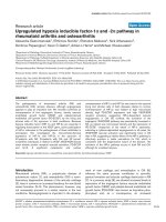
To investigate whether human articular chondrocytes express
15-LOX-1 and -2, total RNA from cultured chondrocytes,
derived from four different OA patients, was subjected to
reverse transcription PCR analysis using specific primers for
15-LOX-1 and -2. As shown in Figure 1a, the expression of 15-
LOX-1 and -2 mRNAs was detected in the four chondrocyte
preparations. No PCR products were obtained with control
reactions performed in the absence of the cDNA or reverse
transcriptase (Figure 1a). To further confirm the expression of
15-LOX-1 and -2 in chondrocytes, we analyzed their expres-
sion at the protein level. Western blot analysis with total pro-
tein extracts revealed the presence of both isoforms in all
examined chondrocyte preparations (Figure 1b, c).
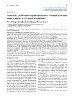
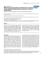
To examine whether chondrocytes express 15-LOX-1 and -2
in vivo, we performed immunohistochemical analysis using OA
cartilage. The positive immunostaining for 15-LOX-1 (Figure
2a) and 15-LOX-2 (Figure 2d) was located mainly in the super-
ficial and intermediate zones of the cartilage. Statistical evalu-
ation of the cell score revealed lower immunostaining for 15-
LOX-1 (mean ± SD: 36.2% ± 17.6%) than for 15-LOX-2
(mean ± SD: 43.7% ± 19.2%), but these differences were not
significant. The specificity of staining was confirmed using
nonimmune control IgG (Figure 2c, f). These observations
demonstrate the in vivo expression of 15-LOX-1 and -2 pro-
teins in OA cartilage.
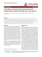
13-HODE and 15-HETE inhibited IL-1β-induced MMP-1
and MMP-13 expression in chondrocytes
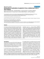
To examine the effects of 15-LOX-1 and -2 metabolites on
MMP-1 and MMP-13 release, chondrocytes were stimulated
with IL-1β in the absence or presence of increasing concen-
trations of 13-HODE or 15-HETE, and the levels of MMP-1
and MMP-13 proteins in conditioned media were determined
Available online />Page 5 of 12
(page number not for citation purposes)
by ELISA. As shown in Figure 3a, b, the production of MMP-1
and MMP-13 was dose dependently reduced in the presence
of 13-HODE or 15-HETE. The concentrations of 13-HODE
and 15-HETE utilized did not affect chondrocyte viability, as
judged using the MTT (3- [4,5-dimethylthiazol-2-yl]-2,5-diphe-
nyltetrazolium bromide) assay (data not shown). Taken
together, these findings suggest that 15-LOX metabolites may
constitute novel endogenous negative regulators of MMP-1
annd MMP-13 expression in chondrocytes.
In addition to IL-1, the pro-inflammatory cytokines TNF-α and
IL-17 also contribute to the pathogenesis of OA and are
potent inducers of MMP-1 and MMP-13. Therefore, we exam-
ined whether 13-HODE and 15-HETE could also attenuate
TNF-α and IL-17-induced MMP-1 and MMP-13 production in
chondrocytes. As shown in Figure 3c–e, the induction of
MMP-1 and MMP-13 production by TNF-α or IL-17 was dose
dependently diminished in the presence of 13-HODE or 15-
HETE. These data suggest that the suppressive effect of 13-
HODE and 15-HETE is not specific to IL-1, and is independ-
ent of the nature of the stimulus that triggers MMP-1 and
MMP-13 production.
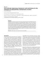
13-HODE and 15-HETE suppress IL-1-induced type II
collagen cleavage
Next, we assessed the effects of 13-HODE and 15-HETE on
IL-1-induced type II collagen cleavage. Cartilage explants
were treated with IL-1β in the absence or presence of increas-
ing concentrations of 13-HODE or 15-HETE for 5 days, and
type II collagen degradation was determined using a specific
commercial kit that measures C2C epitopes of type II colla-
gen. As shown in Figure 4, treatment with 13-HODE or 15-
HETE dose-dependently prevented IL-1-induced type II colla-
gen cleavage.
Suppression of IL-1β-induced MMP-1 and MMP-13
expression by 13-HODE and 15-HETE does not require
de novo protein synthesis
To investigate the effects of 13-HODE and 15-HETE on IL-1β-
induced MMP-1 and MMP-13 mRNA expression, we used
real-time PCR. Consistent with their effects on MMP-1 and
MMP-13 protein production, 13-HODE and 15-HETE dose-
dependently suppressed IL-1β-induced MMP-1 and MMP-13
mRNA expression (Figure 5a, b), suggesting that these effects
occur at the transcriptional level.
To evaluate whether the effect of 13-HODE and 15-HETE on
IL-1β-induced MMP-1 and MMP-13 expression is direct or
indirect, we tested the impact of the protein synthesis inhibitor
cycloheximide. Chondrocytes were pretreated with cyclohex-
imide for 30 minutes and stimulated with IL-1β alone or in com-
bination with either 13-HODE or 15-HETE for 8 hours. The
levels of MMP-1 and MMP-13 mRNAs were analyzed by real-
time PCR. As shown in Figure 5c, pretreatment with cyclohex-
imide did not affect 13-HODE and 15-HETE-mediated inhibi-
tion of IL-1β-induced MMP-1 and MMP-13 expression,
suggesting that their effect was a direct primary effect through
pre-existing factors and was not dependent on de novo pro-
tein synthesis.
13-HODE and 15-HETE suppressed IL-1β-induced MMP-
1 and MMP-13 production in a PPARγ dependent manner
The 15-LOX metabolites 13-HODE and 15-HETE are ligands
for PPARγ, and PPARγ activation was reported to suppress IL-
1β-induced MMP-1 and MMP-13 production [26,27]. To test
the possibility that PPARγ is involved in the suppressive effect
of 13-HODE and 15-HETE on MMP-1 and MMP-13 produc-
tion, we first examined their effects on the transcriptional activ-
ity of endogenous PPARγ in chondrocytes. Chondrocytes
were transiently transfected with a luciferase reporter con-
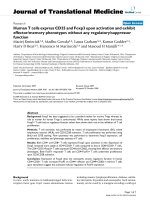
Figure 1
Human articular chondrocytes express both 15-LOX-1 and 15-LOX-2Human articular chondrocytes express both 15-LOX-1 and 15-LOX-2.
(a) Chondrocytes were isolated from OA knee cartilage and maintained
as monolayer culture for 7 to 10 days. Total RNA was prepared,
reverse transcribed into cDNA, and processed for PCR using specific
primers for 15-LOX-1, 15-LOX-2, and GAPDH. PCR products were
resolved on a 1.8% agarose gel and stained with ethidium bromide. C-
RT and C-PCR are negative controls for the reverse transcription and
PCR reaction, respectively. (b, c) Chondrocytes were isolated from OA
knee cartilage and lysates were prepared after 7 to 10 days in culture.
Samples with equal amounts of total proteins (20 μg per lane) were
immunoblotted with specific anti-15-LOX-1 (panel b) and anti-15-LOX-
2 (panel c) antibodies (upper sections). The blots were stripped and
reprobed with a specific anti-β-actin antibody (lower sections). bp,
base pairs; GAPDH, glyceraldehyde-3-phosphate dehydrogenase;
LOX, lipoxygenase; OA, osteoarthritis.
Arthritis Research & Therapy Vol 11 No 2 Chabane et al.
Page 6 of 12
(page number not for citation purposes)
struct containing three copies of a consensus PPRE, and
treated with increasing concentrations of 13-HODE and 15-
HETE. As illustrated in Figure 6a, treatment with 13-HODE
and 15-HETE dose dependently increased the activity of the
synthetic promoter. These data confirm the presence of induc-
ible PPARγ-dependent transcriptional responses in chondro-
cytes. Next, we examined the effect of GW9662, a selective
and irreversible PPARγ antagonist. Chondrocytes were pre-
incubated with increasing concentrations of GW9662 before
addition of 13-HODE or 15-HETE and were subsequently
stimulated with IL-1β. As shown in Figure 6b, GW9662 dose-
dependently relieved the suppressive effect of 13-HODE and
15-HETE on IL-1β-induced MMP-1 and MMP-13 protein pro-
duction. Taken together, these results strongly suggest that
13-HODE and 15-HETE inhibit IL-1β-induced MMP-1 and
MMP-13 production through a PPARγ-dependent mechanism.
Discussion
In the present study, we report for the first time that articular
OA chondrocytes express 15-LOX-1 and -2. Treatment with
13-HODE and 15-HETE, the major products of 15-LOX-1 and
-2, respectively, suppressed IL-1β-induced MMP-1 and MMP-
13 expression and type II collagen degradation. Taken
together, these findings strongly suggest a chondroprotective
role for 15-LOXs by negatively regulating the expression of
MMP-1 and MMP-13.
In addition to their chondroprotective properties observed in
this study, 15-LOX metabolites were shown to exhibit potent
anti-inflammatory effects. For instance, 15-HETE inhibits poly-
morphonuclear neutrophil degranulation and superoxide pro-
duction elicited by N-formylmethionylleucylphenylalaline,
platelet-activating factor and leukotriene B
4
[17]. In addition,
15-HETE prevents polymorphonuclear neutrophil migration
across IL-1β or TNF-α-activated endothelium [32] and TNF-α-
induced expression of several adhesion molecules, including
intercellular adhesion molecule-1, vascular cell adhesion mol-
ecule-1 and E-selectin [33]. On the other hand, 13-HODE
attenuates the production of reactive oxygen species in mac-
rophages [34], the production of IL-8 in colonic epithelial cells
[18], and the ability of dendritic cells to activate interferon-γ
secretion by T lymphocytes [35]. Moreover, 13-HODE and 15-
HETE were shown to mediate the suppressive effect of the
anti-inflammatory cytokine IL-4 on inducible nitric oxide syn-
thase expression in macrophages [21] and IL-2 production in
T lymphocytes [22]. In addition to 13-HODE and 15-HETE for-
mation, 15-LOXs are involved in the generation of the potent
anti-inflammatory molecules lipoxins, resolvings, and pro-
tectins [36]. Thus, 15-LOXs can dampen inflammation
through production of distinct classes of anti-inflammatory and
pro-resolution lipid mediators.
The protective effect of 15-LOXs is further supported by
results from studies using transgenic animals. Over-expres-
sion of 15-LOX in rabbits reduced inflammation and tissue
damage in atherosclerosis [37] and peritonitis [38]. In rats,
over-expression of 15-LOX suppressed renal inflammation
and preserved organ function in experimental glomerulone-
phritis [39]. These data, together with our findings that 15-
LOX metabolites block MMP production, suggest that these
lipids may have protective effects in OA in vivo. Further stud-
ies using cartilage-specific 15-LOX-null mice will be required
to elucidate the role of 15-LOXs in cartilage integrity and the
pathogenesis of OA.
Figure 2
Expression of 15-LOX-1 and 15-LOX-2 in human OA cartilageExpression of 15-LOX-1 and 15-LOX-2 in human OA cartilage. Representative immunostaining of human osteoarthritis (OA) cartilage for (a) 15-
LOX-1 and (d) 15-LOX-2. (b, e) Higher magnification views of the area indicated within the broken line rectangle in panels a and d, respectively. (c,
f) Cartilage treated with nonimmune control IgG at the same concentration as the primary antibody (control for staining specificity). (Magnification:
×100 for panels a, c, d and f; ×250 for panels b and e). The results are representative of four separate experiments performed with cartilage samples
from four different donors. LOX, lipoxygenase.
Available online />Page 7 of 12
(page number not for citation purposes)
Figure 3
13-HODE and 15-HETE downregulate induction of MMP-1/MMP-13 protein synthesis by IL-1β, TNF-α and IL-1713-HODE and 15-HETE downregulate induction of MMP-1/MMP-13 protein synthesis by IL-1β, TNF-α and IL-17. (a, b) Chondrocytes were stimu-
lated with IL-1β (100 pg/ml), (c, d) TNF-α (0.1 ng/ml), or (e, f) IL-17 (10 ng/ml) in the presence of vehicle (dimethyl sulfoxide at a maximum concen-
tration of 0.14%) or increasing concentrations of 13-HODE (panels a, c, and e) or 15-HETE (panels b, d, and f) for 24 hours. The levels of MMP-1
and MMP-13 proteins in conditioned media were measured using ELISA. Results are expressed as the percentage of control, considering 100% as
the value of cells treated with IL-1β, TNF-α or IL-17 alone, and are the mean ± standard deviation of at least three independent experiments. *P <
0.05 versus cells treated with IL-1β, TNF-α, or IL-17 alone. HETE, hydroxyeicosatetraenoic acid; HODE, hydroxy octadecadienoic acid; MMP, matrix
metalloproteinase; TNF, tumor necrosis factor.
Arthritis Research & Therapy Vol 11 No 2 Chabane et al.
Page 8 of 12
(page number not for citation purposes)
Several factors are known to modulate 15-LOX expression.
For instance, IL-4 and IL-13, increase the expression of 15-
LOX-1 and -2 in a number of cell types, including monocytes/
macrophages, T lymphocytes and several cancer cell lines
[40-45]. Moreover, chromatin modifications that play pivotal
roles in the regulation of gene expression were reported to
modulate 15-LOX expression. Histone acetylation appears to
upregulate 15-LOX expression [46] whereas DNA methylation
downregulates 15-LOX expression [47]. Whether these fac-
tors and conditions contribute to the modulation of 15-LOX
expression in chondrocytes is among our ongoing research
projects.
13-HODE and 15-HETE are potent endogenous activators
and ligands for PPARγ [23,24]. Using a PPRE reporter plas-
mid in transient transfection experiments, we confirmed the
capability of the above 15-LOX products to activate PPARγ in
human chondrocytes. We also showed that pretreatment with
an irreversible pharmacological PPARγ antagonist GW9662
overcame the inhibitory effect of 13-HODE and 15-HETE on
IL-1β-induced MMP release. These results are consistent with
previous findings showing that PPARγ activation suppresses
MMP production in several cell types, including chondrocytes
[26] and synovial fibroblasts [27]. Altogether, these data
strongly suggest that 13-HODE and 15-HETE suppress IL-
1β-induced MMP-1 and MMP-13 by chondrocytes through
activation of PPARγ. The expression of MMP-1 and MMP-13
are essentially regulated by the transcription factors activator
protein (AP)-1 and nuclear factor-κB (NF-κB), and analysis of
the 5'-flanking regions of these genes has demonstrated the
presence of numerous putative binding sites for AP-1 and NF-
κB [3]. On the other hand, previous studies showed that acti-
Figure 4
13-HODE and 15-HETE downregulate IL-1β-induced type II collagen degradation cleavage13-HODE and 15-HETE downregulate IL-1β-induced type II collagen
degradation cleavage. Cartilage explants were stimulated with 1 ng/ml
IL-1β in the presence of the control vehicle dimethyl sulfoxide or
increasing concentrations of 13-HODE or 15-HETE for 5 days. Type II
collagen degradation was assessed by quantification of C2C epitopes
of type II collagen in cartilage explants. Data are the mean ± standard
deviation of three independent experiments. *P < 0.05 versus cartilage
explants treated with IL-1β alone. HETE, hydroxyeicosatetraenoic acid;
HODE, hydroxy octadecadienoic acid.
Figure 5
Downregulation of IL-1β-induced MMP-1/MMP-13 expression by 13-HODE and 15-HETE does not require de novo protein synthesisDownregulation of IL-1β-induced MMP-1/MMP-13 expression by 13-
HODE and 15-HETE does not require de novo protein synthesis. (a, b)
Chondrocytes were treated with 100 pg/ml IL-1β in the presence of the
control vehicle dimethyl sulfoxide or increasing concentrations of 13-
HODE (panel a) or 15-HETE (panel b) for 8 hours. (c) Chondrocytes
were pretreated with control vehicle dimethyl sulfoxide or cyclohex-
imide (10 μg/ml) for 30 minutes before stimulation with 100 pg/ml IL-
1β in the absence or presence of 50 μmol/l 13-HODE or 15-HETE for
8 hours. Total RNA was isolated, reverse transcribed into cDNA, and
MMP-1 and MMP-13 mRNAs were quantified using real-time PCR. The
housekeeping gene GAPDH was used for normalization. All experi-
ments were performed in triplicate, and negative controls without tem-
plate RNA were included in each experiment. Results are expressed as
fold changes, considering 1 as the value of untreated cells, and are the
mean ± standard deviation of three independent experiments. *P <
0.05 versus cells treated with IL-1β alone. CHX, cycloheximide;
GAPDH, glyceraldehyde-3-phosphate dehydrogenase; HETE, hydrox-
yeicosatetraenoic acid; HODE, hydroxy octadecadienoic acid; MMP,
matrix metalloproteinase; TNF, tumor necrosis factor.
Available online />Page 9 of 12
(page number not for citation purposes)
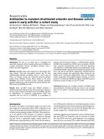
vation of PPARγ suppresses the transcriptional activity of AP-
1 and NF-κB [48]. Therefore, it is possible that activation of
PPARγ by 13-HODE and 15-HETE reduces transcriptional
activity of AP-1 and NF-κB, leading to diminished production
of MMP-1 and MMP-13 (Figure 7). Another possible mecha-
nism through which 13-HODE and 15-HETE may downregu-
late MMP expression could involve the promotion of mRNA
decay. Indeed, 15-LOX metabolites were reported to down-
modulate lipopolysaccharide-induced TNF-α expression by
enhancing mRNA decay [19]. Alternatively, 15-LOX products
could prevent IL-1β-induced MMP-1 and MMP-13 expression
by interfering with key signalling pathways. In this context, 15-
LOX metabolites were shown to inhibit protein kinase C activ-
ity and translocation [20,49], and protein kinase C was shown
to contribute to MMP-1 and MMP-13 expression [50,51].
15-HETE and 13-HODE are synthesized by a number of cell
types such as macrophages, neutrophils and chondrocytes
[52]. They have also been detected in vivo in several patho-
physiological fluids, including sputum from chronic bronchitis
patients [53], cerebrospinal fluid from patients with Alzhe-
imer's disease [54], bronchoalveolar lavage fluids from
patients with asthma [55] and scleroderma lung disease [56].
Apart from a report by Walenga and coworkers [57], who
found that the levels of 15-HETE increase to about 1 μmol/l in
blood stimulated with various agents, the concentrations of
15-HETE and 13-HODE detected in most pathophysiological
fluids (1 to 100 nmol/l) were lower than those used in the
present study (1 to 50 μmol/l). However, it should be noted
that, like other eicosanoids, 13-HODE and 15-HETE function
as autocrine and paracrine molecules and can readily reach
pharmacological levels in the microenvironment of cells that
produce them. Moreover, synovial fibroblasts [58] and osteob-
lasts [59] express 15-LOX and may represent additional
sources for the production of 15-LOX metabolites within the
joint. Also, we cannot exclude the possibility that low concen-
trations of 13-HODE and 15-HETE can synergize with each
other or with other 15-LOX derivatives to suppress inflamma-
tory and catabolic responses in the joint.
Conclusions
We demonstrated that 15-LOX-1 and -2 are expressed in OA
articular chondrocytes. Treatment with 13-HODE and 15-
HETE, the respective metabolites of 15-LOX-1 and -2, sup-
pressed IL-1β-induced MMP-1 and MMP-13 production.
These effects do not require protein synthesis and are mediate
by PPARγ. These data suggest that 15-LOXs and their metab-
olites may have therapeutic promise in OA by preventing the
production of cartilage-degrading enzymes.
Competing interests
The authors declare that they have no competing interests.
Figure 6
13-HODE and 15-HETE suppressed IL-1β-induced MMP-1/MMP-13 production in a PPARγ dependent manner13-HODE and 15-HETE suppressed IL-1β-induced MMP-1/MMP-13
production in a PPARγ dependent manner. (a) 13-HODE and 15-HETE
activate endogenous PPARγ in human chondrocytes. Chondrocytes
were transiently transfected with a reporter construct containing three
copies of a consensus PPRE placed upstream from the Tk-luciferase
reporter (PPRE
3
-Tk-Luc) along with the internal control pSV40-β-gal
using FuGene 6 transfection reagent. Six hours later, the cells were
washed and changed to medium containing 0.5% fetal calf serum for
an additional 18 hours. Transfected cells were then treated with the
control vehicle dimethyl sulfoxide or increasing concentrations of 13-
HODE or 15-HETE for 18 hours. Luciferase activity values were deter-
mined and normalized to β-galactosidase activity. Results are
expressed as fold changes, considering 1 as the value of unstimulated
cells, and are the mean ± standard deviation of three independent
experiments. *P < 0.05 versus unstimulated cells. (b) PPARγ antago-
nist (GW9662) prevented the suppressive effect of 13-HODE and 15-
HETE on IL-1β-induced MMP-1 and MMP-13 release. Chondrocytes
were pretreated with increasing concentrations (1, 5, and 10 μmol/l) of
GW9662 for 30 minutes. Then, the cells were treated with or without
IL-1β (100 pg/ml) for 24 hours in the absence or the presence of 50
μmol/l 13-HODE (panel a) or 50 μmol/l 15-HETE (panel b). The levels
of MMP-1 and MMP-13 proteins in conditioned media were measured
using ELISA. Results are expressed as the percentage of control, con-
sidering 100% as the value of cells treated with IL-1β alone, and are
the mean ± standard deviation of four independent experiments. *P <
0.05 versus cells treated with IL-1β and 13-HODE or 15-HETE. HETE,
hydroxyeicosatetraenoic acid; HODE, hydroxy octadecadienoic acid;
MMP, matrix metalloproteinase; PPAR, peroxisome proliferator-acti-
vated receptor; PPRE, peroxisome proliferator-activated receptor-
responsive element.
Arthritis Research & Therapy Vol 11 No 2 Chabane et al.
Page 10 of 12
(page number not for citation purposes)
Authors' contributions
NC conceived the study, designed and carried out cell and
real-time reverse transcription PCR experiments and some
immunohistochemistry experiments. NZ contributed to the
study design, carried out immunoassays and some cell exper-
iments. MB participated in the study design and data analysis.
JM-P, J-PP and ND helped to obtain tissues, and participated
in the study design and in some immunohistochemistry exper-
iments. HF conceived, designed and coordinated the study,
carried out some cell experiments, and drafted the manuscript.
All authors read and approved the final manuscript.
Acknowledgements
This work was supported by the Canadian Institutes of Health Research
(CIHR) Grant MOP-84282, and the Fonds de la Recherche du Centre
de Recherche du Centre Hospitalier de l'Université de Montréal
(CHUM). HF is a Research Scholar of the Fonds de Recherche en
Santé du Québec (FRSQ).
References
1. Goldring MB, Goldring SR: Osteoarthritis. J Cell Physiol 2007,
213:626-634.
2. Pelletier JP, Martel-Pelletier J, Abramson SB: Osteoarthritis, an
inflammatory disease: potential implication for the selection of
new therapeutic targets. Arthritis Rheum 2001, 44:1237-1247.
3. Burrage PS, Mix KS, Brinckerhoff CE: Matrix metalloproteinases:
role in arthritis. Front Biosci 2006, 11:529-543.
4. Welgus HG, Jeffrey JJ, Eisen AZ: The collagen substrate specif-
icity of human skin fibroblast collagenase. J Biol Chem 1981,
256:9511-9515.
5. Knauper V, Lopez-Otin C, Smith B, Knight G, Murphy G: Bio-
chemical characterization of human collagenase-3. J Biol
Chem 1996, 271:1544-1550.
6. Firestein GS, Paine MM, Littman BH: Gene expression (colla-
genase, tissue inhibitor of metalloproteinases, complement,
and HLA-DR) in rheumatoid arthritis and osteoarthritis syn-
ovium. Quantitative analysis and effect of intraarticular corti-
costeroids. Arthritis Rheum 1991, 34:1094-1105.
7. Reboul P, Pelletier JP, Tardif G, Cloutier JM, Martel-Pelletier J: The
new collagenase, collagenase-3, is expressed and synthe-
sized by human chondrocytes but not by synoviocytes. A role
in osteoarthritis. J Clin Invest 1996, 97:2011-2019.
8. Borden P, Solymar D, Sucharczuk A, Lindman B, Cannon P, Heller
RA: Cytokine control of interstitial collagenase and colla-
genase-3 gene expression in human chondrocytes. J Biol
Chem 1996, 271:23577-23581.
9. Hutchinson JW, Tierney GM, Parsons SL, Davis TR: Dupuytren's
disease and frozen shoulder induced by treatment with a
matrix metalloproteinase inhibitor. J Bone Joint Surg Br 1998,
80:907-908.
10. Coussens LM, Fingleton B, Matrisian LM: Matrix metalloprotein-
ase inhibitors and cancer: trials and tribulations. Science
2002, 295:2387-2392.
11. Thabet MM, Huizinga TW: Drug evaluation: apratastat, a novel
TACE/MMP inhibitor for rheumatoid arthritis. Curr Opin Inves-
tig Drugs 2006, 7:1014-9.
12. Wittwer J, Hersberger M: The two faces of the 15-lipoxygenase
in atherosclerosis. Prostaglandins Leukot Essent Fatty Acids
2007, 77:67-77.
13. Pidgeon GP, Lysaght J, Krishnamoorthy S, Reynolds JV, O'Byrne
K, Nie D, Honn KV: Lipoxygenase metabolism: roles in tumor
Figure 7
Schematic representation of the suppressive effect of 15-LOX metabolites on MMP-1/MMP-13 expressionSchematic representation of the suppressive effect of 15-LOX metabolites on MMP-1/MMP-13 expression. Pro-inflammatory cytokines such as IL-1
interact with their respective receptors that activate MAPK signalling and downstream transcription factors, resulting in the transcription of MMP-1
and MMP-13 genes. 15-LOX convert AA and LA to 15-HETE and 13-HODE, which then activate PPARγ. Activated PPARγ antagonizes the tran-
scriptional activity of AP-1, NF-κB and PEA3, which results in the inhibition of the expression of their target genes (for instance, MMP-1 and MMP-
13). AA, arachidonic acid; AP, activator protein; HETE, hydroxyeicosatetraenoic acid; HODE, hydroxy octadecadienoic acid; LA, linoeic acid; LOX,
lipoxygenase; MAPK, mitogen-activated protein kinase; MMP, matrix metalloproteinase; NF-κB, nuclear factor-κB; PEA3, Polyoma Enhancer Activa-
tor 3; PPAR, peroxisome proliferator-activated receptor.
Available online />Page 11 of 12
(page number not for citation purposes)
progression and survival. Cancer Metastasis Rev 2007,
26:503-524.
14. Funk CD: The molecular biology of mammalian lipoxygenases
and the quest for eicosanoid functions using lipoxygenase-
deficient mice. Biochim Biophys Acta 1996, 1304:65-84.
15. Shappell SB, Boeglin WE, Olson SJ, Kasper S, Brash AR: 15-
lipoxygenase-2 (15-LOX-2) is expressed in benign prostatic
epithelium and reduced in prostate adenocarcinoma. Am J
Pathol 1999, 155:235-245.
16. Brash AR, Boeglin WE, Chang MS: Discovery of a second 15S-
lipoxygenase in humans. Proc Natl Acad Sci USA 1997,
94:6148-6152.
17. Smith RJ, Justen JM, Nidy EG, Sam LM, Bleasdale JE: Transmem-
brane signaling in human polymorphonuclear neutrophils:
15(S)-hydroxy-(5Z, 8Z, 11Z, 13E)-eicosatetraenoic acid modu-
lates receptor agonist-triggered cell activation. Proc Natl Acad
Sci USA 1993, 90:7270-7274.
18. Altmann R, Hausmann M, Spottl T, Gruber M, Bull AW, Menzel K,
Vogl D, Herfarth H, Scholmerich J, Falk W, Rogler G: 13-Oxo-
ODE is an endogenous ligand for PPARgamma in human
colonic epithelial cells. Biochem Pharmacol 2007, 74:612-622.
19. Ferrante JV, Ferrante A: Novel role of lipoxygenases in the
inflammatory response: promotion of TNF mRNA decay by 15-
hydroperoxyeicosatetraenoic acid in a monocytic cell line. J
Immunol 2005, 174:3169-3172.
20. Ferrante JV, Huang ZH, Nandoskar M, Hii CS, Robinson BS,
Rathjen DA, Poulos A, Morris CP, Ferrante A: Altered responses
of human macrophages to lipopolysaccharide by hydroperoxy
eicosatetraenoic acid, hydroxy eicosatetraenoic acid, and ara-
chidonic acid. Inhibition of tumor necrosis factor production. J
Clin Invest 1997, 99:1445-1452.
21. Ricote M, Welch JS, Glass CK: Regulation of macrophage gene
expression by the peroxisome proliferator-activated receptor-
gamma. Horm Res 2000, 54:275-280.
22. Yang XY, Wang LH, Mihalic K, Xiao W, Chen T, Li P, Wahl LM, Far-
rar WL: Interleukin (IL)-4 indirectly suppresses IL-2 production
by human T lymphocytes via peroxisome proliferator-acti-
vated receptor gamma activated by macrophage-derived 12/
15-lipoxygenase ligands. J Biol Chem 2002, 277:3973-3978.
23. Nagy L, Tontonoz P, Alvarez JG, Chen H, Evans RM: Oxidized LDL
regulates macrophage gene expression through ligand activa-
tion of PPARgamma.
Cell 1998, 93:229-240.
24. Shappell SB, Gupta RA, Manning S, Whitehead R, Boeglin WE,
Schneider C, Case T, Price J, Jack GS, Wheeler TM, Matusik RJ,
Brash AR, Dubois RN: 15S-Hydroxyeicosatetraenoic acid acti-
vates peroxisome proliferator-activated receptor gamma and
inhibits proliferation in PC3 prostate carcinoma cells. Cancer
Res 2001, 61:497-503.
25. Fahmi H, Pelletier JP, Martel-Pelletier J: PPARgamma ligands as
modulators of inflammatory and catabolic responses on
arthritis. An overview. J Rheumatol 2002, 29:3-14.
26. Fahmi H, Di Battista JA, Pelletier JP, Mineau F, Ranger P, Martel-
Pelletier J: Peroxisome proliferator-activated receptor gamma
activators inhibit interleukin-1beta-induced nitric oxide and
matrix metalloproteinase 13 production in human chondro-
cytes. Arthritis Rheum 2001, 44:595-607.
27. Fahmi H, Pelletier JP, Di Battista JA, Cheung HS, Fernandes J, Mar-
tel-Pelletier J: Peroxisome proliferator-activated receptor
gamma acitvators inhibit MMP-1 production in human synovial
fibroblasts by reducing the activity of the activator protein 1.
Osteoarthritis Cartilage 2002, 10:100-108.
28. Bianchi A, Moulin D, Sebillaud S, Koufany M, Galteau MM, Netter
P, Terlain B, Jouzeau JY: Contrasting effects of peroxisome-pro-
liferator-activated receptor (PPAR)gamma agonists on mem-
brane-associated prostaglandin E2 synthase-1 in IL-1beta-
stimulated rat chondrocytes: evidence for PPARgamma-inde-
pendent inhibition by 15-deoxy-Delta12,14prostaglandin J2.
Arthritis Res Ther 2005, 7:R1325-R1337.
29. Boileau C, Martel-Pelletier J, Fahmi H, Mineau F, Boily M, Pelletier
JP: The peroxisome proliferator-activated receptor gamma
agonist pioglitazone reduces the development of cartilage
lesions in an experimental dog model of osteoarthritis: in vivo
protective effects mediated through the inhibition of key sign-
aling and catabolic pathways. Arthritis Rheum 2007,
56:2288-2298.
30. Altman RD: Criteria for the classification of osteoarthritis of the
knee and hip. Scand J Rheumatol Suppl 1987, 65:31-39.
31. Billinghurst RC, Dahlberg L, Ionescu M, Reiner A, Bourne R,
Rorabeck C, Mitchell P, Hambor J, Diekmann O, Tschesche H,
Chen J, Van Wart H, Poole AR: Enhanced cleavage of type II col-
lagen by collagenases in osteoarthritic articular cartilage. J
Clin Invest 1997, 99:1534-1545.
32. Takata S, Papayianni A, Matsubara M, Jimenez W, Pronovost PH,
Brady HR: 15-Hydroxyeicosatetraenoic acid inhibits neutrophil
migration across cytokine-activated endothelium.
Am J Pathol
1994, 145:541-549.
33. Huang ZH, Bates EJ, Ferrante JV, Hii CS, Poulos A, Robinson BS,
Ferrante A: Inhibition of stimulus-induced endothelial cell
intercellular adhesion molecule-1, E-selectin, and vascular
cellular adhesion molecule-1 expression by arachidonic acid
and its hydroxy and hydroperoxy derivatives. Circ Res 1997,
80:149-158.
34. Fischer B, von Knethen A, Brune B: Dualism of oxidized lipopro-
teins in provoking and attenuating the oxidative burst in mac-
rophages: role of peroxisome proliferator-activated receptor-
gamma. J Immunol 2002, 168:2828-2834.
35. Coutant F, Agaugue S, Perrin-Cocon L, Andre P, Lotteau V: Sens-
ing environmental lipids by dendritic cell modulates its func-
tion. J Immunol 2004, 172:54-60.
36. Serhan CN, Chiang N, Van Dyke TE: Resolving inflammation:
dual anti-inflammatory and pro-resolution lipid mediators. Nat
Rev Immunol 2008, 8:349-361.
37. Shen J, Herderick E, Cornhill JF, Zsigmond E, Kim HS, Kuhn H,
Guevara NV, Chan L: Macrophage-mediated 15-lipoxygenase
expression protects against atherosclerosis development. J
Clin Invest 1996, 98:2201-2208.
38. Serhan CN, Jain A, Marleau S, Clish C, Kantarci A, Behbehani B,
Colgan SP, Stahl GL, Merched A, Petasis NA, Chan L, Van Dyke
TE: Reduced inflammation and tissue damage in transgenic
rabbits overexpressing 15-lipoxygenase and endogenous
anti-inflammatory lipid mediators. J Immunol 2003,
171:6856-6865.
39. Munger KA, Montero A, Fukunaga M, Uda S, Yura T, Imai E,
Kaneda Y, Valdivielso JM, Badr KF: Transfection of rat kidney
with human 15-lipoxygenase suppresses inflammation and
preserves function in experimental glomerulonephritis. Proc
Natl Acad Sci USA 1999, 96:13375-13380.
40. Brinckmann R, Topp MS, Zalan I, Heydeck D, Ludwig P, Kuhn H,
Berdel WE, Habenicht JR: Regulation of 15-lipoxygenase
expression in lung epithelial cells by interleukin-4. Biochem J
1996, 318:305-312.
41. Nassar GM, Morrow JD, Roberts LJ II, Lakkis FG, Badr KF: Induc-
tion of 15-lipoxygenase by interleukin-13 in human blood
monocytes. J Biol Chem
1994, 269:27631-27634.
42. Heydeck D, Thomas L, Schnurr K, Trebus F, Thierfelder WE, Ihle
JN, Kuhn H: Interleukin-4 and -13 induce upregulation of the
murine macrophage 12/15-lipoxygenase activity: evidence for
the involvement of transcription factor STAT6. Blood 1998,
92:2503-2510.
43. Roy B, Cathcart MK: Induction of 15-lipoxygenase expression
by IL-13 requires tyrosine phosphorylation of Jak2 and Tyk2 in
human monocytes. J Biol Chem 1998, 273:32023-32029.
44. Spanbroek R, Hildner M, Kohler A, Muller A, Zintl F, Kuhn H, Rad-
mark O, Samuelsson B, Habenicht AJ: IL-4 determines eicosa-
noid formation in dendritic cells by down-regulation of 5-
lipoxygenase and up-regulation of 15-lipoxygenase 1 expres-
sion. Proc Natl Acad Sci USA 2001, 98:5152-5157.
45. Lee YW, Kuhn H, Kaiser S, Hennig B, Daugherty A, Toborek M:
Interleukin 4 induces transcription of the 15-lipoxygenase I
gene in human endothelial cells. J Lipid Res 2001, 42:783-791.
46. Shankaranarayanan P, Chaitidis P, Kuhn H, Nigam S: Acetylation
by histone acetyltransferase CREB-binding protein/p300 of
STAT6 is required for transcriptional activation of the 15-lipox-
ygenase-1 gene. J Biol Chem 2001, 276:42753-42760.
47. Liu C, Xu D, Sjoberg J, Forsell P, Bjorkholm M, Claesson HE: Tran-
scriptional regulation of 15-lipoxygenase expression by pro-
moter methylation. Exp Cell Res 2004, 297:61-67.
48. Ricote M, Li AC, Willson TM, Kelly CJ, Glass CK: The peroxisome
proliferator-activated receptor-gamma is a negative regulator
of macrophage activation. Nature 1998, 391:79-82.
49. Pongracz J, Lord JM: The lipoxygenase product 13-hydroxy-
octadecadienoic acid (13-HODE) is a selective inhibitor of
classical PKC isoenzymes. Biochem Biophys Res Commun
1999, 256:269-272.
Arthritis Research & Therapy Vol 11 No 2 Chabane et al.
Page 12 of 12
(page number not for citation purposes)
50. Hussain S, Assender JW, Bond M, Wong LF, Murphy D, Newby
AC: Activation of protein kinase Czeta is essential for
cytokine-induced metalloproteinase-1, -3, and -9 secretion
from rabbit smooth muscle cells and inhibits proliferation. J
Biol Chem 2002, 277:27345-27352.
51. Reuben PM, Brogley MA, Sun Y, Cheung HS: Molecular mecha-
nism of the induction of metalloproteinases 1 and 3 in human
fibroblasts by basic calcium phosphate crystals. Role of cal-
cium-dependent protein kinase C alpha. J Biol Chem 2002,
277:15190-15198.
52. Amat M, Diaz C, Vila L: Leukotriene A4 hydrolase and leukot-
riene C4 synthase activities in human chondrocytes: transcel-
lular biosynthesis of Leukotrienes during granulocyte-
chondrocyte interaction. Arthritis Rheum 1998, 41:1645-1651.
53. Profita M, Sala A, Riccobono L, Pace E, Paterno A, Zarini S, Siena
L, Mirabella A, Bonsignore G, Vignola AM: 15(S)-HETE modu-
lates LTB(4) production and neutrophil chemotaxis in chronic
bronchitis. Am J Physiol Cell Physiol 2000, 279:C1249-C1258.
54. Yao Y, Clark CM, Trojanowski JQ, Lee VM, Pratico D: Elevation of
12/15 lipoxygenase products in AD and mild cognitive impair-
ment. Ann Neurol 2005, 58:623-626.
55. Murray JJ, Tonnel AB, Brash AR, Roberts LJ II, Gosset P, Workman
R, Capron A, Oates JA: Release of prostaglandin D2 into human
airways during acute antigen challenge. N Engl J Med 1986,
315:800-804.
56. Kowal-Bielecka O, Kowal K, Distler O, Rojewska J, Bodzenta-
Lukaszyk A, Michel BA, Gay RE, Gay S, Sierakowski S: Cycloox-
ygenase- and lipoxygenase-derived eicosanoids in bronchoal-
veolar lavage fluid from patients with scleroderma lung
disease: an imbalance between proinflammatory and antiin-
flammatory lipid mediators. Arthritis Rheum 2005,
52:3783-3791.
57. Walenga RW, Boone S, Stuart MJ: Analysis of blood HETE lev-
els by selected ion monitoring with ricinoleic acid as the inter-
nal standard. Prostaglandins 1987, 34:733-748.
58. Liagre B, Vergne P, Rigaud M, Beneytout JL: Arachidonate 15-
lipoxygenase of reticulocyte-type in human rheumatoid arthri-
tis type B synoviocytes and modulation of its activity by proin-
flammatory cytokines. J Rheumatol 1999, 26:1044-1051.
59. Klein RF, Allard J, Avnur Z, Nikolcheva T, Rotstein D, Carlos AS,
Shea M, Waters RV, Belknap JK, Peltz G, Orwoll ES: Regulation
of bone mass in mice by the lipoxygenase gene Alox15. Sci-
ence 2004,
303:229-232.