Critical care medicine - part 8 docx
Bạn đang xem bản rút gọn của tài liệu. Xem và tải ngay bản đầy đủ của tài liệu tại đây (89.05 KB, 15 trang )
108 Toxicologic Syndromes
D. Hemodialysis: Indications include ingestion of phenobarbital, theophylline,
chloral hydrate, salicylate, ethanol, lithium, ethylene glycol, isopropyl
alcohol, procainamide, and methanol, or severe metabolic acidosis.
E. Hemoperfusion: May be more effective than hemodialysis except for
bromides, heavy metals, lithium, and ethylene glycol. Hemoperfusion is
effective for disopyramide, phenytoin, barbiturates, theophylline.
Toxicologic Syndromes
I. Characteristics of common toxicologic syndromes
A. Cholinergic poisoning: Salivation, bradycardia, defecation, lacrimation,
emesis, urination, miosis.
B. Anticholinergic poisoning: Dry skin, flushing, fever, urinary retention,
mydriasis, thirst, delirium, conduction delays, tachycardia, ileus.
C. Sympathomimetic poisoning: Agitation, hypertension, seizure,
tachycardia, mydriasis, vasoconstriction.
D. Narcotic poisoning: Lethargy, hypotension, hypoventilation, miosis,
coma, ileus.
E. Withdrawal syndrome: Diarrhea, lacrimation, mydriasis, cramps,
tachycardia, hallucination.
F. Salicylate poisoning: Fever, respiratory alkalosis, or mixed acid-base
disturbance, hyperpnea, hypokalemia, tinnitus.
G. Causes of toxic seizures: Amoxapine, anticholinergics, camphor,
carbon monoxide, cocaine, ergotamine, isoniazid, lead, lindane, lithium,
LSD, parathion, phencyclidine, phenothiazines, propoxyphene
propranolol, strychnine, theophylline, tricyclic antidepressants,
normeperidine (metabolite of meperidine), thiocyanate.
H. Causes of toxic cardiac arrhythmias: Arsenic, beta-blockers, chloral
hydrate, chloroquine, clonidine, calcium channel blockers, cocaine,
cyanide, carbon monoxide, digitalis, ethanol, phenol, phenothiazine,
tricyclics.
I. Extrapyramidal syndromes: Dysphagia, dysphonia, trismus, rigidity,
torticollis, laryngospasm.
Acetaminophen Overdose
I. Clinical features
A. Acute lethal dose = 13-25 g. Acetaminophen is partly metabolized to
N-acetyl-p-benzoquinonimine which is conjugated by glutathione. Hepatic
glutathione stores can be depleted in acetaminophen overdose, leading to
centrilobular hepatic necrosis.
B. Liver failure occurs 3 days after ingestion if untreated. Liver failure
presents with right upper quadrant pain, elevated liver function tests,
coagulopathy, hypoglycemia, renal failure and encephalopathy.
II. Treatment
A. Gastrointestinal decontamination should consist of gastric lavage
followed by activated charcoal. Residual charcoal should be removed with
saline lavage prior to giving N-acetyl-cysteine (NAC).
Acetaminophen Overdose 109
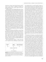
B. Check acetaminophen level 4 hours after ingestion. A nomogram should
be used to determine if treatment is necessary (see next page). Start
treatment if level is above the nontoxic range or if the level is potentially
toxic but the time of ingestion is unknown.
C. Therapy must start no later than 8-12 hours after ingestion. Treatment after
16-24 hours of non-sustained release formulation is significantly less
effective, but should still be accomplished.
D. Oral N-acetyl-cysteine (Mucomyst): 140 mg/kg PO followed by 70 mg/kg
PO q4h x 17 doses (total 1330 mg/kg over 72 h). Repeat loading dose if
emesis occurs Complete all doses even after acetaminophen level falls
below critical value.
E. Hemodialysis and hemoperfusion are somewhat effective, but should not
take the place of NAC treatment.
5
1 5 20 25 30 3 5
1
2
3
4
5
7
8
20
30
40
50
70
80
90
150
200
250
300
9
H ou rs Post In gest io n
6
10
60
100
10
I NT E R P RE TA TIO N O F AC TA M I N O P HE N LE V E L
VS H O UR S PO ST I N G E S TI O N
N o ri sk o f tox ic ity if u n d e r d o u b le l in e s.
P ro b ab le ri sk if a b ove to p li ne.
P o ss ib le r is k i f betw een do uble lines.
Ou tc om e is bes t if tre atm e n t is i nitiate d within 12 h ours o f
inges tion.
Gr a p h applies to non-s u sta in ed re le a se fo rm ulati ons o nly .
110 Cocaine Overdose
Cocaine Overdose
I. Clinical evaluation
A. Cocaine can be used intravenously, smoked, ingested, or inhaled nasally.
Street cocaine often is cut with other substances including amphetamines,
LSD, PCP, heroin, strychnine, lidocaine, talc, and quinine.
B. One-third of fatalities occur within 1 hour, with another third occurring 6-24
hours later.
C. Persons maytransport cocaine by swallowing wrapped packets, and some
users may hastily swallow packets of cocaine to avoid arrest.
II. Clinical features
A. CNS: Sympathetic stimulation, agitation, seizures, tremor, headache,
subarachnoid hemorrhage, ischemic cerebral stoke, psychosis, hallucina-
tions, fever, mydriasis, formication (sensation of insects crawling on skin).
B. Cardiovascular: Atrial and ventricular arrhythmias, myocardial infarction,
hypertension, hypotension, myocarditis, aortic rupture, cardiomyopathy.
C. Pulmonary: Noncardiogenic pulmonary edema, pneumomediastinum,
alveolar hemorrhage, hypersensitivity pneumonitis, bronchiolitis obliterans.
D. Other: Rhabdomyolysis, mesenteric ischemia, hepatitis.
III. Treatment
A. Treatment consists of supportive care because no antidote exists. GI
decontamination, including repeated activated charcoal, whole bowel
irrigation and endoscopic evaluation is provided if oral ingestion is
suspected.
B. Hyperadrenergic symptoms should be treated with benzodiazepines, such
as lorazepam.
C. Seizures: Treat with lorazepam, phenytoin, or phenobarbital.
D. Arrhythmias
1. Treat hyperadrenergic state and supraventricular tachycardia with
lorazepam and propranolol.
2. Ventricular arrhythmias are treated with lidocaine or propranolol.
E. Hypertension
1. Use lorazepam first for tachycardia and hypertension.
2. If no response, use labetalol because it has alpha and beta blocking
effects.
3. If hypertension remains severe, administer sodium nitroprusside or
esmolol drip.
F. Myocardial ischemia and infarction: Treat with thrombolysis, heparin,
aspirin, beta-blockers, nitroglycerin. Control hypertension and exclude
CNS bleeding before using thrombolytic therapy.
Cyclic Antidepressant Overdose 111
Cyclic Antidepressant Overdose
I. Clinical features
A. Antidepressants have prolonged body clearance rates, and cannot be
removal by forced diuresis, hemodialysis, and hemoperfusion. Delayed
absorption is common because of decreased GI motility from
anticholinergic effects. Cyclic antidepressants undergo extensive
enterohepatic recirculation.
B. CNS: Lethargy, coma, hallucinations, seizures, myoclonic jerks.
C. Anticholinergic crises: Blurred vision, dilated pupils, urinary retention,
dry mouth, ileus, hyperthermia.
D. Cardiac: Hypotension, ventricular tachyarrhythmias, sinus tachycardia.
E. ECG: Sinus tachycardia, right bundle branch block, right axis deviation,
increased PR and QT interval, QRS >100 msec, or right axis deviation.
Prolongation of the QRS width is a more reliable predictor of CNS and
cardiac toxicity than the serum level.
II. Treatment
A. Gastrointestinal decontamination and systemic drug removal
1. Magnesium citrate 300 mL via nasogastric tube x 1 dose.
2. Activated charcoal premixed with sorbitol 50 gm via nasogastric tube
q4-6h around-the-clock until the serum level decreases to therapeutic
range. Maintain the head-of-bed at a 30-45 degree angle to prevent
aspiration.
3. Cardiac toxicity
a. Alkalinization is a cardioprotective measure and it has no influence
on drug elimination. The goal of treatment is to achieve an arterial
pH of 7.50-7.55. If mechanical ventilation is necessary, hyperventi-
late to maintain desired pH.
b. Administer sodium bicarbonate 50-100 mEq (1-2 amps or 1-2
mEq/kg) IV over 5-10 min. Followed by infusion of sodium bicarbon-
ate, 2 amps in 1 liter of D5W at 100-150 cc/h. Adjust IV rate to
maintain desired pH.
4. Seizures
a. Administer lorazepam or diazepam IV followed by phenytoin.
b. Physostigmine, 1-2 mg slow IV over 3-4 min, is necessary if seizures
continue.
Digoxin Overdose
I. Clinical features
A. The therapeutic window of digoxin is 0.8-2.0 ng/mL. Drugs that increase
digoxin levels include verapamil, quinidine, amiodarone, flecainide,
erythromycin, and tetracycline. Hypokalemia, hypomagnesemia and
hypercalcemia enhance digoxin toxicity.
B. CNS: Confusion, lethargy; yellow-green visual halo.
C. Cardiac: Common dysrhythmias include ventricular tachycardia or
fibrillation; variable atrioventricular block, atrioventricular dissociation;
sinus bradycardia, junctional tachycardia, premature ventricular contrac-
tions.
D. GI: Nausea, vomiting.
112 Ethylene Glycol Ingestion
E. Metabolic: Hypokalemia enhances the toxic effects of digoxin on the
myocardial tissue and may be present in patients on diuretics.
II. Treatment
A. Gastrointestinal decontamination: Gastric lavage, followed by repeated
doses of activated charcoal, is effective; hemodialysis is ineffective.
B. Treat bradycardia with atropine, isoproterenol, and cardiac pacing.
C. Treat ventricular arrhythmias with lidocaine or phenytoin. Avoid
procainamide and quinidine because theyare proarrhythmic and slow AV
conduction.
D. Electrical DC cardioversion may be dangerous in severe toxicity.
Hypomagnesemia and hypokalemia should be corrected.
E. Digibind (Digoxin - specific Fab antibody fragment)
1. Indication: Life-threatening arrhythmias refractory to conventional
therapy.
2. Dosage of Digoxin immune Fab:
(number of 40 mg vials)= Digoxin level (ng/mL) x body weight (kg)
100
3. Dissolve the digoxin immune Fab in 100-150 mL of NS and infuse IV
over 15-30 minutes. A 0.22 micron in-line filter should be used during
infusion.
4. Hypokalemia, heart failure, and anaphylaxis may occur. The complex
is renally excreted; after administration, serum digoxin levels may be
artificially high because both free and bound digoxin is measured.
Ethylene Glycol Ingestion
I. Clinical features
A. Ethylene glycol is found in antifreeze, detergents, and polishes.
B. Toxicity: Half-life 3-5 hours; the half-life increases to 17 hours if
coingested with alcohol. The minimal lethal dose is 1.0-1.5 cc/kg, and the
lethal blood level is 200 mg/dL.
C. Anion gap metabolic acidosis and severe osmolar gap is often present.
CNS depression and cranial nerve dysfunction (facial and
vestibulocochlear palsies) are common.
D. GI symptoms such as flank pain. Oxalate crystals may be seen in the
urine sediment. Other findings may include hypocalcemia (due to calcium
oxalate formation); tetany, seizures, and prolonged QT.
II. Treatment
A. Fomepizole (Antizol) loading dose 15 mg/kg IV; then 10 mg/kg IV q12h
x 4, then 15 mg/kg IV q12h until ethylene glycol level is <20 mg/dL.
B. Pyridoxine 100 mg IV qid x 2 days and thiamine 100 mg IV qid x 2 days.
C. If definitive therapy is not immediately available, 3-4 ounces of whiskey
(or equivalent) may be given orally.
D. Hemodialysis indications: Severe refractory metabolic acidosis,
crystalluria, serum ethylene glycol level >50 mg/dL; keep glycol level <10
mg/dL.
Gamma-hydroxybutyrate Ingestion 113
Gamma-hydroxybutyrate Ingestion
I. Clinical features
A. Gamma-hydroxybutyrate (GHB) was used as an anesthetic agent but was
banned because of the occurrence of seizures. Gamma-hydroxybutyrate
is now an abused substance at dance clubs because of the euphoric
effects of the drug. It is also abused by body builders because of a
mistaken belief that it has anabolic properties. Gamma-hydroxybutyrate
is a clear, odorless, oily, salty liquid. It is rapidly absorbed within 20-40
minutes of ingestion and metabolized in the liver. The half-life of GHB is
20-30 min.
B. Gamma-hydroxybutyrate is not routinely included on toxicological
screens, but it can be detected in the blood and urine by gas chromatog-
raphy within 12 hours of ingestion. Gamma hydroxybutyrate may cause
respiratory depression, coma, seizures, and severe agitation. Cardiac
effects include hypotension, cardiac arrest, and severe vomiting.
II. Treatment
A. Gastric lavage is not indicated due to rapid absorption of GHB.
B. Immediate care consists of support of ventilation and circulation.
Agitation should be treated with benzodiazepines, haloperidol, or
propofol. Seizures should be treated with lorazepam, phenytoin, or
valproic acid.
Iron Overdose
I. Clinical features
A. Toxicity is caused by free radical organ damage to the GI mucosa, liver,
kidney, heart, and lungs. The cause of death is usually shock and liver
failure.
Toxic dosages and serum levels
Nontoxic <10-20 mg/kg of elemental iron (0-100 mcg/dL)
Toxic >20 mg/kg of elemental iron (350-1000 mcg/dL)
Lethal >180-300 mg/kg of elemental iron (>1000 mcg/dL)
B. Two hours after ingestion: Severe hemorrhagic gastritis; vomiting,
diarrhea, lethargy, tachycardia, and hypotension.
C. Twelve hours after ingestion: Improvement and stabilization.
D. 12-48 hours after ingestion: GI bleeding, coma, seizures, pulmonary
edema, circulatory collapse, hepatic and renal failure, coagulopathy,
hypoglycemia, and severe metabolic acidosis.
II. Treatment
A. Administer deferoxamine if iron levels reach toxic values. Deferoxamine
100 mg binds 9 mg of free elemental iron. The deferoxamine dosage is
10-15 mg/kg/hr IV infusion.
114 Isopropyl Alcohol Ingestion
B. Treat until 24 hours after vin rose colored urine clears. Serum iron levels
during chelation are not accurate. Deferoxamine can cause hypotension,
allergic reactions such as pruritus, urticarial wheals, rash, anaphylaxis,
tachycardia, fever, and leg cramps.
C. Gastrointestinal decontamination
1. Charcoal is not effective in absorbing elemental iron. Abdominal x-rays
should be evaluated for remaining iron tablets. Consider whole bowel
lavage if iron pills are past the stomach and cannot be removed by
gastric lavage (see page 105).
2. Hemodialysis is indicated for severe toxicity.
Isopropyl Alcohol Ingestion
I. Clinical features
A. Isopropyl alcohol is found in rubbing alcohol, solvents, and antifreeze.
B. Toxicity: Lethal dose: 3-4 g/kg
1. Lethal blood level: 400 mg/dL
2. Half-life = 3 hours
C. Metabolism: Isopropyl alcohol is metabolized to acetone. Toxicity is
characterized by an anion gap metabolic acidosis with high serum ketone
level; mild osmolar gap; mildly elevated glucose.
D. CNS depression, headache, nystagmus; cardiovascular depression,
abdominal pain and vomiting, and pulmonary edema may occur.
II. Treatment
A. Treatment consists of supportive care. No antidote is available; ethanol is
not indicated.
B. Hemodialysis: Indications: refractory hypotension, coma, potentially lethal
blood levels.
Lithium Overdose
I. Clinical features
A. Lithium has a narrow therapeutic window of 0.8-1.2 mEq/L.
B. Drugs that will increase lithium level include NSAIDs, phenothiazines,
thiazide and loop diuretics (by causing hyponatremia).
C. Toxicity
1.5-3.0 mEq/L = moderate toxicity
3.0-4.0 mEq/L = severe toxicity
D. Toxicity in chronic lithium users occurs at much lower serum levels than
with acute ingestions.
E. Common manifestations include seizures, encephalopathy, hyperreflexia,
tremor, nausea, vomiting, diarrhea, hypotension. Nephrogenic diabetes
insipidus and hypothyroidism may also occur. Conduction block and
dysrhythmias are rare, but reversible T-wave depression may occur.
II. Treatment
A. Correct hyponatremia with aggressive normal saline hydration. Follow
lithium levels until <1.0 mEq/L.
Methanol Ingestion 115
B. Forced solute diuresis: Hydrate with normal saline infusion to maintain
urine output at 2-4 cc/kg/hr; use furosemide (Lasix) 40-80 mg IV doses as
needed.
C. GI decontamination
1. Administer gastric lavage. Activated charcoal is ineffective. Whole
bowel irrigation may be useful.
2. Indications for hemodialysis: Level >4 mEq/L; CNS or cardiovascular
impairment with level of 2.5-4.0 mEq/L.
Methanol Ingestion
I. Clinical features
A. Methanol is found in antifreeze, Sterno, cleaners, and paints.
B. Toxicity
1. 10 cc causes blindness
2. Minimal lethal dose = 1-5 g/kg
3. Lethal blood level = 80 mg/dL
4. Symptomatic in 40 minutes to 72 hours.
C. Signs and Symptoms
1. Severe osmolar and anion gap metabolic acidosis.
2. Visual changes occur because of optic nerve toxicity, leading to
blindness.
3. Nausea, vomiting, abdominal pain, pancreatitis, and altered mental
status.
II. Treatment
A. Ethanol 10% is infuse in D5W as 7.5 cc/kg load then 1.4 cc/kg/h drip to
keep blood alcohol level between 100-150 mg/dL. Continue therapy until
the methanol level is below 20-25 mg/dL.
B. Give folate 50 mg IV q4h to enhance formic acid metabolism.
C. Correct acidosis and electrolyte imbalances.
D. Hemodialysis: Indications: peak methanol level >50 mg/dL; formic acid
level >20 mg/dL; severe metabolic acidosis; acute renal failure; any visual
compromise.
Salicylate Overdose
I. Clinical features
A. Toxicity
150-300 mg/kg - mild toxicity
300-500 mg/kg - moderate toxicity
>500 mg/kg - severe toxicity
B. Chronic use can cause toxicity at much lower levels (ie, 25 mg/dL) than
occurs with acute use.
C. Acid/Base Abnormalities: Patients present initially with a respiratory
alkalosis because of central hyperventilation. Later an anion gap metabolic
acidosis occurs.
D. CNS: Tinnitus, lethargy, irritability, seizures, coma, cerebral edema.
E. GI: Nausea, vomiting, liver failure, GI bleeding.
116 Theophylline Toxicity
F. Cardiac: Hypotension, sinus tachycardia, AV block, wide complex
tachycardia.
G. Pulmonary: Non-cardiogenic pulmonary edema, adult respiratory distress
syndrome.
H. Metabolic: Renal failure; coagulopathy because of decreased factor VII;
hyperthermia because of uncoupled oxidative phosphorylation.
Hypoglycemia may occur in children, but it is rare in adults.
II. Treatment
A. Provide supportive care and GI decontamination. Aspirin may form
concretions or drug bezoars, and ingestion of enteric coated preparations
may lead to delayed toxicity.
B. Multiple dose activated charcoal, whole bowel irrigation, and serial
salicylate levels are indicated. Hypotension should be treated vigorously
with fluids. Abnormalities should be corrected, especially hypokalemia.
Urine output should be maintained at 200 cc/h or more. Metabolic acidosis
should be treated with bicarbonate 50-100 mEq (1-2 amps) IVP.
C. Renal clearance is increased by alkalinization of urine with a bicarbonate
infusion (2-3 amps in 1 liter of D5W IV at 150-200 mL/h), keeping the urine
pH at 7.5-8.5.
D. Hemodialysis is indicated for seizures, cardiac or renal failure, intractable
acidosis, acute salicylate level >120 mg/dL or chronic level >50 mg/dL
(therapeutic level 15-25 mg/dL).
Theophylline Toxicity
I. Clinical features
A. Drug interactions can increase serum theophylline level, including
quinolone and macrolide antibiotics, propranolol, cimetidine, and oral
contraceptives. Liver disease or heart failure will decrease clearance.
B. Serum toxicity levels
20-40 mg/dL - mild
40-70 mg/dL - moderate
>70 mg/dL - life threatening
C. Toxicity in chronic users occurs at lower serum levels than with short-term
users. Seizures and arrhythmias can occur at therapeutic or minimally
supra-therapeutic levels.
D. CNS: Hyperventilation, agitation, and tonic-clonic seizures.
E. Cardiac: Sinus tachycardia, multi-focal atrial tachycardia, supraventricular
tachycardia, ventricular tachycardia and fibrillation, premature ventricular
contractions, hypotension or hypertension.
F. Gastrointestinal: Vomiting, diarrhea, hematemesis.
G. Musculoskeletal: Tremor, myoclonic jerks
H. Metabolic: Hypokalemia, hypomagnesemia, hypophosphatemia, hyper-
glycemia, and hypercalcemia.
II. Treatment
A. Gastrointestinal decontamination and systemic drug removal
1. Activated charcoal premixed with sorbitol, 50 gm PO or via nasogastric
tube q4h around-the-clock until theophylline level is less than 20
mcg/mL. Maintain head-of-bed at 30 degrees to prevent charcoal
aspiration.
Warfarin Overdose 117
2. Hemodialysis is as effective as repeated oral doses of activated
charcoal and should be used when charcoal hemoperfusion is not
feasible.
3. Indications for charcoal hemoperfusion: Coma, seizures,
hemodynamic instability, theophylline level >60 mcg/mL; rebound in
serum levels may occur after discontinuation of hemoperfusion.
4. Seizures are generally refractory to anticonvulsants. High doses of
lorazepam, diazepam or phenobarbital should be used; phenytoin is
less effective.
5. Treatment of hypotension
a. Normal saline fluid bolus.
b. Norepinephrine 8-12 mcg/min IV infusion or
c. Phenylephrine 20-200 mcg/min IV infusion.
6. Treatment of ventricular arrhythmias
a. Amiodarone 150-300 mg IV over 10 min, then 1 mg/min x 6 hours,
followed by 0.5 mg/min IV infusion. Lidocaine should be avoided
because it has epileptogenic properties.
b. Esmolol (Brevibloc) 500 mcg/kg/min loading dose, then 50-300
mcg/kg/min continuous IV drip.
Warfarin (Coumadin) Overdose
I. Clinical management
A. Elimination measures: Gastric lavage and activated charcoal if recent
oral ingestion of warfarin (Coumadin).
B. Reversal of coumadin anticoagulation: Coagulopathy should be
corrected rapidly or slowly depending on the following factors: 1) Intensity
of hypocoagulability, 2) severity or risk of bleeding, 3) need for reinstitution
of anticoagulation.
C. Emergent reversal
1. Fresh frozen plasma: Replace vitamin K dependent factors with FFP
2-4 units; repeat in 4 hours if prothrombin time remains prolonged.
2. Vitamin K, 25 mg in 50 cc NS, to infuse no faster than 1 mg/min; risk
of anaphylactoid reactions and shock; slow infusion minimizes risk.
D. Reversal over 24-48 Hours: Vitamin K 10-25 mg subcutaneously. Full
reversal of anticoagulation will result in resistance to further Coumadin
therapy for several days.
E. Partial correction: Lower dose vitamin K (0.5-1.0 mg) will lower
prothrombin time without interfering with reinitiation of Coumadin.
References
Craig K. Gomez H, et al: Severe Gamma-Hydroxybutyrate Withdrawal: A case report and
literature review. J of Emergency Medicine, 2000; 18:65-70.
The American Academy of Clinical Toxicology and the European Association of Poison
Control Centers and Clinical Toxicologists position statement on gut decontamination in
acute poisoning. J Toxicol-Clin Toxicol 1997; 35:695-762.
Kelly RA, Smith TW: Recognition and management of digitalis toxicity. The American
Journal of Cardiology,69:108G, 1992
Markenson D, Greenberg MD: Cyclic Antidepressant overdose: mechanism to management.
Emergency Medicine, 25:49, 1993.
118 Warfarin Overdose
Spiller HA, Kreuzelok EP, Grand GA, et al.: A prospective evaluation of the effect of
activated charcoal before oral n-acetylcysteine in acetaminophen overdose. Annals of
Emergency Medicine, 23:519-523, 1994.
Ischemic Stroke 119
Neurologic Disorders
Hans Poggemeyer, MD
Ischemic Stroke
Ischemic stroke is the third leading cause of death in the United States and the
most common cause of neurologic disability in adults. Approximately 85 percent
of strokes are ischemic in nature.
I. Clinical evaluation of the stroke patient
A. A rapid evaluation should determine the time when symptoms started.
Other diseases that may mimic a stroke, such as seizure disorders,
metabolic abnormalities, hypoglycemia, complex migraine, dysrhythmia
or syncope, infection, should be excluded.
B. Markers of vascular disease such as diabetes, angina pectoris and
intermittent claudication, are suggestive of ischemic stroke. A history of
atrial fibrillation or MI suggests a cardiac embolic stroke.
II. Physical examination
A. Assessment should determine whether the patient's condition is acutely
deteriorating or relatively stable. Airway and circulatory stabilization take
precedence over diagnostic and therapeutic interventions.
B. Neurologic exam. Evaluation should include the level of consciousness,
orientation; ability to speak and understand language; cranial nerve
function, especially eye movements, pupil reflexes and facial paresis;
neglect, gaze preference, arm and leg strength, sensation, and walking
ability.
C. A semiconscious or unconscious patient probably has a hemorrhage. A
patient with an ischemic stroke may be drowsy but is unlikely to lose
consciousness unless the infarcted area is large.
III. CT scanning and diagnostic studies
A. All patients with signs of stroke should undergo a noncontrast head CT
to screen for bleeding and to rule out expanding lesions, such as subdural
hematomas, or epidural hematomas.
B. CT scanning usually does not show signs of acute ischemia. Within 3
hours of onset, the sensitivity is 30 %, within 24 hours 60%, and 100% at
7 days. Early CT changes include effacement of sulci or ventricles,
blurring of the basal ganglia, mass effect, and loss of the normal gray-
white junction in the insula.
C. A complete blood count including platelets, international normalized ratio,
activated partial thromboplastin time, serum electrolytes, and a rapid
blood glucose should be obtained. ECG, and chest x-ray should be
ordered. Arterial blood gas and lumbar puncture should be obtained when
indicated.
IV. Management of ischemic stroke
A. Tissue plasminogen activator (t-PA, Activase). Use of t-PA within 3
hours of onset of stroke results incomplete or near-complete neurological
recovery in one-third more patients, compared to placebo, at three and 12
months. The incidence of brain hemorrhage is 10 times higher with t-PA
(6.4% vs 0.6% placebo) and occurs predominantly in those patients with
120 Ischemic Stroke
gross neurological deficit, and in the presence of cerebral edema or
mass.
B. The CT scan must document the absence of intracranial bleeding before
treatment.
Criteria for Thrombolysis of Patients with Acute Ischemic Stroke
Using Tissue Plasminogen Activator
Inclusion criteria
Age greater than 18 years
Clinical diagnosis of ischemic stroke, with onset of symptoms within three
hours of initiation of treatment
Noncontrast CT scan with no evidence of hemorrhage
Exclusion criteria
History
Stroke or head trauma in previous three months
History of intracranial hemorrhage that may increase risk of recurrent
hemorrhage
Major surgery or other serious trauma in previous 14 days
Gastrointestinal or genitourinary bleeding in previous 21 days
Arterial puncture in previous seven days
Pregnant or lactating patient
Clinical findings
Rapidly improving stroke symptoms
Seizure at onset of stroke
Symptoms suggestive of subarachnoid hemorrhage, even if CT scan is
normal
Persistent systolic pressure greater than 185 mm Hg or diastolic pres-
sure greater than 110 mm Hg, or patient is requiring aggressive therapy
to control blood pressure
Clinical presentation consistent with acute myocardial infarction or
postmyocardial infarction pericarditis requires cardiologic evaluation
before treatment
Imaging results
CT scan with evidence of hemorrhage
CT scan with evidence of hypodensity and/or effacement of cerebral sulci
in more than one-third of middle cerebral artery territory
Laboratory findings
Glucose level less than 50 mg per dL or greater than 400 mg per dL
Platelet count less than 100,000 per mm
3
Warfarin therapy with an international normalized ratio $1.7
Patient has received heparin within 48 hours, and partial thromboplastin
time is increased
Ischemic Stroke 121
Antiplatelet Agents for Prevention of Ischemic Stoke
• Enteric-coated aspirin (Ecotrin) 325 mg PO qd
• Clopidogrel (Plavix) 75 mg PO qd
• Extended-release aspirin 25 mg with dipyridamole 200 mg (Aggrenox)
one tab PO qd
C. Antiplatelet agents minimally reduce in the risk of death or disability by
about one case per 100 patients. Aspirin, clopidogrel (Plavix), or a
combination of low-dose aspirin plus extended-release dipyridamole
(Aggrenox) should be initiated after a repeat CT scan (24 hours after t-
PA) has excluded hemorrhage due to thrombolysis.
D. Antithrombotic therapy. Heparin has no proven benefit in the treatment
of ischemic stroke. In cases of cardioembolic stroke or carotid stenosis,
heparin may be initiated 24 hours after t-PA, providing a repeat CT scan
at that time shows no hemorrhage. Heparin should be gradually
transitioned to warfarin for long-term treatment.
E. Cytoprotective agents. These agents increase the tolerance of neurons
to ischemia. Large trials evaluating citicoline, clomethiazole and glycine
antagonist should be completed soon.
Initial Management of Acute Stroke
Determine whether stroke is ischemic or hemorrhagic by computed
tomography
Consider administration of t-PA if less than three hours from stroke onset
General management:
• Blood pressure (avoid hypotension)
• Assure adequate oxygenation
• Administer intravenous glucose
• Take dysphagia/aspiration precautions
• Consider prophylaxis for venous thrombosis if the patient is unable to
walk
• Suppress fever, if present
• Assess stroke mechanism (eg, atrial fibrillation, hypertension)
• Consider aspirin or clopidogrel (Plavix) therapy if ischemic stroke and
no contraindications (begin 24 hours after t-PA).
F. Thrombolytic therapy. The dose of t-PA for acute ischemic stroke is 0.9
mg/kg with a maximum dose of 90 mg. Ten percent of the dose is given
as a bolus dose, and the remainder is given over 60 minutes. No heparin
or anti-platelet agents (aspirin) should be administered until 24 hours
after initiation of t-PA treatment and a scan 24 hours after the stroke CT
has ruled out intracranial hemorrhage.
G. Blood pressure management in thrombolytic therapy
1. Arterial blood pressure should be kept just below 185 mm Hg during
the first 24 hours.
2. Severe hypertension should be controlled with labetalol, administered
at an initial dose of 10 mg IV over 1-2 minutes. The dose may be
repeated or doubled every 10-20 minutes if needed, or an IV infusion
122 Elevated Intracranial Pressure
of 2 mg/min may be initiated. If the response is unsatisfactory, then an
infusion of sodium nitroprusside starting at an initial dose of 0.1
mcg/kg/min is recommended.
Elevated Intracranial Pressure
Cerebrospinal fluid (CSF) pressure in excess of 250 mm CSF is usually a
manifestation of serious neurologic disease. Intracranial hypertension is most
often associated with rapidly expanding mass lesions, CSF outflow obstruction,
or cerebral venous congestion.
I. Clinical evaluation
A. Increased intracranial pressure may manifest as headache caused by
traction on pain-sensitive cerebral blood vessels or dura mater.
B. Papilledema is the most reliable sign of ICP, although it fails to develop
in many patients with increased ICP. Retinal venous pulsations, when
present, imply that CSF pressure is normal or not significantly elevated.
Patients with increased ICP often complain of worsening headache, in the
morning.
Causes of Increased Intracranial Pressure
Diffuse cerebral edema
Meningitis
Encephalitis
Hepatic encephalopathy
Reye's syndrome
Acute liver failure
Electrolyte shifts
Dialysis
Hypertensive encephalopathy
Posthypoxic brain injury
Lead encephalopathy
Uncompensated hypercarbia
Head trauma
Diffuse axonal injury
Space-occupying lesions
Intracerebral hemorrhage
Epidural hemorrhage
Subdural hemorrhage
Tumor
Abscess
Hydrocephalus
Subarachnoid hemorrhage
Meningitis
Aqueductal stenosis
Idiopathic
Miscellaneous
Pseudotumor cerebri
Craniosynostosis
Venous sinus thrombosis
II. Intracranial Pressure Monitoring
A. Clinical signs of elevated ICP, such as the Cushing response (systemic
hypertension, bradycardia, and irregular respirations), are usually a late
findings and may never even occur; therefore, ICP should be directly
measured with an invasive device.
B. Normal intracranial pressures range from approximately 10-20 cm H
2
O (or
about 5 to 15 mm Hg). Ventricular catheterization involves insertion of a
sterile catheter into the lateral ventricle.