Vital Signs and Resuscitation - part 4 docx
Bạn đang xem bản rút gọn của tài liệu. Xem và tải ngay bản đầy đủ của tài liệu tại đây (286.05 KB, 18 trang )
46 Vital Signs and Resuscitation
3
normal atrial depolarization (the P-wave), the EKG shows fibrillatory waves
accompanied by irregular QRS-complexes. Treatment: if the condition is
recent (<48 hours), amiodarone (Cordarone) 150 mg IV over 10 min, or
ibutilide (Corvert) 0.1 mg/kg IV over 10 min), is effective. If the condition
is old and the rate normal, because of the high risk for embolization antico-
agulation is begun with coumadin (target INR of 2.5) or aspirin for 3 weeks,
depending on risk factors and age. This is followed by electrical or pharma-
cologic cardioversion (Fig. 3.18).
Premature supraventricular contractions are extra beats originating from
either the atria (premature atrial contractions—PAC’s) or the AV node (junc-
tional premature beats). They occur in patients with and without heart dis-
Fig. 3.16. Bradycardia Algorithm. Reprinted with permission from: Guidelines for
2000 for Cardiopulmonary Resuscitation and Emergency Cardiovascular Care, Ameri-
can Heart Association.
47Vital Sign #2: Heart Rate/Pulse
3
ease. The EKG shows a premature P-wave in the first, and no P-wave in the
latter. Treatment: usually none is required (Fig. 3.19).
Premature ventricular contractions (PVC’s) are extra beats originating
from a single focus (unifocal) or different foci (multifocal) in the ventricle.
Many older citizens have occasional PVC’s. The condition is aggravated by
caffeine, smoking, stress and heart disease. Some cardiac drugs may cause
PVC’s. The EKG shows a wide QRS without a P-wave, and the complexes
are different in configuration from the normal QRS. Treatment: efforts
should be undertaken to alleviate the underlying cause(s) (Fig. 3.20).
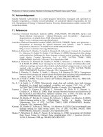
Fig. 3.19. Premature Supraventricular Contraction. Reprinted with permission from:
Conway, A Pocket Atlas of Arrhythmias. © 1974 Year Book Medical Pub.
Fig. 3.18. Atrial Fibrillation. Reprinted with permission from: Merck, Sharp & Dohm,
Division of Merck & Co., Inc.
Fig. 3.17. Sinus Arrhythmia. Reprinted with permission from: Conway, A Pocket
Atlas of Arrhythmias. © 1974 Year Book Medical Pub.
48 Vital Signs and Resuscitation
3
Multifocal atrial tachycardia (MAT), or “wandering pacemaker”, is seen
in elderly patients with chronic obstructive lung disease. In addition to the
SA node, two or more different areas of the atrium act as pacemakers (ectopic
foci). The EKG shows P-waves of varying morphology and changing PR
intervals. Treatment: oxygen and bronchodilators. In those with fast rates,
magnesium sulfate 2 g IV over 1 minute is sometimes effective (Fig. 3.21).
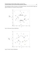
Fig. 3.20. Premature Ventricular Contraction (PVC). Reprinted with permission from:
Merck, Sharp & Dohm, Division of Merck & Co., Inc.
Fig. 3.21. Wandering Pacemaker. Reprinted with permission from: Merck, Sharp &
Dohm, Division of Merck & Co., Inc.
49Vital Sign #2: Heart Rate/Pulse
3
Special Cases
Blood/Fluid Loss
Abnormal orthostatic vital signs are initial indicators of significant blood/
body-fluid loss. After about a liter deficit (about 20% of body fluid) an
increase in heart rate occurs to compensate for the decreased volume. A
patient has orthostatic tachycardia if the heart-rate increases by 30 or more
beats per minute or the person becomes light-headed from supine to stand-
ing. Only when about 30% of blood-body-fluid loss occurs—about 2 li-
ters—does the systolic pressure begin to drop. An exception to tachycardia
with blood loss is sometimes seen. Bradycardia may occur (paradoxical
bradycardia) because of stimulation of afferent vagal fibers in the left ven-
tricle from cardiac contraction around a reduced blood volume (orthostatic
vital signs and paradoxical bradycardia are discussed in detail in Chapter 5).
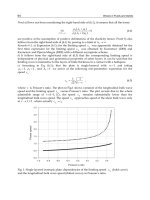
Doppler Pulse
If one cannot hear the heart or palpate the pulse, a Doppler or ultrasound
device may be used. The Doppler transducer, or flowmeter, is a transmitter
and receiver and detects the movement of red blood cells, converting the
frequency shift of the reflected ultrasound to an audible signal. Acoustic gel
is applied. After contact with the skin, the probe is angled in different direc-
tions over the artery until an optimum sound is heard with earphones or
speaker (Fig. 3.22).
Fig. 3.22. Doppler Stethoscope.
50 Vital Signs and Resuscitation
3
Fetal heart tones (FHTs) may be heard with a regular stethoscope after
about 18-20 weeks and with a Doppler stethoscope after about 12 weeks.
The mother’s heart rate is auscultated prior to the Doppler to avoid confu-
sion. After application of a conducting gel, it is pressed firmly against the
abdominal wall. Normal FHTs range from 120-160 beats per minute. Above
160 or below 120 requires urgent obstetric consultation. If fetal bradycardia
is present (indicating fetal distress) the mother is placed in the left lateral
decubitus position and supplemental oxygen is administered.
Abnormal Heart Sounds
Several normal and abnormal heart sounds are common and may be
recognized:
Splitting of the 1st sound. One may hear both AV valves close sepa-
rately. Usually splitting of the first sound has little clinical significance.
Splitting of the 2nd sound. The aortic valve closes before the pulmo-
nary, sometimes heard in inspiration. If it is heard during expiration it may
indicate heart disease.
Third sound. This is a weak sound, heard occasionally after the 2nd and
caused by distention of the ventricles during filling. It is loud in heart failure
because of overfilling of the failing ventricle. Three sounds are heard in car-
diac failure and resemble a galloping horse (gallop rhythm).
Fourth sound. This sound precedes the first and is caused by vibrations
and decreased compliance of the left ventricle. It is sometimes heard in myo-
cardial infarction.
A click may be heard after the 1st or before the 2nd sound, indicating a
normal opening of the semilunar valves, aortic valve disease, pulmonic dis-
ease or mitral valve prolapse.
An opening snap is sometimes heard after the 2nd sound. It is caused by
the opening of a narrowed mitral valve.
A friction rub, often heard in infectious pericarditis, is a squeaky or
scratchy sound caused by the rubbing together of the dry epicardium against
the parietal pericardium.
A heart murmur is the “swooshing” sound of blood heard before, during
or after (or all three) the heart sounds. It may originate from several
mechanisms: 1. increased velocity of blood flow, as in exercise, 2. normal
velocity with lessened viscosity, as in anemia, 3. obstruction to flow, as in
valvular disease, 4. flow into a dilated chamber, as in an aortic aneurysm and
5. flow through an abnormal opening, as in a congenital heart defect. It is
commonly heard in valves that are damaged and do not open properly (nar-
rowing, or stenosis), or close properly, letting blood back up through the
valve (regurgitation, or insufficiency).
Murmurs occurring after the first heart sound are systolic. Those occur-
ring after the second heart sound are diastolic. The loudness of the murmur
51Vital Sign #2: Heart Rate/Pulse
3
from grade 1 (barely heard) to grade 6 (loudest) and its location is recorded
(see Fig. 3.4).
Examples
1. In the setting of an acute myocardial infarction, a new systolic
murmur may signal papillary muscle dysfunction or ventricular
septal rupture: 3/6 systolic murmur at apex.
2. One may hear the diastolic murmur of aortic regurgitation in a
dissecting aortic aneurysm: 5/6 diastolic murmur right sternal
border (RSB).
The Pulse
Evaluation of the Pulse
Blood forced into the aorta during systole sets up a pressure wave that
travels down the arteries. The wave expands arterial walls. The expansion
wave is palpated with the fingertips as the pulse. In contrast to the heart rate,
where two sounds are heard with each beat, one beat is felt with the pulse.
Palpation is done with the tips of the first two fingers, not the fatty parts,
since the digital arteries for each finger anastomose at the fingerpad and
using the fatty parts may result in the examiner’s own pulse being recorded.
The heart rate may differ from the pulse rate. This is a pulse deficit, seen
in atrial fibrillation and occasionally in premature ventricular contractions.
It occurs in fast rates when some ventricular contractions fail to generate a
palpable pulse. One beat is so close to another that the ventricle does not
have time to fill and not enough blood is available to produce a pulse wave
in the artery. The pulse rate is thus lower than the heart rate. It is discovered
when the heart rate is auscultated and the radial pulse is palpated, or when
the pulse rate differs from the rate on the cardiac monitor. It is seen in arter-
ies removed from the heart, such as the radial.
Trauma patients and those suspected of having critical conditions such as
myocardial infarction, dissecting aortic aneurysm and acute abdominal
aneurysm should have pulses assessed in all extremities.
Although various pulse magnitudes and contours exist (i.e., pulsus
bigeminus, pulsus bisferiens, Corrigan or water-hammer pulse, etc.), demon-
strated by the sphygmograph, the usefulness of these as vital sign parameters is
weak. The possible exceptions are pulsus alternans and pulsus paradoxus.
Pulsus alternans is an alternating weak and strong pulse. It is seen in
advanced heart failure.
A paradoxical pulse (pulsus paradoxus) is an exaggeration of the nor-
mal decrease in amplitude of the pulse during inspiration. During inspira-
tion, vessels of the lungs increase in size because of increased negative pressure
in the thorax. Blood collects in the lungs, and the stroke volume decreases.
52 Vital Signs and Resuscitation
3
Expiration has the opposite effect. Kussmaul described the condition in 1873
after treating several patients with pericardial effusion. The pulse decreased
during inspiration (and in some cases disappeared). However, the heart was
obviously still beating, so Kussmaul named the condition “der paradoxe Puls”
(see Chapter 1).
A paradoxial pulse is seen when cardiac output is blocked, as in cardiac
tamponade, but also when lung compliance is decreased, as in COPD. A
blood-pressure apparatus was not yet invented in 1873 (Korotkoff first used
the Riva-Rocci cuff in 1905) and a link between pulse and blood pressure
was not made until Gauchat and Katz at Western Reserve University did so
in 1924. Thus, although pulsus paradoxus currently is considered a blood
pressure sign, it is actually a pulse sign (the blood-pressure counterpart of
the sign is described in Chapter 5). In a busy emergency setting where nuances
of change in blood-pressure readings are difficult to detect, reversion to
Kussmaul’s palpation of an artery is more useful. As an example, in the trauma
setting when a penetrating injury to the chest is present, gradual disappearance
of the radial pulse on inspiration may herald an impending cardiac tamponade.
Peripheral Pulses
The artery commonly used for pulse-taking is the radial, lying lateral to
the flexor carpi radialis tendon on the distal radius (Fig. 3.23). It is some-
times difficult to find.
The second most useful is the brachial, because of blood pressure taking.
Its location sometimes surprises people (Fig. 3.24). It is more easily palpable
medial, not lateral, to the biceps tendon and superior to, not in, the antecu-
bital fossa (cubital is forearm; antecubital is volar forearm. The differences
have become obscured and the two terms are often used synonymously). In
the antecubital fossa, the brachial artery divides into the radial and ulnar
arteries. The ulnar goes deep and the radial crosses the biceps tendon and
runs laterally down the forearm. If the stethoscope is placed in the antecu-
bital fossa, the blood pressure is being measured in the proximal portion of
the radial artery, not the brachial. Accurate palpation of the brachial artery
alleviates multiple attempts at blood-pressure taking.
The common carotid (Fig. 3.25) artery lies deep and slightly anterior to
the sternocleidomastoid muscle. One must be careful to lightly palpate the
artery, since sustained pressure will activate the baroreceptor mechanism and
slow the heart rate. Do not palpate both carotid arteries at the same time or
fainting may occur.
The femoral artery (Fig. 3.26), the largest of the pulse-taking arteries, is
located at the midpoint of the inguinal ligament between the anterior supe-
rior iliac spine and the pubic symphysis. It is the more useful for palpation
in infants, the obese, the elderly and during cardiopulmonary resuscitation.
53Vital Sign #2: Heart Rate/Pulse
3
Fig. 3.23. Radial Pulse.
Fig. 3.24. Brachial Artery.
54 Vital Signs and Resuscitation
3
The popliteal artery is the continuation of the femoral at the popliteal
fossa. It lies deep and medial to the popliteal vein and tibial nerve and is
frequently difficult, if not impossible, to find. Searching for it is unnecessary
if good femoral and pedal pulses are present. Popliteal palpation evaluates
patency when foot arteries are unavailable.
In the foot, the posterior tibial artery is the continuation of the popliteal
and is sometimes difficult to locate. It lies behind and below the medial
malleolus. Often an easier one to find is the dorsal pedis on the dorsum of the
foot at the junction of the first two extensor tendons (extensor hallucis longus
and brevis—hallus: Latin—great toe). It is helpful to mark the area with an “X”
for a difficult-to-find dorsal pedis pulse (or any other) (Figs. 3.27, 3.28).
Fig. 3.25. Carotid Artery.
55Vital Sign #2: Heart Rate/Pulse
3
Fig. 3.26. Femoral Artery.
Fig. 3.27. Dorsal pedis artery.
56 Vital Signs and Resuscitation
3
Practical Points
•Record the rate and rhythm (regular, irregular), as well as the qual-
ity and strength of the pulse (weak, strong, thready). Examples:
1. L radial—54, reg, weak.
2. R femoral—130, irreg, thready.
• Always auscultate the heart and palpate the pulse. The rates may
differ (pulse deficit). Example: HR—120, L radial P—66.
•Never be satisfied with one set of vitals.
•In a patient with a possible vascular event, such as a dissecting
thoracic aneurysm, take pulses in all extremities.
•Sometimes in the obese patient and others a radial and brachial
pulse cannot be felt, and one cannot hear a heart beat. If the pa-
tient is comatose, palpate the femoral. It is the easiest to find (in
the elderly the carotid is sometimes stenosed and difficult to find).
Use a Doppler if necessary.
Fig. 3.28. Posterior tibial artery (right leg).
57Vital Sign #2: Heart Rate/Pulse
3
•Never rely on a monitor or any electronic device for the heart rate.
Murphy’s Law will exert its inexorable effect and although a nor-
mal sinus rhythm will be showing on the monitor the patient will
have no pulse and will be moribund or dead.
•Do not auscultate the heart over clothing.
References
1. American Heart Association and the International Liaison Committee on Resusci-
tation (ILCOR): Guidelines 2000 for cardiopulmonary resuscitation and emer-
gency cardiovascular care. Baltimore: Lippincott, Williams & Wilkins, 2000.
2. Barach P. Pulsus paradoxus. Hosp Phys 2000; 36:49.
3. Bolton E. Disturbances of cardiac rhythm and conduction. In: Tintinalli et al, eds.
Emergency Medicine: A Comprehensive Study Guide. New York: McGraw-Hill,
2000.
4. Conway N. A Pocket Atlas of Arrhythmias. Chicago: Year Book Medical Pub, 1974.
5. DeGowan R et al. Bedside diagnostic examination, New York: Macmillan Pub.
Co., 2000.
6. Hoffman B. Adrenoceptor-activating drugs. In: Katzung B, ed. Basic and Clinical
Pharmacology. Norwalk: Appleton & Lange, 1989.
7. Koziol-McLain J et al. Orthostatic vital signs in emergency department patients.
Ann Emerg Med 1991; 20:6.
8. Kussmaul A. Ueber schweilige Mediastino-Pericarditis und den paradoxen Puls.
Berl Klin Wochenschr 1873; 10:37-39.
9. Lewinter J et al. Vital sign measurement procedures. In: Roberts J, Hedges J, eds.
Clinical Procedures in Emergency Medicine. Philadelphia: WB Saunders, 1998.
10. McGregor M. Pulsus paradoxus. N Engl J Med 1979; 301:480.
11. Miles W et al. Arrhythmias. In: Andreoli et al, eds. Cecil Essentials of Medicine,
Philadelphia, 1997.
12. Niemann J. The cardiomyopathies, myocarditis and pericardial disease. In: Tintinalli
et al, editors: Emergency Medicine: A Comprehensive Study Guide. New York:
McGraw-Hill, 2000.
13. O’Rourke M et al. The Arterial Pulse. Philadelphia: Lea & Febiger, 1992.
14. O’Rourke R. The measurement of systemic blood pressure; normal and abnormal
pulsations of the arteries and veins. In: Hurst J et al. The Heart. New York: McGraw-
Hill, 1990.
15. Shabetai R et al. Pulsus paradoxus. J Clin Invest 1965; 44:11.
16. Stewart J. Clinical Anatomy and Physiology for the Angry Health Professional.
Miami: MedMaster Inc., 2001.
17. Volgman A. Managing atrial fibrillation: What is the value of adding aspirin to
warfarin therapy? J Crit Ill 2000; 15:185.
18. Wo C et al. Unreliability of blood pressure and heart rate to evaluate cardiac output
in emergency resuscitation and critical illness. Crit Care Med 1993; 21:2.
19. Yealy D, Delbridge T. Dysrhythmias. In: Rosen P et al, eds. Emergency Medicine:
Concepts and Clinical Practice. St. Louis: Mosby Year Book, 1998.
20. Zide R, Tsapatsaris N. Use of anticoagulation: Addressing atrial fibrillation and
deep venous thrombosis. Res & Staff Phys 1999; 45:21.
58 Vital Signs and Resuscitation
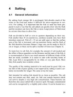
4
Vital Signs and Resuscitation, by Joseph V. Stewart. ©2003 Landes Bioscience.
CHAPTER 4
Vital Sign #3: Respiration
The name of the vital sign proposed by Edward Seguin in 1866 was “res-
pirations”. Over the years the name changed to “respiratory rate” (RR), the
original emphasis of the sign. Today in many charts it is back to
“respiration(s)”, indicating a more thorough evaluation.
Respiration is the more critical of the vital signs, since the heart and brain
require a definite amount of oxygen in order to function. In emergencies,
the airway is first addressed, then breathing, then circulation—the ABCs of
resuscitation. Apnea for more than 7-10 minutes usually means irreversible
brain damage. Exceptions exist, including infants, and isolated cases of
hypothermia and drowning.
A protocol was developed a few years ago for the Advanced Trauma Life
Support (ATLS) course also, relevant to the nontrauma patient: Primary
Survey (ABCDs), Resuscitative Measures and Secondary Survey (history
and physical exam).
The Airway is opened with jaw thrust or chin lift, oxygen is adminis-
tered, pulse oximetry is monitored, the airway is secured by intubation or
cricothyrotomy and the cervical spine is cleared. Breathing is assisted with
bag-valve-mask or ventilator, tension pneumothorax is decompressed by
needle, a chest tube is placed for pneumothorax or hemothorax and pulmo-
nary edema is treated. Circulation: IV access is obtained, a cardiac monitor
is placed, dysrhythmias and shock are treated, and Disability: a brief neuro-
logical exam is assessed. If decreased level of consciousness is present, a
chemstrip glucose is obtained (or if unavailable glucose is given) and
thiamine and naloxone are administered. The above protocol appears as
Figure 8.15.
A thorough discussion of Disability is found in Chapter 6, and Resusci-
tation in Chapter 8.
Anatomy and Physiology
The Mechanics of Breathing
During inspiration, the external intercostal muscles contract, lifting the
lower ribs up and out. The diaphragm moves down, increasing the volume
of the pleural cavity. Elastic fibers in the alveolar walls stretch, and the air
sacs of the lungs expand. During expiration, the external intercostals and
diaphragm relax. In diseases such as asthma and COPD accessory muscles
59Vital Sign #3: Respiration
4
of respiration may be used: inspiration is assisted by the sternocleidomastoids
and scalene muscles; expiration is aided by the internal intercostals and
abdominal muscles.
Lung Volumes and Pulmonary Function Testing
Breathing in and out creates volumes that can be measured. In earlier
times this was done with a spirometer, a revolving drum with a writing lever
that recorded lung volumes on graph paper. Today a computer program is
used. The patient blows into a small hand-held device (flow sensor) and the
results are displayed on a computer screen. Key elements are the forced vital
capacity (FVC)—the maximum volume one can forcibly expel after a maxi-
mum inspiration (i.e., 4800 ml) and the forced expiratory volume in 1 sec-
ond (FEV-1)—the amount of air forcibly expelled in 1 second (i.e., 83%).
The tidal volume (TV) is the amount of air moved during normal respira-
tion. An asthmatic, for example, often has a normal TV and FVC but
decreased FEV. In emphysema all three are usually decreased. The peak
expiratory flow rate (PEFR), or peak flow, is a method of respiratory evalu-
ation that can be done quickly in the emergency setting. A small hand-held
device (peak flow meter) is used. The maneuver is similar to the FVC but
Fig. 4.1. Lung Volumes.
60 Vital Signs and Resuscitation
4
recorded in liters per minute (i.e., 550 L/min). A reading below 200 L/min
usually indicates respiratory compromise (Figs. 4.1, 4.2).
The Normal Respiratory Rate
The respiratory rate in adults is 12-18 breaths per minute. In the new-
born it is about 40 and decreases to adult values at age 18. When a person
realizes that the respiratory rate will be observed, he becomes self-conscious
and begins to breathe in an odd fashion. Thus, the usual practice is to exam-
ine the heart or pulse and observe respirations without mentioning it.
The Physiology of Respiration
Oxygen from air enters the lungs and diffuses through the alveolar and
capillary membranes into the bloodstream. The pulmonary veins return oxy-
gen-rich blood to the left side of the heart, where it is pumped to the rest of
the body via the aorta. Oxygen is transported in the blood as oxyhemoglo-
bin in the red cells.
In the tissues, red cells move into the capillaries. At the arteriole end of
the capillary, oxygen diffuses through the red cells, then through the capil-
lary membrane into the tissue fluid. It then diffuses through the tissue cell
membrane to be used as fuel for cellular metabolism.
Fig. 4.2. Peak Flow Meters.
61Vital Sign #3: Respiration
4
Carbon dioxide moves out of tissue cells in the reverse direction into red
cells, where most is converted to bicarbonate ion (HCO
3
-). Bicarbonate is
transported in the plasma. The large veins bring oxygen-poor blood to the
right side of the heart where it is transported via the pulmonary arteries to
the lungs. In the lungs the process is reversed in the alveoli, and carbon
dioxide is blown off (Fig. 4.3).
Two tools are available for the analysis of the pH, oxygen and carbon
dioxide content of blood. The pulse oximeter is a computer and probe con-
sisting of 2 photodiodes and photodetector that attaches to the fingertip and
measures the oxygen saturation of arterial blood. Blood gas analyzers,
using blood gas and pH electrodes, measure the partial pressures of oxygen,
carbon dioxide and pH of blood. Other values, such as oxygen saturation
(Sa0
2
) and bicarb level (HC0
3
-) are calculated. Normal arterial blood gas
values are as follows (each lab may differ slightly):
1. pO
2
= 85—105 mmHg
2. pCO
2
= 35—45 mmHg
3. pH = 7.35—7.45
4. HCO
3
- = 21—26 meq/l
5. SaO
2
= 95—100%
The normal oxygen saturation is between 97 and 100%. Below 94%
represents hypoxia. Severe hypoxia is present with an oxygen saturation of
90%. As seen in the oxyhemoglobin dissociation curve, an oxygen satura-
tion of 90% represents a p02 of only 60 mm Hg (Fig. 4.4).
Regulation of Respiration
Respiration is controlled by the respiratory center, nerve cells in the
reticular formation of the pons and medulla. Impulses from the cerebral
cortex modify respirations, as do changes in the oxygen content, carbon
dioxide content and the pH of blood. Cells sensitive to these changes are
chemoreceptors, located in the medulla, the arch of the aorta (aortic bod-
ies) and junction of the internal and external carotid arteries (carotid bod-
ies). Low oxygen, high carbon dioxide or low pH activates the chemoreceptors
and causes the respiratory rate to increase. Low carbon dioxide or a high pH
has the opposite effect. Impulses from the aortic and carotid bodies travel to
the respiratory center in the brainstem via the vagus and glossopharyngeal
nerves (Fig. 4.6).
Fig. 4.3. Bicarbonate buffer equation.
62 Vital Signs and Resuscitation
4
Acid/Base Conditions
The bicarbonate buffer equation illustrates the effects of various physi-
ological conditions. Carbonic acid is transiently formed and carbon dioxide
is blown off during expiration (Fig. 4.5).
In metabolic acidosis, a common problem, the body produces an
increased amount of acid (H
+
), which combines with bicarbonate to form
CO
2
and water. In compensated metabolic acidosis, the body adjusts to keep
the pH within normal limits. In uncompensated metabolic acidosis the body
is unable to cope with the acid load and the pH begins to fall. The low pH
stimulates the respiratory center and the equation shifts to the right. As H
+
continuously combines with HCO
3
-
, the HCO
3
-
begins to fall. As CO
2
ac-
cumulates, the body breathes deeper and/or faster to eliminate it. This is
seen in diabetic ketoacidosis. An accumulation of acidic substances in kid-
ney disease or aspirin overdose (impairment of oxidative phosphylation, a
Fig. 4.5. Bicarbonate Buffer Equation.
Fig. 4.4. Oxyhemoglobin-Dissociation Curve.
63Vital Sign #3: Respiration
4
major buffer of H
+
) may also cause metabolic acidosis. Treatment: The un-
derlying cause is treated. If the pH is less than 7 or the HCO
3
-
is becoming
depleted, 1 or 2 amps of bicarbonate are given intravenously (Fig. 4.7).
In metabolic alkalosis, a rare condition, acid is lost, the equation shifts
to the left to restore acid, and ventilation decreases. This is seen occasionally
in those who have been vomiting for a long time (depletion of HCl from the
Fig. 4.6. Baroreceptors/chemoreceptors.