Vital Signs and Resuscitation - part 5 doc
Bạn đang xem bản rút gọn của tài liệu. Xem và tải ngay bản đầy đủ của tài liệu tại đây (161.97 KB, 18 trang )
64 Vital Signs and Resuscitation
4
stomach) and in those using diuretics (loss of H
+
from the kidney). Treat-
ment: correcting the condition (Fig. 4.8).
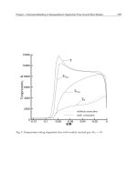
Respiratory acidosis is seen in severe asthma, chronic obstructive pul-
monary disease (COPD) and in conditions in which ventilation is poor,
such as congestive heart failure and pneumonia. The person is unable to
blow off CO
2
, which accumulates. The equation shifts to the left resulting
in rising acidity. The respiratory rate increases in an effort to blow off accu-
mulated CO
2
. Treatment: increasing ventilation with bronchodilators. Oc-
casionally intubation and assisted ventilation are required (Fig. 4.9).
Respiratory alkalosis occurs as a compensatory reaction to metabolic
acidosis and in anxiety reactions (hyperventilation syndrome—see next sec-
tion). Treatment for hyperventilation is a quiet setting to restore CO
2
, and
in the case of metabolic acidosis (i.e. ketoacidosis) the underlying condition
is treated (Fig. 4.10).
Fig. 4.9. Respiratory Acidosis.
Fig. 4.7. Metabolic Acidosis.
Fig. 4.8. Metabolic Alkalosis.
Fig. 4.10. Respiratory Alkalosis.
65Vital Sign #3: Respiration
4
Often two acid-base conditions occur together. In diabetic ketoacidosis,
as mentioned, the metabolic acidosis triggers a compensatory respiratory
alkalosis. In this case, the HCO
3
-
is low, but so is the CO
2
. The pH will be close
to normal. The person with COPD in respiratory acidosis may also have devel-
oped an additional metabolic acidosis. In this case, instead of the HCO
3
-
being
close to normal, it will fall. In general, if the pH is close to normal, and the CO
2
and/or HCO
3
-
are abnormal, one may assume a mixed condition.
Atypical Breathing
Fast Breathing (Tachypnea)
Tachypnea is usually significant at rates above 20. A low oxygen, a high
CO
2
, or a low pH (or combinations) may cause tachypnea, seen in asthma,
pneumonia, CHF, exacerbations of COPD and DKA. Other conditions
causing tachypnea are emotional reactions (i.e., hyperventilation), pulmo-
nary embolism, pneumothorax, obesity (increased vessel resistance), pain
(increased nervous stimulation), anemia (decreased oxygen) and hyperthy-
roidism (increased metabolic rate). An increased respiratory rate is also seen
with sympathomimetric drugs, as well as aspirin, methanol, ethylene glycol
and carbon monoxide poisonings. Treatment is directed at correcting the
underlying condition.
Although hyperventilation is usually the result of anxiety, life-threaten-
ing conditions such as pulmonary embolism, diabetic ketoacidosis and sep-
sis must first be ruled out. Carbon dioxide is blown off, creating respiratory
alkalosis. The person presents with a feeling of shortness of breath, light-
headedness and tingling in the hands, feet and mouth (decreased CO
2
causes
cerebral vasoconstriction, reduced cerebral blood flow and paresthesias). The
alkalosis causes increased binding of calcium to plasma protein, decreasing
the amount of ionized calcium in the bloodstream. The low calcium results
in spasms of skeletal muscles (tetany), and the person often arrives in the
emergency department in carpal spasm. This reverses as the CO
2
returns to
normal. Treatment: carbon dioxide is restored by decreasing the respiratory
rate in a quiet environment (breathing into a paper bag should be avoided
because of the potential for hypoxia).
Slow Breathing (Bradypnea)
A slow respiratory rate is usually significant at a rate of 8 or less per minute.
Often this is an emergency and requires immediate therapy. Conditions caus-
ing bradypnea are the ingestion of drugs (i.e., alcohol, narcotics, sedative-
hypnotics), increased intracranial pressure from trauma and hemorrhage
(pressure on the respiratory center), severe respiratory depression (i.e., CO
2
narcosis) and coma from any cause. It is seen in many pre-arrest and end-stage
66 Vital Signs and Resuscitation
4
conditions. Treatment: assisted ventilation is often required with a bag-valve-
mask (BVM). Endotracheal intubation is frequently necessary.
Irregular Breathing
Cheyne-Stokes Breathing
This type of irregular respiratory pattern is observed in terminal situa-
tions where tachypnea alternates with apnea. It is seen in severe central ner-
vous system injuries such as stroke, hypertensive encephalopathy, brain
swelling from trauma with impending herniation (increased intracranial pres-
sure) and in severe heart failure. The cause is an altered cerebral response to
CO
2
. Overbreathing is present when the CO
2
is elevated, then apnea occurs
to restore the CO
2
. In severe heart failure, the sluggish circulation causes a
delay and overcorrection of the acid-base status.
Kussmaul Breathing
Described in 1873 by the German physician Adolf Kussmaul,
“Lufthunger” or “air hunger” is the deep breathing seen in diabetic ketoacidosis
and uremia to blow off carbon dioxide produced in metabolic acidosis. The
rate may be slow, regular or fast. Treatment is directed at the underlying cause.
Sleep Apnea
In some obese individuals, drowsy episodes accompanied by snoring and
apneic spells occur. This obstructive sleep apnea is caused by one or more
anatomic abnormalities. The tongue falls back during sleep and blocks the
airway. Treatment involves weight loss, avoidance of alcohol and nasal con-
tinuous positive airway pressure (nasal CPAP) at night. Resection of pha-
ryngeal soft tissue may be required.
Abnormal Respiratory Sounds
1. Snoring respirations are sometimes caused by the tongue falling
back in the throat, partially obstructing the upper airway. A jaw
thrust or chin lift corrects the situation.
2. Stridor is the high-pitched sound of air moving through a partially
obstructed upper airway.
3. Decreased breath sounds in a portion of a lung (usually the base)
may be caused by a pneumothorax, hemothorax or a large pleural
effusion.
4. Rales (pronounced “rahls”, also called crackles) are sounds like tis-
sue paper being squeezed, indicating fluid in the small airways and
alveoli.
5. Rhonchi are rattling sounds from mucous and fluid in the large
airways (bronchi).
6. Wheezes are musical sounds produced by air moving through nar-
rowed bronchi and bronchioles.
67Vital Sign #3: Respiration
4
Labored Breathing (Dyspnea)
Upper Airway
The most common cause of upper airway obstruction is a decreased level
of consciousness from any cause. The tongue falls back in the mouth, par-
tially obstructing the airway. Treatment is a jaw thrust or chin lift, and inser-
tion of a nasopharyngeal or oropharyngeal airway (Fig. 4.11).
Signs of upper airway obstruction include snoring respirations, shortness
of breath, cyanosis, hoarseness, difficulty in swallowing (dysphagia) or speak-
ing, stridor, coughing, grunting or tachypnea in any combination. In the
pediatric population, tachypnea, chest retractions and nasal flaring are often
prominent. Causes are foreign bodies, trauma, allergic reactions and infec-
tion. These are frequently medical emergencies. Treatment depends on the
specific problem. Foreign bodies may be removed manually. With trauma
patients, if intubation is not possible, a cricothyrotomy is performed. An
allergic reaction involving the upper airway (angioedema) or a systemic
reaction (anaphylaxis) is treated with epinephrine, antihistamines and ste-
roids (see Chapter 5, Anaphylactic Shock).
Lower Airway
Common lower airway problems causing dyspnea are asthma, COPD,
pneumonia, pulmonary edema, pulmonary embolism/infarction and pneu-
mothorax (see following section).
Fig. 4.11. Jaw Thrust.
68 Vital Signs and Resuscitation
4
Common Examples of Labored Breathing
Asthma
Asthma is an allergic disorder affecting bronchi and bronchioles. Smooth
muscle constricts and glands of the bronchi secrete increased amounts of
mucous. Air enters the alveoli but leaves with difficulty. The result is wheez-
ing. Wheezing is not critical unless the patient is using accessory muscles to
force air out of the lungs. Occasionally in a tiring patient, wheezes diminish
and little air is moved, heralding respiratory failure and requiring endotra-
cheal intubation and mechanical ventilation. Tests reflecting the patient’s
respiratory status are pulse oximetry and peak flow (see earlier section).
Blood gases are usually not required (they show a partial respiratory alkalo-
sis—CO
2
is blown off). However, if done, a normal CO
2
in a tiring asth-
matic indicates impending respiratory failure. A peak flow of less than 200
L/min after several nebulizer treatments is usually an indication for hospital-
ization (normal peak flow is over 400 liters per minute).
Treatment:
1. oxygen by cannula or mask,
2. nebulizer therapy: a predominantly beta-2 agent such as albuterol
(Ventolin), an anticholinergic bronchodilator such as ipratropium
(Atrovent), combinations, or a more beta-2 selective agent such as
levalbuterol (Xopinex) is administered,
3. an intravenous steroid such as methylprednisolone (Solu-Medrol)
125 mg is given. In addition to having delayed long-acting anti-
inflammatory effects, steroids act synergistically with beta-2 aero-
sols to abort some of the bronchospasm of asthma.
It is important to remember that, from a vital sign standpoint, a person
may have a normal respiratory rate, not be wheezing and be in severe respi-
ratory failure, as signaled by use of accessory muscles, sweating, tiring, dete-
rioration of mental status and movement of little air. In this case endotracheal
intubation is required (see respiratory failure, Chapter 8; see also Pediatric
Asthma, Chapter 7).
Chronic Obstructive Pulmonary Disease
Chronic obstructive pulmonary disease (COPD) refers to two disease
entities sharing characteristics of long term obstruction to air flow: chronic
bronchitis and emphysema. Smoking is often a component in both dis-
eases. In bronchitis, the bronchial mucosa is swollen and red, mucous is
secreted by the glands, and the sputum may be green or yellow. In emphy-
sema (Gr: “to puff up”) air is trapped in the alveoli because of long-term irrita-
tion of the bronchi, and mucous and pus accumulate. When pressure in the
alveoli exceeds the elastic limit, they become permanently ballooned-out and
nonelastic. This produces the barrel-chested person sometimes requiring use of
69Vital Sign #3: Respiration
4
accessory muscles of respiration to breathe. Wheezes are often heard in both
situations. Pulse oximentry usually shows chronic hypoxemia. A peak flow is
less useful than in asthma, and is usually measured against the patient’s
baseline. Treatment:
1. low-flow oxygen at 2 L/min (high-flow may abolish the hypoxic
ventilatory drive and lead to respiratory arrest), or 28% by Venturi
mask,
2. bronchospasm is treated with a beta-agonist such as albuterol 2.5
mg, levalbuterol (Xopinex), or an anticholinergic agent such as
iprotropium (Atrovent) 500 µg in 2 ml normal saline by nebulizer,
3. inflammation is treated with a steroid such as methylprednisolone
125 mg IV,
4. an antibiotic (amoxicillin or trimethoprim-sulfamethoxazole) is ad-
ministered since the exacerbation is usually the result of an infec-
tion, and
5. stopping smoking helps dramatically.
Pneumonia
Signs and symptoms of pneumonia are fever, chills, cough, production of
rust-colored sputum, chest pain, tachypnea, dyspnea, decreased breath sounds
and rales. The CBC shows a leukocytosis and a chest x-ray usually reveals an
infiltrate. Treatment: viruses require no therapy. A bacterial infection is treated
with an appropriate antibiotic based on gram stain or probable etiology.
Pulmonary Edema
Acute pulmonary edema is a life-threatening sequel of congestive heart
failure, often triggered by failure to take appropriate medication and some-
times by an acute myocardial infarction. Because of inadequate pumping
action of the left ventricle, fluid backs up in the lungs. Cough, orthopnea
and chest pain are common symptoms. Anxiety, dyspnea, tachypnea, rales,
wheezes, tachycardia with an S-3 gallop rhythm, jugular venous distention
(JVD), peripheral edema and diaphoresis may be present. Blood gases show
hypoxia and sometimes hypercapnia. A chest x-ray reveals diffuse infiltrates
in both lungs. Treatment:
1. upright position,
2. high flow oxygen by mask,
3. a diuretic such as furosemide 80 mg IV to remove fluid,
4. nitroglycerine (NTG) 10 µg per minute by intravenous infusion
for vasodilation, reducing preload (and some afterload),
5. morphine, 2 mg IV, although controversial, slightly reduces
afterload, cardiac work and produces a sedative effect,
6. a systolic pressure <100 mmHg may require the administration of
dopamine (5 µg/kg/min).
70 Vital Signs and Resuscitation
4
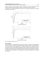
7. severe hypertension not responding to NTG may require nitro-
prusside 0.5 µg/kg/min. Intubation is frequently required. First-
line therapy may be remembered by the mnemonic: L (lasix), M
(morphine), N (nitrates), O (oxygen), P (position) (Fig. 4.12).
Pulmonary Embolism/Infarction
In pulmonary embolism, a clot from a pelvic or deep leg vein detaches
and travels to the lung. The clot impacts in a branch of the pulmonary artery
causing chest pain, dyspnea, tachypnea and sometimes syncope, anxiety,
cough and hemoptysis. A large embolus occluding a major branch of the
pulmonary artery (infarction) may cause shock and even death. Predispos-
ing factors include a previous pulmonary embolism, deep vein thrombosis
(DVT), CHF, MI, obesity, recent surgery, immobilization, trauma, preg-
nancy and malignancy. Diagnosis is made by lung scan (normal ventilation,
with perfusion defects), or a spiral CT scan. If results are equivocal, a pul-
monary angiogram is done. Doppler studies of the leg are often positive.
Treatment:
1. oxygen by cannula or mask to maintain an oxygen saturation at
95%,
2. heparin 10,000 unit IV bolus and 1000 units per hour, or low
molecular weight heparin such as enoxaparin (Lovenox) at 1 mg/
kg subq q 12 hours,
3. IV normal saline,
4. dopamine (5 µg/kg/min) may be required for hypotension. Long
term therapy includes an oral anticoagulant such as warfarin
(Coumadin).
Note: pulmonary embolism is one of the more missed diagnoses. An
increased respiratory rate with some hypoxia is almost always present.
Pneumothorax
A pneumothorax is air between visceral and parietal pleurae. It occurs
from rupture of a pulmonary bleb in the lung of the asthmatic/COPDer, in
the trauma patient (particularly knife or bullet wound) and sometimes in
cancer patients. Symptoms are sharp chest pain and cough. Occasionally
dyspnea is present. Unless the pneumothorax is quite small, breath sounds
are decreased on one side. A chest x-ray is usually diagnostic. Occasionally,
air may compress the mediastinum and vena cavae, resulting in severe respi-
ratory distress, tachycardia and hypotension (tension pneumothorax). Treat-
ment: close observation for a small nontraumatic pneumothorax. A greater
than 20% pneumothorax usually requires a chest tube (tube thoracostomy).
For tension pneumothorax, a 14/16-gauge needle/catheter is inserted in the
second interspace, midclavicular line, followed by tube thoracostomy (Fig.
4.13; see also Fig. 8.7).
71Vital Sign #3: Respiration
4
Practical Points
•First, the ABCs of resuscitation are followed (see also Chapter 8).
• The respiratory rate is counted per minute while taking heart rate
and blood pressure.
•Note any pattern (i.e., respiratory rate 30, followed by apnea for
10 seconds), the depth of respirations (strong, shallow) and abnor-
mal sounds (labored, snoring, stridor, rales, wheezes).
Fig. 4.12. Pulmonary Edema, Hypotension, Shock Algorithm. Reprinted with per-
mission from: Guidelines for 2000 for Cardiopulmonary Resuscitation and Emer-
gency Cardiovascular Care, American Heart Association.
72 Vital Signs and Resuscitation
4
•Indicate if breath sounds are diminished on one side (if so, a needle
or chest tube may be immediately required).
•Record use of accessory muscles, and whether the person has to sit
up to breathe. Note respiratory distress or dyspnea (mild, moder-
ate, severe).
•Do not auscultate the lungs over clothing.
• Examples:
1. RR 12/breath sounds strong and equal bilaterally
2. RR 6/shallow respirations/moderate resp distress
3. RR 25/bilateral wheezes/mild distress/use of accessory muscles.
4. RR 30/bilateral basilar rales
5. RR 38/shallow resp/accessory muscles/moderate distress
6. RR 10/labored breathing/no breath sounds on left
References
1. ACC/AHA Task Force. Guidelines for the evaluation and management of heart fail-
ure. Circulation 1995; 92:2764.
2. American Heart Association and the International Liaison Committee on Resuscita-
tion (ILCOR). Guidelines 2000 for cardiopulmonary resuscitation and emergency
cardiovascular care. Baltimore: Lippincott, Williams & Wilkins, 2000.
3. Baker W et al. Noninvasive assessment and support of oxygenation and ventilation.
In: Hedges R. Clinical Procedures in Emergency Medicine. Philadelphia: WB Saunders,
1998.
4. Barrett S. Dyspnea and shortness of breath. In: Rosen P et al. Emergency Medicine:
Concepts and Clinical Practice. St. Louis: Mosby Year Book, 1998.
Fig. 4.13. Pneumothorax.
73Vital Sign #3: Respiration
4
5. Baumann M, Strange C. The clinician’s perspective on pneumothorax management.
Chest 1997; 112:822.
6. Carpenter L, Verdile V. Arterial blood gas analysis. In: Wolfson A, Paris P. Diagnostic
Testing in Emergency Medicine. Philadelphia: WB Saunders, 1996.
7. Crapo R. Pulmonary-function testing. N Eng J Med 1994; 331:1.
8. Cydulka R and Khandelwal S. Chronic obstructive pulmonary disease. In: Tintinalli
J et al. Emergency Medicine: A Comprehensive Study Guide. New York: McGraw-
Hill, 2000.
9. Expert Panel Report. Guidelines for the diagnosis and management of asthma.
Bethesda: National Institutes of Health, 1991.
10. Grifoni S et al. Short term clinical outcome of patients with acute pulmonary embo-
lism. Circulation 2000; 101:2817.
11. LeConte P et al. Prognostic factors in acute cardiogenic pulmonary edema. Am J
Emerg Med 1999; 17:329.
12. Lin R et al. Rapid improvement of peak flow in asthmatic patients treated with
parenteral methylprednisolone in the emergency department: A randomized con-
trolled study. Ann Emerg Med 1999; 33:487.
13. Manning H, Schwartzstein R. Pathophysiology of dyspnea. N Engl J Med 1995;
333:1547.
14. Manthous C. Management of acute asthma. Res & Staff Phys 2000; 46:11.
15. Mihm F, Halperin B. Noninvasive detection of profound arterial desaturations using
a pulse oximetry device. Anesthesiology 1985; 62:1.
16. Nicolaou D and Kelen G. Acid-base disorders. In: Tintinalli J et al. Emergency Medi-
cine: A Comprehensive Study Guide. New York: McGraw-Hill, 2000.
17. Paape K, Fry W. Spontaneous pneumothorax. Chest 1994; 4:517.
18. Shapiro B, Cane R. Blood gas monitoring: Yesterday, today and tomorrow. Crit Care
Med 1989; 17:966.
19. Sherter C, Hill D. Update on the treatment of asthma. Res & Staff Phys 2000; 46:5.
20. Stein P et al. Clinical characteristics of patients with acute pulmonary embolism. Am
J Cardiol 1991; 68:1723.
21. Tapson V. Management of the critically ill patient with pulmonary embolism. J Crit
Illness 2000; 15:S18.
22. Vines D et al. Current respiratory care, Part 1: Oxygen therapy, oximetry, bronchial
hygiene. J Crit Illness 2000; 15:507.
23. Ward K. Pulse oximetry. In: Wolfson A, Paris P, eds. Diagnostic Testing in Emergency
Medicine. Philadelphia: WB Saunders, 1996.
24. Yelderman M, New W. Evaluation of pulse oximetry. Anesthesiology 1983; 59:4.
74 Vital Signs and Resuscitation
5
CHAPTER 5
Vital Sign #4: Blood Pressure
Anatomy and Physiology
Blood pressure is highest in thick-walled arteries nearest the heart and
lowest in veins which are thinner and away from the heart. A pressure of
100 mmHg in the brachial artery, close to the heart, means a force sufficient
to push a column of Mercury up 100 millimeters in a manometer tube.
Regulation of Blood Pressure
Sympathetic fibers (efferents) from the vasomotor center in the medulla
innervate arterioles. Arterioles regulate blood pressure. Normally they are in
a state of partial constriction, or arteriolar tone. Several types of adrenergic
receptors exist in the autonomic nervous system. Alpha, beta-1 and beta-2
are found on many organs. Alpha receptors are predominant in smooth muscle
of arterioles. Stimulation of the sympathetic portion of the vasomotor cen-
ter causes vasoconstriction and a rise in blood pressure; nonstimulation causes
vasodilation and a fall in blood pressure. Factors affecting the vasomotor
center and blood pressure are:
1. Cardiac status. In general, factors causing an increase and decrease
in heart rate cause an increase and decrease in blood pressure. Ta-
chycardia elevates blood pressure. Bradycardia or severe tachycar-
dia lowers blood pressure because of decreased cardiac output.
2. Baroreceptors are nerve endings sensitive to pressure, or stretch
(see Fig. 4.6, Chapter 4). The more important are located near
chemoreceptors at the arch of the aorta and at the bifurcation of
carotid arteries. Afferents run to the vasomotor center via the
glossopharygeal and vagus nerves. An increase in blood pressure
stimulates the vagal portion of the vasomotor center in the
brainstem, the blood pressure decreases and the heart rate slows.
Baroreceptor stimulation by massaging the carotid artery area on
one side (carotid massage) in the emergence department may con-
vert supraventricular tachycardia to a normal sinus rhythm. In hy-
potension, baroreceptors are not stretched and sympathetic output
is increased, causing alpha and beta-1 stimulation, vasoconstric-
tion and tachycardia.
Vital Signs and Resuscitation, by Joseph V. Stewart. ©2003 Landes Bioscience.
75Vital Sign #4: Blood Pressure
5
3. Other factors may act on the vasomotor center to increase or de-
crease blood pressure. Emotions play a role: nerve fibers from the
cortex synapse in centers in the hypothalamus, and stimulation
may cause an increase or decrease in blood pressure. Stimulation of
the lateral spinothalamic tract (pain and temperature pathway to
the brain) causes a rise in blood pressure. When oxygen is low and
carbon dioxide is high, as in hypotension, chemoreceptors are acti-
vated. Chemoreceptor afferents pass with baroreceptor fibers to
the vasomotor center, assisting in elevating the blood pressure. An
increase in carbon dioxide causes vasodilation of the vessels, par-
ticularly in the brain and skin. A decrease causes vasoconstriction.
Normal Blood Pressure
In the average adult, a pressure greater than 160/90 mmHg is considered
high (hypertension) and a pressure less than 90/60 mmHg low (hypoten-
sion). Blood pressure may be measured directly by arterial cannulation, or
indirectly using a sphygmomanometer. Direct monitoring is performed on
critically ill patients: an angiocatheter is inserted into the radial artery and
connected to a pressure transducer/monitor. With the exception of severe
hypotension, indirect monitoring is usually within 10 mmHg of direct
monitoring.
The mean pressure, or mean arterial pressure (MAP) is the average of
pressures recorded over a period of time (not an average of systolic and dias-
tolic pressures). One-third of the pulse pressure is added to the diastolic
pressure. Example: what is the mean pressure of 140/80 mmHg? The pulse
pressure is 40 mmHg. 1/3 of 40 = 13, and 13 + 80 = 93. Answer: the mean
pressure is 93 mmHg.
Blood Pressure Devices
1. The mercury sphygmomanometer (sphygmo—Gr: pulse) is the
usual apparatus for measuring blood pressure. It consists of an in-
flatable sleeve or cuff of various widths and lengths attached by
tubing to a manometer tube containing Mercury. It measures the
pressure required to move a column of Mercury up the tube a cer-
tain distance.
2. The aneroid (sphygmo) manometer (aneros—Gr: no liquid) uses
a similar cuff to the above but air pressure elongates a metal bel-
lows that transmits motion to the needle in a small round device
that one may hang from the cuff. These are popular with physicians.
However, many are inaccurate because of infrequent calibrations.
3. The automatic electronic oscillometric instrument (i.e., Dinamap)
is widely used in hospitals today. The cuff is connected to a moni-
tor which may also display oxygen saturation and cardiac rhythm.
76 Vital Signs and Resuscitation
5
A microprocessor senses the amplitude of arterial wall oscillations.
As the cuff deflates, the first increase in amplitude is the systolic pres-
sure; a quick decrease is the diastolic pressure. The mean pressure is
the strongest amplitude. The devise is statistically more accurate than
the mercury sphygomomanometer. Recent portable devices
Fig. 5.1. Blood-pressure Reading.
77Vital Sign #4: Blood Pressure
5
have arm, wrist or finger cuffs (the latter two are prone to er-
ror). A built-in algorithm calculates blood pressure based on
arterial vibration patterns. Some portable monitors also have
oxygen saturation capabilities (i.e., OscilloMate NIBP Monitor,
CASE Medical Systems, Inc.)
Indirect Measurement of Blood Pressure
Blood pressure should ideally be assessed in both arms. The patient is
positioned so that the elbow is flexed and perhaps supported on a pillow.
The cuff of the mercury or aneroid manometer is placed so that the lower
edge is about an inch above the antecubital fossa. Do not wrap the cuff over
clothing or the reading will be falsely high. A 5 inch cuff is used in adults.
The bladder of the cuff should encircle at least 2/3 of the arm. In an obese
person, it is wise to use a large or leg cuff 8 inches wide or the reading will be
10-15 mmHg higher than the actual value. In infants and small children, a
1 1/2 inch cuff is used. Ages 2-5 years require a 3 inch cuff (see Chapter 7).
Palpate the radial artery and inflate the cuff until pulsations disappear.
Note the pressure. Deflate the cuff, palpate and place the stethoscope on the
brachial artery at heart level. An elevated position above the heart will give
a falsely low reading; one below the heart will give a falsely high reading.
Inflate the cuff 20-30 mmHg above where radial artery pulsations disap-
pear, then lower the pressure slowly until sounds appear. These are the sounds
of Korotkoff and are divided into several phases. Phase I is when they are
first heard, and is the systolic pressure. Phase II corresponds to Phase I with
a swishing sound, Phase III occurs when the sounds are loudest, Phase IV is
the muffling of sounds and Phase V is when sounds are no longer heard.
Controversy exists over whether Phase IV or V is the true diastolic
pressure. The point at which sounds are no longer heard is usually recorded.
If the difference between the muffling and disappearance of sounds is less
than 10 mmHg, it makes little difference which is recorded. If greater than
10, it is prudent to record both (i.e., 160/80/60). Sometimes sounds are
heard to 0. Obviously a diastolic pressure of 0 is impossible, so the muffling
of sounds is the diastolic pressure. This is seen in such conditions as hyper-
thyroidism and aortic regurgitation.
Occasionally, while deflating the cuff, sounds appear, then disappear, then
reappear. This auscultatory gap, caused by diminished blood flow to the
extremity, is seen in conditions such as arteriosclerotic disease, hypertension
and aortic stenosis. Sounds are actually present, but inaudible. The gap has
important consequences for the blood-pressure taker. If the cuff is only
inflated to the gap, the systolic pressure will be falsely low. If the systolic
pressure is noted correctly and the first disappearance of sounds is taken as
the diastolic pressure, the diastolic will be falsely high. This is overcome by
palpating the radial artery while the cuff is deflated, making sure sounds are
78 Vital Signs and Resuscitation
5
auscultated for a sufficient amount of time. The gap is usually within 40
mmHg of the systolic pressure.
The opposite arm is used if an IV is running, and the affected arm is not
used in trauma and in the postmastectomy and renal patient with an arterio-
venous fistula. Lower extremity arteries are used when trauma, including
burns, is present in both upper extremities. Each time blood pressure is taken
the cuff should be completely deflated. Multiple attempts at blood pressure
taking without deflating the cuff not only irritates the patient but produces
a falsely elevated reading.
Blood-Pressure by Palpation
In some critically ill patients, it is occasionally not possible to auscultate
a blood pressure. Palpation may be performed, similar to auscultation: the
cuff is inflated to 20 mmHg above the level at which a palpable brachial
pulse disappears and deflated until it appears. The result is an estimated
systolic pressure.
Forearm and Leg Blood-Pressures
If the brachial artery is unavailable, the cuff may be placed around the
forearm and the radial artery auscultated. The systolic pressure is 10 mmHg
lower than the brachial. If the arm is unavailable, the thigh or leg may be
used. The person lies on his stomach, or on his back with the knee flexed,
and an 8 inch cuff is wrapped around the thigh. Inflate the cuff as the popliteal
artery is auscultated (a difficult artery to palpate). The systolic pressure is
20 mmHg higher than the brachial systolic pressure. The diastolic is the
same. The cuff may be wrapped around the leg just above the malleoli. Either
the dorsal pedis or posterior tibial artery may be used. Systolic and diastolic
values are the same as for the brachial artery (Fig. 5.2).
Doppler Stethoscope
The Doppler Stethoscope is a transducer with a high-frequency output
that measures flow. A weak nonpalpable pulse may be auscultated, and sys-
tolic pressures as low as 30 mmHg may be detected (the diastolic cannot be
measured). The cuff is placed around the arm and inflated, the Doppler is
placed over the artery and the systolic pressure is noted when pulsatile sounds
are first heard. Because it evaluates flow rather than pulsations, the Doppler
is more sensitive than a regular stethoscope (see Fig. 3.22).
Approximation of Blood-Pressure from Pulse
In the past, it was alleged but never substantiated by invasive monitoring
that a palpable radial, femoral and carotid pulse meant a systolic pressure of
>80 mmHg, a carotid and femoral pulse represented a pressure between 70
and 80 mmHg and a palpable carotid pulse indicated a pressure between 60
79Vital Sign #4: Blood Pressure
5
and 70 mmHg. Since invasive confirmation was never obtained and since
pressures seemed to vary widely the concept faded.
Recently, a British anesthetist chose to challenge the 1985 ATLS guide-
lines showing this relationship, not realizing that the current version of the
ATLS manual (1997) no longer includes it. He performed invasive moni-
toring on 30 patients with hypotension secondary to hypovolemic shock,
grouping them as follows: group 1—radial, femoral and carotid pulses, group
2—femoral and carotid pulses, and group 3—carotid pulse only. In group 1
the high pressure was 88 mmHg with a mean of 72.5 mmHg, group 2 had
a high of 78 mmHg with a mean of 66.4 mmHg, and group 3 had a high of
56 mmHg with a mean of 50 mmHg. A fourth group showed no palpable
pulses, but had systolic pressures of 52, 54 and 76 mmHg.
The results indicate a lower than expected correlation, with a wide varia-
tion in predicted pressures from pulses. In summary, in this study a pal-
pable radial pulse indicated a pressure between 53 and 88 mmHg, a femoral
indicated a pressure between 48 and 78 mmHg, and a carotid indicated a
Fig. 5.2. Leg Blood-Pressure.
80 Vital Signs and Resuscitation
5
pressure between 38 and 58 mmHg. The interesting facet of this investiga-
tion is that a substantial blood pressure existed in the absence of any pal-
pable pulse.
Pulsus Paradoxus
As mentioned in Chapter 1, Kussmaul in 1873 noticed that in patients
with pericardial effusion the pulse decreased and sometimes disappeared
during inspiration. Later, after development of the blood-pressure cuff, it
was found that a decrease in blood-pressure accompanied the weak pulse. In
a normal person a slight waxing and waning of blood pressure exists during
inspiration—up to 10 mmHg. Today a pulsus paradoxus is defined as an
inspiratory fall in systolic blood-pressure greater than 10 mmHg. It is mea-
sured as follows: using normal blood-pressure protocol, the cuff is inflated
above systolic pressure, then deflated to Korotkoff sounds during expira-
tion. While the pressure is maintained the person inhales. The cuff pressure
is lowered until Korotkoff sounds reappear. If this exceeds 10 mmHg, a
paradoxical pulse is present. It is seen in conditions such as cardiac tampon-
ade, severe asthma, COPD and heart failure. Its usefulness in a busy emer-
gency setting is limited. Palpation of the radial or femoral pulse during
inspiration is more practical, particularly if cardiac tamponade is suspected
(see Chapter 3, Paradoxical Pulse).
Increased Pulse Pressure
The pulse pressure is the difference between systolic and diastolic pres-
sures and reflects cardiac output. The systolic pressure rises in exercise, fever
and conditions such as hyperthyroidism. Attention to the pulse pressure is
important in head injuries, because a widening pulse pressure may indicate
rising intracranial pressure from edema or bleeding (see Chapter 6). Aortic
regurgitation may cause a widened pulse pressure. When the aortic valve is
damaged, some aortic blood flows backwards at diastole, lowering the dias-
tolic pressure. The left ventricle now pumps more blood during systole, rais-
ing the systolic pressure. Treatment: valve replacement.
In patent ductus arteriosus, the connection between the pulmonary
artery and aorta remains open in the adult. In the fetus, the ductus arterio-
sus carries blood from the pulmonary artery to the aorta, bypassing the lungs,
which are inactive. If the ductus fails to close at birth, blood is shunted from
the high-pressure aorta back into the pulmonary artery. The result is high
output heart failure (the left ventricle is pumping two or more times the
normal cardiac output). A continuous “machinery” murmur is heard over
the pulmonary area. Treatment: surgical correction.
81Vital Sign #4: Blood Pressure
5
Decreased Pulse Pressure
A narrowed pulse pressure is seen in conditions such as hypovolemic
shock and cardiac tamponade. It is sometimes seen in heart failure from
increased peripheral resistance and in severe aortic stenosis from decreased
stroke volume.
High Blood Pressure (Hypertension)
Causes of Hypertension
The World Health Organization’s criterion for a diagnosis of hyperten-
sion is a blood pressure of 160/95 mmHg or greater. Pressures between this
and 140/90 are borderline. Most physicians treat diastolic pressures of
95 mmHg. Three readings are usually recorded over several days before treat-
ment is begun. Factors such as anger, anxiety, stress and exercise may cause a
transient increase in blood pressure so the person should rest for several min-
utes before the blood pressure is taken.
Primary Hypertension
In most cases (95%) the cause of hypertension is unknown (primary or
“essential” hypertension). A genetic component appears to be present in-
volving excessive sensitivity to dietary salt and an overstimulation of the
sympathetic nervous system. The overstimulation of sympathetic activity
causes arteriolar spasm which creates hypertrophy of the muscle layer of the
arteriole, narrowing of the vessel, and an elevation of the blood pressure.
The baroreceptor mechanism is reset to a higher level. The kidney appears
to be involved. Treatment: weight reduction, salt restriction, and the dis-
continuance of alcohol and smoking. A stepped-care approach is often used:
the patient is started on a diuretic, beta-blocker or calcium-channel blocker,
and other agents are added as needed, such as one of the above or an angio-
tensin-converting enzyme (ACE) inhibitor.
Hypertensive Emergencies
Chronic uncontrolled or poorly controlled hypertension predisposes to a
cerebrovascular event, such as hemorrhage, transient ischemic attack (TIA),
thrombosis and hypertensive encephalopathy, as well as aortic dissection,
cardiac disease (angina, myocardial infarction, congestive heart failure) and
renal failure.
Hypertensive Encephalopathy
Hypertensive encephalopathy is seen when the blood pressure exceeds
the limits of autoregulation by the blood-brain barrier. Blood enters brain
tissue causing cerebral edema. The process develops over hours to days, with
symptoms of headache, nausea, vomiting and an altered mental status ranging