Gastrointestinal microbiology - part 4 ppt
Bạn đang xem bản rút gọn của tài liệu. Xem và tải ngay bản đầy đủ của tài liệu tại đây (441.53 KB, 43 trang )
microbiota of an adult human lacked the adjuvant ability to stimulate the sIgA anti-
rotavirus response in gnotobiotic mice but, on the contrary, exerted a suppressive effect as
do E. coli (Table 2) (79). Thus, the modulating effect of Bifidobacterium is strain-
dependent, as it has also been described for different Lactobacillus strains used as
probiotics in other mice studies (80). Taken together, these data suggest that it is important
to define the modulatory effect of the strains of bifidobacteria either normally colonizing
the digestive tract of babies after birth or given as probiotics, to modulate in a good
protective way a specific intestinal immune response.
In conclusion, and on the basis of the experimental and clinical data, we may
consider that the presence of certain bacterial strains in the infantile intestinal microbiota,
namely some strains of Bifidobacterium, or some transiting strains of probiotics, enable
activation of the mechanisms that result in optimization of the anti-rotavirus protective
IgA Ab response. Elucidation of the immunomodulatory mechanisms must now
be pursued.
Regulation of the Immune Responses
Tolerance to Soluble Proteins: Oral Tolerance
The role of the intestinal microbiota on the OT process has been demonstrated by various
experimental studies using GF mice. Results depend on the immune response considered,
oral Ag, and experimental schedule used. In these experiments, immune responses to
a specific Ag are compared in two groups of mice: the tolerant group where mice are fed
with an Ag prior to the peripheral immunization with the same Ag, and the control group
fed with only the buffer before the same peripheral immunization. Specific immune
responses to the Ag used are then evaluated (Ab responses in serum or cellular response
by delayed-type hypersensitivity) in both groups. The tolerant state is present when
peripheral immune responses to the Ag are abolished or significantly decreased in the
group Ag-fed as compared with the control group.
In an initial study, Wannemuehler and coworkers (81) showed that, in contrast to
what is observed with the CV mice, gavage of GF mice with a particular antigen, sheep red
blood cells (SRBC), does not enable suppression of immune responses to SRBC in serum.
However, the OT process was re-established when LPS was administered orally prior
to gavage. The authors concluded that Gram-negative bacteria play a fundamental role in
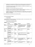
Table 2 The Gut Colonization of Different Bacterial Strains Modulates the Intestinal
Anti-rotavirus IgA Antibody Response Measured in Gnotobiotic Mice
Intestinal microflora of gnotobiotic mice
Anti-rotavirus sIgA antibody
level (AU/g of feces)
Bifidobacterium bifidum (from baby) 31G7
a
[
Bifidobacterium DN 173 010 (a commercial strain) 21G3
a
[
Germ-free (control) 11G2
Bifidobacterium infantisCB. pseudocatenulatumC
B. angulatumCB. sp (from human adult)
4G1
a
Y
E. coli (from infants) or Bacteroides vulgatus (from
human adult)
4G1
a
Y
a
Significant difference with germ-free mice (p!0.01).
Abbreviation: AU, arbitrary units.
Source: From Refs. 72, 79.
Immune Modulation by the Intestinal Microbiota 109
the mechanisms responsible for OT. Subsequently, other experiments using adult GF mice
fed with a soluble protein, OVA, in order to study the immune suppression of anti-OVA
serum IgG response, demonstrated that it was possible to induce OT in GF mice. However,
in contrast to what is observed with CV mice, the suppression was of very short duration,
about 10–15 days, versus more than 5 months in CV mice (82). Similar results were
obtained in human-microbiota-associated gnotobiotic mice (60). Colonization of the
intestinal tract with E. coli alone prior to gavage was sufficient to restore lasting
suppression (83), and the same results were obtained with another Gram-negative bacteria,
Bacteroides (unpublished personal data), while in our experimental conditions, adult GF
colonized with the strain of Bifidobacterium bifidum isolated from a baby’s feces, had no
effect on the serum IgG anti-OVA suppression (83).
Recently, in their experimental conditions, Sudo and coworkers (84) showed that in
OVA-fed mice, the GF state does not allow suppression of the systemic anti-OVA IgE
response in serum in contrast to what is observed with CV mice. Colonization of the
intestinal tract by a strain of Bifidobacterium infantis restored the suppression but only
when the strain colonized the intestinal tract of the mouse from birth. The importance of
the presence of intestinal bacteria from birth in the optimization of the immune processes
has also been suggested in a more recent study (60).
It is interesting to compare these experimental results to those described in human
neonates by Lodinova-Zadnikova and coworkers (85). In their study, they colonized
the digestive tract of babies just after birth with a given strain of E. coli. In these conditions
E. coli is able to establish durably in the digestive tract of newborns as described
previously (86). After 10 years (preterm infants) and 20 years (full-term infants),
differences in occurrence of food allergies between colonized and control subjects were
statistically significant; 21% versus 53%, and 36% versus 51% respectively. Furthermore,
recent clinical trials using ingestion of a strain of probiotic, Lactobacillus rhamnosus GG,
during the last month of pregnancy to women and after birth to babies during 6 months,
reduced the incidence of atopic eczema in at-risk children during the first 4 years of
life (87). However, in this case, IgE levels were not decreased in the treated group as
compared with the placebo group. The protective mechanisms of these interventions are
not elucidated.
All these experimental data show the importance that a single bacterial strain present
in the intestinal digestive microbiota of infants may have with respect to the establishment
of tolerance mechanisms. Are there E. coli, Bacteroides or some strains of Bifidobacterium
which play this important role? First, as suggested by previous studies, it is not sure
whether the mechanisms are the same for suppression of the various isotypes IgG and IgE
(45,88), and consequently that the same bacteria are operating on them. Secondly, as
described previously, all the strains belonging to the same bacterial genus have not the
same immunoregulatory properties and it is conceivable that some Bifidobacterium strains
may have regulatory properties on suppressive immune processes.
The cellular ways by which the bacteria are acting, and the exact bacterial
components involved are not known. However, from an ecological point of view, it is
important to note that some experimental data point out the importance of the neonatal
period with respect to the ability to recognize bacterial messages.
Tolerance to the Intestinal Microbiota
An important question is why the intestinal microbiota does not mount an inflammatory
response in the gut while this state is broken in pathologic conditions such as IBD?
Moreau110
The mechanisms by which commensal and non-pathogenic bacteria are tolerated
by the IIS is beginning to be understood and may result from a cross-talk between
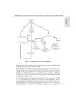
bacteria, epithelium, and immune cells. In an interesting experimental study, Neish
and co-workers (89) demonstrated, using an in vitro model of cultured human intestinal
epithelial cells, that a non-pathogenic strain of Salmonella directly influenced the
intestinal epithelium to limit inflammatory cytokine production. They showed that the
immunosuppressive effect was due to the inhibition of the NFk-B activation pathway by
blockage of IkB-a degradation. Another interesting conclusion from this study was that
non-pathogenic bacteria, which do not belong to the commensal intestinal microbiota,
are unable to induce inflammatory responses. Another study converges to an opposite
conclusion (90). In several intestinal epithelial cell lines, the authors demonstrated that
a commensal bacterial strain, Bacteroides vulgatus, was able to activate the NF-kB
signaling pathway through IkB-a degradation and ReIA phosphorylation. However, the
presence of TGF-b1 cytokine inhibits B. vulgatus-mediated NF-kB transcriptional
activity showing that the responsiveness of intestinal epithelial cells to luminal enteric
bacteria depends on a network of communication between immune and epithelial cells
and their secreted mediators.
Recently, it was shown in vivo in mice, that the intestinal microbiota itself plays
a regulatory role with respect to inhibition of the NFk-B activation pathway, by the way of
another inhibitory factor, the peroxisome proliferator-activated receptor (PPARg) (61).
The latter is highly expressed in the colon and its activation has anti-inflammatory effects,
with protection against colitis. PPARg activators are able to limit inflammatory cytokine
production through the inhibition of the NF-kB pathway. It has been suggested that PPARg
could play an important role in homeostasis of the gut, especially in the colon. In patients
with IBD, impaired expression of PPARg in colon epithelial cells was observed (61).
In the same work, in vivo observations showed that the intestinal microbiota and TLR-4
regulates PPARg expression by epithelial cells of the colon. Indeed, it is highly expressed
in CV mice while it is barely detectable in GF mice. When TLR-4 transfected CaCo-2 cells
were incubated with LPS, an increase of PPARg expression was observed showing
the involvement of TLR-4 in this process and suggesting that PPARg may be a regu-
latory factor able to shut down the TLR-4 signaling given by bacterial LPS abundant in
the colon (61).
Taken together, these data provide evidence that the cross-talk existing between the
IIS and intestinal microbiota pass through regulatory processes preventing inflammatory
responses induced by activation of some nuclear factors, such as NF-kB, which could be
different, or predominant, according to the intestinal site. They are mediated through the
actions of commensal bacteria, but also through exogenous non-pathogenic bacteria action
and this data is of importance in terms of nutrition. Indeed, we can ingest billions of
exogenous bacteria in some foods such as fermented milks and some cheeses, without
detrimental consequences. In terms of pathology, a lot of other questions concerning the
mechanisms and origin of IBD have yet to be answered. Why is an activation of the NF-kB
pathway observed in IBD? Is it due to some subsets of the intestinal microbiota, which are
suddenly dominant in an unbalanced microbiota? Is it due to enteropathogens which can
interact with the NF-kB pathway during infection? Or, is it due to a decrease and
modification of mucus secretion allowing excessive adhesion of commensal bacteria?
All these factors, and others, may be responsible.
It is interesting to give recent clinical results concerning oral administration of
probiotics on the maintenance of the remission phase in IBD, either the use of a mixture
of 8 strains of lactic-acid bacteria used as probiotics (VSL#3) in chronic pouchitis (91), or
a yeast strain, Saccharomyces boulardii (92) or the E. coli Nissle 1917 (93) in ulcerative
Immune Modulation by the Intestinal Microbiota 111
colitis. The mechanisms underlying such beneficial effects are still not known and they are
multifactorial. From experimental data it has been suggested that a stimulation of the non-
inflammatory IL-10 cytokine production by ingestion of probiotics may be involved
in such protective effect (94). Further experimental and clinical studies need to be
conducted to further elucidate the mechanisms involved in the epithelium-bacterial
cross talk.
RELATIONSHIPS BETWEEN THE PERIPHERAL IMMUNE SYSTEM
AND INTESTINAL MICROBIOTA
Activation of the Immune System
Innate immunity plays a very important role in the activation of the immune system and
the ability to develop specific acquired immune responses. Through their Ag-presenting
activity and the synthesis of numerous pro-inflammatory chemokines and cytokines (IL-8,
IL-1, IL-6, TNF-a, and IL-12), macrophages, and DCs play a key role in the regulation
of immune responses. They are the gatekeepers of the host, generating innate resistance to
pathogens, and specific immune responses by the stimulation of T-cell-acquired immunity
and regulation of the TH1/Th2 balance.
It has been postulated that the immune defects in neonates may result from
a developmental immaturity of APC functions (78), and bacterial components resulting
from intestinal colonization could be an important factor for maturation of APCs (95).
Recently, Sun and coworkers (96) investigated the ontogeny of peripheral DCs and their
capacity to provide innate responses to microbial stimuli in early life. They show that
neonatal murine spleen DCs have intrinsic capacity to produce bioactive IL-12. Moreover,
after microbial stimulation given in vitro by LPS, they are able to up-regulate MHC and
costimulatory molecule expression required for productive interaction with naive T cells.
Thus, neonatal DCs could be fully competent in their innate functions but they need to be
activated, through TLR recognition as described previously, by bacterial stimuli afforded
by the intestinal microbiota. Another interesting study supports this hypothesis. Nicaise
and coworkers (97) demonstrated that the presence of the intestinal microbiota underlies
IL-12 synthesis by macrophages derived from splenic precursors.
On the basis of those experimental data, one can wonder whether the first bacteria
colonizing the intestinal tract, E. coli, rich in LPS, and subsequently bifidobacteria rich
in peptidoglycan and CpG dinucleotides, do not play such crucial activating roles? It is
conceivable that in newborns, the abrupt colonization of the intestinal tract by the
microbiota may induce a physiological inflammatory reaction with, as a consequence, an
increase in intestinal permeability, bacterial translocation and systemic activation of
immune cells, especially APCs. Experimental evidence supports that hypothesis. Studies
in mice have shown that the presence of the intestinal microbiota induces the synthesis of
pro-inflammatory cytokines IL-1, IL-6, and TNF-a by peritoneal macrophages. Such
effects can be reproduced in gnotobiotic mice colonized with E. coli alone while
a Bifidobacterium bifidum strain isolated from baby’s feces had no effect (Table 3) (98).
Other non-specific resistance factors play an important role in host defense
mechanisms to infection. GF and gnotobiotic animal models have showed that some
functional parameters involved in innate immunity, phagocytosis, complement system,
and opsonins, are expressed to a lesser extent than in CV animals (99).
Moreau112
Modulation and Regulation of Immune Responses
Balance Th1/Th2
Experimental results, epidemiological studies and clinical trials strongly argue for the fact
that bacterial environment plays a crucial role in the Th1/Th2 balance via different
mechanisms of which cytokine synthesis by innate immune cells, especially IL-12, and
IFN-g, could play a decisive role.
The prenatal period and early childhood are considered to be critical for the
establishment and maintenance of a normal Th1/Th2 balance. It has been described that
the immune context at birth is mainly Th2, while Th1 responses are partially suppressed,
enabling non-rejection of the fetus during gestation. After birth, neonates must rapidly
restore the balance by developing the potential to induce Th1-type responses (100).
Various studies have shown that, in atopic infants, the switch does not occur, and the infant
is in a context of an imbalance toward Th2 with a predisposition to development of IgE
responses (101,102). The neonatal period is thus considered to be extremely important in
enabling regulation of the Th1/Th2 balance to become operative, and the switch could
occur during the first 5 years of life especially during the first year of life (103).
The Th2/Th1 switch is dependent on multiple factors whose relative importance
has yet to be elucidated. Bacterial stimuli are considered to play a considerable role, and
some years ago it had been claimed that infections might prevent the development of atopic
diseases. This is referred to as the “hygiene hypothesis” (13), but it is now a matter of
debate. From a recent study (104), authors did not find any evidence that exposure
to infections in infants reduces the incidence of allergic disease, but, in contrast, exposure to
antibiotics may be associated with an increased risk of developing allergic disease. Today,
accumulating evidence suggests that rather than infections, alteration of the composition of
the intestinal microbiota early in life may be an important determinant of atopic status
(13,105). Experimental studies have supported this hypothesis. Thus, in one-week-old rats,
peripheral immunization leads to a Th2-biased memory response. However, when the rats
are concomitantly administered a bacterial extract by the oral route with immunization, the
memory response switches to both Th1 and Th2 (106). Another study showed how, in three-
week-old mice, the disturbance in intestinal bacterial equilibrium following ingestion of an
antibiotic, kanamycin, promoted a shift in the Th1/Th2 balance toward a Th2-dominant
immunity, while it became Th1 and Th2 in non-treated growing CV mice (107). Ingestion
of intestinal bacteria such as Enterococcus faecalis five days after antibiotic treatment
again permitted the shift back towards the Th1/Th2 balance (108).
Table 3 Influence of Intestinal Bacteria on the Inflammatory Cytokine Production by Peritoneal
Macrophages
Gnotobiotic mice Cytokines (units/ml)
IL-1 IL-6 TNF-a
Conventional 18200 6,33 72
Germ-free 8300
a
2,62
a
!50
a
Bifidobacterium bifidum 8000
a
2,46
a
!50
a
Escherichia coli 15350
b
7,24
b
108
b
a
Significant difference with conventional mice (p!0.01).
b
Not significant.
Abbreviations: IL, interleukin; TNF, tumor necrosis factor.
Source: From Ref. 98.
Immune Modulation by the Intestinal Microbiota 113
From an epidemiological point of view, very interesting studies argue in favor of
the important role of the bacterial environment in the first year of life in order to ensure the
good orientation of immune responses preventing the short- and long-term development of
atopic diseases (13,101,103,109–111). Recent comparative studies have been conducted
in children living in the same allergenic environment but under different life-style
conditions, urban and farming environments. Results showed that substantial protection
against development of asthma, hay fever, and allergic sensitization was seen only in
children exposed to stables, farm raw milk, or both in their first year of life (103). Authors
also found that prenatal exposure of women had a substantial protective effect.
Bacteria that are responsible for such effects are not known. Gram-negative bacteria
rich in LPS have been suggested to be important in that phenomenon (85,109,112) but it is
also possible that Gram-positive bacteria, such as bifidobacteria and Lactobacillus, are
involved. The comparative study between Swedish and Estonian children (105) has
suggested a specific role of the intestinal microbiota, regarding its nature, diversity and
changes with time. Besides genetic factors, which are known to play an important role in
the development of allergic diseases, all these data suggest that the infant intestinal
microbiota normally rich in Gram-negative (LPS-producing) and Gram-positive bacteria
may not be well-balanced in atopic children. Depending on the microbial environment
associated with the life-style, especially during the first year of life, a restoration of the
normal balance could be achieved.
Clinical trials using probiotics to treat or prevent atopic eczema in infants have also
generated arguments suggesting that the infantile intestinal microbiota balance plays an
important role in the good orientation of immune responses. In a recent double-blind trial,
Kallioma
¨
ki and coworkers (87) have shown that the supplementation of pregnant women
one month before delivery followed by 6 months post-parturition (mother or baby) with
a probiotic strain, Lactobacillus rhamnosus GG, lead to a significant decrease in the
incidence of atopic eczema in babies with a family history of atopic disease. At two years
of age, atopic eczema was diagnosed in 23% of treated babies versus 46% in the placebo
group. The preventive effect of L. rhamnosus GG extends to the age of 4 years follow-up
treatment (87). The mechanisms involved in such a protection are unknown. Indeed, the
frequencies of positive skin-prick test reactivity (measuring the specific IgE levels) were
comparable between treated and placebo groups. Further studies are necessary to elucidate
the mechanisms responsible for these interesting protective effects.
On the basis of all the above data, questions arise with respect to delivery conditions,
infant feeding, and antibiotic treatments to be administered during infancy in order to
enable and optimally establish and maintain integrity of the intestinal microbiota.
Probiotics may also be considered as good palliative agents with respect to impaired
equilibrium of the intestinal microbiota. Knowledge of the immunoregulatory
mechanisms driven by the intestinal microbiota of infants, as well as the bacterial
components which are involved, are crucial to prevent some pathologies which are
dramatically increasing today.
Natural IgG
In the absence of immunization, there is a natural level of immunoglobulins (Ig) in serum
named “natural Ig” or “natural Abs.” The roles of those Abs in the immune responses have
yet to be completely elucidated but it is known that they play important regulatory roles in
humoral immune responses, especially in immune responses to self-Ag (113). It has also
been demonstrated in mice that they intervene with the development of the B
repertoire at peripheral level (spleen), enabling expansion of the Ab response towards
Moreau114
thymo-dependant Ags (114,115). In man, the role of these natural Abs is under
investigation in the context of research on certain autoimmune disease (116).
Intrinsic and extrinsic factors, especially the intestinal microbiota, act on the natural
Ig levels, depending on isotypes and sub-classes. Thus, GF mice had normal serum IgM
levels, but IgG, and IgA levels are approximately 5% of conventionally reared littermates
(114). It has been established in mice that one of the roles of the natural IgG is to expand
B cell repertoire. The latter can be evaluated through the expression of some genes coding
for the variable part of the heavy chain of Ig (VH gene) using probes. Analysis of a VH
gene expression has provided a quantitative tool for the global assessment of Ab
repertoire, and a preferential use of the gene means that the repertoire is poorly diversified.
Early in ontogeny, a high frequency of B cells could bind to multiple Ags, among
which auto-Ags are found, in neonatal CV mice. This fact has been correlated with
preferential use of VH gene family, namely VH7183. In CV adult mice these multi-
reactive B cells are much less frequent coinciding with a random usage of VH genes, as
seen by the decreased utilization of VH7173 gene family, showing a diversified repertoire.
Thus, there is a maturation of the immune system of adult CV mice. This fact is not present
in adult GF mice where a high percentage of B cells expressing VH 7138 genes is found as
in neonatal CV mice (115). The injection of purified natural IgG Ig from serum adult CV
mice into GF mice reduced the use of the VH7183 gene family in the peripheral B-cells, as
in CV mice (115). From these data authors concluded that if a genetic program leading to
non-random position-dependent preference of rearrangement and expression initially
controls the establishment of the VH repertoire, a broader utilization of the B-cell
repertoire is thereafter stimulated by environmental Ags and Igs. The finding that GF mice
maintain a “fetal-like” VH repertoire that can be modified by the administration of pooled
Igs from normal unimmunized CV mice establishes the crucial role of the intestinal
microbiota in this function.
This data may have clinical relevance. Many reports have described the beneficial
results of intravenous injection of normal human IgG in treatment of autoimmune
disease (116).
The mechanism by which exogenous antigenic stimulation can influence the
expression of VH gene remains unclear. Exogenous Ags may play an important role in
the final modulation of the expressed repertoires either by direct stimulation of Ag-specific
clones or indirectly by idiotype interactions mediated by the Abs produced in those
responses (113–115).
Autoimmune Diseases
One example of the regulatory effect exerted by intestinal microbiota on an autoimmune
disease has been reported by Van der Broek and co-workers (117). Streptococcal cell wall
(SCW)-induced arthritis is a chronic erosive polyarthritis, which can be induced in
susceptible rats by a single intra-peritonal injection of a sterile aqueous suspension of
SCW. The acute phase of the disease develops within a few days, the second, chronic
phase, which mainly involves peripheral joint inflammation, develops from 10 days after.
The second phase is dependent on functional T lymphocytes. F344 rats are genetically
described as resistant to the second chronic phase, while in contrast another strain of rats,
Lewis rats, are described as susceptible. These data suggest that a T-cell unresponsiveness
due to immune tolerance to SCW may be the mechanism underlying resistance to SCW-
induced arthritis of F344 rats, while Lewis rats are defective in their tolerance. When
F344 rats are reared in GF conditions, they become susceptible to SCW-induced arthritis
as are Lewis rats. There was a correlation between the susceptibility of the disease and the
Immune Modulation by the Intestinal Microbiota 115
T cell proliferation response to SCW measured in vitro. In CV Lewis and GF-F344 rats,
a proliferation was measured while it was not present in CV F-344 rats. This concept that
disease might result from a similarity between naturally occurring cell surface Ags of the
host and those expressed on some commensal or pathogenic micro-organisms have been
referred to as the “molecular mimicry hypothesis.” Mono-association of GF F344 rats
with E. coli resulted in resistance, which equaled that in CV F344 rats whereas
mono-association with a Lactobacillus strain did not really affect susceptibility. Thus,
in CV F-344 rats, a state of tolerance to arthritogenic epitopes is induced during the
neonatal period of life and maintained through life by the bacterial microbiota, resulting
in resistance to SCW-induced arthritis. In Lewis rats, this tolerant state is deficient and/or
easily broken.
Bacterial effects have been suggested in other autoimmune diseases. Thus, oral
antibiotic treatment after adjuvant-induced arthritis (AIA) induction in rats significantly
decreased clinical symptoms of AIA while, concomitantly, E. coli levels increased in the
distal ileum of antibiotic-treated rats (118). In addition, it has been described that
Mycobacterial infections profoundly inhibit the development of diabetes in non-obese
diabetic (NOD) mice (119).
CONCLUSION
From all the experimental epidemiological and clinical results presented here, the
digestive microbiota can be considered as an organ: it is specifically tolerated by
the host and in turn, it exerts many continuous regulatory effects on intestinal and
peripheral host’s immune responses. Consequently, it plays fundamental roles in health.
It is very important to develop knowledge about its composition, the bacterial components
and metabolites that participate to such immunoregulatory effects, and the exact
mechanisms involved.
Studies from GF animals have demonstrated the importance of the digestive
microbiota on intestinal and peripheral immune systems. In some cases, the entire
digestive microbiota is needed to obtain the complete effect while other immunoregulatory
effects can be reproduced with only one bacterium and sometimes with only specific
strains. Because the intestinal microbiota is a dynamic community which modifies from
birth to old age in predominant bacteria composition, specific targeted interests have to be
defined for the study of relationships between the intestinal microbiota and the host,
according to age. Indeed, bacterial species found in the predominant microbiota are not
constantly the same throughout life and several studies have demonstrated the strain-
dependant immunomodulatory effect of bacteria. For instance, some strains of
bifidobacteria, such as B. breve, are more commonly found in infants but less in adults
(120). Other studies from adult GF animals have demonstrated that some bacterial effects
are only obtained when the bacteria colonized the intestinal tract from birth indicating that
the bacterial effects need some characteristics of the neonate immune system. A number of
indirect findings converge toward the idea that the neonatal period is crucial for the infant
with respect to setting up the regulatory mechanisms which will play an important role in
the good orientation of immune responses throughout life. Because of the long-term
consequence of the establishment of appropriate immunoregulatory networks, it is very
important to develop knowledge on the cross-talk between the intestinal microbiota and
immune system early in life. In this context, recent studies of the innate responses to
bacterial constituents should generate decisive information in support of the role of the
intestinal microbiota.
Moreau116
In adults, regulation of immune responses seems to be constantly reshaped by
persistent interactions between the host and its digestive microbiota.
Today, an increasing challenge for researchers studying immunity (IIS as well as
oral or peripheral immune responses after Ag vaccination, pro-, and prebiotic effects) is
that the intestinal microbiota of experimental rodents used is not defined and can differ
between breeders because of the great variety in housing conditions. Since the
development of knock-out mice, which are very sensitive to infections, the microbial
status required by experimenters has led to the production of highly clean animals which
carry a commensal microbiota with reduced diversity. This fact has probably a significant
impact on the development of the immune responses. Thus, because results could not
reflect the exact conditions of microbial stimulation, the interpretation of experiments
may be completely different according to different laboratories. Some controversial
results obtained in mice and humans might also be explained by such paucity of mouse
microbiota existing in pathogen-free mouse breeding-care units. Now, it is crucial to
develop animal models in which the commensal microbiota will be better defined and
designed to allow the maintenance of biological features relevant in the field of
immunological investigations.
A more comprehensive understanding of the relationships between the intestinal
microbiota and innate and acquired immune systems should offer new approaches for the
therapy of some diseases such as allergies and IBD and for the design of oral vaccinations,
and the maintenance of health. Beneficial micro-organisms such as probiotics, and dietary
ingredients such as prebiotics, that act on the digestive microbiota, show promise for
treatment in these immune-related intestinal disorders. Researchers addressing those
subjects have to consider the digestive microbiota in their investigations.
All of the studies presented here clearly indicate the close relationship between the
prokaryotic and eucaryotic worlds, and the intricacy and complexity of the relationships.
Much work remains to be done and much is left to discover about our intestinal microbiota
and immunity. It is to be hoped that the current enthusiasm with respect to the interest in
the action of intestinal microbiota on immunity will continue to increase. The practical
applications that can emerge in terms of human health can be highly significant.
REFERENCES
1. Sanders ME. Probiotic bacteria: implications for human health. J Nutr 2000; 130:384S–390S.
2. Isolauri E, Sutas Y, Kankaanpaa P, Arvilommi H, Salminen S. Probiotics: effects on
immunity. Am J Clin Nutr 2001; 73:444S–450S.
3. Erickson KL, Hubbard NE. Probiotic immunomodulation in health and disease. J Nutr 2000;
130:403S–409S.
4. Dutchmann R, Kaiser I, Hermann E, Mayet W, Ewe K, Meyer zum Buschenfelde KH.
Tolerance exists towards commensal intestinal flora but is broken in active inflammatory
bowel disease. Clin Exp Immunol 1995; 102:448–455.
5. Guaner F, Malagelada JR. Gut flora in health and disease. The Lancet 2003; 360:512–519.
6. Umesaki Y, Setoyama H. Structure of the intestinal flora responsible for development of the
gut immune system in a rodent model. Microbes Infect 2000; 2:1343–1351.
7. Moreau MC, Gaboriau-Routhiau V. Influence of commensal intestinal microflora on the
development and functions of the gut-associated lymphoid tissue. Microb Ecol Health Dis
2001; 13:65–86.
8. Hooper LV, Gordon JI. Commensal host-bacterial relationships in the gut. Science 2001;
292:1115–1118.
9. Rubaltelli FE, Biadaioli R, Pecile P, Nicoletti P. Intestinal flora in breast-fed and bottle-fed
infants. J Perinat Med 1998; 26:186–191.
Immune Modulation by the Intestinal Microbiota 117
10. Raibaud P. Factors controlling the bacterial colonization of the neonatal intestine. In:
Hanson LA, ed. Biology of human milk. New-York Nestec Ltd.: Vevey/Raven Press,
1988:205–219.
11. Favier CF, Vaughan EE, DeVos WM. Akkermans ADL. Molecular monitoring of succession
of bacterial communities in human neonates. Appl Environ Microbiol 2002; 68:219–226.
12. Kelly D, Coutts AGP. Early nutrition and the development of immune function in the neonate.
Proc Nutr Soc 2000; 59:177–185.
13. Wold AE. The hygiene hypothesis revised: is the rising frequency of allergy due to changes
in the intestinal flora ? Allergy 1998; 46:20–25.
14. Salminen S, Bouley C, Boutron-Ruault MC, et al. Functional food science and gastrointestinal
physiology and function. Br J Nutr 1998; 80:S147–S171.
15. Hopkins MJ, Sharp R, Macfarlane GT. Age and disease related changes in intestinal bacterial
populations assessed by cell culture, 16S rRNA abundance, and community cellular fatty acid
profiles. Gut 2001; 48:198–205.
16. Tannock GW. Molecular assessment of intestinal microflora. Am J Clin Nutr 2001;
73:410S–414S.
17. Vasselon T, Detmers PA. Toll receptors: a central element in innate immune responses.
Inf Immun 2002; 70:1033–1041.
18. Uderhill DM, Ozinsky A. Toll-like receptors: key mediators of microbe detection. Curr Opin
Immunol 2002; 14:103–110.
19. Hemmi H, Takeuchi O, Kawai T, et al. A toll-like receptor recognizes bacterial DNA. Nature
2000; 408:740–745.
20. Cario E, Rosenberg IM, Brandwein SL, Beck PL, Reinecker HC, Podolsky DK.
Lipolopysaccharide activate distinct signaling pathways in intestinal epithelial cell lines
expressing toll-like receptors. J Immunol 2000; 164:966–972.
21. Neish AS. The gut microflora and epithelial cells: a continuing dialogue. Microbes Infec
2002; 4:309–317.
22. Wu MX, Ao Z, Prasad KV, Wu R, Schlossman SF. IEX-1L, an apoptosis inhibitor involved in
NF-kB-mediated cell survival. Science 1998; 281:998–1001.
23. Xavier RJ, Podolsky DK. How to get along-friendly microbes in a hostile world. Science
2000; 289:1483–1484.
24. Ricote M, Li AC, Willson TM, Kelly CJ, Glass CK. The peroxisome proliferator-
activated receptor gamma is a negative regulator of macrophage activation. Nature 1998;
391:79–82.
25. Tato CM, Hunter CA. Host-pathogen interactions: subversion and utilization of the NF-kappa
B pathway during infection. Inf Immun 2002; 70:3311–3317.
26. Rescigno M, Granucci F, Cittero S, Foti M, Ricciardi-Castagnoli P. Coordinated events during
bacteria-induced DC maturation. Immunol Today 1999; 20:200–204.
27. Mosmann TR, Cherwinski H, Bond W, Giedlin A, Coffman RL. Two types of murine helper T
cell clone. Definition according to profiles of lymphokine activities and secreted proteins.
J Immunol 1986; 136:2348–2357.
28. Miller A, Lider O, Roberts AB, Spom MB, Weiner HL. Suppressor T cells generated by oral
tolerization to myelin basic protein suppress both in vitro and in vivo immune reponses by the
release of transforming growth factor b after antigen-specific triggering. PNAS USA 1992;
89:421–425.
29. Pretolani M, Goldman M. IL-10: a potential therapy for allergic inflammation? Immunol
Today 1997; 6:277–280.
30. Mowat AMc, Viney JL. The anatomical basis of intestinal immunity. Immunol Rev 1997;
156:145–166.
31. Mowatt AMc. Anatomical basis of tolerance and immunity to intestinal antigens. Nat Rev
Immunol 2003; 3:331–341.
32. Brandtzaeg P. Development and basic mechanisms of human gut immunity. Nutr Rev 1998;
56:S5–S18.
33. Boursier L, Farstad IN, Mellembakken JR, Brandtzaeg P, Spencer J. IgVH gene analysis
suggests that peritoneal B cells do not contribute to the gut immune system in man.
Eur J Immunol 2002; 32:2427–2436.
Moreau118
34. MacDonald TT, Monteleone G. IL-12 and Th1 immune responses in human Peyer’s patches.
Trends Immunol 2001; 22:244–247.
35. Guy-Grand D, Vassali P. Gut intraepithelial lymphocyte development. Curr Opin Immunol
2002; 14:255–259.
36. Bu P, Keshavarzian A, Stone DD, et al. Apotosis: one of the possible mechanisms that
maintains unresponsiveness of the intestinal mucosal immune system. J Immunol 2001;
166:6399–6403.
37. Johansen FE, Braathen R, Brandtzaeg P. The J chain is essential for polymeric Ig recepetor-
mediated epitheliall transport of IgA. J Immunol 2001; 167:5185–5192.
38. Iwasaki A, Kelsall BL. Unique functions of CD11bCCD8C, and double negative Peyer’s
patch dendritic cells. J Immunol 2001; 166:4884–4890.
39. George A. Generation of gamma interferon responses in murine Peyer’s patches following
oral immunisation. Infect Immun 1996; 64:4606–4611.
40. Becker C, Wirtz S, Blessing M, et al. M.F. Constitutive p40 promoter activation and IL-23
production in the terminal ileum mediated by dendritic cells. J Clin Invest 2003; 112:693–706.
41. Veazey RS, Marx PA, Lacker AA. The mucosal immune system primary target for HIV
infection and AIDS. Trends Immunol 2001; 22:626–633.
42. Jump RL, Levine AD. Murine Peyer’s patches favor development of an IL-10 secreting,
regulatory T cell population. J Immunol 2002; 168:6113–6119.
43. Husby S, Mestecky J, Moldoveanu Z, Holland S, Elson CO. Oral tolerance in humans—T cell
but not B cell tolerance after antigen feeding. J Immunol 1994; 152:4663–4670.
44. Strobel S, Ferguson A. Persistence of oral tolerance in mice fed ovalbumin is different for
humoral and cell-mediated immune responses. Immunology 1987; 60:317–318.
45. Strobel S, Mowat AMc. Immune responses to dietary antigens: oral tolerance. Immunol
Today 1998; 19:173–181.
46. Alpan O, Rudomen G, Matzinger P. The role of dendritic cells, B cells, and M cells in gut-
oriented immune responses. J Immunol 2001; 166:4843–4852.
47. Vincent-Schneider H, Stumptner-Cuvelette P, Lankar D. Exosomes bearing HLA-DR1
molecules need dendritic cells to efficiently stimulate specific T cells. Int Immunol 2002;
14:713–722.
48. Viney JL, Mowat AM, O’Malley JM, Williamson E, Fanger NA. Expanding dendritic cells
in vivo enhances the induction of oral tolerance. J Immunol 1998; 160:5815–5825.
49. Groux H, O’Garra A, Bigler M, et al. A CD4CT-cell subset inhibits antigen-specific T-cell
responses and prevent colitis. Nature 1997; 389:737–742.
50. Khoo UY, Proctor IE, Macpherson AJ. CD4- Tcells down-regulation in human intestinal
mucosa: evidence for intestinal tolerance to luminal bacterial antigens. J Immunol 1997;
158:3626–3634.
51. Smith PD, Smythies LE, Mosteller-Barnum M, et al. Intestinal macrophages lack CD14 and
CD89 and consequently are down-regulated for LPS- and IgA-mediated activities. J Immunol
2001; 167:2651–2656.
52. Macpherson AJ, Hunziker L, McCoy K, Lamarre A. IgA responses in the intestinal
mucosa against pathogenic and non-pathogenic microorganisms. Microbes Infect 2001;
3:1021–1035.
53. Phalipon A, Cardona A, Kraehenbuhl JP, Edelman L, Sansonetti PJ, Corthesy B. Secretory
component: a new role in secretory IgA-mediated immune exclusion in vivo. Immunity 2002;
17:107–115.
54. Macpherson AJ, Gatto D, Sainsbury E, Harriman GR, Hengartner H, Zinkernagel RM.
A primitive T cell-independant mechanism of intestinal mucosal IgA response to commensal
bacteria. Science 2000; 288:2222–2226.
55. Madsen K, Cornish A, Soper P, et al. Probiotic bacteria enhance murine and human intestinal
epithelial barrier function. Gastroenterology 2001; 121:580–591.
56. Bry L, Falk PG, Midtvedt T, Gordon JI. A model of host-microbial interactions in an open
mammalian ecosystem. Science 1996; 273:1380–1383.
57. Freitas M, Axelsson LG, Cayuela C, Midtvedt T, Trugnan G. Microbial-host interactions
specifically control the glycosylation pattern in intestinal mouse mucosa. Histochem Cell Biol
2002; 118:149–161.
Immune Modulation by the Intestinal Microbiota 119
58. Hooper LV, Stappenbeck TS, Hong CV, Gordon JI. Angiogenins: a new class of microbicidal
proteins involved in innate immunity. Nature Immunol 2003; 4:269–273.
59. Hooper LV, Wong MH, Thelin A, Hansson L, Falk PG, Gordon JI. Molecular
analysis of commensal host-microbial relationships in the intestine. Science 2001;
291:881–884.
60. Gaboriau-Routhiau V, Raibaud P, Dubuquoy C, Moreau MC. Colonization of gnotobiotic
mice with human microflora at birth protects against Escherichia coli heat-labile enterotoxin-
mediated abrogation of oral tolerance. Pediatr Res 2003; 54:739–746.
61. Dubuquoy L, Jansson EA, Deeb S, et al. Impaired expression of peroxisome proliferator-
activated recepetor g in ulcerative colitis. Gastroenterology 2003; 124:1265–1276.
62. Moreau MC, Raibaud P, Muller MC. Relation entre le de
´
veloppement du syste
`
me
immunitaire intestinal a
`
IgA et l’e
´
tablissement de la flore microbienne dans le tube digestif
du souriceau holoxe
´
nique. Ann Immunol (Inst Pasteur) 1982; 133D:29–39.
63. Moreau MC, Ducluzeau R, Guy-Grand D, Muller MC. Increase in the population of duodenal
IgA plasmocytes in axenic mice monoassociated with different living or dead bacterial strains
of intestinal origin. Infect Immun 1978; 21:532–539.
64. Macpherson GG, Liu MM. Dendritic cells and Langherans cells in the uptake of mucosal
antigens. Curr Top Microbiol Immunol 1999; 236:33–53.
65. MacWilliam AS, Holt PG. Mucosal dendritic cells in the respiratory tract. Mucosal Immunol
Update 1997; 5:21–25.
66. Newberry RD, Stenson WF, Lorenz RG. Cyclooxygenase-2-dependent prostaglandin E2
production by stromal cells in the murine small intestine lamina propria: directing the tone of
the intestinal immune response. J Immunol 2001; 166:4465–4472.
67. Harizi H, Jusan M, Pitard V, Moreau JF, Gualde N. Cyclooxygenase-2- issued prostaglandine
E2 enhances the production of endogenous IL-10, which down-regulates dendritic cell
functions. J Immunol 2002; 168:2255–2263.
68. Uhlig HH, Powrie F. Dendritic cells and the intestinal bacterial flora: a role for localized
mucosal immune responses. J Clin Invest 2003; 112:648–651.
69. Wold AE, Hanson LA. Defense factors in human milk. Curr Opin Gastroenterol 1994;
10:652–658.
70. Mastretta E, Longo P, Laccisaglia A, et al. Effect of Lactobacillus GG and breast-feeding in
the prevention of rotavirus nosocomial infection. J Pediatr Gastroenterol Nutr 2002;
35:527–531.
71. Goldman AS. Modulation of the gastrointestinal tract of infants by human milk. Interfaces and
interactions. An evolutionary perspective. J Nutr 2000; 130:426S–431S.
72. Moreau MC. Effet immunomodulateur des bacte
´
ries intestinales: ro
ˆ
le des bifidobacte
´
ries.
JPe
´
diatr Pue
´
riculture 2001; 14:135–139.
73. Isolauri E, Juntenen M, Rautanen T, Sillanaukee P, Koivula T. A human Lactobacillus strain
(Lactobacillus casei sp strain GG) promotes recovery from acute diarrhea in children.
Pediatrics 1991; 88:90–97.
74. Kaila M, Isolauri E, Soppi E, Virtanen V, Laine S, Arvilommi H. Enhancement of the
circulating antibody secreting cell response in human diarrhea by a human Lactobacillus
strain. Pediatr Res 1992; 32:141–144.
75. Herias MV, Midtvedt T, Hanson LA, Wold AE. Increased antibody production against gut-
colonizing. E. coli in the presence of the anaerobic bacterium Peptostreptococcus. Scand
J Immunol 1998; 48:277–282.
76. Flo J, Goldman H, Roux ME, Massoud E. Oral administration of a bacterial immunomodulator
enhances the immune response to cholera toxin. Vaccine 1996; 14:1167–1173.
77. Cebra JJ, Bos NA, Cebra ER, Kramer DR, Kroese FGM, Schrader CE. Cellular and molecular
biologic approaches for analyzing the in vivo development and maintenance of gut mucosal
IgA responses. In: Mestecky et al. eds. Advances in Mucosal Immunology. New-York:
Plenum press, 1995:429–434.
78. Bona C, Bot A. Neonatal immunoresponsiveness. Immunologist 1997; 5:5–9.
79. Moreau MC, Gaboriau-Routhiau V, Guiard G, Bouley. Strain-dependent immunomodulatory
properties of lactic acid bacteria: experimental data from Bifidobacterium strains and
Lactobacillus strains. 17th International Congress of Nutrition, Modern aspects of nutrition,
Vienna, 27–31 August 2001.
Moreau120
80. Maassen CBM, Van Holten-Neelen C, Balk F, et al. Strain-dependent induction of cytokine
profiles in the gut by orally administered Lactobacillus strains. Vaccine 2000; 18:2613–2623.
81. Wannemuehler MJ, Kiyono H, Babb JL, Michalek SM, McGhee JR. Lipopolysaccharide
(LPS) regulation of the immune response: LPS converts germfree mice to sensitivity to oral
tolerance induction. J Immunol 1982; 129:959–965.
82. Moreau MC, Gaboriau-Routhiau V. The absence of gut flora, the doses of antigen ingested,
and aging affect the long-term peripheral tolerance induced by ovalbumin feeding in mice.
Immunol Res 1996; 147:49–59.
83. Moreau MC, Gaboriau-Routhiau V, Dubuquoy C, Bisetti N, Bouley C, Prevoteau H.
Modulating properties of intestinal bacterial strains, Escherichia coli, and Bifidobacterium,on
two specific immune responses generated by the gut, i.e., oral tolerance to ovalbumin, and
intestinal IgA anti-rotavirus response, in gnotobiotic mice. The 10th International Congress of
Immunology, New-Dehli. In: Talwar GP, Nath I, Ganguly NK, Rao KVS, eds. Bologna:
Monduzzi Editore, 1998:407–411.
84. Sudo N, Sawamura SA, Tanaka K, Aiba Y, Kubo C, Koga Y. The requirement of intestinal
bacterial flora for the development of an IgE production system fully susceptible to oral
tolerance induction. J Immunol 1997; 159:1739–1745.
85. Lodinova-Zadnikova R, Cukrowska B, Tlaskalova-Hogenova H. Oral administration of
probiotic Escherichia coli after birth reduces frequency of allergies and repeated infections
later in life (after 10 and 20 years). Int Arch Allergy Immunol 2003; 131:209–211.
86. Duval-Iflah Y, Ouriet MF, Moreau MC, Daniel N, Gabilan JC, Raibaud P. Implantation
pre
´
coce d’une souche de Escherichia coli dans l’intestin de nouveau-ne
´
s humains: effet de
barrie
`
re vis-a
`
-vis de souches de E. coli antibiore
´
sistantes. Ann Microbiol (Inst Pasteur), 133A
1982; 133A:393–408.
87. Kalliomaki M, Salminen S, Poussa T, Arvilommi H, Isolauri E. Probiotics and prevention of
atopic disease: 4-year follow-up of a randomised placebo-controlled trial. Lancet 2003;
361:1869–1871.
88. McMenamin C, McKersey M, Kuhnlein P, Hunig T, Holt PG. Gamma-delta T cells down-
regulate primary IgE responses in rats to inhaled soluble protein antigens. J Immunol 1995;
154:4390–4394.
89. Neish AS, Gewirtz A, Zeng H, et al. Procaryotic regulation of epithelial responses by
inhibition of IkB-aubiquition. Science 2000; 289:1560–1563.
90. Haller D, Russo MP, Sartor RB, Jobin C. IKK beta and phosphatidylinositol 3-kinase/Akt in non-
pathogenic Gram negative enteric bacteria-induced ReIA phosphorylation and NF-kB activation
in both primary and intestinal epithelial cell lines. J Biol Chem 2002; 277:38168–38178.
91. Gionchetti P, Amadini C, Rizello F, Venturi A, Poggioli G, Campieri M. Probiotic for the
treatment of postoperative complications following intestinal surgery. Best Pract Res Clin
Gastroenterol 2003; 17:821–831.
92. Guslandi M, Giollo P, Testoni PA. A pilot trial of Saccharomyces boulardii in ulcerative
colitis. Eur J Gastroenterol Hepatol 2003; 15:697–698.
93. Rembacken BJ, Snelling AM, Hawkey P, Chalmers DM, Axon TR. Non pathogenic
Escherichia coli versus mesalazine for the treatment of ulcerative colitis: a randomised trial.
Lancet 1999; 354:635–639.
94. Madsen K, Doyle JS, Jewell LD, Tavernini M, Fedorak RN. Lactobacillus species prevents
colitis in interleukin-10 gene-deficient mice. Gastroenterology 1999; 116:1107–1114.
95. Ridge JP, Fuchs EJ, Matzinger P. Neonatal tolerance revisited: turning on newborn T cells
with dendritic cells. Science 1996; 271:1723–1726.
96. Sun CM, Fiette L, Tanguy M, Leclerc C, Lo-Man R. Ontogeny and innate properties of
neonatal dendritic cells. Blood 2003; 102:585–591.
97. Nicaise P, Gleizes A, Sandre C, et al. The intestinal microflora regulates cytokine production
positively in spleen-derived macrophages but negatively in bone marrow-derived
macrophages. Eur Cytokine Net 1999; 10:365–372.
98. Nicaise P, Gleizes A, Forestier F, Sandre C, Quero AM, Labarre C. The influence of E. coli
implantation in axenic mice on cytokine production by peritoneal and bone marrow-derived
macrophages. Cytokine 1995; 7:713–719.
99. Podoprigora G. The role of microbial factors in non-specific resistance of the host to infection.
Microecol Ther 1996; 24:207–217.
Immune Modulation by the Intestinal Microbiota 121
100. Adkins B, Bu YR, Guevara P. Murine neonatal CD4C lymph nodes are highly deficient in the
development of antigen-specific Th1 function in adoptive adult hosts. J Immunol 2002;
169:4998–5004.
101. Renz H, vonMutius E, llli S, Wolkers F, Hirsh T, Wieland SK. T(H)1/T(H)2 immune
responses profiles differ between atopic children in eastern and western Germany. J Allergy
Clin Immunol 2002; 109:338–342.
102. Marodi L. Down-regulation of Th1 responses in human neonates. Clin Exp Immunol 2002;
128:1–2.
103. Riedler J, Braun-Fahrla
¨
nder C, Eder W, et al. The ALEX study team. Exposure to farming in
early life and development of asthma and allergy: a cross-sectional survey. Lancet 2001;
358:1129–1133.
104. McKeever TM, Lewis SA, Smith C, et al. Early exposure to infections and antibiotics and the
incidence of allergic disease: a birth cohort study with the West Midlands general practice
research database. J Allergy Clin Immunol 2002; 109:43–50.
105. Bjorksten B, Naaber P, Sepp E, Mikelsaar M. The intestinal microflora in allergic Estonian
and Swedish 2-year-old children. Clin Exp Allergy 1999; 29:342–346.
106. Bowman LM, Holt PG. Selective enhancement of systemic Th1 immunity in immunologically
immature rats with anorally administered bacterial extract. Infect Immun 2001; 69:3719–3727.
107. Oyama N, Sudo N, Sogawa H, Kubo C. Antibiotic use during infancy promotes a shift in the
Th1/Th2 balance towards Th2-dominant immunity in mice. J Allergy Clin Immunol 2001;
107:153–159.
108. Sudo N, Yu XN, Aiba Y, et al. An oral introduction of intestinal bacteria prevents the
development of a long-term Th2-skewed immunological memory induced by neonatal
antibiotic treatment in mice. Clin Exp Allergy 2002; 32:1112–1116.
109. Braun-Fahrlander C, Riedler J, Herz U, et al. Environmental exposure to endotoxin and its
relation to asthma in school-age children. N Engl J Med 2002; 347:869–877.
110. Strannegard O, Strannegard IL. The causes of the increasing prevalence of allergy: is atopy
a microbial privation? Allergy 2001; 56:91–102.
111. Von Mutius E. Environmental factors influencing the development and progression of
pediatric asthma. J Allergy Clin Immunol 2002; 109:525S–532S.
112. Cukrowska B, Lodinova-Zadnikova R, Enders C, Sonnenborn U, Schulze J, Tlaskalova-
Hogenova H. Specific proliferative and antibody responses of premature infants to intestinal
colonization with non-pathogenic E. coli strain Nissle 1917. Scand J Immunol 2002;
55:204–209.
113. Avrameas S. Natural antibodies: from “horror autotoxicus” to “gnothi seauton”. Immunol
Today 1991; 123:154–159.
114. Bos NA, Meeuswen CG, Wostmann BS, Pleasants JR, Benner R. The influence of exogenous
stimulation on the specificity repertoire of background immunoglobulin-secreting cells of
different isotypes. Cell Immunol 1988; 112:371–380.
115. Freitas AA, Viale AC, Sunblad A, Heusser C, Coutinho A. Normal serum immunoglobulins
participate in the selection of peripheral B-cell repertoires. PNAS 1991; 88:5640–5644.
116. Kaveri SV, Lacroix-Desmazes S, Mouthon L, Kazatchine MD. Human natural antibodies:
lessons from physiology and prospects for therapy. Immunologist 1998; 6:227–233.
117. Van der Broek MF, Van Bruggen MCJ, Koopman JP, Hazenberg MP, Van den Berg WB. Gut
flora induces and maintains resistance against streptococcal cell wall-induced arthritis in F344
rats. Clin Exp Immunol 1992; 88:313–317.
118. Nieuwenhuis EES, Visser MR, Kavelaars A, et al. Oral antibiotics as novel therapy for arthritis.
Evidence for a beneficial effect of intestinal E. coli. Arthritis Rheum 2000; 43:2583–2589.
119. Martins TC, Aguas A. Mechanisms of Mycobacterium avium-induced resistance against
insulin-dependant diabetes melitus (DDM) in non-obese diabetic (NOD) mice: role of Fas and
Th1 cells. Clin Exp Immunol 1999; 115:248–254.
120. Gavini F, Cayuela C, Antoine JM, et al. Differences on the distribution of bifidobacterial and
enterobacterial species in human faecal microflora of three different (children, adults, elderly)
age groups. Microb Ecol Health Dis 2001; 13:40–45.
Moreau122
6
Mucosal Interactions and Gastrointestinal
Microbiota
Wai Ling Chow and Yuan-Kun Lee
National University of Singapore, Department of Microbiology,
Faculty of Medicine, Singapore
INTRODUCTION
The human gut harbors a complex and diverse microbiota. The numbers of microorganisms in
the upper gastrointestinal (GI) tract are kept low by the actions of gastric acid, pancreatic
enzymes, bile, and a propulsive motor pattern. The colonic population of microbes is
estimated to be 10
12
organisms/gram with at least 400 possible species. The above figure
was obtained by traditional culture-based methods. Modern molecular methods such as 16S
ribosomal RNA clone libraries that are discussed in Chapter 1 indicate that the number of
species will be even higher. The composition of the intestinal microbiota varies from human
to human. These differences in the composition of the microbiota are affected by
physiological, chemical, and environmental factors. The common intestinal microbiota in
humans includes predominantly members of genera Clostridium, Eubacterium, Bacter-
oides, Atopobium and Bifidobacterium spp. and many others to a lesser extent. There is an
approximation that almost 90% of the cells in our body are microbial, whereas only 10%
are human.
The bacteria that colonize the gut must be able to proliferate at a rate that resists
washout. Adherence to the intestinal mucosal surface is an important factor in intestinal
bacterial colonization. In healthy individuals, a layer of mucus is found to line the gut. It is
composed mostly of glycoproteins and serves as a lubricant and a protective lining over
the mucosa. Microbiota degradation of the mucin polymeric glycoprotein results in the
release of monosaccharides such as N-acetylglucosamine and fucose amongst others,
which the microbiota use to support their growth (2). Furthermore, under the mucus the
surfaces of intestinal epithelial cells are covered with an abundance of terminally
fucosylated glycoproteins and glycolipids which are induced by members of the intestinal
microbiota (3). In particular, it was demonstrated that Bacteroides thetaiotaomicron
cleaves L-fucose moieties from the host’s surface and internalizes them for use as an
energy source. This commensal microbe modulates the production of the fucose by the
host with its requirement needs, which gives it a competitive colonization advantage
123
within the intestinal niche (68). Thus, the interaction of microorganisms with the mucosa
is a complex one, which involves cross-talk between the microbes, and between the
microbes and the host.
In this chapter, we provide some insights about the development and regulation of
the gastrointestinal microbiota as well as the interaction of the microbes with the intestinal
mucosal layer. The majority of research on the molecular interactions between microbes
and the mucosa relate to pathogen-enterocyte interaction, and consequently, this field is
also occasionally referred to.
FEATURES OF THE GASTROINTESTINAL TRACT
Structure and Function of the Small Intestine
The small intestine is the principal site of food digestion, nutrient absorption as well as
endocrine secretion. It is the longest component of the alimentary tract, measuring over
6 meters, and is divided into three anatomic regions: duodenum, jejunum and ileum. The
duodenum begins at the pylorus of the stomach and is the proximal 20–25 cm of the small
intestine. The jejunum spans about 2.5 meters in length. The ileum is approximately
3.5 meters long and an extension of the jejunum.
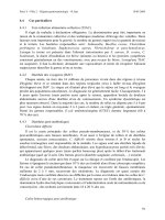
The absorptive surface area of the small intestine is greatly increased by tissue and
cell specializations such as plicae circulares, villi and microvilli (Fig. 1). Plicae circulares
are permanent transverse folds of the mucosa, forming semicircular or spiral elevations.
They are abundant in the distal duodenum and beginning of the jejunum. Intestinal villi are
finger-like outgrowths of mucosa protruding into the lumen of the small intestine.
Microvilli are protrusions of the apical plasmalemma of the epithelial cells covering the
intestinal villi, increasing the surface area of the small intestine 20 times. Therefore, these
modifications immensely amplify the absorptive and interactive (with intestinal content,
including the microbiota) surface area of the small intestine.
The mucosa comprises the lining epithelium, a lamina propria that houses glands and
muscularis mucosa. There are at least 5 types of cells found in the intestinal mucosal
Mucous
epithelium of
pylorus
Goblet cell
Lamina
propria
Intestinal
epithelium
Villi
Muscularis
mucosae
Paneth
cell
Lymphoid
cell
Figure 1 Schematic diagram of the mucosa, villi, and component cells of the small intestine.
Chow and Lee124
epithelium. They include enterocytes, goblet cells, Paneth cells, enteroendocrine cells and
M cells (microfold cells). Both the enterocytes and the goblet cells line the villus and are the
major cell types in the epithelium. The enterocytes are columnar in shape and have brush
borders composed of microvilli which help to enhance the water ions and nutrient absorbing
surface area. Goblet cells are unicellular mucin-secreting glands which produce mucinogen
and mucin, a component of mucus. The number of goblet cells increases progressively down
the gastrointestinal tract from the duodenum, to jejunum, ileum and colon, where they are
most abundant. The Paneth cells’ role is to maintain the innate immunity by secreting
antimicrobial substances such as a-defensins (4,69). Enteroendocrine cells are present only in
small numbers (w 1%) and their functions include the production of panacrine and endocrine
hormones (5). M cells are modified enterocytes overlying the enlarged lymphatic nodules in
the lamina propria. Their function is to phagocytose and transport antigens present in the
intestinal lumen to the underlying macrophages and lymphoid cells, which then migrate to
other compartments of the lymphoid nodes, where immune responses to foreign antigens are
initiated (5).
The lamina propria is rich in lymphoid cells, which will protect the intestinal lining
from bacterial invasion. The loose connective tissue of lamina propria forms the main part
of the villi, extending down to the muscularis mucosa. The epithelium may invaginate into
the lamina propria between the villi to form glands, termed the crypts of Lieberku
¨
hn.
These tubular glands consist of enterocytes, goblet cells, regenerative cells, enteroendo-
crine cells and Paneth cells. The rate at which the regenerative cells proliferate is high and
they are capable of replacing other cell types in the intestinal epithelium. As mentioned
above, the pyramidal-shaped Paneth cells secrete antibacterial agents, such as lysozyme
and a-defensins or cryptdins, and internalized extracellular matter such as bacteria and
immunoglobulin. Therefore, it is postulated that these cells help in regulation of the
bacterial microenvironment in the small intestine.
Structure and Function of the Large Intestine
The large intestine is a continuation of the ileum and is usually divided into three
regions: the colon, rectum and anal canal. The colon accounts for nearly the full length
of the large intestine. The colon absorbs water and electrolytes (approximately 1400 ml
per day). It also compacts and eliminates feces (about 100 ml per day). Feces are
composed of water (75%), dead bacteria (7%), roughage (5%), inorganic substances
(5%), and undigested protein, dead cells and bile pigment (1%). Bacterial products,
including the vitamins riboflavin, thiamin, vitamin B12 and vitamin K, are also
excreted in the feces (5).
The colonic mucosal membrane does not have any folds due to an absence of villi
(Fig. 2). The intestinal glands are long and characterized by a great abundance of goblet
and absorptive cells, and a small number of enteroendocrine cells. The large intestinal
epithelium is specialized for mucos secretion, salt and water absorption.
The histology of the rectum is identical to that of the colon except that the crypts of
Lieberku
¨
hn are deeper and fewer in number. The rectum is about 12–18 cm in length and
is continuous with the anal canal, which spans about 3 to 4 cm. The mucosa of the anal
region displays a series of longitudinal folds, the rectal columns of Morgagni. These rectal
columns meet one another to form pouch-like outpocketings, the anal valves with
intervening anal sinuses. The anal valves assist in supporting the column of feces (5). The
epithelial cells of the entire gastrointestinal tract are constantly shed. They are replaced
with stem cells that have undergone mitosis. The high turnover rate of the epithelial cells
may explain why the small intestine is affected rapidly by the administration of
Mucosal Interactions and Gastrointestinal Microbiota 125
anti-mitotic drugs, as in cancer chemotherapy. The epithelial cells continue to be lost at the
tip of the villi, but drugs inhibit cell proliferation (6).
Mucus
The gastrointestinal tract contains tremendous numbers of microorganisms and some of
these microorganisms are pathogenic in nature under certain conditions. Therefore a
function of the mucus is to protect the underlying epithelial cells by keeping the microbes
and toxins at bay, on the outer mucosal surfaces. The mucus layer is comprised of various
mucosal secretions including mucins, trefoil peptides, and surfactant phospholipids.
Mucus occurs in two distinct physical forms: (1) a thin layer of stable, water
insoluble mucus gel firmly adhering to the gastroduodenal mucosal surface, (2) and as
soluble mucus which is quite viscous but mixes with the luminal juice (7).
The layer of mucus that is bound to the surface of the gastrointestinal tract is
resistant to its removal from the mucosa. It is approximately 50–450 mm thick in humans
and about twofold less in rats. This adherent mucus functions to support and define
the mucosal ecosystem since it is the outermost sensory “organ” of the mucosal immune
system. The mucus gel plays a role in providing surface neutralization by having the
HCO
K
3
barrier to the gastric acid. The surfactant lipids maintain surface hydrophobicity on
the mucus. The adherent mucus also serves as a stable protective barrier that prevents the
entry of luminal pepsin to the underlying epithelial cells.
The soluble mucus plays a role in maintaining the protective barrier because it is not
physically attached to the mucosa and can be removed from the mucosa by gentle washing.
Due to the viscous nature of the soluble mucus, the soluble mucus makes an excellent
lubricant which allows easy movement of solid material in the lumen. This helps to
prevent the damage to the underlying epithelial cells as well as minimize the tearing of the
adherent layer of mucus from the mucosal surface (7).
Lamina
propria
Muscularis
mucosae
Submucosa
Muscularis
externa
Colonic
epithelium
Figure 2 Schematic diagram of the colonic epithelium and associated cells.
Chow and Lee126
The main structural component of the mucus layer are the mucins or glycoproteins
of molecular weight ranging from one to several million daltons. When concentrated,
these glycoprotein macromolecules (M
r
R2!10
6
) polymerise to form gels. Mucin
molecules consist of carbohydrate side chains (70–80%) bound to a protein skeleton. The
O-linked oligosaccharide chains contain a restricted number of monosaccharides,
including galactose, fucose, N-acetylgalactosamine, N-acetylglucosamine and often
terminated with sialic acids or sulfate groups, which account for the polyanionic nature of
mucins at a neutral pH (7,8). Oligosaccharides chains are successively added on to mucins
specifically by membrane bound glycosyltransferases. The biochemistry of the intestinal
mucins confers their protective nature: the protein backbone has a high O-linked
oligosaccharide content (O80% carbohydrate by mass) that provides lectin-binding
capacity, whereas the ability of the protein core to form multimers (through disulphide
bonds) causes polymerization into gels and bestows viscoelasticity and lubrication (9).
The trefoil peptides also facilitate the mucins to confer visoelasticity on the mucus (10).
The composition of the mucus is constantly regulated by the varying secretion rates
of the mucin types, ions, lipids, proteins and water. The variation in the composition of the
mucus is also dependent on the development stage of the host as well as the host’s diet and
the interaction of the commensals and pathogens (10). Commensals rapidly colonize the
individual soon after birth and some play a role in inhibiting the growth of pathogenic
bacteria. However, many commensals are capable of becoming opportunistic pathogens
by overgrowing when the stable gastrointestinal ecosystem is disturbed. Thus, the mucus
has to be continuously secreted and then shed, discarded, digested or recycled. This form
of protective mechanism keeps the numbers of both pathogens and commensals in check
by blocking the bacterial adherence to the epithelial cells.
MICROBIOTA AND GASTROINTESTINAL SYSTEM
Distribution of Microbiota
The mucosal surface of the human body, including the gastrointestinal tract, the
respiratory tract and the urogenital tract, has a total surface area of more than 400 m
2
(11).
The gastrointestinal tract’s surface area is about 200–300 m
2
and is colonized by 10
13–14
bacteria with hundreds of bacterial species and subspecies.
The normal microbiota of the gastrointestinal tract has been grouped and defined into
two categories, the autochthonous (indigenous) and the allochthonous (nonindigenous)
species (12). The autochthonous microbes (1) are always present in the normal adult’s
gastrointestinal tract, (2) play a role in maintaining the stable bacterial populations in
the gastrointestinal tract, (3) colonize particular parts of the tract, (4) can grow
anaerobically, (5) colonize their habitats in succession in infants, and (6) often associate
intimately with the gastrointestinal mucosal epithelium.
On the other hand, allochthonous species are not characteristic of the normal habitat.
Allochthonous microbiota is defined as transient microbes which will not be established
but would just be passing through, having arrived in the habitat in food, in water, from
another habitat in the gastrointestinal tract, or from elsewhere in the body. These microbes
either cannot or find it very challenging to establish themselves since they cannot compete
in the various niches or may be killed by host or bacterial factors.
However, the allochthonous microorganisms might colonize the habitats vacated by
the autochthonous microbes in the disturbed gastrointestinal system (13). This was
evidently seen in the administration of antibiotics which caused severe disturbance in the
gastrointestinal microbiota leading to undesirable effects, such as the overgrowth and
Mucosal Interactions and Gastrointestinal Microbiota 127
superinfection with allochthonous microorganisms like yeast (14,15); see also chapter 18
by Sullivan and Nord in this book.
Thus, the main difference between autochthonous and allochthonous species is that
an autochthonous microbe naturally colonizes the habitat, whereas an allochthonous one
cannot colonize it except under abnormal or atypical situations (13).
In a steady gastrointestinal ecosystem, all the niches are probably occupied by
indigenous microbes. The number of microorganisms in the stomach and the upper
two-thirds of the small intestine is very scarce: a maximum of 10
4
per milliliter of intestinal
contents. The relatively low number of microbes is due to the low pH (approximately pH 2)
of the intestinal contents resulting from gastric acid production and the relatively swift flow
(transit time of 4–6 hours) of digesta through the stomach and small intestine. Culturing
studies indicate that lactobacilli and streptococci are commonly found microbes in the small
intestine (16). Unlike the bulk of the microbes within the gastrointestinal tract, both the
lactobacilli and streptococci are acid-tolerant bacteria, and are capable of surviving the
passage through the stomach.
The ileum contains larger numbers of microbes (10
8
–10
9
bacteria per ml of intestinal
contents) in comparison to the upper regions of the gastrointestinal tract. The higher
bacterial numbers in the ileum are the result of a lower peristalsis and low
oxidation-reduction potential. Therefore, lactobacilli, streptococci, enterobacteriacae
and anaerobic bacteria are able to establish themselves in the distal region of the small
intestine. The main site of microbial colonization in the gastrointestinal tract is the colon.
The slow intestinal motility in the colon with a transit time of up to 60 hours and low
oxidation-reduction potential are responsible for the large numbers of bacteria present.
The colon contains 10
11
–10
12
bacteria per gram of intestinal contents. More than 99% of
the colonic microbiota are obligate anaerobes such as Bacteroides spp., Eubacterium,
Bifidobacterium and Clostridium spp. (17).
Enteric Pathogens
Most intestinal bacterial infections are caused by enteric pathogens. The clinical
symptoms usually associated with the intestinal infections include fever, abdominal pain
and diarrhea. Enteric bacteria are capable of evading host defense factors such as gastric
acidity, intestinal motility, the normal indigenous microbiota, mucus secretion, and
specific mucosal and systemic immune mechanisms.
In order for ingested pathogenic bacteria to infect the colon, they produce virulence
factors. Enteric bacteria can be divided into four main categories based on the virulence
factors that enable them to overcome the host defense. The first group of bacterial
pathogens consists of Campylobacter jejuni, Yersinia enterocolitica, Shigella and
Salmonella species. Their mechanism of virulence involves the mucosal invasion with
intraepithelial cell multiplication resulting in cell death. The second group comprises
enteric pathogens that produce cytotoxins which will in turn cause cell injury and
inflammation. Microorganisms that produce cytotoxins include Clostridium difficile,
enteropathogenic Escherichia coli (EPEC) and enterohemorrhagic E. coli (EHEC). The
third class of pathogens secretes enterotoxins which will alter intestinal salt and water
balance without affecting mucosal morphology. Vibrio cholerae, Shigella and
enterotoxigenic E. coli produce such enterotoxins. The last category of enteric pathogens
can only cause disease when they tightly adhere to the intestinal surface. The classic
enteropathogenic E. coli as well as the enteroadherent E. coli is typical of this group. Both
the small intestine and colon are primary sites for enteroadhesion (18).
Chow and Lee128
DEVELOPMENT OF GI TRACT NORMAL MICROBIOTA IN HUMANS
The fetus in utero is sterile until birth. Colonization of the human body with a heterogenous
collection of microorganisms from the birth canal begins at delivery. The Lactobacillus
species constitute the major population of the vaginal microbiota and thus provide the initial
inoculum to the infant during birth. In the case of caesarean section or premature infants,
most microbes that are transferred to the newborn can be traced from the environment, i.e.,
from other infants via the air, equipment and nursing staff (19). Therefore, the type of
delivery (passage through the birth canal versus caesarean section) as well as the type of diet
(breast versus formula feeding) might affect the pattern of microbial colonization.
The general pattern observed was that the facultative microorganisms appeared first
and were subsequently followed by a limited number of anaerobes during the first two
weeks (20). The types of bacterial strains that are capable of populating the GI tract are
regulated through the limitation of the intestinal milieu, which changes with the successive
establishment of the different bacteria. Hence, bacteria that are capable of oxidative
metabolism, such as enterobacteria, streptococci and staphylococci, are among the first to
proliferate in the gut. As the numbers of the facultative bacteria increase, they consume
oxygen and lower the redox potential to negative values. These conditions are favorable
for the anaerobic bacteria to multiply and reach much higher levels than that of the first
week. Populations of bifidobacteria, Bacteroides and clostridia, the commonly found
anaerobes, increase with subsequent change of conditions in the GI tract. By the fourth
week, the fecal microbiota of the breast-fed infants consists mainly of bifidobacteria and
other groups to a lesser extent including enterobacteria, clostridia, and Bacteroides.
However, in formula-fed infants, bifidobacteria do not beome so dominant and a more
complex microbiota develops. The differences between the breast-fed and formula-fed
infants gradually disappear with the intake of solid food. By the twelfth month, the number
of facultative anaerobes declines as the anaerobes begin to increase and form a stable
population, resembling that of adults in numbers and in composition. By the age of two,
the profile resembles that of an adult (19). In adults, the ratio of anaerobic to aerobic
bacteria is 1000:1 (21).
Adhesion of Bacteria
The colonization of microorganisms in various niches is dependent on their ability to
adhere to surfaces and substratum. Adhesion or adherence is defined as the measurable
union between a bacterium and substratum. A bacterium is considered to have adhered to a
substratum when energy is required to separate the bacterium from the substratum (22).
Adhesion of a bacterium to a substratum, its colonization and finally possible invasion
of the tissue is a multi-step process. It usually involves two or more kinetic steps. Firstly, the
bacterium approaches the substratum via long distance interactions, such as van der Waals
forces and electrostatic forces and becomes loosely attached (22). Complementary
adhesion-receptor interaction leads to the formation of a bacterium-cell complex:
Bacteria C Intestinal cell%
k
1
k
K1
BacteriumKIntestinal cell complex (1)
where k
1
and k
K1
are dissociation constants for the above reaction. At equilibrium, the
concentration of the adhered bacteria (e
x
) can be expressed as:
e
x
Z e
m
,x=ðk
x
CxÞ (2)
where e
m
is the maximum value of e
x
at saturated bacterial concentration (23). The value
Mucosal Interactions and Gastrointestinal Microbiota 129
of e
m
is equivalent to the concentration of adhesion sites on the mucosal surface and x is the
concentration of bacterial cells present around the adhesion site. The dissociation constant,
k
x
determines the affinity the bacterial cells have for the adhesion sites on the mucosal
surfaces. Thus, the adhesion of a bacterium to the substratum is determined by two major
properties: the concentration of the bacterium in the vicinity of the cell receptor (x in the
above equation) and the affinity of the bacterium for the receptor (k
x
in the equation).
Bacterial adhesion is crucial for invasive pathogenic microbes and may be important
for certain commensals, prior to colonization of the intestinal mucosa. The receptors for
bacterial adhesins are found in three groups of membrane consitituents: integral, peripheral
and cell surface coat components. These receptors are chemically proteins, glycoproteins or
glycolipids. They fulfill the criteria of a biological receptor because they exhibit specific
binding followed by physiologically relevant responses. An example would be membrane-
associated fibronectin acting as a receptor molecule for streptococci (22).
Bacterial adhesion to substrata receptors could involve the specific adhesin-receptor
interaction and non-specific interactions. The specific adhesion is defined as the association
between the bacteria and substratum that requires rigid stereochemical constraints (22).
Many bacteria have the ability to produce lectins (24), carbohydrate-specific proteins,
which are usually expressed on the bacterial surfaces. Lectins are a subset of adhesins that
recognize and bind to a defined carbohydrate sequence present on host glycoproteins.
Previous studies reported that there were three main types of adhesin-receptor interactions.
The first type was based on the carbohydrate-lectin recognition, the second kind involved
protein-protein interaction and the third class, which is the least characterized, involved the
binding interactions between hydrophobic moieties of proteins and lipids (25).
A well-established example is the type 1 fimbriae (carrying adhesins) of E. coli which
recognize D-mannose as the receptor site on the host mucosal surface (26). Binding
of some Lactobacillus to human colonic cells is a mannose-specific adherence mechanism
(27,28). Their similarity in binding specificity may contribute to competitive exclusion of
enteropathogens by some strains of probiotic lactic acid bacteria. Lactic acid bacteria
have been shown to exclude enteropathogens from the mucosal surface in in vitro
studies (29–32).
On the other hand, the non-specific adhesion is also an association between a
bacterium and substratum that may involve the same forces involved in the specific
adhesion. However, in non-specific adhesion, a precise stereochemical fit is not necessary.
Non-specific interaction comprises the physiochemical forces such as van der Waals,
electrostatic forces (33), hydrogen bonding (34), and hydrophobic interactions (35).
The synthesis of adhesins can be switched on and off by the bacteria, depending on
the environmental conditions, a process called phase variation (36). Phase variation has
been demonstrated in Gram-negative bacteria. However, the environmental regulation of
adhesin expression is likely to be present in some commensal and lactic acid bacteria also,
since bacteria that are unable to regulate their adhesin expression are often inefficient
colonizers (37,38). It has been suggested that the mucosal adhesive properties of the
lactic acid bacteria is strain and host dependent, and the mucosal binding of human lactic
acid bacteria are strain- and host specific (39,40). The adhesion and colonization of
bifidobacteria have been suggested to be disease (allergy, cancer) dependent (41,42). The
adhesion to the intestinal mucus of the fecal bifidobacteria from healthy infants was
significantly higher than for allergic infants, suggesting a correlation between allergic
disease and the composition of the bifidobacteria (41). Surprisingly, bifidobacteria,
amongst other bacteria, were generally positively associated with increased risk of colon
cancer in a study involving native Japanese and African patients (42). The ability of
intestinal bacteria to persist on the intestinal mucosal surface may ultimately be determined
Chow and Lee130
by their doubling time in the intestine to maintain a high local concentration. Slowly-
dividing bacteria would be expected to be out-competed or washed-out with the intestinal
contents (43).
CROSS-TALK BETWEEN BACTERIA AND INTESTINAL
EPITHELIAL CELLS
As discussed in chapter 5, some ingested probiotic bacteria have shown immunomo-
dulatory properties (44–46). Both commensal and pathogenic bacteria possess recognized
structures named pathogen-associated molecular patterns (PAMPS). These recognized
structures are essential for the microbe, mostly constitutively expressed and shared by the
same group of microorganisms. PAMPS that are characterized to date include
N-formylated peptide (47), lipopolysaccharides (LPS) (48), and lipopeptides (49), more
recently described PAMPS are flagellin (50) and unmethylated segments of CpG DNA
(51). Even though unmethylated segments of CpG DNA are not a cell surface structure, it
serves to differentiate the microorganism from the host. Therefore, they epitomize the
ideal targets for the innate immune system to identify the presence of infectious agents
with a limited numbers of receptors.
The best studied of the PAMPS is the glycolipid LPS, an important component of the
outer membrane of Gram-negative bacteria. LPS is recognized by Toll-like receptor
(TLR) 4, the first described member of the family of transmembrane TLR molecules that
play a central role in the transcription activation of host defense mechanisms, such as
chemokine and cytokine secretion, and the expression of costimulatory molecules (52).
TLRs are transmembrane receptors defined by the presence of leucine-rich repeats in the
extracellular portion of the molecule and a Toll/IL-IR/resistance (TIR) cytoplasmic
domain. The extracellular leucine-rich repeats are thought to function in ligand recognition,
whereas the TIR domain works in signaling. Leucine-rich repeat domains are common to
proteins that are involved in the recognition of foreign proteins. There are currently 10
identified members of the mammalian TLR family (52). From recent publications (53), it
has been shown that some types of intestinal epithelial cells express TLR 4.
Upon activation of TLR 4 by LPS, a series of events lead to the activation of
ubiquitin ligase TRAF6 by a unique self-polyubiquitination reaction. TRAF6 then
activates the TAK1 complex (54). This step leads to the phosphorylation and activation of
mitogen-activated protein kinase and the inhibitor kB kinase (IKK) complex (54,55). The
IKK complex comprises two kinases, IKKa and IKKb, and one protein, NEMO. When
activated, IKKb phosphorylates IkBa, triggering its polyubiquitination and degradation
(56,57). In the unstimulated state, the IkBa interacts and traps NFkb in the cytosol.
Degradation of IkBa releases the NFkb to translocate into the nucleus and to activate
proinflammatory and prosurvival gene expression. Therefore, TLR 4 activates multiple
signaling pathways which will eventually lead to the production of cytokines and other
factors to protect the host against infection (58). The expression level of TLR 4 in the
intestine of patients with inflammatory bowel disease was found to be strongly
up-regulated compared to the TLR 4 expression in healthy individuals.
As for the other PAMPS such as N-formylated peptides, the cell surface receptors
that recognized them are the heterotrimeric G-protein coupled receptors (59).
N-formylated peptides play an important role in recruiting and activating inflammatory
cells (60). They will eventually activate the NFkb pathway the same way as the TLR.
On the other hand, enteric pathogens have also evolved mechanisms to evade the
immune recognition and defense. Helicobacter pylori, the etiological agent of gastritis and
Mucosal Interactions and Gastrointestinal Microbiota 131
stomach cancer, expresses hypoacylated LPS to avoid recognition by the human
TLR4/MD2 module (61). Other pathogens like Yersinia pseudotuberculosis have
developed ways to down-regulate TLR 4 signaling by injecting proteins to abolish the
signaling leading to NFkb activation (52).
At the beginning of the chapter, we mentioned that the gastrointestinal tract is
colonized by huge, complex and dynamic populations of microorganisms. Hence, the
molecular pattern recognition of the epithelial cells of the gastrointestinal mucosa needs to
be tightly regulated so as to avoid an extreme immune response and uncontrolled
inflammatory reaction. The exact mechanism by which they do this still remains to be
elucidated. However, recent studies have shed light into this area of interest. The
mechanism by which one TLR, TLR 5, achieved this feat is due to the fact that gut
epithelial cells express TLR 5 only on their basolateral surfaces. Therefore only those
bacteria that breached the epithelial cells or have translocated flagellin across the epithelia
will activate the receptor (62).
Using a gnotobiotic mouse model it was shown that Bacteroides thetaiotaomicron is
able to induce the production of a-L fucose on intestinal epithelial cells via a regulator,
FucR, as a molecular sensor of L-fucose availability (3,68). FucR coordinates expression
of an operon encoding enzymes in the L-fucose metabolic pathway in the bacteria with
expression of another locus that regulates production of fucosylated glycans in the
intestinal enterocytes. By tightly coordinating presentation of host-derived fucose with
the rate of fucose utilization, an excess of epithelial fucose is avoided. This may minimize
the risk of encroachment by pathogens that use fucosylated glycans as receptors for their
adhesins (69).
Certain pathogenic bacteria require intimate contact with the host to cause disease.
E. coli (EPEC) is one such pathogen which requires intimate attachment to the host cells for
maximum virulence to occur. There are a few factors which facilitate the cross-talk between
the microorganism and the host epithelial cells and this involves the EPEC-secreted
proteins, the type-three secretion system and the expression of outer membrane protein,
intimin (64,65). The release of extracellular protein via the type-three secretion system is
necessary for the formation of attaching lesions by EPEC. The attachment of bacteria is by
means of intimin binding to a 90 kDa tyrosine phoshorylated protein in the host membrane.
This receptor is known as translocated intimin receptor (Tir) and is of bacterial origin; it is
translocated on to the host membrane where its tyrosine residues become phosphorylated
and binds to intimin. Subsequent signal transduction events that occur within the host cells
are the activation of protein kinase C, inositol triphosphate and calcium release. This leads
to the formation of an actin-rich pedestal that forms a dome-like anchoring site for the
bacteria which is an essential feature of EPEC pathogenesis (63).
There is evidence to suggest that in some strains of Lactobacillus reuteri, mucus-
binding adhesion could be induced by the presence of mucin glycoproteins and solid
substratum (66).
CONCLUSION
The gastrointestinal tract is a highly dynamic ecosystem where interaction of the
microbiota with the host mucosa plays an important role. Thus, it not only functions to
digest food and absorb nutrients; it is also the major site where communication between
microbes, and also between microbiota and their host takes place.
Probiotics and prebiotics offer dietary means to support the balance of intestinal
microbiota. They may be used to counteract local immunological dysfunctions, to stabilize
Chow and Lee132
the gut mucosal barrier function, to prevent infectious succession of pathogenic
microorganisms or to influence intestinal metabolism. However, many of the proposed
mechanisms still need to be validated in human clinical trials (67). Future research on
commensal microbiota interactions with mucosal surfaces of the host should focus on the
cross-talk and determining the signaling mechanisms involved.
REFERENCES
1. Mitsuoka T. The human gastroinstestinal tract. In: Brian JB Wood, ed. In: Lactic Acid Bacteria,
Vol. 1. London and New York: Wood Publisher Elseivier Applied Science, 1992:69–114.
2. Hoskins LC. Mucin degradation in the human gastrointestinal tract and its significance to
enteric microbial ecology. Eur J Gastroenterol Hepatol 1993; 5:205.
3. Bry L, Falk PG, Midtvedt T, Gordon JI. A model of host-microbial interactions in an open
mammalian ecosystem. Science 1996; 273:1380–1383.
4. Ross MH, Kaye GI, Pawlina W. Digestive system III: esophagus and gastrointestinal tract.
Histology: A Text and Atlas. Philadelphia: Lippincott Williams & Wilkins, 2003 pp. 474–531.
5. Gartner LP, Hiatt JL. Digestive system III— alimentary canal. Color Textbook of Histology.
Philadelphia: W.B. Saunders Company, 1997 pp. 325–335.
6. Junqueria LC, Carneiro J, Contopoulos AN. Digestive tract. In: Junqueria LC, Carneiro J,
Contopoulos AN, eds. Basic Histology: Text and Atlas. Los Altos. CA: Lange Medical
Publications, 1998 pp. 272–303.
7. Allen A, Carroll NJ. Adherent and soluble mucus in the stomach and duodenum. Dig Dis Sci
1985; 30:55S–62S.
8. Forstner JF, Oliver MG, Sylvester FA. Production, structure, and biologic relevance of
gastrointestinal mucins. In: Blaser MJ, Smith PD, Ravdin JI, Greenberg HB, Guerrant RL, eds.
Infections of the Gastrointestinal Tract. New York: Raven Press, 1995:71–88.
9. Belley A, Keller K, Gottke M, Chadee K, Gottke M. Intestinal mucins in colonization and host
defense against pathogens. Am J Trop Med Hyg 1999; 60:10–15.
10. Cone RA. Mucus. In: Orga PL, Mestecky J, Lamm ME, Strober W, Bienenstock J, McGhee JR,
eds. Mucosal Immunology. New York: Academic Press, 1999:43–60.
11. Kraehenbuhl JP, Neutra MR. Molecular and cellular basis of immune protection of mucosal
surfaces. Physiol Rev 1992; 72:853–879.
12. Savage DC. Microbial ecology of the gastrointestinal tract. Ann Rev Microbiol 1977;
6:155–178.
13. Alexander M. Microbial Ecology. New York: Wiley, 1971 pp. 3–21.
14. Nord CE, Hermdah IA, Kager L. Antimicrobial induced alteration of the human oropharyngeal
and intestinal microflora. Scand J Infect Dis Suppl 1986; 49:64–72.
15. Trenschel R, Peceny R, Runde V, et al. Fungal colonization and invasive fungal infections
following allogeneic BMT using metronidazole, ciprofloxacin and fluconazole or ciprofloxacin
and fluconazole as intestinal decontamination. Bone Marrow Transplant 2000; 26:993–997.
16. Tannock GW. Normal Microflora: An Introduction to Microbes Inhabiting the Human Body.
U.K.: Chapman and Hall, 1995.
17. Herbert MK, Holzer P, Roewer N. Problems of the Gastrointestinal Tract in Anesthesia, the
Perioperative Period, and Intensive Care. New York; Berlin: Springer, 1999.
18. Cohen MB, Giannella RA. Bacterial infections: pathophysiology, clinical features and
treatment. In: Philips SF, Permberton JH, Shorter RG, eds. The Large Intestine: Physiology,
Pathophysiology and Disease. New York: Raven Press, 1991:395–428.
19. Conway PL. Microbial ecology of the human large intestine. In: Gibson GR, Macfarlane GT, eds.
Human Colonic Bacteria: Nutrition, Physiology, and Pathology. Boca Raton: CRC, 1995:1–24.
20. Stark PL, Lee A. The microbial ecology of the large bowel of the breast-fed and formula-fed
infants during the first year of life. J Med Microbiol 1982; 15:189–203.
21. Berg RD. The indigenous gastrointestinal microflora. Trends Microbiol 1996; 4:430–435.
Mucosal Interactions and Gastrointestinal Microbiota 133