Báo cáo y học: "Preliminary evidence for a change in spectral sensitivity of the circadian system at night" potx
Bạn đang xem bản rút gọn của tài liệu. Xem và tải ngay bản đầy đủ của tài liệu tại đây (295.16 KB, 9 trang )
BioMed Central
Page 1 of 9
(page number not for citation purposes)
Journal of Circadian Rhythms
Open Access
Research
Preliminary evidence for a change in spectral sensitivity of the
circadian system at night
Mariana G Figueiro
1
, John D Bullough
1
, Robert H Parsons
2
and Mark S Rea*
1
Address:
1
Lighting Research Center, Rensselaer Polytechnic Institute, 21 Union Street, Troy, NY 12180, USA and
2
Department of Biology,
Rensselaer Polytechnic Institute, 110 8th Street, Troy, NY 12180, USA
Email: Mariana G Figueiro - ; John D Bullough - ; Robert H Parsons - ;
Mark S Rea* -
* Corresponding author
Abstract
Background: It is well established that the absolute sensitivity of the suprachiasmatic nucleus to
photic stimulation received through the retino-hypothalamic tract changes throughout the 24-hour
day. It is also believed that a combination of classical photoreceptors (rods and cones) and
melanopsin-containing retinal ganglion cells participate in circadian phototransduction, with a
spectral sensitivity peaking between 440 and 500 nm. It is still unknown, however, whether the
spectral sensitivity of the circadian system also changes throughout the solar day. Reported here is
a new study that was designed to determine whether the spectral sensitivity of the circadian retinal
phototransduction mechanism, measured through melatonin suppression and iris constriction,
varies at night.
Methods: Human adult males were exposed to a high-pressure mercury lamp [450 lux (170 µW/
cm
2
) at the cornea] and an array of blue light emitting diodes [18 lux (29 µW/cm
2
) at the cornea]
during two nighttime experimental sessions. Both melatonin suppression and iris constriction were
measured during and after a one-hour light exposure just after midnight and just before dawn.
Results: An increase in the percentage of melatonin suppression and an increase in pupil
constriction for the mercury source relative to the blue light source at night were found, suggesting
a temporal change in the contribution of photoreceptor mechanisms leading to melatonin
suppression and, possibly, iris constriction by light in humans.
Conclusion: The preliminary data presented here suggest a change in the spectral sensitivity of
circadian phototransduction mechanisms at two different times of the night. These findings are
hypothesized to be the result of a change in the sensitivity of the melanopsin-expressing retinal
ganglion cells to light during the night.
Background
It is well established that the absolute sensitivity of the
suprachiasmatic nucleus (SCN) to photic stimulation
received through the retino-hypothalamic tract (RHT)
changes along the 24-hour day [1-4]. Conceivably,
changes in the sensitivity of the circadian system to light/
dark patterns could be driven by the master clock in the
SCN, by a peripheral clock in the retina, or by both.
Published: 11 December 2005
Journal of Circadian Rhythms 2005, 3:14 doi:10.1186/1740-3391-3-14
Received: 03 October 2005
Accepted: 11 December 2005
This article is available from: />© 2005 Figueiro et al; licensee BioMed Central Ltd.
This is an Open Access article distributed under the terms of the Creative Commons Attribution License ( />),
which permits unrestricted use, distribution, and reproduction in any medium, provided the original work is properly cited.
Journal of Circadian Rhythms 2005, 3:14 />Page 2 of 9
(page number not for citation purposes)
Jagota et al. [4] showed that neural activity in the hamster
SCN varied over the 24-hour cycle, suggesting the exist-
ence of a morning and an evening oscillator in the SCN.
Changes in photoperiod affected the two SCN peak activ-
ity periods differently, demonstrating that the phases of
the two peaks are not locked but are independently linked
to the environmental cycle of dusk and dawn. Moreover,
they showed that the two peaks responded differently to a
pulse of glutamate (the neurotransmitter that conveys
light information from the eye to the SCN). Glutamate,
when given after dusk, delayed the evening peak but not
the morning peak; when glutamate was given before
dawn, the early peak was advanced but the evening peak
was unaffected. Pevet et al. [1] also demonstrated that the
duration of the SCN phase sensitivity to light is closely
related to the length of the night. The SCN phase sensitiv-
ity to light was measured in terms of the expression of Fos
protein, which is considered a marker of SCN cell
response to light stimuli. The findings of Jagota et al. [4]
and of Pevet et al. [1] reinforce the growing evidence for
temporal changes in the SCN's sensitivity to light.
Unknown, however, is whether there is temporal varia-
tion in the sensitivity of the circadian phototransduction
mechanism itself throughout the 24-hour cycle.
Lucas et al. [5] have shown that light can reset the circa-
dian clock as well as stimulate the iris light reflex of genet-
ically-manipulated mice without classical photoreceptors
(rods and cones). Berson et al. [6] showed that a subset of
retinal ganglion cells (RGCs) innervating the SCN were
directly photosensitive and able to convert electromag-
netic radiation into neural signals. Melanopsin, a photo-
pigment based on vitamin A, was found in these RGCs
and is the strongest candidate for the circadian photopig-
ment within these cells [7]. Genetically-manipulated mice
that do not have melanopsin still show phase shifting by
light exposure, although to a lesser degree [8]. This result,
as well as more recent data from Hattar et al. [9], Panda et
al. [10] and from Bullough et al. [11] seem to demonstrate
that classical photoreceptors (rods and cones) as well as
melanopsin-expressing RGCs participate in circadian
phototransduction of mammals.
The spectral sensitivity of the human circadian system
peaks between 440 and 500 nm [12,13]. Those data
[12,13] are consistent with the conclusion that, overall,
human melatonin suppression is dominated by at least
two (not just one) opsins. However, the two studies
[12,13] were conducted at similar times of the night, mak-
ing it impossible to ascertain whether the spectral sensitiv-
ity of the circadian system changes at night. Two studies
conducted in our own laboratory suggest that this might
be true. Human adult males were exposed to a combina-
tion of two light levels and two broadband spectral power
distributions (SPDs) from fluorescent lamps every two
hours (at 00:00, 02:00, 04:00 and 06:00) for four nights
in a counterbalanced order [14,15]. The results suggested
that the spectral sensitivity of melatonin suppression may
change during the night, because the relative contribution
of the candidate photopigments (traditional photorecep-
tors and melanopsin-expressing RGCs) to best fit the sup-
pression data seemed to systematically change during the
night. The data obtained from broadband fluorescent
light were not sufficiently precise, however, to determine
which of several possible combinations of retinal photo-
pigments participated in the circadian response to light.
In addition to the studies of the circadian system's
response to light, and perhaps of direct relevance, several
studies have shown that the absolute sensitivity of the vis-
ual system changes over the course of the night [16-18].
Increment thresholds to visual targets are apparently low-
est just before dark and highest just before dawn [18].
Dacey et al. [19] have recently shown that in macaque
(and, therefore, probably in humans as well), photosensi-
tive melanopsin-expressing RGCs have input to the lateral
geniculate nucleus (LGN), a major neural relay station
from the retina to the visual cortex. If the overall sensitiv-
ity to light increases over the course of the night in this
newly discovered class of RGCs, two results could occur.
First, these cells could, in effect, set a higher luminous
background on which a visual target must be detected,
thus, increasing increment thresholds in the early morn-
ing relative to the early night. Second, the spectral sensi-
tivity of the visual and circadian systems could shift to
shorter wavelengths as the melanopsin-expressing RGCs
become more dominant because their peak spectral
response is at or near 480 nm.
Although a change in absolute sensitivity of the visual sys-
tem over the course of the 24-hour day has been studied,
there are no comparable studies for a change in the abso-
lute sensitivity of the circadian phototransduction system.
In part at least, this may be the result of the inherent
nature of the outcome measures used in most studies of
the circadian system. Changes in nocturnal melatonin
production, core body temperature and phase shifting,
the most common outcome measures used to evaluate the
circadian system's response to light, can be the result of
changes in the circadian phototransduction mechanism
in the retina, the circadian clock in the SCN, or both.
Changes in the relative values of these outcome measures
to two different lights at two different times of night
could, however, indicate a change in the circadian pho-
totransduction mechanism. The experiment reported here
was designed to investigate, using the relative difference in
melatonin suppression and iris construction by two differ-
ent light spectra, whether the spectral sensitivity of the cir-
cadian system changes at two different times of the night
and thereby determine whether there was evidence for a
Journal of Circadian Rhythms 2005, 3:14 />Page 3 of 9
(page number not for citation purposes)
temporal change in the retinal circadian phototransduc-
tion mechanism. The data used as the basis for this report
are the same as those previously published suggesting
spectral opponency in the human circadian phototrans-
duction system [20,21].
Methods
Both melatonin suppression and iris constriction were
measured during and after a one-hour light exposure just
after midnight and just before dawn. A clear, high-pres-
sure mercury (Hg) lamp and an array of blue (λ
max
= 470
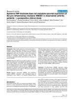
nm) light emitting diodes (LEDs) were used (Figure 1).
The Hg lamp provided 450 lx (170 µW/cm
2
) at the cornea
and the set of LEDs provided 18 lx (29 µW/cm
2
) at the cor-
nea. These light sources and light levels were selected to
ensure that the suppression of melatonin for either light
source was not high enough to produce asymptotic mela-
tonin suppression [21].
Four male subjects, 20 or 21 years of age, participated in
the study during two nights in May 2003. Each session
lasted 8.5 h (from 22:30 to 07:00 h). All subjects signed a
consent form approved by Rensselaer's Institute Review
Board (IRB).
Each subject was seated in front of a 0.6 × 0.6 × 0.6 m ply-
wood and matte-white painted box resting atop a small
table, 0.76 m above the floor. The fronts of the boxes con-
tained square 0.45 × 0.45 m apertures and chin rests so
that every subject's face was inside one of the boxes. The
backs of the boxes also had a square 0.3 × 0.3 m aperture
behind which a computer monitor was placed. The com-
puter monitors were adjusted so that only the red phos-
phor was used and provided no more than 3 lx at subjects'
eyes when they sat at the boxes. Another small hole in the
back of each box accommodated the zoom lens of a dig-
ital video camera, which was used to measure pupil size,
as descried below.
The roofs of two boxes supported an uncoated, 175 W
high-pressure Hg lamp (General Electric HR175A39) and
ballast. When energized, the Hg lamps provided diffuse
Relative spectral power distributions of the light sources used in the experimentFigure 1
Relative spectral power distributions of the light sources used in the experiment.
Journal of Circadian Rhythms 2005, 3:14 />Page 4 of 9
(page number not for citation purposes)
illumination throughout the box; light levels were con-
trolled with mechanical filters and a neutral density
acrylic filter (25% transmission). The inside front faces of
the other two boxes were lined with an array of blue LEDs
(Color Kinetics iCove) which provided diffuse illumina-
tion throughout the box; light levels were controlled elec-
tronically. As previously stated, each Hg lamp provided
450 lx (170 µW/cm
2
) at the cornea when a subject was
seated at the table supporting the box and positioned in
the chin rest; the set of LEDs provided 18 lx (29 µW/cm
2
)
at the cornea.
All subjects followed their normal routine but refrained
from consuming caffeinated products for 12 h before each
session. Upon arrival at the facility, a registered nurse
inserted a catheter into an arm vein of each subject. At
23:30, the first session of the night began by extinguishing
all light in the laboratory except that from two red LED
traffic signal lights that provided dim (<3 lx at the eye)
ambient illumination throughout the laboratory. At mid-
night subjects were assigned to a light box for the entire
night and asked to sit in front of it while wearing dark
glasses, and before the Hg and LED light sources were
energized. Subjects that were assigned to an Hg-illumi-
nated box on the first night were assigned to an LED-illu-
minated box on the second night, and vice versa. During
this time and throughout the night, subjects could interact
with the modified computer monitor by playing video
games or corresponding with friends on the Internet while
their heads were positioned in the chin rest.
During the first session, three sets of three blood samples
(3 ml each) were collected in the dark every 15 min., start-
ing at 00:30. At 01:00 the light sources were energized,
and four sets of three blood samples were collected every
15 min. from every subject until 02:00, at which time the
lights were either extinguished or the subjects were asked
to close their eyes. Three more sets of three blood samples
were collected in the dark every 15 min. until 02:45.
Because the catheter was flushed with saline each time
before blood samples were collected, the first blood sam-
ple collected in every set was always discarded. The two
remaining samples in each set were immediately spun in
a centrifuge at 3200 rpm (approximately 1000 × g) to
obtain the plasma, which was then frozen at -85°C. Fro-
zen samples were subsequently sent to an independent
laboratory (Neuroscience Inc., Osceola, WI) for mela-
tonin radioimmunoassay (Melatonin
direct
I-125 RIA). The
limit of detection of the assay was 1.5 pg/ml. The intra
assay coefficients of variation (CVs) were 12.1% at 16.5
pg/ml, 5.7% at 68.7 pg/ml, and 9.8% at 162.7 pg/ml. The
inter assay CVs were 13.2% at 17.3 pg/ml, 8.4% at 69 pg/
ml, and 9.2% at 164.7 pg/ml.
Between blood sample collections, the irises of the subject
were videotaped for one minute twice in the dark prior to
light exposure, three times during light exposure, and,
again, twice in the dark following the light exposure. Dur-
ing videotaping, subjects looked at a fixation point on the
computer monitor so that pupil size did not vary with
accommodation. Subsequently, images were digitized
and pupil sizes were measured. After the experiment was
completed, a video-editing program (Adobe Premiere
6.0) was used to capture six video images every 10 s from
each subject at each experimental condition. If the subject
blinked at the moment of video-frame capture, the video
capture was sampled just before or just after the blink
occurred. These captured images were then used for the
pupil measurements. Pupil measurements were per-
formed using MatLab 6.5. This program's unit of measure
was pixels and pupil measurements were based on the
relationship between the pupil and iris area because the
position of the eye was not constant among the subjects.
For each of the six images captured, three measurements
of the pupil diameter and three measurements of the iris
diameter were taken. It was assumed that a circle could
mathematically represent both the pupil and iris. A rela-
tive measurement, referred to as relative pupil area (rPA)
was obtained by dividing the pupil area by the iris area.
Only the rPA values obtained during the light exposure
periods were analyzed. All of the rPA values for a one-
minute recording session were averaged to give a single
estimate of pupil size during that period. Thus, three esti-
mates of pupil size were obtained for both lighting condi-
tions for every subject on both nights.
At 04:00, session two of the night began by asking subjects
to again be seated in front of their assigned boxes. As
before, ten blood samples were collected every 15 minutes
and subjects' irises were videotaped between blood sam-
ple collections. After completion of the second session,
the catheters were removed. Subjects left the laboratory at
07:00.
Results
Figures 2 and 3 show the average melatonin concentra-
tions (pg/ml) under each combination of session and
lighting conditions. Melatonin concentrations were
totaled so that an overall suppression of melatonin for
each combination could be determined. Average mela-
tonin suppression (in percent) and standard error of the
mean (S.E.M.) under Hg and LED lighting conditions for
each session were then calculated using the melatonin
concentrations from the last two measurements before
light onset and melatonin concentrations from the last
two measurements before light offset (Figure 4). A
repeated-measures analysis of variance (ANOVA) showed
a significant main effect for lighting condition (F
1,7
= 8.48,
p = 0.02). Overall, melatonin suppression for the LED
Journal of Circadian Rhythms 2005, 3:14 />Page 5 of 9
(page number not for citation purposes)
condition was significantly greater than it was for the Hg
lighting condition. The main effect for time of night was
not significant (F
1,7
= 4.71, p = 0.07) although melatonin
levels during the dark periods prior to any light exposure
were higher in session 2 than in session 1 (Figures 2 and
3). Post-hoc statistical tests were conducted to determine
whether there was a significant change in melatonin sup-
pression from session 1 to session 2 for each lighting con-
dition, LED and Hg (Figure 4). A paired, one-tailed
Student's t-test showed significantly more suppression of
melatonin in session 2 than in session 1 for the Hg light-
ing condition (t
7
= 3.11, p = 0.008), but there was no sig-
nificant difference in melatonin suppression for the LED
lighting condition at the two times of night (t
7
= 0.96, p =
0.2). Mann-Whitney nonparametric statistical tests were
also conducted and revealed similar results as the para-
metric tests (i.e., significant main effect of lighting condi-
tion, but not of session time).
The pupil size results showed similar trends, but in the
opposite direction. Figure 5 shows the rPA (calculated as
described above as the proportion of the iris area, normal-
ized to unity) for the Hg and LED lighting conditions in
sessions 1 and 2. A repeated-measures ANOVA showed a
significant main effect for lighting condition (F1,5 =
12.63, p = 0.02). Overall, pupil size for the LED condition
was significantly smaller than it was for the Hg lighting
condition. Overall, pupil sizes were larger in session 1
than in session 2 (Figure 5). Post-hoc statistical tests were
conducted to determine whether there was a significant
change in pupil size from session 1 to session 2 for each
lighting condition, LED and Hg. A paired, one-tailed Stu-
dent's t-test showed that pupil area in session 1 was signif-
icantly larger than in session 2 for Hg (t5 = 1.96, p = 0.05),
but there was no significant difference in pupil size for the
LED lighting condition at the two times of night (t5 =
0.62, p = 0.3). These results suggest that the iris light reflex
is less affected by the Hg lighting condition in session 1
(resulting in a larger pupil size) than in session 2. As with
the melatonin suppression data, Mann-Whitney nonpara-
metric statistical tests were conducted and revealed similar
results.
It should further be noted that overall suppression of
melatonin for the LED lighting condition was signifi-
cantly higher than for the Hg lighting condition, even
though the pupil areas for the LED lighting condition
were smaller. Since pupil size is determined, in part, by
the retinal exposure to light and, thus influences the
amount of melatonin suppression [22], the difference
Average melatonin concentrations (pg/ml ± S.E.M.) under the Hg lighting conditionFigure 2
Average melatonin concentrations (pg/ml ± S.E.M.) under the Hg lighting condition.
Journal of Circadian Rhythms 2005, 3:14 />Page 6 of 9
(page number not for citation purposes)
between melatonin suppression by the Hg and LED spec-
tra would have been relatively larger if pupil sizes were
held constant throughout the experiment.
Discussion
The increase in melatonin suppression and in iris constric-
tion for the Hg source relative to the LED source at night
suggests a temporal change in the photoreceptor mecha-
nisms contributing to the circadian system phototrans-
duction. Although a discussion of the recently published
model of human circadian phototransduction by Rea et
al. [23] is beyond the scope of this short communication,
the model does predict greater overall melatonin suppres-
sion from the blue LED source at 18 lx (29 µW/cm
2
) at the
cornea than from the Hg source at 450 lx (170 µW/cm
2
)
at the cornea. The model, based upon retinal neuroanat-
omy and electrophysiology, incorporates input from con-
ventional photoreceptors and from melanopsin-
expressing RGCs to predict the circadian light stimulus
from both monochromatic and polychromatic light
sources. It does not, however, make provision for a change
in the spectral sensitivity of circadian phototransduction
at different times of the night. The model could accom-
modate a change in spectral sensitivity through a tempo-
rally dependent coefficient modulating the relative
magnitude of the contribution to the overall spectral sen-
sitivity by the melanopsin-expressing RGCs. Indeed, to
model an increasing contribution of the melanopsin-
expressing RGCs at different times of the night, we
increased the value of that coefficient and found that the
Hg and LED sources would have much closer predicted
circadian stimulus values and would, thus, produce simi-
lar levels of melatonin suppression. In other words, a sim-
ple increase in the relative contribution of the
melanopsin-expressing RGCs near morning would
account for the smaller difference in melatonin suppres-
sion between the Hg and the LED conditions in session 2
(between 04:00 and 05:00) than the difference between
those two lighting conditions in session 1 (between 01:00
and 02:00). It should be noted that the model by Rea et
al. [23] does not deal quantitatively with circadian input
to the light reflex of the iris, including a possible change
in the spectral sensitivity of the iris light reflex at night.
Two observations might initially be offered in contradic-
tion to the inference that the spectral sensitivity of the cir-
cadian system changes at night. First, the difference in
relative suppression might be an artifact of having differ-
ent amounts of melatonin to suppress throughout the
night (i.e., there was more melatonin to suppress later at
night than early in the night). Under no experimental con-
dition, however, was light intensity strong enough to sup-
Average melatonin concentrations (pg/ml ± S.E.M.) under the LED lighting conditionFigure 3
Average melatonin concentrations (pg/ml ± S.E.M.) under the LED lighting condition.
Journal of Circadian Rhythms 2005, 3:14 />Page 7 of 9
(page number not for citation purposes)
press melatonin to daytime levels (below 12 pg/ml). Since
there was always melatonin to be suppressed by light, the
percent melatonin suppression should be unrelated to the
absolute level of melatonin available. Certainly, percent
suppression is accepted in the literature as a measure of
the impact of light on the circadian system [12-15]. Given
enough melatonin to suppress under all lighting condi-
tions, the differential effects of the two spectra at the two
times of the night strongly suggest that there is a change in
the spectral sensitivity of the retinal phototransduction
mechanisms of the circadian system. Second, the absolute
reduction in pupil size at night might be the result of
increased fatigue [24-26]. This interpretation is also likely
incomplete because, again, of the relative impact on pupil
constriction by the two sources at the two times of the
night and because neither lighting condition produced
maximum constriction of the pupil. Moreover, pupil con-
striction mirrored the percentage of melatonin suppres-
sion for the two sources over time, indicating similar
underlying phototransduction mechanism. In this con-
text, it should be recalled that Lucas et al. [5] showed that
in mice, pupil constriction, as well as phase shifting, is
influenced by melanopsin-expressing, intrinsically photo-
sensitive RGCs. Nevertheless, the pupil size data pre-
sented here should be interpreted with caution. Human
pupil size is notoriously variable [27,28], especially
because the balance between sympathetic and parasympa-
thetic input to the iris response can vary moment to
moment, at different times of day, and for different tasks
[24-26].
These data suggest, despite inherent uncertainty in the
pupil size measurements, that the mirrored changes in
pupil constriction and melatonin suppression reflect
changes in the relative photoreceptor contributions to the
circadian phototransduction system at night and that
these changes could be related to increased participation
by melanopsin-expressing RGCs closer to the morning. As
discussed in the introduction, the earlier data by Rea et al.
[14,15] are consistent with this interpretation as, at least
indirectly, are some recent evidence from Hannibal et al.
[29] suggesting that gene expression of melanopsin in the
photosensitive RGCs of the albino Wistar rat follow a cir-
cadian pattern. Finally, these findings might provide addi-
tional insight into the reported changes in visual
thresholds at night [18].
Melatonin suppression (mean ± S.E.M.) for sessions 1 and 2 for each lighting condition (Hg and LED)Figure 4
Melatonin suppression (mean ± S.E.M.) for sessions 1 and 2 for each lighting condition (Hg and LED).
Journal of Circadian Rhythms 2005, 3:14 />Page 8 of 9
(page number not for citation purposes)
Conclusion
The results presented here are the first to suggest a tempo-
ral change in spectral sensitivity of the human circadian
system phototransduction at two different times during
the night, measured through nocturnal melatonin sup-
pression, and with less certainty (owing to the inherent
variability of the iris constriction response) through pupil
area.
Competing interests
The author(s) declare that they have no competing inter-
ests.
Authors' contributions
MGF helped with the conception and design of the exper-
iment, collected the data, participated in the data analyses
and interpretation, and helped to draft the manuscript.
JDB helped with the conception and design of the experi-
ment, helped to collect the data, participated in the data
analyses and interpretation, and helped to draft the man-
uscript.
RHP helped with the conception and design of the exper-
iment and participated in the data analyses and interpre-
tation.
MSR conceived the study, helped to collect the data, par-
ticipated in the data analyses and interpretation, and
helped to draft the manuscript.
All authors read and approved the final manuscript.
Acknowledgements
This study was sponsored by the Lighting Research Center and by seed
funds from Rensselaer Polytechnic Institute's Office of Vice-President for
Research. General Electric Lighting donated the mercury vapor lamps and
ballasts for this study.
References
1. Pevet P, Jacob N, Lakhdar-Ghazal N, Vuillez P: How do the supra-
chiasmatic nuclei of the hypothalamus integrate photoperi-
odic information? Biol Cell 1997, 89:569-577.
2. Meijer JH, Watanabe K, Detari L, Schaap J: Circadian rhythm in
light response in suprachiasmatic nucleus neurons of freely
moving rats. Brain Res 1996, 741:352-355.
3. Meijer JH, Schwartz WJ: In search of the pathways for light-
induced pacemaker resetting in the suprachiasmatic
nucleus. J Biol Rhythms 2003, 18:235-249.
Relative pupil area (calculated as a proportion of the iris area, normalized to unity, ± S.E.M.) for sessions 1 and 2 for each light-ing condition (Hg and LED)Figure 5
Relative pupil area (calculated as a proportion of the iris area, normalized to unity, ± S.E.M.) for sessions 1 and 2 for each light-
ing condition (Hg and LED).
Publish with BioMed Central and every
scientist can read your work free of charge
"BioMed Central will be the most significant development for
disseminating the results of biomedical research in our lifetime."
Sir Paul Nurse, Cancer Research UK
Your research papers will be:
available free of charge to the entire biomedical community
peer reviewed and published immediately upon acceptance
cited in PubMed and archived on PubMed Central
yours — you keep the copyright
Submit your manuscript here:
/>BioMedcentral
Journal of Circadian Rhythms 2005, 3:14 />Page 9 of 9
(page number not for citation purposes)
4. Jagota A, de la Iglesia HO, Schwartz WJ: Morning and evening cir-
cadian oscillations in the suprachiasmatic nucleus in vitro.
Nat Neurosci 2000, 3:372-376.
5. Lucas RJ, Douglas RH, Foster RG: Characterization of an ocular
photopigment capable of driving pupillary constriction in
mice. Nat Neurosci 2001, 4:621-626.
6. Berson DM, Dunn FA, Takao M: Phototransduction by retinal
ganglion cells that set the circadian clock. Science 2002,
295:1070-1073.
7. Fu Y, Liao HW, Do MT, Yau KW: Non-image-forming ocular
photoreception in vertebrates. Curr Opin Neurobiol 2005,
15:415-422.
8. Panda S, Sato TK, Castrucci AM, Rollag MD, DeGrip WJ, Hogenesch
JB, Provencio I, Kay SA: Melanopsin (Opn4) requirement for
normal light-induced circadian phase shifting. Science 2002,
298:2213-2216.
9. Hattar S, Lucas RJ, Mrosovsky N, Thompson S, Douglas RH, Hankins
MW, Lem J, Biel M, Hofmann F, Foster RG, Yau KW: Melanopsin
and rod-cone photoreceptive systems account for all major
accessory visual functions in mice. Nature 2003, 424:75-81.
10. Panda S, Provencio I, Tu DC, Pires SS, Rollag MD, Castrucci AM,
Pletcher MT, Sato TK, Wiltshire T, Andahazy M, Kay SA, Van Gelder
RN, Hogenesch JB: Melanopsin is required for non-image-form-
ing photic responses in blind mice. Science 2003, 301:525-527.
11. Bullough JD, Figueiro MG, Possidente BP, Parsons RH, Rea MS: Addi-
tivity in murine circadian phototransduction. Zoolog Sci 2005,
22:223-227.
12. Brainard GC, Hanifin JP, Greeson JM, Byrne B, Glickman G, Gerner
E, Rollag MD: Action spectrum for melatonin regulation in
humans: evidence for a novel circadian photoreceptor. J Neu-
rosci 2001, 21:6405-6412.
13. Thapan K, Arendt J, Skene DJ: An action spectrum for melatonin
suppression: evidence for a novel non-rod, non-cone pho-
toreceptor system in humans. J Physiol 2001, 535:261-267.
14. Rea MS, Bullough JD, Figueiro MG: Human melatonin suppres-
sion by light: a case for scotopic efficiency. Neurosci Lett 2001,
299:45-48.
15. Rea MS, Bullough JD, Figueiro MG: Phototransduction for human
melatonin suppression. J Pineal Res 2002, 32:209-213.
16. Bassi CJ, Powers MK: Circadian rhythm in goldfish visual sensi-
tivity. Invest Ophthalmol Vis Sci 1987, 28:1811-1815.
17. Li L, Dowling JE: Zebrafish visual sensitivity is regulated by a
circadian clock. Vis Neurosci 1998, 15:851-857.
18. Tassi P, Pellerin N, Moessinger M, Hoeft A, Muzet A: Visual resolu-
tion in humans fluctuates over the 24 h period. Chronbiol Int
2000, 17:187-195.
19. Dacey DM, Liao HW, Peterson BB, Robinson FR, Smith VC, Pokorny
J, Yau KW, Gamlin PD: Melanopsin-expressing ganglion cells in
primate retina signal colour and irradiance and project to
the LGN. Nature 2005, 433:749-754.
20. Figueiro MG: Using light to understand better the mecha-
nisms of the circadian system. In PhD thesis Rensselaer Polytech-
nic Institute, Multidisciplinary Science; 2004.
21. Figueiro MG, Bullough JD, Possidente BP, Parsons RH, Rea MS: Pre-
liminary evidence for spectral opponency in the suppression
of melatonin by light in humans. NeuroReport 2004, 15:313-316.
22. Gaddy JR, Rollag MD, Brainard GC: Pupil size regulation of
threshold of light-induced melatonin suppression. J Clin Endo-
crinol Metab 1993, 77:1398-1401.
23. Rea MS, Figueiro MG, Bullough JD, Bierman A: A model of pho-
totransduction by the human circadian system. Brain Res Rev
2005, 50:213-228.
24. Morad Y, Lemberg H, Yofe N, Dagan Y: Pupillography as an objec-
tive indicator of fatigue. Curr Eye Res 2000, 21:535-542.
25. Wilhelm B, Wilhelm H, Ludtke H, Streicher P, Adler M: Pupillo-
graphic assessment of sleepiness in sleep-deprived healthy
subjects. Sleep 1998, 21:258-265.
26. Yoss RE, Moyer NJ, Hollenhorst RW: Pupil size and spontaneous
pupillary waves associated with alertness, drowsiness, and
sleep. Neurology 1970, 20:545-554.
27. Loewenfeld IE: The Pupil: Anatomy, Physiology and Clinical Application
Boston: Butterworth-Heinman; 1999.
28. Loving RT, Kripke DF, Glazner LK: Circadian rhythms in the
human pupil and eyelid. Am J Physiol 1996, 271:R320-R324.
29. Hannibal J, Georg B, Hindersson P, Fahrenkrug J: Light and dark-
ness regulate melanopsin in the retinal ganglion cells of the
albino Wistar rat. J Mol Neurosci 2005, 27:147-156.