Báo cáo y học: "Sternal plate fixation for sternal wound reconstruction: initial experience (Retrospective study)" potx
Bạn đang xem bản rút gọn của tài liệu. Xem và tải ngay bản đầy đủ của tài liệu tại đây (1.07 MB, 7 trang )
RESEARCH ARTICLE Open Access
Sternal plate fixation for sternal wound
reconstruction: initial experience
(Retrospective study)
Hosam Fawzy
1*
, Kannin Osei-Tutu
2
, Lee Errett
1
, David Latter
1
, Daniel Bonneau
1
, Melinda Musgrave
2
and
James Mahoney
2
Abstract
Background: Median sternotomy infection and bony nonunion are two commonly described complications which
occur in 0.4 - 5.1% of cardiac procedures. Although relatively infrequent, these complications can lead to significant
morbidity and mortality. The aim of this retrospective study is to evaluate the initial experience of a transverse
plate fixation system following wound complications associated with sternal dehiscence with or without infection
following cardiac surgery.
Methods: A retrospective chart review of 40 consecutive patients who required sternal wound reconstruction post
sternotomy was performed. Soft tissue debridement with removal of all compromised tissue was performed.
Sternal debridement was carried using ronguers to healthy bleeding bone. All patients underwent sternal fixation
using three rib plates combined with a single manubrial plate (Titanium Sternal Fixation System
®
, Synthes).
Incisions were closed in a layered fashion with the pectoral muscles being advanced to the midline. Data were
expressed as mean ± SD, Median (range) or number (%). Statistical analyses were made by using Excel 2003 for
Windows (Microsoft, Redmond, WA, USA).
Results: There were 40 consecutive patients, 31 males and 9 females. Twenty two patients (55%) were diagnosed
with sternal dehiscence alone and 18 patients (45%) with associated wound discharge. Thirty eight patients went on
to heal their wounds. Two patients developed recurrent wound infection and required VAC therapy. Both were
immunocompromised. Median post-op ICU stay was one day with the median hospital stay of 18 days after plating.
Conclusion: Sternal plating appears to be an effective option for the treatment of sternal wound dehiscence
associated with sternal instability. Long-term follow-up and further larger studies are needed to address the
indications, benefits and complications of sternal plating.
Keywords: Sternal Plating Sternal Dehiscence
Background
The median sternotomy incision remai ns appealing
because it offers advantages paramount to cardiac sur-
gery. It can be performed quickly, provides excellent
exposure of vital chest structures, affords the safety of
central cannulation for cardiopulmonary bypass, and is
well tolerated by most patients [1].
Since Julian re-introd uced Milton’s operation for med-
ian sternotomy in 1957 [2], numerous methods for ster-
nal fixation have been described. The common
mechanism leading to major and minor sternal compli-
cations is the inability to maintain stabilization of the
sternotomy closure site. The curre nt standard technique
for sternal closure remains the cerclage stainless steel
wires. This technique under normal physiologic loads
can lead to inadequate fixation and sternal dehiscence
[3]. This happens when mechanical stresses are concen-
trated at the steel wires, causing them to cut into the
bone, and allowing variable degrees of motion to occur
* Correspondence:
1
Division of Cardiovascular and Thoracic Surgery, Department of Surgery,
Terrence Donnelly Heart Center, Keenan Research Center in the Li Ka Shing
Knowledge Institute of St. Michael’s Hospital, University of Toronto, 30 Bond
Street, Toronto, Ontario, M5B 1W8, Canada
Full list of author information is available at the end of the article
Fawzy et al. Journal of Cardiothoracic Surgery 2011, 6:63
/>© 2011 Fawzy et al; licensee Bi oMed Ce ntral Ltd. This is an Open Access article distributed under the terms of the Creative Commons
Attribution License (http://creativecommo ns.org/licenses/by/2.0), which permits unrestricted use, distribution, and reproduction in
any medium, provided the original work is properly cited.
at the closure site. A sternal wound complication follow-
ing cardiac procedures is a multifactorial problem,
including numerous patient related variables as well as
operative and postoperative factors. Bacterial contamina-
tion in the face of sternal separation and instability can
then progress to deep sternal wound infection and med-
iastinitis. Off centre sternotomy, osteoporosis and
advancing age may contribute to the inability of w ire
fixation to maintain stability leading to sternal non-
union. Effective rigid closure of the sternum may pre-
vent post-sternotomy mediastinitis by affording greater
stability and promoting primary healing of the sternum.
The aim of this study is to determine whether the
transverse sternal plate fixation system is an effective
treatment f or postoperative wound complications asso-
ciated with sternal dehiscence secondly to see if it
improves the mechanical stability of sternal closure and
to evaluate its initial outcome.
Patients and methods
Patients
With approval of the S t. Michael’s Hospital Research
Ethics Board and the University of Toronto, this retro-
spective review study included consecutive patients who
underwent sternal plating during the period from July
2004 to January 2008. Patients were considered for plate
fixation after failure of the primary standard wire closure
associated with wound dehiscence. Informed consent was
obtained for every procedure. Follow up includes all in
hospital complications and following discharge up until
one year with no patients lost to follow up.
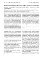
Operative procedure: (Figure 1)
Under general anesthesia with en dotracheal intubation,
the patient was placed in the supine position with both
arms tucked along the sides to avoid stretchi ng on th e
pectoralis muscles and to facilitate reducing the sternal
separation. Debridement was begun with excision of all
wound edges, including skin, subcutaneous tissue and
any necrotic tissue until they were free of the devitalized
tissue and down to the bleeding tissue. Hemostasis was
obtained. The old sternal wires were removed and all
nonviable bone and cartilage was removed down to the
level of bony cortex and marrow cavity. Bone biopsy
was sent to microbiology for culture.
To expose the ribs bilaterally, pectoralis major muscles
were elevated with overlying soft tissue from the midline
to the level of the mid-clavicular line to create flaps and
permit later approximation in the midline. The intercos-
tals perforating vessels were divided with cautery. It was
not necessary to perform a second incision at the
shoulder to release the pectoral muscle insertion. Subse-
quently, the entire wound was lavaged with 3 liters of
warm normal saline. The two sternal halves were
brought together using two large reduction forceps on
both the superior and inferior aspects of the sternum.
Four titanium plat es (Synthes Titanium Sternal Fixa-
tion System, Synthes CMF, West Chester, PA, USA)
(A) Debridement (B) Sternal Plates fixation
Figure 1 Surgical technique of sternal plates’ fi xa tion . It shows wound debridement and pectoral flap development f ollowed by sternal
fixation using four plates.
Fawzy et al. Journal of Cardiothoracic Surgery 2011, 6:63
/>Page 2 of 7
were placed transver sely across the two sternal halves at
the level of the second, third, fourth ribs, and one man-
ubrial plate in most circums tances. Using a templat e,
the plates were contoured. The appropriate length of
the plate was selected to allow a minimum of four lock-
ing screws on each side. The holes were drilled in bone
and cartilage with the aid of the drill guide to precisely
create the drill hole depth avoiding injury to the under-
lying structures. Depth was assessed at times with the
depth gauge but recently by analysis of sternal and rib
thickn ess by CT scan. Using the measureme nt tool ster-
nal and rib thickness can be assessed preoperatively. It
was also important to avoid the inferior margin of the
rib to avoid injury to the intercostals vessels and nerves.
Screw lengths varied among patients and ranged from
12 to 18 mm in length, with 12 mm being the most
commonly used size. Once the plates were secured in
place, the reduction forceps were removed.
Two Jackson-Pratt no. 10 drains were placed one
under each muscle flap, through two separate small inci-
sions along the lower edge of the sternotomy wound.
The muscles were approximated at the mid line with
interrupted no. 1 Vicryl sutures. Superficial muscle fas-
cia and subcutaneous tissues were closed with 2-0 Vicryl
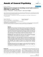
sutures. The skin was closed with staples. A postopera-
tive chest radiograph was obtained routi nely in every
patient (figure 2) to confirm the position of the plates
and exclude pneumothorax.
Sternal precautions were observed for the first 6 week
postoperatively with, avoidance of activity that would
place stress on the pectoral region. Drains were removed
when output was consistently less than 25 cc per drain
per day. An antibiotic course for 4-6 weeks was com-
pleted post-operatively under the direction of the infec-
tious disease service.
Statistical Analysis
All pre-operative patients’ demographic data together
with operative characteristics and post-operative data
were imported into Excel worksheets, for organizational
purposes. Data were expressed as mean ± SD, Median
(range)ornumber(%)using Excel 2003 for Windows
(Microsoft, Redmond, WA, USA).
Results
During the two years of the study, 40 patients under-
went sternal plating. There were 31 males and 9
females. The average age was 69.7 ± 9.4 years with BMI
of 47.1 ± 4.8 Kg/m
2
. Fourteen patients (35%) were dia-
betics, 27 (67.5%) hypertensive, 10(25%) smokers, 8
(20%) had COPD and 11 (27.5%) had renal failure. A
summary of patients’ characteristics is shown in Table 1.
Most often following CABG (70%), AVR (12.5%),
Mitral valve repair (5%) or combined procedures
(12.5%). LIMA was used in all CABG patients but 2.
The average time from the heart procedure to sternal
plating was 9 da ys (range 4-420 days). Primary opera-
tive characteristics of the patients are shown in Table
2. The decision to proceed with plating was made
based on clinical assessmentofthesternumorgross
infection in the operating room. All patients were pre-
sented by pain and wound dehiscence. Twenty two
patients (55%) were presented by sternal instability
alone while 18 patients (45%) with associated wound
discharge. All patients had gross instability at the time
of plating. All patients had cultures from the wound at
thetimeofsurgery.Themostcommonpathogens
were coagulase -negative staphylococci ( 35%) and Sta-
phylococcus aureus (17.5%). Operative cultures o f eight
patients (20%) showed no growth. Table 3 summarizes
the different organisms detected.
In addition to surgical treatment, intravenous antibio-
tics were administrated for the infection, most com-
monly Cloxacillin (60%), followed by Vancomycin (20%).
Mean duration of intravenous antibiotic treatment was
28 days following surgery, followed by oral antibiotics.
Mean operative time was 122.5 minutes. All patients
healed. Postoperative wound complications included: one
patient (2.5%) has post-operative bleeding. One patient
(2.5%) developed postoperative seroma after 16 days. Six
patients (15%) developed post-operative superficial
wound dehiscence with discharge. They all subsequently
healed. Four patients (10%) developed postoperative
pleural effusion that was successfully drained by thoraco-
centesis. One patient (2.5%) developed post-operative
pneumothorax that was drained by an intercostal chest
tube. Two patients (5%) developed recurrent wound
infect ion and healed with negative pressure wound ther-
apy. Both were immunocompromised. Post-o perative
complications are shown in Table 4.
Figure 2 Post-operative Chest X-ray following sternal plates’
fixation. It demonstrates sternal union.
Fawzy et al. Journal of Cardiothoracic Surgery 2011, 6:63
/>Page 3 of 7
Our first few patients were kept sedated and ventilated
for 48 hours before extubation was attempted to mini-
mize excessive movements that might affect the wound.
Later, we have modified our post-operative care proto-
col, so all patients were extubated immediately after sur-
gery unless haemodynamically unstable. Seventy five
percent of the patients were extubated immediately after
surgery, 10% extubated in the first 24 hours and 15%
were late (> 24 hours). Those of late extubation com-
promised a group of six patients with severe COPD that
were on the ventilator for a long time and who ulti-
mately required tracheostomy. Median pos t-op ICU stay
was one day (range 1-29 days). Total median hospital
stay was 18 days, with a range from 3-88 days after ster-
nal plating. The wide variability was mostly due to pro-
longed stay of few patients due to other medical
problems not related to the sternum. There was one
death unrelated to the sternal closure that had infective
endocarditits of his prosthetic valve and died of refrac-
tory septic shock. Post-operative hospital course is
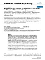
shown in Table 5. The median follow-up time at one
year revealed thoracic stability in all patients (figure 3).
No patient showed clinically significant restrictive pul-
monary compromise, although formal postoperative pul-
monary function measurements were not obtained.
Postoperative chest pain disappeared in the majority of
the patients. Chronic postoperative pain was reported in
two patients, the first one was well managed with oral
nonsteroidal medications. The second patient required
plate removal to relieve his pain.
Comment
Since the introduction of median sternotomy as an
approach to perform open heart operations, it remains
the preferred approach allowing better exposure and
Table 1 Pre-operative Patients’ Demographics
Item Patients
Age (y) 69.7 ± 9.4
Male/Female 31/9
Weight (Kg) 77 ± 9.5
BMI (Kg/m
2
) 47.1 ± 4.8
Obesity (BMI > 30 Kg/m
2
) 27 (67.5%)
Hypertension 27 (67.5%)
Diabetes 14 (35%)
Smoking 10 (25%)
Hypothyroidism 2 (5%)
Chronic Renal Failure 11 (27.5%)
COPD 8 (20%)
PVD 1 (2.5%)
Corticosteroid use 1 (2.5%)
CHF 10 (25%)
Re-operation 3 (7.5%)
Values are means ± SD or Number (%) where shown.
BMI: Body Mass Index.
COPD: Chronic Obstructive Pulmonary Disease.
CHF: Congestive Heart Failure.
PVD: Peripheral Vascular Disease.
Table 2 Primary operative characteristics of the Patients
Operation Patients
CABG 28 (70%)
AVR 5 (12.5%)
CABG + AVR 4 (10%)
CABG + MVR 1 (2.5%)
MV Repair 2 (5%)
Total No. of grafts 3.4 ± 0.5
Use of LIMA/RIMA 26 (65%)
TBT (min) 86.9 ± 43.5
OR Time (min) 238.8 ± 58.7
Re-operation for bleeding 7 (17.5%)
Pts. required massive blood transfusion 1 (2.5%)
Values are means ± SD or Number (%) where shown.
AVR: Aortic Valve Replacement.
CABG: Coronary Artery Bypass Grafting.
LIMA: Left Internal Mammary Artery.
MVR: Mitral Valve Replacement.
Min: minutes.
OR Time: Operative Time.
RIMA: Right Internal Mammary Artery.
TBT: Total Bypass Time.
Table 3 Organisms detected
Organisms Patients
Coagulase-negative Staph. 14 (35%)
Staph. Aureus` 7 (17.5%)
Gram-negative rods 5 (12.5%)
Enterococcus 2 (5%)
Coliform Bacilli 3 (7.5%)
Diphteroid Bacilli 1 (2.5%)
No Pathogens identified 8 (20%)
Values are Number (%) where shown.
Table 4 Post-operative Complications
Complications Patients
Post-op bleeding 1 (2.5%)
Seroma 1 (2.5%)
Superficial Wound Dehiscence 6 (15%)
Recurrent infection 2 (5%)
Hard-ware removal 2 (5%)
Pleural effusion 4 (10%)
Pneumothorax 1 (2.5%)
Values are Number (%) where shown.
Fawzy et al. Journal of Cardiothoracic Surgery 2011, 6:63
/>Page 4 of 7
easy access of the heart and mediastinal structures. Two
well-described complications with this type of incision
are sternal dehiscence and wound infection [4-6].
Despite the relative infrequency of t hese complications
(0.4% - 5.1%), they carry high morbidity and mortality.
Many techniques have been developed to break the
vicious circle of ster nal stability and infection. Continu-
ous antibiotic irrigation was first used by Shumacker
and Mandelbanum [6]. Debridement with removal of all
infected tissue is emphasized no matter what the recon-
structive technique is utilized. Negative pressure wound
therapy has also be en utilized both as a treatme nt for
wound dehiscence and as a temporary measure to help
treat the infection before sternal reconstruction. Soft tis-
sue reconstruction has emphasized obliteration of dead
space with health well vascularized tissue initially using
pectoralis major flaps [4]. Subsequently, many techni-
ques have been used that include advancement flaps,
rotational flaps, and turnover flaps [7-10]. Large sternal
defects associated with partial and total sternectomy
have also been covered using the omentum, rectus mus-
cle and latissimus muscle flap [11-14]. Muscle flap s can
be used alone or in combination with sternal rewiring.
Robicszek parasternal weave is still the standard techni-
que used for sternal rewiring in many centers [15]. As a
supplement to sternal wire s, longitudinal plates had
been used to fix the sternum together with circumferen-
tial wire [16-18]. Other techniques have in cluded the X-
shaped and box-shaped plates over the sternum with
two figures of 8 wires plac ed around the manub rium
and the xiphisternal junction [18].
Chest wall defects seen after complete or partial ster-
nectomy can res ult in paradoxical chest wall mov ement
and thoracic instability that is difficult to address by
muscle flaps alo ne. Restoration of sternal or chest wall
stability can be achieved with transverse locking plate
fixation system by distributing the force laterally over
the ribs on both sides. It relies on rib and sternal fixa-
tion when available to provide chest wall stability. Using
the described technique, pla te stabilization is achieved
on the anterior surface of the ribs and no dissection is
necessary at the deep aspect of the sternum, avoiding
the risk of injury to the underlying heart structures. It is
also feasible in such cases with sternal loss with little
residual sternum left for fixation. However, it is not
without risks or complications. Our postoperative com-
plication rate is low particularly involving seroma as
seen with other authors. We only have one patient
(2.5%) in our series that developed post- op seroma
Table 5 Post-operative Hospital Course
Item Patients
Ventilation time > 24 Hrs. 6 (15%)
ICU stay (days) 1 (1-29)
Hospital stay (days) 18 (3-88)
Hospital Mortality 1 (2.5%)
Values are Number (%) or Median (range) where shown.
Pre-operative Post-operative
Figure 3 Pre and Post-op erative chest CT scan following sternal plates’ fixation. It demonstrates resolving of mediastinitis and sternal
union.
Fawzy et al. Journal of Cardiothoracic Surgery 2011, 6:63
/>Page 5 of 7
requirin g treatment. Cicilioni et al [19] reporte d higher
rate of seroma formation in 5 patients (10%) in their
series of 50 consecutive sternal wound reconstructions
using transverse plate fixation. Hugo et al. [7] used pec-
toralis muscle flaps only and reported the highest rate
of seroma formation (24%) in their series of 74 patients.
It is obvious that seroma formation is directly related to
extensive pectoral muscle dissection rather than the pre-
sence of the metal plates. This creates a dead space
underneath the muscles where seroma can form. Our
low rate of seroma might be related to limited pectoral
muscle dissection and routine placement of two sub
pectoral Jackson -Pratt drains. We left the drains in situ
untilthedailydrainageisless25mlbeforeremoval.In
addition we have used a technique where the deep pec-
toral fixation sutures are fixed to the plate, helping to
obliterate dead space. Drilling too deep or using long
screws carry the risk of injury to the internal mammary
artery and vein and the intercostal vessels as well as the
lung s and mediastinal struct ures. We avoid this compli-
cation by measuring the sternal and ribs thickness preo-
peratively using chest CT scan that helped in accurately
choice the proper size screw. Using the drill guide and
an accurately measured screw length with the depth
gauge intraoperatively at time played an important role
in minimizing post-operative bleeding in our series. We
only have one case (2.5%) of post-op bleeding and that
was not related to plates or screws. The bleeder was
found in the subcutaneous tissue that was surgically
ligated. Cicilioni [19] reported two post-op bleeding
(4%) in his series. Both bleeding events were from an
intercostals and a pectoral vessel and thus was not
plate-related. They w ere easily rec ognized and treated.
Violation of the pleural space can occur duri ng the deb-
ridement, drilling or during the insertion, and this
occurred in one of our patients. Huh [20] had no inci-
dence of pneumothora x or injury to the underly ing
heart or vascular structures.
In addition to restoration of sternal stability following
sternal plating, prevention o f post-operative infection
requires excellent debridement, lavage irrigation with
saline and aggressive postoperative antibiotic coverage.
We have made it our practice to aggressively excise all
wound edges, including skin, subcutaneous tissue, any
necrotic -appearing tissue including chronic granulation
tissue present down to the level of the sternal bone. The
goal of debridement is to convert the chronic wound
into an acute one. All nonviable bone and cartilage were
removed down to a level that bleeds. Subsequently, the
entire wound was vigorously lavaged with 3 liters of
warm normal saline. We had only 2 patients developed
postoperative wound infec tion. The first patient had end
stage renal failure with chronic hemodialysis, uncon-
trolled IDDM and PVD that required bilateral leg
amputation. During his hospital course, he developed
sternal wound infection that initially required VAC ther-
apy and subsequently underwent sternal fixation. He
required hardware removal. At the time of his hardware
removal, the sternal bone was found to be well healed.
The soft tissue wound was successfully managed with
VAC therapy. He was treated with a second course of
IV antibiotics and went on to complete healing. The
second patient was on chro nicsteroidusebecauseof
long standing chronic asthma. His wound responded
well to wound treatment and antib iotics. Cicilioni et al.,
[19] encountered only a 2.7% incidence of recurrent
infection and suggest that titanium plates have a bacter-
iostatic property.
We achieved thoracic stability i n all our patients.
Using traditional strategies, such as parasternal Robicsek
weave with or without muscle flap, Olbrecht [15],
noticed that 20% of his patients had post-operative ster-
nal dehiscence.
We did not r emove any plates because of loosening
although others had had this complication [19,21]. One
author Huh et al., [20] had to remove the plates in two
patients (14%) associated with infection in one and plate
fracture in another. Plate fracture leading to instability
is a concern. Repeated bending weaken the strength of a
plate at one point may be a factor.
Patients complain of chro nic pain following surgery
for treatment of sternal dehiscenc e and sternal osteo-
myelitis.Weonlyhavetwopatients(5%)whohad
chronic post-operative pain. The first patient responded
well to medical management and the second one
required plate removal to relieve his pain.
However, one author [22] removed 50% of the trans-
verse placed plates , due to persisting pain. Another [23]
reported that 51% of their p atients complained of
chronic chest or shoulder pain following sternal wound
reconstruction using muscle or omental flaps.
Limitations of the study
The number of the patients in this series is relatively
small group of the 1200 cases undergoing open he art
surgery at our institution every year. However, this
approach to sternal infection/dehiscence has changed
our practice. Presently, sternectomy is rarely performed
and more extensive soft tissue reconstruction techniques
are not required. Preservation of the anatomical and
functional aspects of the sternum is possible. However,
the physiologic effect of this approach in preserving the
sternum and the effect of the plating itself, require
further study. In additio n, being a ret rospe ctive review
study in one centre, the benef it and integration of this
appr oach into present practice requires further study. A
larger multi-center prospective control trials using larger
number of patients, comparing different techniques of
Fawzy et al. Journal of Cardiothoracic Surgery 2011, 6:63
/>Page 6 of 7
sternal reconstruction are needed to address the indica -
tions, benefits and potential complications of this
approach.
Conclusions
Sternal plating appears to be an effective option for the
treatment of sternal dehiscence as it yields a stable ster-
num. It is simple and safe technique without risks.
Long-term follow-up and further larger studies are
needed to address the indications, benefits and compli-
cations of sternal plating.
Acknowledgements
The study was not financi ally supported. The authors are thankful to Dr.
Wegdan Mawlana, research assistant at St. Michael’s hospital for her valuable
assistance in chart revision and data collection.
The study has been presented at the Annual Meeting of the American
Society of Plastic Surgeons, November 2-5, 2008, Chicago, U.S.
Author details
1
Division of Cardiovascular and Thoracic Surgery, Department of Surgery,
Terrence Donnelly Heart Center, Keenan Research Center in the Li Ka Shing
Knowledge Institute of St. Michael’s Hospital, University of Toronto, 30 Bond
Street, Toronto, Ontario, M5B 1W8, Canada.
2
Division of Plastic Surgery,
Department of Surgery, Terrence Donnelly Heart Center, Keenan Research
Center in the Li Ka Shing Knowledge Institute of St. Michael ’ s Hospital,
University of Toronto, 30 Bond Street, Toronto, Ontario, M5B 1W8, Canada.
Authors’ contributions
HF conceived of the study, participated in its design, carried out the
operations, collecting and analyzing the data, writing, reviewing and
submitting the manuscript. KO participated in collecting and analyzing the
data. LE participated in reviewing the manuscript. DL participated in
reviewing the manuscript. DB participated in reviewing the manuscript. MM
participated in performing the operations and reviewing the manuscript. JM
conceived of the study, participated in its design, carried out the operations,
writing and reviewing the manuscript. All authors read and approved the
final manuscript.
Competing interests
The authors declare that they have no competing interests.
Received: 8 February 2011 Accepted: 29 April 2011
Published: 29 April 2011
References
1. McGregor WE, Payne M, Trumble DR, Farkas KM, Magovern JA:
Improvement of sternal closure stability with reinforced steel wires. Ann
Thorac Surg 2003, 76:1631-4.
2. Dalton ML, Connally SR, Sealy WC: Julian’s re-introduction of Milton’s
operation. Ann Thorac Surg 1992, 53(3):532-3.
3. Tremble DR, McGregor WE, Magovern JA: Validation of a bone analog
model for studies of sternal closure. Ann Thorac Surg 2002, 74(3):739-44.
4. Jurkiewicz MJ, Bostwick J, Hester TR, Bishop JB, Craver J: Infected median
Strenotomy wound: successful treatment by muscle flaps. Ann Surg 1980,
191:738.
5. Culliford AT, Cunningham JN, Zeff RH, Isom OW, Teiko P, Spencer FC:
Sternal and costochondral infections following open heart surgery: A
review of 2594 cases. J Thorac Cardiovasc Surg 1976, 72:714.
6. Shumacker HB, Mandelbaum I: Continuous antibiotic irrigation in the
treatment of infection. Arch Surg 1963, 86:384.
7. Hugo NE, Sultan MR, Ascherman JA, Patsis MC, Smith CR, Rose EA: Single
-stage management of 74 consecutive sternal wound complications
with pectoralis major myocutaneous advancement flaps. Plast Reconstr
Surg 1994, 93:1433-41.
8. Nahai F, Morales L, Bone DK, Bostwick J: Pectoralis major muscle turnover
flaps for closure of the infected sternotomy wound with preservation of
form and function. Plast Reconstr Surg 1982, 70:1982.
9. Lopez-Monjardin H, de la Pena-Salcedo , Mendoza-Munoz M, Lopez-Yanez-
de la Pena A, Palacio-Lopez E, Lopez-Garcia A: Omentum flap versus
pectoralis major flap in the treatment of mediastnitis. Plast Reconstr Surg
1998, 101:1481.
10. Herrera HR, Ginsburg ME: The pectoralis major myocutaneous flap and
omental transposition for closure of infected median strenectomy
wounds. Plast Reconstr Surg 1982, 70:465.
11. Ishii CH: Double-pedicle transverse rectus abdominis myocutaenous flap
for unilateral breast and chest wall reconstruction. Plast Reconstr Surg
1985, 76:901.
12. Hultman CS, Culbertson JH, Jones GE, Losken A, Kumar AV, Carlson GW,
Bostwick J, Jurkiewicz MJ: Thoracic reconstruction with the omentum:
Indications, complications, and results. Ann Plast Surg 2001, 46:242.
13. Tizian C, Borst HG, Berger A: Treatment of total necrosis using the
latissimus dorsi muscle flap. Plast Reconstr Surg 1985, 76 :703.
14. Solomon MP, Granick MS: Bipedicle muscle flaps in sternal wound repair.
Plast Reconstr Surg
1988, 101:356.
15. Olbrecht VA, Barreiro Cj, Bonde PN, Williams JA, Baumgartner WA, Gott VL,
Conte JV: Clinical outcomes of noninfectious sternal dehiscence after
median sternotomy. Ann Thorac Surg 2006, 82:902-7.
16. Wu LC, Renucci JD, Song DH: Sternal nonunion: a review of current
treatments and a new method of rigid fixation. Ann Plast Surg 2005,
54(1):55-8, Review.
17. Mitra A, Elahi MM, Tariq GB, Mir H, Powell R, Spears J: Composite plate and
wire fixation for complicated sternal closure. Ann Plast Surg 2004,
53(3):217-21.
18. Jaishankar R, David S, David HS: Rigid Plate Fixation of the Sternum. Ann
Thorac Surg 2007, 84:1056-8.
19. Cicilioni OJ Jr, Stieg FH III, Papanicolaou G: Sternal wound reconstruction
with transverse plate fixation. Plast Reconstr Surg 2005, 115(5):1297-303.
20. Huh J, Bakaeen F, Chu D, Wall M: Transverse sternal plating in secondary
sternal reconstruction. J Thorac Cardiovasc Surg 2008, 136(6):1476-1480.
21. Hallock GG, Szydlowski GW: Rigid fixation of the sternum using a new
coupled titanium transverse plate fixation system. Ann Plast Surg 2007,
58:640-644.
22. Voss B, Bauernschmitt R, Will A, Krane M, Kross R, Brockmann G, Libera P,
Lange R: Sternal reconstruction with titanium plates in complicated
sternal dehiscence. Eur J Cardiothorac Surg 2007, 32:391-3.
23. Ringelman PR, Vander Kolk CA, Cameron D, Baumgartner WA, Manson PN:
Long-term results of flap reconstruction in median sternotomy wound
infections. Plast Reconstr Surg 1994, 93:1208.
doi:10.1186/1749-8090-6-63
Cite this article as: Fawzy et al.: Sternal plate fixation for sternal wound
reconstruction: initial experience (Retrospective study). Journal of
Cardiothoracic Surgery 2011 6:63.
Submit your next manuscript to BioMed Central
and take full advantage of:
• Convenient online submission
• Thorough peer review
• No space constraints or color figure charges
• Immediate publication on acceptance
• Inclusion in PubMed, CAS, Scopus and Google Scholar
• Research which is freely available for redistribution
Submit your manuscript at
www.biomedcentral.com/submit
Fawzy et al. Journal of Cardiothoracic Surgery 2011, 6:63
/>Page 7 of 7