Báo cáo y học: "An interesting journey of an ingested needle: a case report and review of the literature on extraabdominal migration of ingested Foreign bodies" pdf
Bạn đang xem bản rút gọn của tài liệu. Xem và tải ngay bản đầy đủ của tài liệu tại đây (1.12 MB, 4 trang )
REVIE W Open Access
An interesting journey of an ingested needle: a
case report and review of the literature on extra-
abdominal migration of ingested Foreign bodies
Zeynep Ozkan
1
, Metin Kement
1*
, Ahmet B Kargı
2
, Zafer Censur
1
, Fazli C Gezen
1
, Selahattin Vural
1
and
Mustafa Oncel
1
Abstract
Swallowed foreign bodies encounter a major problem espe cially in children, but fortunately they mostly do not
cause any related complication and are easily passed with the stool. In this paper, an interesting journey of a
needle is presented. A 20-year old female admitted to our emergency service after she had swallowed a sewing
machine needle, which is initially observed in the stomach in the plain abdominal radiography. During the follow-
up period, the needle traveled through bowels, and surprisingly was observed in the left lung on 10
th
day of the
follow-up. It was removed with a thoracotomy and pneumotomy under the fluoroscopic guidance. The
postoperative period was uneventful and the patient was discharged from the hospital on the day 5. We also
review the literature on interesting extra-abdominal migrations of swallowing foreign bodies.
Keywords: Foreign body, migration, pneumotomy
Background
The foreign body ingest ion occurs usually in chil dren.
Although it is detected rarely in adults, prisoners, mentally
retarded people and young girls with turba n in Islamic
countries are commonly affected [1-3]. Foreign bodies
generally pass spontaneously through the gastrointestinal
tract (GI tract) and do not result in any complications, but
very sharp or pointed ob jects may ca use perforations
along the gastrointestinal tract. In additi on, retained for-
eign bodies may cause gastrointestinal erosions and abra-
sions, which result i n bleeding. The rate of complication
from foreign body ingestion is estimated less than 1 %.
Complications due to foreign bodies in the stomach and
small intestine typically involve perforation associated with
peritonitis. Foreign bodies account for 15% to 35% of all
bowel perforations. These cases require surgical interven-
tion. Although migration of foreign bodies from esophagus
to mediastinum and thorax may lead to very serious com-
plications including pneumomediastinum, mediastinitis,
pneumothorax, pericarditis, cardiac tamponade, or even
horrific lethal vascular injuries to the aorta or pulmonary
vasculature, migration of foreign bodies from the colon to
the lung is not reported before [4-8].
In t his report, we present an interesting journal of an
ingested sewing machine needle which migrated from
the transverse colon to the lung in a young woman. We
also review the literatur e on interesting extra-abdominal
migrations of swallowing foreign bodies.
Case
A 20-year old female was admitted to our emergency
service immediately after accidental swallowing of a
sewing machine needle. On admission, she had no
symptoms such as abdominal pain, vomiting o r dyspha-
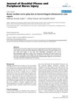
gia. A plain abdominal radiography (PAR) revealed a
needle located in the upper abdomen (Figure 1). A
fiber diet was prescribed and a daily routine out-
patient follow-up with PAR’ s was planned. The two
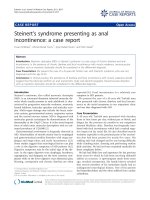
PAR’s taken on the days 3 and 7 showed that the nee-
dle had passed to the terminal ileum and transverse
colon (Figure 2). However, on the day 10, PAR showed
that needle migrated into the thorax (Figure 3). She
did not have any symptoms or signs of peritonitis. An
emergent computerized tomography (CT) confirmed
* Correspondence:
1
General Surgery Department, Kartal Education and Research Hospital,
Istanbul, Turkey
Full list of author information is available at the end of the article
Ozkan et al. Journal of Cardiothoracic Surgery 2011, 6:77
/>© 2011 Ozkan et al; licensee BioMed Central Ltd. This is an Open Access article distributed und er the terms of the Creative Commons
Attribution License ( ), which permits unrestricted use, distribution, and reproduction in
any medium, provided the original work is properly cited.
that the needle was located in the lower lobe of the left
lung (Figure 4). Also, there was no intestinal contrast
leakage in CT. There fore, it was decided to perform an
emergent thoracotomy. Fluoroscopy and finger palpa-
tion were used to verify the exact location of the n ee-
dle during the operation and the needle was removed
after pneumotomy. The postoperative period was
uneventful and the patient discharged from the hospital
on the day 5.
Discussion
The incidence of foreign body ingestions is unknown.
The most common causes of foreign body ingestion are
accidental swallowing of objects. Children usually put
any object they find into their mouths and may acciden-
tally swallow them. In healthy adults, accidental swal-
lowing often involves toothpicks, dentures and turban
pins. Psychiatric patients may swallow a wide variety of
objects, including large and bizarre items. Although the
majority of foreign bodies pass harmlessly through the
GI tract and conservative management is generally
recommended, 10% to 20% of them will require non-
operative intervention such as endoscopy, and approxi-
mately 1% of them will req uire surgery [9-11]. An
estimated 1500 deaths occur annu ally from foreign body
ingestion in USA [12].
A foreign body lodged in the gastrointestinal tract may
cause local inflammation leading to pain, bleeding, scar-
ring and obstruction, or it may erode through the GI
tract. The site of perforation due to foreign bodies
appears to be variable in the GI tract. Although
Figure 1 A plain abdominal radiography (PAR) revealed a
needle located in the upper abdomen.
Figure 2 Theneedlehadpassedtotheterminalileumand
transverse colon.
Figure 3 PAR showed that needle migrated into the thorax.
Ozkan et al. Journal of Cardiothoracic Surgery 2011, 6:77
/>Page 2 of 4
McManus et al. identified ileocecal region as the most
common site of perforation secondary to foreign body
ingestion [13], duodenum has been reported as the most
common site by Spitz et al. and Gracia et al. [14,15].
Foreign body ingestions necessitate careful and contin-
ued observation due to the possibility of serious
complications.
Children and the mentally impaired, or the psychiatric
patients may present with refusal to eat, vomiting, chok-
ing, drooling, wheezing, blood-stained saliva, or respira-
tory distress. Erythema, tenderness, or crepitus in the
neck may be present with oropharyngeal or esophageal
perforation. The abdomen should be examined for clini-
cal evidences of peritonitis. These conditions will
require emergent surgical intervention. Ventilation, air-
way compromise and the risk of aspiration should also
be assessed. If the swallowed object is radio-opaque, a
single frontal radiograph that includes the neck, chest,
and entire abdomen is usually sufficient to locate the
object. The plain radiography is effective in localizing
most of radio-opaque objects [16]. CT scan or MRI is
rarely indicated but may enhance the detection of for-
eign bodies or complications (e.g., perforations, migra-
tions) in special cases.
Migration of foreign bodies from the abdomen or pel-
vis to the lung is very rare but well-defined entity. Diag-
nostic catheters, venous shunts and bullets have been
reported in this context, but differently from our case,
most of them have migrated hematogenously [17-19].
To our knowledge, our case is unique i n that we have
described a foreign body migration from the transverse
colon to the lung parenchym a. Interestingly, that migra-
tion did not have lead peritonitis and the patient was
able to manage without the need for a laparotomy. We
think, the perforation in the transverse colon wall was
too small to cause a significant bowel leakage. Also, the
route of the journey after transverse colon is not certain.
In our opinion, there are two possibilities for the needle
to pass thorax: either, it penetrated the diaphragm or
passed through esophageal hiatus.
In reviewing the literature on extra-abdominal migra-
tion of swallowing foreign bodies, Macchi at al. repo rted
a case of a 48-year-old man with esophageal perforation,
mediastinitis, and evidence of perf oration of the ascend-
ing aorta during surgical drainage of the mediastinum.
They reported finding a fish bone unde r the aortic arch
at autopsy [ 5]. Kunishige et al. presented a 79-year-old
woman who had referred to hospital with chest pain
after swallowing a fish bone. The bone had been
removed by esophagoscopy. Eleven days later she had
presented because of hematemesis. Computed tomogra-
phy and angiography had confirmed a diagnosis of eso-
phageal perforation leading to mediastinitis and the
presence of an infected pseudoaneurysm. The infected
pseudoaneurysm had been completely resected [6].
Cekirdekci et al. and Vesna et al. reported two different
cases with cardiac tamponade due to migration of sew-
ing needle from the esophagus [7]. On the contrary,
Graffstädt et al. presented a journey of a wandering nee-
dle from bronchus to intestine in a 14-year-old girl. The
needle had been excreted naturally on the third day
[20]. Ozsunar et al. presented an interesting unique cas e
in which a needle had been accidentally swallowed and
then migrated into the vertebral body [21]. Chen et al.
reported a 50-year-old woman who had been diagnosed
with thyroid abscess secondary to a swallowing fish
bone [22].
As a conclusion, an ingested foreign body infrequently
causes severe problems, however complications such as
perforation and migration should be always keep in
mind and close follow up should be done. In addition,
we have to be certain to detect that foreign bodies have
left the body.
Consent
Written informed consent was obtained from the patient
for publication of this case report. A copy of the written
consent is available for review by the Editor-in-Chief of
this journal.
Author details
1
General Surgery Department, Kartal Education and Research Hospital,
Istanbul, Turkey.
2
Thoracic Surgery Department, Kartal Education and
Research Hospital, Istanbul, Turkey.
Authors’ contributions
ZO, MK and ZC were involved in patient care. ZO and MK reviewed the
literature and wrote the manuscript. ABK performed operation. CG and MO
supervised the manuscript. All authors read and approved the final
manuscript.
Figure 4 An emergent computarized tomography confirmed
that the needle was located in the lower lobe of the left lung.
Ozkan et al. Journal of Cardiothoracic Surgery 2011, 6:77
/>Page 3 of 4
Competing interests
The authors declare that they have no competing interests.
Received: 24 February 2011 Accepted: 26 May 2011
Published: 26 May 2011
References
1. Stack LB, Munter DW: Foreign bodies in the gastrointestinal tract. Emerg
Med Clin North Am 1996, 14:493-521.
2. Cheng W, Tam PK: Foreign-body ingestion in children: experience with
1,265 cases. J Pediatr Surg 1999, 34:1472-1476.
3. Conway WC, Sugawa C, Ono H, Lucas CE: Upper GI foreign body: an adult
urban emergency hospital experience. Surg Endosc 2007, 21:455-460.
4. Ghimire A, Bhattarai M, Kumar M, Wakode PT: Descending necrotizing
mediastinitis: a fatal complication of neglected esophageal foreign
body. Kathmandu Univ Med J (KUMJ) 2007, 5:98-101.
5. Macchi V, Porzionato A, Bardini R, Parenti A, De Caro R: Rupture of
ascending aorta secondary to esophageal perforation by fish bone. J
Forensic Sci 2008, 53:1181-1184.
6. Kunishige H, Myojin K, Ishibashi Y, Ishii K, Kawasaki M, Oka J: Perforation of
the esophagus by a fish bone leading to an infected pseudoaneurysm
of the thoracic aorta. Gen Thorac Cardiovasc Surg 2008, 56:427-429.
7. Cekirdekci A, Ayan E, Ilkay E, Yildirim H: Cardiac tamponade caused by an
ingested sewing needle. A case report. J Cardiovasc Surg (Torino) 2003,
44:745-746.
8. Vesna D, Tatjana A, Slobodan S, Slobodan N: Cardiac tamponade caused
by migration of a swallowed sewing needle. Forensic Sci Int 2004,
139:237-239.
9. Webb WA: Management of foreign bodies of the upper gastrointestinal
tract: Update. Gastrointest Endosc 1995, 41:39-51.
10. Nandi P, Ong GB: Foreign body in the esophagus: Review of 2394 cases.
British Journal of Surgery 1978, 65:5-9.
11. Vizcarrondo FJ, Brady PG, Nord HJ: Foreign bodies of the upper
gastrointestinal tract. Gastro-intest Endosc 1983, 29:208-210.
12. Stack LB, Munter DW: Foreign bodies in the gastrointestinal tract. Emerg
Med Clin North Am 1996, 14:493-521.
13. MacManus JE: Perforations of the intestine by ingested foreign bodies.
Am J Surg 1941, 53:393.
14. Spitz L: Management of ingested foreign bodies in childhood. Br Med J
1971, 4:469-472.
15. Gracia C, Frey CF, Bodai BI: Diagnosis and management of ingested
foreign bodies: A Ten Years Experience. Annals of emergency medicine
1984, 13:159.
16. Suita S, Ohgami H, Nagasaki A, Yakabe S: Management of pediatric
patients who have swallowed foreign objects. Am J Surg 1989, 55:585.
17. Gupta AK, Dogra VS, Ahmad I, DelBalso AM: Missile emboli to the
pulmonary artery [letter]. Am J Emerg Med 1997, 15:213-214.
18. Doughty IM, David TJ: Migration of fine bore Silastic catheter to
pulmonary artery [letter]. Arch Dis Child 1994, 70 :451.
19. Planas R, Domenech E, Montana X, Pilar Rodriguez M, Rodriguez N, Cabré E,
Gassull MA: Rupture and migration of the venous segment of LeVeen
shunt: an unreported complication. Am J Gastroenterol 1993,
88:1101-1103.
20. Graffstädt H, Dieckow B, Grüber C, Stöver B, Niggemann B: Christmas
surprise: the unnoticed journey of a needle-from bronchus to intestine.
Respir Med 2005, 99:1600-1602.
21. Ozsunar Y, Tali ET, Kilic K: Unusual migration of a foreign body from the
gut to a vertebral body. Neuroradiology 1997, 40:673-674.
22. Chen CY, Peng JP: Esophageal fish bone migration induced thyroid
abscess: case report and review of the literature. Am J Otolaryngol 2010,
4:29.
doi:10.1186/1749-8090-6-77
Cite this article as: Ozkan et al.: An interesting journey of an ingested
needle: a case report and review of the literature on extra-abdominal
migration of inges ted Foreign bodies. Journal of Cardiothoracic Surgery
2011 6:77.
Submit your next manuscript to BioMed Central
and take full advantage of:
• Convenient online submission
• Thorough peer review
• No space constraints or color figure charges
• Immediate publication on acceptance
• Inclusion in PubMed, CAS, Scopus and Google Scholar
• Research which is freely available for redistribution
Submit your manuscript at
www.biomedcentral.com/submit
Ozkan et al. Journal of Cardiothoracic Surgery 2011, 6:77
/>Page 4 of 4