Báo cáo y học: "Changes in Two Point Discrimination and the law of mobility in Diabetes Mellitus patients" pps
Bạn đang xem bản rút gọn của tài liệu. Xem và tải ngay bản đầy đủ của tài liệu tại đây (201.04 KB, 6 trang )
BioMed Central
Page 1 of 6
(page number not for citation purposes)
Journal of Brachial Plexus and
Peripheral Nerve Injury
Open Access
Research article
Changes in Two Point Discrimination and the law of mobility in
Diabetes Mellitus patients
R Periyasamy
†1
, M Manivannan
†1
and Vengesana Balakrish
Raja Narayanamurthy*
2
Address:
1
Biomedical Engineering Group, Department of Applied Mechanics, Indian Institute of Technology Madras, Chennai, 600036, India and
2
Diabetic Foot Clinic, Sundaram Medical Foundation, Chennai, 600040, India
Email: R Periyasamy - ; M Manivannan - ; Vengesana Balakrish
Raja Narayanamurthy* -
* Corresponding author †Equal contributors
Abstract
Background: Diabetic neuropathy is a family of nerve disorders with progressive loss of nerve
function in 15% of diabetes mellitus (DM) subjects. Two-point discrimination (TPD) is one method
of quantitatively testing for loss of nerve function. The law of mobility for TPD is known for normal
subjects in earlier studies but has not been studied for diabetic subjects. This is a pilot study to
evaluate and plot the law of mobility for TPD among DM subjects.
Methods: The Semmes Weinstein monofilament (SWMF) was used to measure the loss of
protective sensation. An Aesthesiometer was used to find the TPD of several areas in upper and
lower extremities for normal and diabetic subjects. All the subjects were screened for peripheral
artery occlusive disease with ankle brachial pressure index (0.9 or above).
Results: TPD of normal and diabetic subjects for different areas of hands and legs from proximal
to distal is evaluated for 18 subjects. TPD values decrease from proximal to distal areas. Vierodt's
law of mobility for TPD holds good for normal subjects in the hand and foot areas. The law of
mobility for TPD in DM subjects holds well in the hand but doesn't hold well in foot areas with or
without sensation.
Conclusion: TPD is a quantitative and direct measure of sensory loss. The TPD value of diabetic
subjects reveals that the law of mobility do not hold well for Diabetic subjects in foot areas. The
significance of this result is that the TPD of the diabetic subjects could provide direct, cost effective
and quantitative measure of neuropathy.
Background
Foot problems are the most common reason for hospital-
ization of diabetes mellitus (DM) patients. Neuropathy
and impaired blood supply are the chief causes for foot
ulceration in DM patients. In the presence of neuropathy,
the primary factor for ulcer formation in diabetes is the
loss of protective sensation [1,2]. Evaluation of sensibility
on the feet of diabetic patients is important in order to
properly identify the group with neuropathy and to estab-
lish prevention of ulceration for those at risk. Various
modalities of sensation like temperature, vibration, point
localization and two point discrimination (TPD) have
Published: 29 January 2008
Journal of Brachial Plexus and Peripheral Nerve Injury 2008, 3:3 doi:10.1186/1749-7221-3-3
Received: 2 November 2007
Accepted: 29 January 2008
This article is available from: />© 2008 Periyasamy et al; licensee BioMed Central Ltd.
This is an Open Access article distributed under the terms of the Creative Commons Attribution License ( />),
which permits unrestricted use, distribution, and reproduction in any medium, provided the original work is properly cited.
Journal of Brachial Plexus and Peripheral Nerve Injury 2008, 3:3 />Page 2 of 6
(page number not for citation purposes)
been used to measure sensory loss. Semmes-Weinstein
monofilament testing (10 gm) divides the huge popula-
tion of DM subjects into subjects who are at risk but does
not precisely determine the degree of progressive loss of
sensation, or suggest degree of nerve compression and
axonal loss. Specialist clinics may quantify neuropathy
with biothesiometry, plantar foot pressure measurement,
and assess lower extremity vascular status with handheld
Doppler systems and ankle-brachial blood pressure indi-
ces (ABPI).
Though monofilament testing is one of the primary
screening methods for measuring cutaneous sensibility
clinically, the sensibility can be only qualitatively assessed
as normal touch, diminished light touch, diminished pro-
tective sensation, and loss of protective sensation [3]
depending upon the size of the filament and force exerted
to buckle the mono-filament. The measurement of cuta-
neous sensation to differentiate one-point from two-point
static touch stimuli may allow identification of ulceration
earlier in the clinical course of diabetic neuropathy [4].
Static and moving TPD have been used as tools to meas-
ure sensory loss in DM patients. Although the method is
subjective, as the patient must report whether or not the
pressure is felt, it is more reliable than the previously
available methods and it is a quantitative measure of the
sensory loss. Both the vascular dysfunction and the neu-
ropathy result in increased TPD in foot areas. Therefore,
increase in TPD does not necessarily indicate either neu-
ropathy or vascular dysfunction [5].
It is well known that TPD obeys the law of mobility in
normal subjects [6], however the applicability of the law
to diabetic subjects is not known. This paper presents the
first systematic study of the law of mobility to assess the
sensibility of diabetic subjects, comparing the law of
mobility of TPD in the upper and lower extremities of DM
patients. We observe that the law of mobility does not
hold well in DM patients.
Law of Mobility
Research on cutaneous sensibility was undertaken in the
nineteenth century by Vierodt [6] and Weber [7]. Weber
introduced the point localization test and the accompany-
ing measures, two-point discrimination (TPD) and locali-
zation error, as measures of cutaneous sensibility. Density
of mechanoreceptors in an area determines the TPD. A
dense population leads to finer TPD and the receptors
have smaller receptive fields. Mapping of the whole body
revealed large differences in the sensibility between differ-
ent parts of the body. Vierodt generalized this observation
into the 'law of mobility', which states that the TPD improves
with the mobility of the body part. TPD correlates with the
Degree of Freedom (DOF) of the body part. It is to be
noted that no exception to this law has been found yet.
After the work of Weber and Vierodt, little attention was
given to this field until the 1950s [8] and 1960s [9]. Wein-
stein observed significant effects of body locus. Lowest
TPD was found for the fingertips (2.5 mm). TPD for the
trunk was approximately 40 mm. Sensitivity decreased
from distal to proximal regions, and thresholds correlated
with the relative size of cortical areas subserving a body
part. Another important observation was that good TPD
did not necessarily mean good sensitivity to pressure, that
is, a low detection threshold.
Static and dynamic Two-Point Discrimination
The minimum distance between two stimulus points on
the skin, which are perceived as distinct points, is defined
as TPD. Among the two types of TPD, static and dynamic
TPD, static two-point discrimination (STPD) is com-
monly used in emergency departments to determine dig-
ital nerve integrity [10]. Static TPD is the current
recommended method for physicians evaluating degree of
sensory loss in diabetic patients. Dynamic TPD (DTPD) is
usually measured with a Disk-criminator [10], moving the
prongs along the surface of the center. Moving TPD values
were smaller in magnitude than stationary TPD values in
all anatomical areas tested. Dynamic TPD is not routinely
used in clinical practices. In this paper we consider only
the static two-point discrimination for the law of mobil-
ity.
Methods to measure TPD
Calipers or an opened paper clip with two parallel ends
are used for finding STPD [11]. An aesthesiometer is a
modified form of vernier caliper useful for determining
the TPD of touch, by moving the prongs into contact with
the portion of the body part and then pressing until the
patient feels a sensation. A disk-criminator, consisting two
rotating plastic disks that are joined together, is useful for
testing DTPD [12]. A set of small grating surfaces recently
introduced for cutaneous spatial resolution measurement.
The gratings are placed on the skin and subjects are
required to identify the orientation of grooves and bars.
The finest grating whose orientation is discriminated reli-
ably provides an estimate of the spatial resolution limit in
the tested area [13]. In the 1990s, Dellon proposed the
Pressure-Specified Sensory Device (PSSD) that could
gather information about static and moving touch in a
continuous form by using a computer [12]. PSSD is a
quantitative sensory device, which consists of one or two
blunt probes and sensitive transducers to measure and
record the perception thresholds of pressure on the sur-
face of the body in gms/sqmm. PSSD is not routinely used
in clinical practices. In this paper we used an aesthesiom-
eter to measure the TPD.
Journal of Brachial Plexus and Peripheral Nerve Injury 2008, 3:3 />Page 3 of 6
(page number not for citation purposes)
Methods
We measured the loss of protective sensation and TPD in
forearm, palm, fingers, lower leg, and foot areas. While
the loss of protective sensation was measured using 10 gm
SWMF [3], TPD was assessed using an aesthesiometer. We
tested the pressure exerted by two prongs of the aesthesi-
ometer using a weighing balance. A total of 50 gm was
exerted on usual pressure.
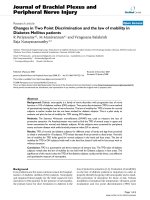
Although in literature the foot is divided into ten standard
significant areas as shown in figure 1(a) as per method
indicated in [1,14], for our analysis, we divided the foot
into four areas as shown in figure 1(b). Hind foot com-
bines areas 1 & 2, mid foot combines areas 3 & 4, fore foot
combines areas 5, 6 & 7, and the big toe is area 8.
We evaluated sensibility of the feet of 18 subjects. Sensi-
bility values for the normal subjects were collected from
the literature and four normal subjects were also included
in our study. The fourteen other subjects were DM
patients.
The test was carried out with the patient in a comfortable
reclining position with eyes closed. SWMF (10 gm) was
used on the different areas of the foot to find the sensa-
tion. An aesthesiometer was used to find the TPD. The two
prong tips of aesthesiometer were made to touch the body
part at the same instant. The subject orally stated whether
he/she perceives the touch as a single point or as two sep-
arate points. Occasionally, without the subject's knowl-
edge, the subject was touched with only one prong. This
prevented the subject from knowing whether or not a two-
point stimulus was always delivered. When the subjects
consistently perceive one point rather than two points, the
TPD is reached and this was recorded in the datasheet.
If the subject had callosity in any of the foot area, the TPD
measurement was taken in the adjacent area to the callos-
ity but within the same area of the foot. The subject's age,
duration of the DM, medication were noted but not con-
sidered for our analysis. Other than the foot area, we also
tested TPD in lower legs and similarly three areas (fore-
arm, palm, fingers) in the upper extremities.
The study period was from Jan'07 to Mar'07. A total of 18
subjects were tested and the details of diabetic subjects are
given below in the table 1. All the subjects were screened
for peripheral artery occlusive disease (PAOD) with the
ankle-brachial-blood pressure index (0.9 or above).
Results
Subjects are classified as with sensation and without sensa-
tion based on their response to SWMF i.e. able to feel 10
gm monofilament. Of the 14 DM subjects, 5 subjects had
sensation and 9 subjects did not have sensation. The val-
ues for TPD used for plotting the graphs represent the
mean value of TPD.
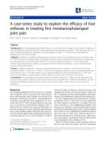
Normal and diabetic TPD for legs
Figures 2 and 3 show TPD of normal and diabetic subjects
for different areas of leg and foot from proximal to distal.
In normal subjects, the TPD value from the leg to big toe
decreases from 3 cm to 0.5 cm. But in diabetic subjects the
TPD value decreases from 3 cm to 2.5 cm. It suggests that
Vierodt's law of mobility holds well in the leg areas for the
normal subjects. The lower leg has less mobility than the
foot. The TPD of the lower leg is more than that of the
hind foot. The foot area i.e. hind foot, mid foot and fore-
foot have similar mobility monotonically increasing from
proximal to distal and their TPD is almost the same. How-
ever, the mobility of the big toe is more and its TPD is sig-
nificantly smaller than that of previous areas. This same
trend is observed in both left and right legs. Therefore, the
law of mobility doesn't hold for the DM subjects with sen-
sation as shown in figure 2. Figure 3 shows TPD values for
DM subject without sensation and the results are similar
to those with sensation.
Normal and diabetic TPD for hands
We tested the law of mobility in the upper extremities. Fig-
ures 4 and 5 show TPD of normal and diabetic subjects for
different areas of the hand from proximal to distal. It is
observed from the graph that TPD values decrease from
proximal to distal areas and that the law of mobility holds
for both normal and DM subjects with and without sensa-
tion in both right hand and left hand.
Discussion
As expected, the TPD of DM subjects is always higher than
that of normal subjects, at least in some areas of the foot.
Though the TPD values expressed here in all the above
graphs are mean of subjects with sensation and without
sensation, the law of mobility is not obeyed even if we
a Standard Division of foot areaFigure 1
a Standard Division of foot area. b Division of foot area for
our study.
Journal of Brachial Plexus and Peripheral Nerve Injury 2008, 3:3 />Page 4 of 6
(page number not for citation purposes)
evaluate individual DM subjects. This is true for both right
and left legs for all the subjects, except for one subject who
had sensation, whose left leg obeys the law of mobility as
shown in figure 6.
This observation in the foot areas with and without sensa-
tion is not true in the hands of DM subjects; the law of
mobility holds well in the hand of the DM subjects. As
Rendell [15] have stressed, the maldistribution between
nutritional and thermoregulatory skin blood flow is
observed in the toes but not the fingers of diabetic
patients. This could be directly related to the development
of ulcers in the feet but not in the hands.
Urbancic [5] describes the role of micro vascular dysfunc-
tion in the development of diabetic foot ulceration and
the differences between the left and right, and the lower
and upper extremities. In another study, Joseph [16]
describes the decline of tactile acuity in aging due to
blood-supply and other factors. Such micro vascular dys-
function could have resulted the differences in the TPD
between left and right, and the lower and upper extremi-
ties of the DM subjects. Measure of TPD and the law of
mobility in DM subjects could then easily reveal such
micro vascular dysfunction and its role in neuropathy.
TPD measurements therefore could give quantitative
measure of axonal loss in compression neuropathy.
One of the subjects had the TPD values merely increased
(from normal value) uniformly over all the areas in the
left leg, still satisfying the law of mobility, typical sign of
micro vascular dysfunction is shown in figure 7. Another
subject had the same TPD values as normal subjects except
in one-foot area, typical sign of neuropathic condition is
shown in figure 8. Although all the subjects were screened
for peripheral artery occlusive disease (PAOD) with the
ankle-brachial-pressure index (0.9 or above), the test does
not completely eliminate the subjects with micro vascular
dysfunction from the experiment. Most of the DM sub-
jects have neuro-ischaemia problems, simultaneously
with neuropathy and micro vascular dysfunction.
In this paper we have presented the law of mobility only
for TPD. It is well-established fact that point localization
has direct correlation with TPD, though not the same [17].
Similarly SWMF has direct correlation with the point
localization, as the later measures the area localisation
error and the SWMF measure pressure threshold in terms
of weight. It is possible that the law of mobility could be
used with point localisation and SWMF as well. The other
sensory measures such as vibration detection threshold
(VDT), cold detection threshold (CDT), warm detection
threshold (WDT), and heat pain onset threshold (HPO)
could be studied for the law of mobility, specifically in
DM subjects.
Table 1: Details of Diabetic Subjects
Diabetic subjects Age of Diabetic subjects Number of subjectswith callosity Duration of diabetes mellitus
Male Female Male Female
5 9 50 – 70 3 5–20 5 – 20
Comparison of Normal with Diabetic TPD – with sensation in Leg areasFigure 2
Comparison of Normal with Diabetic TPD – with sensation
in Leg areas. N → number of subjects.
Comparison of Normal with Diabetic TPD – without sensa-tion in Leg areasFigure 3
Comparison of Normal with Diabetic TPD – without sensa-
tion in Leg areas. N → number of subjects.
Journal of Brachial Plexus and Peripheral Nerve Injury 2008, 3:3 />Page 5 of 6
(page number not for citation purposes)
Conclusion
Although TPD measurement is subjective, it is a well-
established test to measure sensory loss in DM subjects.
This paper presents the first systematic study of the law of
mobility to assess the sensibility of diabetic subjects, com-
paring the law of mobility of TPD in upper and lower
extremities of DM patients. We observe that the law of
mobility does not hold well in DM patients.
The TPD data for diabetic patients reveals that the law of
mobility for diabetic patients does not hold in the foot
areas. The significance of this result is that the TPD of the
diabetic patients could provide direct and quantitative
measure of micro vascular dysfunction and its effect on
neuropathy. Though TPD has been accepted widely as a
measure of sensory loss, we did not find any particular dif-
ference in the applicability of the law for DM subjects with
and without sensation of 10 gm monofilaments.
We screened the subjects for peripheral artery occlusive
disease (PAOD) with the ankle-brachial-pressure index
(0.9 or above), but this test does not completely eliminate
the subjects with micro vascular dysfunction. However
more accurate techniques like laser doppler flow, transcu-
taneous oxygen saturation, skin temperature or any com-
bination of these techniques could better evaluate the use
of law of mobility for microvascular dysfunction.
The law of mobility shows the degree of axonal loss. The
law of mobility for TPD of diabetic subjects may provide
a simple, easy, cost effective clinical tool to evaluate com-
pression neuropathy in patients with or without neuropa-
thy as shown by SWMF and progressive axonal loss due to
microvascular dysfunction subsequent to tarsal tunnel
compression. If the sensory loss is due to microvascular
dysfunction because of compression neuropathy at the
tarsal tunnel, changes of law of mobility towards normal
Comparison of Normal with Diabetic TPD – with sensation in Hand areasFigure 4
Comparison of Normal with Diabetic TPD – with sensation
in Hand areas. N → number of subjects.
Comparison of Normal with Diabetic TPD – without sensa-tion in Hand areasFigure 5
Comparison of Normal with Diabetic TPD – without sensa-
tion in Hand areas. N → number of subjects.
Comparison of Normal with Diabetic TPD for single subject – with sensation in Left Leg areasFigure 6
Comparison of Normal with Diabetic TPD for single subject
– with sensation in Left Leg areas. N → number of subject.
Comparison of Normal with Diabetic TPD for single subject – with sensation in Left Leg areasFigure 7
Comparison of Normal with Diabetic TPD for single subject
– with sensation in Left Leg areas. N → number of subject.
Publish with BioMed Central and every
scientist can read your work free of charge
"BioMed Central will be the most significant development for
disseminating the results of biomedical research in our lifetime."
Sir Paul Nurse, Cancer Research UK
Your research papers will be:
available free of charge to the entire biomedical community
peer reviewed and published immediately upon acceptance
cited in PubMed and archived on PubMed Central
yours — you keep the copyright
Submit your manuscript here:
/>BioMedcentral
Journal of Brachial Plexus and Peripheral Nerve Injury 2008, 3:3 />Page 6 of 6
(page number not for citation purposes)
will indicate success of decompression of the nerve at the
site of compression.
Authors' contributions
All the authors had full access to all data in the study. VBN
carried out the clinical studies and participated in drafting
the manuscript. Both MM and PR conceived of the study
and participated in its design and coordination. All
authors read and approved the final manuscript.
Acknowledgements
We thank Ms. Richa Poddar and Mr. Suresh, physiotherapy department,
Sundaram medical foundation, Chennai, for their help in evaluating the
patients. We also thank Dr. Akila, epidemiologists, Sundaram medical foun-
dation, Chennai, for helping in statistical analysis.
References
1. Cavanagh PR, Simoneau GG, Ulbrecht JS: Ulceration, unsteadi-
ness, and uncertainty: the biomechanical consequences of
diabetes mellitus. J Biomechanics 1993, 26:23-40.
2. Delbridge L, Ctercteko G, Fowler C, Reeve TS, Lequesne LP: The
Aetiology of diabetic neuropathic ulceration of the foot. Brit-
ish Journal of Surgery 1985, 72:1-6.
3. Bell-Krotoski JS, Weinstein S, Weinstein C: Testing sensibility,
including touch-pressure, two point discrimination, point
localization and vibration. J Hand Ther 1993, 6:114.
4. Dellon AL: Computer-assisted sensibility evaluation and sur-
gical treatment of tarsal tunnel syndrome. Adv Podiatry 1996,
2:17-40.
5. Urbancic-Rovan V, Stefanovska A, Bernjak A, Katja Azman-Juvan,
Andreja Kocijancic: Skin blood flow in the upper and lower
extremities of diabetic patients with and without autonomic
neuropathy. J Vasc Res 2004, 41(6):535-45. Epub 2004 Oct 28
6. Vierodt KH: Abhängigkeit der Ausbildung des Raumsinnes der
Haut von der Beweglichkeit der Körpertheile. (Dependence
of the development of the skin's spatial sense on the flexibil-
ity of parts of the body). Zeitschrift für Biologie 1870, 6:53-72.
7. Weber EH: De pulsu, resorptione, auditu et tactu. In E.H.
Weber, on the tactile senses Edited by: Ross HE, Murray DJ. Hove (UK):
Taylor & Francis; 1834.
8. Wilska A: On the vibrational sensitivity in different regions of
the body surface. Acta Physiol Scand 1954, 31:284-289.
9. Weinstein S: Intensive and extensive aspects of tactile sensitiv-
ity as a function of body-part, sex and laterality. In The Skin
Senses Edited by: Kenshalo DR. Springfield, C.C. Thomas;
1968:195-218.
10. Ferreira Marcus Castro, Leandro Rodriguez, Klaus Fels: New
method for evaluation of cutaneous sensibility in diabetic
feet: preliminary report. Rev Hosp Clin Fac Med Sao Paulo 2004,
59():286-290.
11. Finnell JT, Knopp R, Johnson P, Holland PC, Schubert W:
A cali-
brated paper clip is a reliable measure of two-point discrim-
ination. Acad Emerg Med 2004, 11:710-714.
12. Dellon ES, Keller KM, Moratz V, Dellon AL: Validation of cutane-
ous pressure threshold measurements for the evaluation of
hand function. Ann Plast Surg 1997, 38:485-92.
13. Craig James C, Kisner Jayne M: Factors affecting tactile spatial
acuity. Journal Somatosensory and Motor Research 1998, 15(1):29-45.
14. Patil KM, Babu M, Oommen PK, Malaviya GN: On-line system of
measurement and analysis of standing and walking foot pres-
sures in normal and patients with neuropathic feet. Innov
Technol Biol Med, France 1996, 17(5):401-8.
15. Rendell M, Bergman T, O'Donnell G, Drobny E, Borgos J, Bonner RF:
Micro vascular blood flow, volume, and velocity measured by
laser Doppler techniques in IDDM. Diabetes 1989, 38:819-824.
16. Stevens JC, Alvarez-Reeves M, Dipietro L, Mack GW, Green BG:
Decline of tactile acuity in aging: a study of body site, blood
flow, and lifetime habits of smoking and physical activity.
Somato sensory and Motor Research 2003, 20(3 & 4):271-279.
17. Boring Edwin G: The two point limens and the error of locali-
zation. The American journal of physiology 1930, 42(3):446-449.
Comparison of Normal with Diabetic TPD for single subject – without sensation in Leg areasFigure 8
Comparison of Normal with Diabetic TPD for single subject
– without sensation in Leg areas. N → number of subject.