Volume 18 - Friction, Lubrication, and Wear Technology Part 18 potx
Bạn đang xem bản rút gọn của tài liệu. Xem và tải ngay bản đầy đủ của tài liệu tại đây (1.76 MB, 80 trang )
Unusual habits
2-body Saliva Tooth/restoration
Foreign body . . .
Prophylactic causes of wear
Toothbrush and dentifrice
3-body Water Tooth/restoration
Toothbrush Dentifrice
Prophylactic pastes
3-body Water Tooth/restoration
Polishing cup Pumice
Scaling and cleaning instruments
2-body Saliva Tooth/restoration
Instruments . . .
Cutting, finishing, polishing
Cutting burs/diamonds
2-body Water Tooth/restoration
Bur . . .
Finishing burs
2-body Water Tooth/restoration
Bur . . .
Polishing pastes
3-body Water Tooth/restoration
Polishing cup Abrasive slurry
Physiologic Wear
Physiologic wear, or attrition (Ref 5), is caused by processes involving sliding contact wear, contact wear (impact without
sliding), and noncontact wear from food abrasion alone. Sliding contact wear produces the most prominent effects during
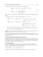
masticatory function. An example of severe attrition is shown in Fig. 1. Attrition occurs because of function (Ref 6) and
occurs only where the opposing teeth come into contact (Ref 7), so it can be distinguished from erosion (see the
discussion of "Pathologic Wear" given below).
Fig. 1 Severe occlusal wear resulting from dental attrition. Source: Ref 4
Attrition in primitive man was often severe (with pupal exposure) owing to the nature of the aboriginal diet of tough meat
and sandy, fibrous plants, as studies on both skeletal and living representatives of aboriginal populations have attested
(Ref 8, 9, 10).
Fundamental research on the degree and types of attrition found in the teeth of ancient, primitive, and modern populations
has dealt mainly with the wear planes produced on molars surfaces (Ref 11, 12, 13) and with the effects of attrition on
facial height (Ref 14, 15, 16, 17) and on the position of the temporomandibular joint (Ref 16, 18). It is apparent that not
all people wear on their teeth the same way. The patterns produced are frequently characteristic of ethnic variations.
Improved tooth care and dietary habits have lessened considerably the occurrence of attrition in modern populations. For
additional information on the effects of food, degree of function, and age on attrition, see Ref 19, 20, 21, 22, 23, 24, 25.
Pathologic Wear
This cause of wear can be particularly destructive to individual teeth or the entire dentition. Xerostomia and bruxism are
the most frequently reported pathologic causes of wear.
Xerostomia. This condition results in dryness of the oral cavity and causes brittleness of the teeth. It has been observed
in women during and after menopause (Ref 26). Investigation of causes of abnormal tooth wear must take salivary factors
into consideration (Ref 27, 28). Mucin-based saliva substitutes lubricate with values comparable to whole human saliva,
whereas substitutes based on carboxymethylcellulose do not appear to lubricate well (Ref 29).
Bruxism. This condition is a nonfunctional mandibular movement that is manifested by occasional or habitual grinding
or clenching of the teeth (Ref 30). The major effects of severe bruxism can be tooth wear and accelerated alveolar bone
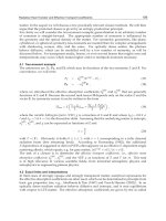
loss. An example of occlusal wear resulting from bruxism is shown in Fig. 2. This abnormal wear rapidly removes the
cusps of teeth. Wear takes place mainly on incisal edges of upper and lower anterior teeth. With time, edges become
highly polished and flattened. In the posterior teeth, wear appears as small saucer-like excavations.
Fig. 2 Occlusal wear resulting from bruxism. Source: Ref 4
Erosion. This condition is the chemical weakening of human enamel resulting from acid decalcification. The pH affects
the rate of decalcification for cleaned, and mechanically abraded enamel (Ref 31). Chemical decalcification may be
caused by environmental pollutants contaminating air and/or saliva of patients. Accelerated wear is observed in people
employed in occupations (for example, mining, or sulfuric acid production) where an unusual or severe atmospheric
environment exists (Ref 32).
Unusual Habits. People who grasp needles or nails with their teeth or smoke pipes may also exhibit localized
pathologic wear.
Prophylactic Wear
Toothbrush and Dentifrice. Oral hygiene is necessary for maintaining a healthy mouth and for social acceptance.
The emphasis in the study of wear of the dentition by toothbrush and dentifrice has been the elimination of overly
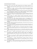
abrasive dentifrice components. Cervical abrasion resulting from improper and excessive toothbrush and dentifrice use is
shown in Fig. 3.
Fig. 3 Cervical abrasion resulting from excessive toothbrush and dentifrice use. Source: Ref 4
The primary function of a dentifrice is to clean and polish the surfaces of the teeth accessible with a toothbrush. During
cleaning, extraneous debris and deposits need to be removed from the tooth surface. These deposits listed in order of
increasing difficulty of removal are (Ref 33): food debris, plaque (a soft, mainly bacterial film), acquired pellicle (a
proteinaceous film of salivary origin), and calculus.
The ideal abrasive should exhibit a maximum cleaning efficiency with minimum tooth abrasion. In addition, a dentifrice
should polish the teeth. Highly polished teeth are not only aesthetically desirable, but they may also be less receptive to
the retention of deposits (Ref 34).
Typical dentifrice abrasives include: calcium carbonate, dibasic calcium phosphate dihydrate, anhydrous dibasic calcium
phosphate, tricalcium phosphate, calcium sulfate, calcium pyrophosphate, insoluble sodium metaphosphate, and hydrated
alumina (Ref 35).
Selection of a dentifrice by a dentist for a patient should be based on: (1) degree of staining, (2) force exerted on the
brush, (3) method of brushing, and (4) amount of exposed dentin and cementum. The Council on Dental Therapeutics of
the American Dental Association published information on the abrasivity of dentifrices in 1970 (Ref 36).
Prophylactic Paste. A dental prophylactic paste should be sufficiently abrasive to remove effectively exogenous
stains, pellicle, materia alba, and oral debris from the tooth surface without causing undue abrasion to the enamel, dentin,
or cementum. Polymeric materials, such as denture base and artificial tooth resins, composite restorations, and pit and
fissure sealants, are particularly susceptible to abrasion because of their low hardness. The undesirable results of wear can
be a reduction in anatomic contours and increased surface roughness.
Abrasives in commercial prophylactic pastes include: recrystallized kaolinite, silicon dioxide, calcined magnesium
silicate, diatomaceous silicon dioxide, pumice, sodium-potassium-aluminum silicate, and zirconium silicate (Ref 37).
Cutting, Finishing, and Polishing Wear
Tooth structure and restorative dental materials are routinely reshaped and smoothened using special instruments for
cutting and finishing. A highly polished surface is then produced by treatment with polishing pastes containing alumina or
diamond abrasive particles less than 1 m in size.
Wear Studies
Traditional wear theory divides observed wear into categories of adhesive, abrasive, corrosive, and fatigue wear;
however, predictions of these wear models depend on the materials behaving in a relatively brittle fashion. Most dental
materials under intraoral circumstances do not behave in this way; therefore, it is difficult to rank dental materials
performance. Most wear tests have not faithfully predicted clinical performance.
Wear information on dental materials has been collected from fundamental studies with simple laboratory tests,
simulation studies with customized machines, and clinical studies. Unfortunately, the fundamental laboratory tests and the
simulation studies have not had much success in correlating with observed clinical wear.
Fundamental Laboratory Studies
In a single-pass sliding technique, fluorapatite single crystals served as a simple model system for human enamel, which
is composed of hydroxyapatite. The wear and friction of fluorapatite single crystals under conditions of single- and
multiple-pass sliding with a diamond hemisphere (360 m in diameter) can be evaluated by interpretation of tangential
force, track width, and surface failure classification data (Ref 38, 39, 40). A failure classification scale (Fig. 4) includes:
• Class 1: entirely ductile
• Class 2: mostly ductile with some tensile cracking
• Class 3: essentially tensile cracking
• Class 4: mostly tensile cracking with chevrons (chipping)
• Class 5: chevrons
Examples of Classes 1, 3, and 5 are shown in Fig. 5.
Fig. 4 Failure classification scale. Source: Ref 40
Fig. 5 Examples of surface failure of fluorapatite single crystals.
(a) Class 1. (b) Class 3. (c) Class 5. Source:
Ref 40
The failure of fluorapatite at a 0.1 N load for single-pass sliding in the <2110> direction is essentially ductile and
progresses toward brittle failure as the load is increased (Ref 41). At 0.5 N loads and higher, failure is characterized by
chevron formation. Track width follows an exponential function, whereas the tangential force (friction) increases linearly
with normal load (Ref 41). The coefficient of friction is not a perfect indicator of wear. The track width data indicate that
the principal mechanisms for the accommodation of strain are elastic deformation and cracking. Sliding in the <0110>
direction results in slightly lower friction but substantially increased surface damage (Table 4).
Table 4 Influence of sliding direction and environment on friction and wear properties of fluorapatite single
crystals for single-pass sliding on the basal plane
Condition Coefficient of
friction ( )
Ductile-to-brittle
transition load, N
<2110> direction in air
0.22 0.75
<0110> direction in air
0.19 0.15
<2110> direction in water
0.24 0.18
<2110> direction in dimethylformamide
0.24 0.62
Source: Ref 41, 42
The ductile-to-brittle transition for sliding of diamond on fluorapatite occurs at higher normal loads in air and
dimethylformamide than in water (Table 4) (Ref 42). The lowering of the transition in water is explained on the basis of
surface hardening as a result of the interaction of polar water molecules or their dissociation products and charged near-
surface species. The interaction results in pinning of dislocations and a reduction in the ability of the lattice to
accommodate strain by slip and thus effectively lowers the stress required to cause fracture.
Single-pass sliding by itself cannot completely describe the wear of fluorapatite single crystals (Ref 43). Wear tracks for a
single and double pass on the same track in the opposite direction under a 0.7 N load are shown in Fig. 6. The effect of
sliding a second pass across a wear track is shown in Fig. 7. When both tracks are made under a 0.5 N load, catastrophic
failure occurs at the intersection of the tracks. Considerable care is necessary in extrapolating single-pass wear data to
repetitive wear measurements.
Fig. 6
Wear tracks on a fluorapatite single crystal for a single and double pass on the same track in the
opposite direction under a 0.7 N load. Source: Ref 43
Fig. 7 Intersecting wear tracks on a fluorapatite single crystal under a 0.5 N load. Source: Ref 43
The frictional behavior and surface failure of human enamel has been studied by sliding with a diamond hemisphere (360
m in diameter) in water (Ref 44, 45). Similar tests have been carried out on a sintered hydroxyapatite ceramic, which
approximates the properties of human enamel (Ref 46). Table 5 compares the properties of these materials.
Table 5 Properties of a sintered hydroxyapatite ceramic compared with human enamel
Property HAP-60K-1200C
Human enamel
Compressive strength, MPa (ksi)
380 (55) 400 (58)
Young's modulus, GPa (psi × 10
6
)
120 (17.4) 80 (11.6)
Knoop hardness, kg/mm
2
450 340
Density, g/cm
3
3.1 2.96
Coefficient of friction
0.24 0.36
Linear coefficient of thermal expansion, 10
-6
/ °C
9.2-11.8 11.4
Source: Ref 46
Simulation Studies
Chewing Machines. An early attempt to study attrition used a machine capable of simulating the actions of the human
mandible during chewing to produce a variety of wear patterns (Ref 47). Much more sophisticated chewing machines
have been developed to examine dental composite wear and are discussed later.
Brushing Machines (Dentifrices). Three main methods used to determine the loss of hard tooth tissue from
brushing are: (1) measuring the amount of tooth tissue abraded from an irradiated tooth by the concentration of
radioactive phosphorus in the wear debris (Ref 36, 48, 49, 50, 51), (2) determining the change in profile of samples (Ref
52), and (3) measuring the change in reflectance of the surface of tooth structure (Ref 53).
Dentifrices used in 1942 were compared to a calcium carbonate standard (Ref 54, 55). Abrasion was found to occur 25
times faster on dentin and 35 times faster on cementum than on the enamel tips of cusps. Abrasive power (percent
abrasion of a calcium carbonate control) increases linearly with particle size (Ref 56, 57). On the other hand, the polishing
ratio of zirconium silicate increases with decreasing particle size, and particle size distribution is important (Ref 53).
Increasing the load on the brush also increases enamel and dentin abrasion scores (Ref 50, 58).
The radiotracer method can measure wear rates of dental tissues by as few as one or two strokes of a brush on dentin with
commercial dentifrices. Use of this method has led to the observation that wear of dental tissues is proportional to
penetration hardness if hard abrasives are used (Ref 49).
Brushing Machines (Toothbrush). A number of studies have attempted to determine the influence of the toothbrush
and its variables on the wear of dental tissues. Plastic toothbrush bristles have little abrasive or polishing power (Ref 59,
60, 61, 62). Other studies have compared automatic versus hand toothbrushes (Ref 63, 64, 65). In general, the mechanical
toothbrushes produce less abrasion of hard tooth tissue than simulated manual brushing, but the forces associated with
manual brushing are usually larger.
Many variables of toothbrushes and dentifrices have been examined with brushing machines. (These include (Ref 4):
• Dentifrice properties: hardness, particle size, and particle distribution of
the abrasive, and composition
and concentration of remaining components
• Toothbrush properties: geometry, hardness, stiffness, and number of bristles
• Substrate properties: orientation, hardness, surface preparation
• Testing conditions: brush load, stroke length, stroke rate, number of strokes, and presence of saliva
Machines for Prophylaxis. Products containing quartz and pumice show higher cleansing values but generally result
in greater abrasion to enamel and dentin (Ref 66). Abrasion of dentin by a pumice slurry is about 20 times greater than
abrasion of enamel under standardized conditions (Ref 67). Increases in treatment time, load and cup speed cause linear
increases in abrasion of irradiated human enamel and dentin (Ref 68). Commercial products containing calcined
magnesium silicate and sodium-potassium-aluminum silicate show best polishing with low abrasivity (Ref 37). No
clinical study has yet correlated the degree of abrasivity with any destructive effect on hard and soft tissues. American
Dental Association Specification No. 37 includes a suggested abrasivity test that uses a radiotracer technique (Ref 69).
Cutting Machines. Cutting of human enamel by a high-speed diamond stone is enhanced by use of a solution of
glycerol, ethanol, and water (2:1:2) when compared to cutting with water alone (Ref 70). Such a chemomechanical effect
is also observed in the cutting of amalgam with diamonds and carbide burs and the cutting of composites with carbide
burs. Cutting of hydroxyapatite blocks with autoclaved tungsten carbide burs is enhanced if the burs are dipped in sodium
nitrate or commercial anticorrosive dips before autoclaving (Ref 71).
Clinical Studies
Clinical studies collect physiologic wear data produced by: (1) sliding contacts or direct contact alone (occlusal-contact-
area wear) and (2) noncontact wear (contact-free wear), which is related to food abrasion. To effectively examine these
events, pathologic and prophylactic wear must be absent or controlled. In addition to the restorative treatment variables in
clinical studies, there are also intraoral variables and patient factors that complicate clinical results and interpretations.
Occlusal-contact-area wear of human enamel has been measured with a computerized three-dimensional measuring
technique on tooth replicas over a period of four years (Ref 72). The steady-state wear rates at enamel occlusal contact
areas are about 29 m/year for molars and about 15 m/year for premolars. These data agree with earlier reports of 33
m/year (Ref 73) and 41 m/year (Ref 74).
Observations of orthodontic patients with arrested carious lesions indicate that functional wear and toothbrushing are
responsible for the arrestment by disturbance and removal of bacterial deposits (Ref 75). Changes in surface enamel
morphology after acid etching are also the result of abrasion rather than the precipitation of mineral from saliva (Ref 62).
Enamel undergoes physiologic wear but is routinely redeposited by nucleation and growth of new hydroxyapatite from
the calcium phosphate present in saliva. This process helps to compensate for losses that occur during physiologic,
prophylactic, and polishing wear. Dentifrice abrasivity of enamel as measured in vivo by a cellulose acetate replication
technique is much lower than abrasion caused by pumice or zirconium silicate (Ref 76).
Dental Amalgam
Dental amalgam is an alloy that results when mercury is mixed with an alloy containing silver, tin, copper, and sometimes
zinc (Table 1). Before it hardens, the freshly mixed mass of amalgam can be packed into a cavity prepared in a tooth.
Amalgams are usually limited to replacement of tooth issue in the posterior teeth and often function in stress-bearing
areas susceptible to occlusal wear. Some properties of dental amalgams are listed in Table 2.
Fundamental Laboratory Studies
Abrasion Tests. Two-body abrasion of dental amalgam has been measured using a Taber abrader (Ref 77), a silicon
carbide two-body abrasion test (Ref 78), and a pin-on-disk test (Ref 79). With the Taber abrader, smearing of amalgam
and clogging of the abrader wheel cause inconsistent ranking with clinical observations; clogging of the abrasive surface
is avoided by abrading at a low load over a fresh abrasive surface on each pass. Ranking of amalgam and composite
restorative materials with two-body abrasion tests is in better agreement with clinical observations than that done with
three-body abrasion tests. Two-body abrasion test result on some typical dental amalgam alloys are given in Table 6. As
indicated by these results, a dispersed high-copper amalgam (Dispersalloy) exhibited better resistance to silicon carbide
two-body abrasion than spherical low-copper amalgams.
Table 6 Material loss on abrasion of dental amalgams
Material loss, 10
-4
mm
3
/
mm of travel
(a)
Material
24 h 1 month
Spher-a-Caps
7.0 7.0
New True Dentalloy
6.5 6.3
Dispersalloy
5.6 4.9
(a)
Load: 0.17 MPa.
The pin-on-disk test (Ref 79) utilizes a cylindrical sample of enamel to rub on a rotating disk of amalgam. Measurements
of wear rate are possible, but transfer of material from the disk to the pin confuses interpretation of the results.
Single-Pass Sliding. The wear of dental amalgam also has been studied by single- and double-pass sliding with a
diamond hemisphere (360 m in diameter) (Ref 80). The dispersed high-copper amalgam has the lowest values of
tangential force and track width. The mode of surface failure under single- and double-pass sliding is ductile with no
evidence of subsurface failure. Smearing of phases for both spherical and dispersed amalgams during sliding does occur,
but the dispersed amalgam is more resistant to smearing. Cracks that occur at higher loads propagate around the stronger
phases. Wear is determined by resistance to penetration and by a ductile mode of surface failure over the load range
tested.
Friction of dental amalgam is altered when any transfer of material from one member of the pair to the other member
occurs (Ref 81). For example, when gold or dental composite slide against amalgam, amalgam material is transferred to
the gold or composite surface and the friction then becomes that of amalgam on amalgam.
Fracture toughness, critical strain energy release rate, and critical stress intensity factor have been determined for
several types of dental amalgams (Ref 82). Data are consistent with surface failure observed in single-pass wear studies.
An equation developed from single-pass studies (Ref 83) has been derived that relates the sliding frictional force (F) to
normal load (N), fracture toughness, modulus of elasticity, yield strength, and slider diameter in the form of F = KN
n
(Table 7). The observed friction is caused primarily by plowing or deformation during single-pass sliding.
Table 7 Friction of dental materials as described by various properties including fracture toughness
Modulus of elasticity
Yield strength
Material
GPa psi × 10
6
MPa ksi
Fracture
toughness,
J/m
2
n K
Amalgams
Spherical, low-copper
12.9 1.87 142 20.6 117 1.17
0.211
Lathe-cut, low-copper
12.8 1.86 119 17.3 247 1.31
0.146
Admixed, high-copper
17.7 2.57 130 18.9 104 1.25
0.124
Composite 7.8 1.13 135 19.6 182 1.23
0.190
Unfilled resin
Acrylic
2.0 0.29 104 15.1 382 1.54
0.141
Diacrylate
1.8 0.26 65 9.4 402 1.19
0.338
Source: Ref 83
Simulation Studies
Amalgam abrasion from dentifrices in brushing machines has been studied by profilometry and laser reflection
techniques, which both indicate that amalgam wears about 10 times more rapidly than gold alloys under the same
conditions of simulated brushing with normal dentifrices (Ref 84).
Clinical Studies
The physiologic wear rates of amalgam are 6 to 15 m/year in contact-free areas and 28 to 58 m/year in occlusal
contact areas (Ref 85). This wear may be compensated by continual amalgam expansion that produces occlusal extrusion.
Therefore, the rate of attrition of amalgam is usually not considered to be a clinical problem.
Amalgam degrades at the tooth-restoration interface by a process called marginal fracture, which is the result of
electrochemical corrosion in the presence of direct-contact stresses (Ref 86). Direct-contact areas on amalgams that are
not stabilized by contacts on enamel may also produce noticeable facets.
Composite Restorative Materials
Composite restorative materials consist of a cross-linked polymer matrix that is chemically bonded by coupling agents to
the surfaces of dispersed silica-based filler particles (Table 1). Composite restorations have the appearance of natural
tooth tissue and can be placed directly into a cavity preparation for in situ hardening. They are recommended for
restorations where occlusal stress is minimal and appearance is crucial. Composites are also available for limited posterior
use in areas of occlusal stress, but they are less durable than amalgam (Ref 87). Some properties are listed in Table 2.
Wear resistance of composite restorations is important for clinical longevity, esthetics, and resistance to dental plaque.
The need for markedly improved wear resistance has been emphasized by recent literature reviews (Ref 88, 89, 90, 91).
The reason that wear-resistant composites have not been developed is primarily because of the lack of understanding of
the mechanisms of clinical wear. This problem is confounded by the lack of reliable and consistent clinical wear data.
Fundamental Laboratory Studies
The problem with all wear theories has been the lack of correlation of clinical wear results with laboratory properties.
Mechanical properties do correlate with filler types and loading levels (Ref 92, 93, 94, 95, 96, 97), but they do not predict
clinical performance. To bridge the gap, a wide range of tests have been performed. Some of these are described below.
Abrasion Tests. Taber abrasion (Ref 81), single-pass sliding on abrasive papers (Ref 98), pin and plate (Ref 99), pin
and disk (Ref 100), metallographic polishing (Ref 101), toothbrushing (Ref 102), and oscillating wear (Ref 103) tests
have shown very little or on success in explaining intraoral wear. With the Taber abrader, smearing of resin and clogging
of the abrader wheel produced apparently better resistance for unfilled resins than for composites; these results are
inconsistent in ranking with clinical observations.
A silicon carbide two-body abrasion test was developed to avoid clogging effects (Ref 78). This test provided some
agreement with early clinical studies, but its primary usefulness was to examine the relative effects of different
formulation variables.
Improvements in the durability of composites depend on a thorough understanding of the wear bahavior of the resin
matrix (Ref 104). The resin with the lowest value of two-body abrasion also showed the lowest coefficient of friction and
the most ductile behavior during single-pass sliding. (Table 8). Abrasion resistance was improved by finishing the cured
surface before testing (Ref 105). Reduced wear of large-particle composites was related to the increased size (relative to
the abrasive), increased hardness, and increased volume fraction of the filler particles (Ref 106, 107).
Table 8 Material loss on abrasion and single-pass wear characteristics of dimethacrylate resins
Material Material loss,
10
-4
mm
3
/mm
of travel
(a)
Coefficient
of friction
( )
Ductile-to-
brittle
transition
load, N
Tetraethylene glycoldimeth-acrylate
22.0 0.45 7.0
Bisphenol A-bis ethylmeth-acrylate
17.7 0.61 5.0
Bisphenol A-bis (2-hydroxypropyl)methacrylate + ethylene glycol-dimethacrylate (1:1)
15.5 0.35 >10
Bisphenol A-bis ethylmeth-acrylate + octafluoro-1-pentyl-methacrylate (9:3)
19.1 0.61 4.0
Bisphenol A-bis ethylmeth-acrylate + octafluoro-1-pentyl-methacrylate (3:9)
32.2 1.48 3.0
Source: Ref 104
(a)
Load: 0.18 MPa.
Pin-on-disk testers were popular at least in part become subsurface damage detected during microdefect analysis of a few
composites recovered from in vivo service was apparently the same as the structure observed in composites subjected to
pin-on-disk wear using stainless steel sliders (Ref 108).
Two-body pin-on-disk abrasion of composite veneering resins was shown to be much higher for rubbing contact against
porcelain versus enamel, gold, or the same veneering resin (Ref 109). Combinations of composite with composite, silver-
reinforced glass ionomer, or porcelain wore excessively (Ref 110).
The enthusiasm for abrasion testing produced a large amount of data, but actual clinical wear mechanisms often proved to
be quite different.
Single-Pass Sliding. The wear of composites also has been studied by single- and double-pass sliding with a diamond
hemisphere (360 m in diameter) (Ref 111, 112). The surface failure for unfilled diacrylate resins was more severe than
that observed for an unfilled acrylic resin. Addition of nonsilanated filler to the diacrylate resin increased resistance to
penetration but did not dramatically change the mode of failure (Fig. 8). Diacrylate resins that contained silanated filler
(commercial composites) were ductile in failure during sliding and showed the higher resistance to penetration. Damage
was more severe for double-pass than for single-pass sliding. Single-pass data did not, however, provide an estimate of
volume lost on repetitive wear of composites.
Fig. 8 Scanning electron micrographs of double-
pass wear scars made under a normal load of 7 N of (A)
unfilled diacrylate resin, (B) unfilled acrylic, (C) diacrylate res
in with silanated filler, (D) diacrylate resin with
nonsilanated filler. Source: Ref 112
Aging and Chemical Softening. The surface degradation of composites caused by accelerated aging in a weathering
chamber was characterized by erosion of the resin matrices and exposure of filler particles (Ref 113). Differences in
surface roughness and profile indicated that the aged composites were eroded at different rates. Surface crazing (Fig. 9)
was observed for some aged composites. Surface degradation also resulted in color changes, particularly for large-particle
composites but much less so for microfilled composites (Ref 114).
Fig. 9 Scanning electron micrographs of composites aged for 900 h showing surface crazing. Source: Ref 113
Single-pass sliding wear of aged, chemically cured, large-particle composites resulted in smaller track widths, lower
tangential forces, easier dislodging of material, and more severe surface failure at lower normal loads than that of unaged
composites (Ref 115). Ductile-to-brittle transitions occurred at lower normal loads (Table 9), and changes in morphology
became more severe (Fig. 10) with increased aging (Ref 116).
Table 9 Influence of increased aging on single-pass wear transitions of composites under conditions of accelerated aging
Ductile-to-brittle transition load, N
Material
Unaged 300 h 900 h
Large-particle composite
Concise
2.8 1.8 1.5
Microfilled composite
Finesse
8.0 3.3 3.0
Isopast
4.3 4.3 4.3
Silar
7.0 6.0 2.0
Source: Ref 116
Fig. 10 Scanning electron micrographs of surface morphology of two large-
particle composites at (a) 0, (b)
300, (c) 600, and (d) 900 h. Source: Ref 116
Preconditioning composites in food-simulating liquids (heptane and several ethanol/water solutions) has been shown to
decrease the hardness with a corresponding increase in pin-on-disk wear (Ref 117). Swelling of the polymer matrix and
surface damage occurs during preconditioning. Increasing the degree of cure of the matrix polymer may inhibit diffusion
of penetrants, and additional cross-linking may reduce swelling and damage by solvents. Evidence of erosion has also
been observed under in vivo conditions (Ref 118).
Fracture toughness, critical strain energy release rate (G
Ic
), and critical stress intensity factor (K
Ic
) have been
determined for experimental and commercial composites (Ref 119). The commercial composite was less resistant to crack
initiation and had a higher K
Ic
than its unfilled diacrylate resin. Data were consistent with surface failure observed in
single-pass wear studies of these resins.
Correlating Abrasion with Hardness and Tensile Data. Hardness and tensile strength of large-particle
composites were not related to measured in vitro abrasion rates (Ref 120). However, clinical abrasion of microfilled
composites and unfilled resins appeared related to indentation hardness (Ref 121).
Simulation Studies
Most simulation studies evaluate physiological wear. Few studies have dealt with prophylactic wear. Microfilled
composites subjected to brushing abrasion with a dentifrice wore 5 to 10 times faster than large-particle composites (Ref
122). Abrasion slowed once the resin-rich layer of a composite was lost or when the composite was postcured by heat.
A limited study was reported in which composites were abraded by an artificial food bolus (Ref 123). Composites were
placed along the edge of a rotating disk immersed in a millet seed suspension and ground against a stainless steel wheel.
Although not perfect, the ranking of composites agreed with clinical studies.
Chewing machines have been developed to simulate the full range of intraoral wear events (Ref 124). To date, the results
have only shown a limited agreement with clinical studies.
Clinical Studies
Many direct clinical evaluations of wear rely on the United States Public Health Service (USPHS) criteria for classifying
the loss of anatomical form of posterior restorations (Ref 125). Unfortunately, the USPHS criteria alone are not sufficient
to detect early loss of material in posterior composites (Ref 126, 127, 128, 129, 130, 131). Because of the crudeness of the
USPHS scale, there was considerable impetus in the early 1980s to develop more precise indirect measurement
techniques.
The first indirect method to become popular was designated the Leinfelder technique. It measured the loss of material
using a stone cast poured from a clinical impression of the restoration to compare it to a series of calibrated stone casts
(Ref 126, 130, 132). Other similar indirect cast methods have tired to refine the Leinfelder approach (Ref 133, 134, 135).
Unfortunately, because of operational differences, these scales have been shown to underestimate actual wear by 50%.
The major advantage of indirect methods employing stone casts and evaluators is their inexpensive nature. All other
methods, particularly computer digitization, are remarkably costly and thus impractical for even small clinical studies.
Electroplated or epoxy resin replicas of restorations have been prepared from impressions and studied by optical,
scanning electron, or reflex microscopy; by Moiré techniques; or with computer digitization (Ref 86, 136). Wear has also
been measured by monitoring the recession of resin on the surfaces of prominent filler particles by scanning electron
microscopy of sequences of impressions of a restoration (Ref 137).
A less-complicated method of clinically evaluating restorations is to place them into denture teeth in a removable denture
(Ref 138, 139, 140, 141, 142). The dentures permit direct measurements of the samples by profilometry or three-
dimensional computerized surface digitization.
Because of the large number of uncontrollable clinical variables involved in these studies, there is a broad distribution of
results at each recall. The results are different for molars and premolars. Differences in operators and techniques among
clinical studies are often so great that the results are not comparable at all. The reported results are often misrepresented
as wear rates rather than total wear. Wear does not occur in a linear fashion.
Published wear rates for composites are 12 to 79 m/year in contact-free areas and 39 to 135 m/year in occlusal contact
areas (Ref 85). For autocured (Ref 143) and ultraviolet light-polymerized (Ref 144, 145) composites, wear has been
shown to occur at decreasing rates during the first three years (Fig. 11). This same pattern of decreasing wear has now
been reported for several newer visible light cured composites (Ref 146, 147, 148). For one composite, about 74% of the
total wear over a three-year period occurred during the first year, followed by 19% in the second year, and 6% in the third
year. This pattern is less obvious if the first six months of data are excluded. The wear rate for the next few years may
appear almost constant (Ref 149). The rate reported for a few microfilled composites appeared to be nearly linear (Ref 85,
150), but early wear may have been hidden by beveling of the restoration margins. Recently, wear rates for posterior
composites have been reported to drop to very low values after 5 to 8 years (Ref 145, 147, 148). Because of this overall
decreasing rate of wear, it is hazardous to project long-term wear rates from any short period of time.
Fig. 11 Decreasing clinical wear rate with time. Source: Ref 148
Wear of posterior conventional composites involves exfoliation of the filler particles as the resin matrix is continually
worn away (Ref 147, 151). This process gives the appearance of a restoration submerging below the surface (Fig. 12).
The wear is reduced in the hybrid composites by reducing the mean filler particle size from a range of 30 to 50 m to a
range of 3 to 5 m and by increasing the amount of filler from 75 to 86 wt% (Ref 128). The use of softer barium glass
(HK 400) to replace quartz filler (HK 600) in the particle size range of 10 to 20 m also results in better wear resistance,
although the glass particles themselves show evidence of wear.
Fig. 12 Scanning electron micrograph of a composite restoration (Concise)
with severe occlusal wear after a
seven-year period. 10×. Source: Ref 188
Other factors leading to breakdown include the degradation of the silane coupling agent, which can cause microcracking
of the resin (Ref 152) and the large mismatch in coefficient of thermal expansion between filler and resin (Ref 86, 118).
Maximizing filler particle-to-particle contacts does not improve wear resistance (Ref 153). Resistance to wear is higher
for unfinished composites than for those finished conventionally with a finishing bur and white stone, suggesting that
finishing at least initially mechanically weakens the surface (Ref 154).
Another problem for clinical research is that different evaluation systems record different wear events. For the most part,
the indirect method relies on relief occurring at margins to create contrasts that can be compared to standards. This
method does not measure wear elsewhere on the occlusal surface. It is not clear where on the restoration direct clinical
evaluation measures wear, but it seems most likely to be at the marginal areas. Profiling methods measure the general
occlusal surface very well but are least accurate at the margins (Ref 155, 156).
A further complication of clinical research studies is that there has always been the wide distribution of wear values for
individual restorations at any recall period. After three years, the wear ratings for composites vary from 0 to 350 m (Ref
148).
In an attempt to understand this variation, the factors contributing to intraoral wear have been collected into categories for
statistical analysis (Ref 157). The categories include factors involving cavity preparation, restoration, manipulation,
intraoral location, and the patient. Although not all of these factors are yet well known, it has been possible to identify the
major ones.
Intraoral location is the most important factor. First molars wear more than second molars, second premolars, and first
premolars, in that order. Restoration width is also important. There are minor effects from arch, gender, and complexity of
the restoration. Differences in formulations of materials have secondary effects (Ref 158). Understanding these effects, it
is possible to normalize differences among clinical studies to pool the clinical data for posterior composites and correct
these variations to an ideal clinical population using Weibull analysis (Ref 159).
Numerous theories of wear have been proposed for dental composites based on their microstructures and resistance to
microstructure. The analysis typically is presented in terms of the filler, coupling agent, or resin matrix being the weakest
link (Fig. 13). The percent conversion of monomer to polymer during curing (Ref 160, 161, 162, 163, 164, 165, 166,
167), the depth of curing (Ref 168, 169, 170, 171, 172, 173, 174), the glass transition temperature of the polymer (Ref
175, 176), and the strength of the polymer (Ref 177, 178, 179, 180, 181, 182) have all been proposed as reasons that the
matrix might be most susceptible to microfracturing and thus wear. Problems with coupling agent effectiveness and
hydrolysis have been proposed as possible weak links (Ref 152). It has been hypothesized that the surfaces of composites
absorb chemicals from food and become predisposed to microfracture, thus permitting wear (Ref 108, 117). Different
wear mechanisms have been theorized for different composite types (Ref 149). None of these mechanisms has been
established, and none of the explanations is predictive.
Fig. 13 Schematic of worn composite restoration indicating possible weak links
A key observation was made by examination of local patterns of wear on composite surfaces (Ref 121, 138). Wear of
resin seemed to occur only when separation of filler particles was greater than approximately 0.1 m. It was proposed
that abrasion was caused principally by abrasive particles in the food bolus and that the smallest effective particles were
larger than 0.1 to 0.2 m. Therefore, composites with interparticle spacings less than that dimension were
microscopically protected against wear. No information has been available about the size, amount, or abrasivity of the
wear-producing particles in food, but they are assumed to be significantly harder than the matrix, softer than the filler, and
larger than 0.1 to 0.2 m. For microfills and many hybrids containing 20 vol% of 0.4 m microfiller particles, the
computed average filler particle separation is <0.2 m. This theory readily explains the apparent resistance to wear of
those composites in vivo. Theoretical calculations of microfiller levels required for microprotection indicate that only a
few percent are required. However, assuming that microfiller tends to be agglomerated, levels of 45 to 50% are necessary.
An extension of this argument has been that macroscopic protection would also exist for small cavity preparations.
Increased exposure of the preparation walls shelters the adjacent composite and causes a decrease in the clinical wear rate.
Most of the observed wear patterns and decreasing wear rates appear to be explainable on the basis of these two
protection mechanisms alone (Fig. 14).
Fig. 14 Schematic of (a) micro- and (b) macroprotection theory. See text for details.
On this basis, most modern wear-resistant composites include high filler levels with sufficient microfiller to reduce the
interparticle spacings to relatively low values. Improvements in coupling agents, strength of the resin matrix, and
hydrophobicity may contribute to wear resistance as well.
Pit and Fissure Sealants
Pit and fissure sealant consist mainly of a polymer matrix with minor amounts of dispersed filler particles, primarily for
coloration (Table 1). Sealants are designed to be more fluid than composites so they will penetrate the pits, fissures, and
etched areas of enamel to produce macroscopic and microscopic mechanical retention. The purpose of sealants is to
penetrate all cracks, pits, and fissures on the occlusal surfaces of deciduous or permanent teeth at risk to dental caries, to
seal these areas, and to provide effective protection against caries-producing bacteria. Some properties of sealants are
listed in Table 2.
Fundamental Laboratory Studies
Two-body abrasion of sealants has been measured using a silicon carbide two-body abrasion test (Ref 183). Weight-loss
data range from 22 × 10
-4
to 24 × 10
-4
mm
3
/mm of travel, which is characteristic of wear of diacrylate resins with little or
no filler. The addition of 40 wt% quartz to a sealer did not affect its resistance to two-body abrasion.
The wear of sealants also has been studied by single-pass sliding with a diamond hemisphere (360 m in diameter) (Ref
183). The sealant with 40 wt% quartz was more resistant to penetration and showed less surface damage from single-pass
sliding than two unfilled diacrylate sealants.
Clinical Studies
Direct clinical observations of 205 restorations after 4 years has shown that physiologic wear of sealants in the non-stress
bearing areas of pits and fissures is only slight (7%) (Ref 184). Wear on other occlusal surface areas where sealant may be
applied inadvertently is apparently irrelevant, because these areas are naturally self-cleansing by abrasion from food.
Therefore, the wear of sealants in that regard is clinically unimportant.
Dental Cements
Various modified zinc oxide-eugenol and zinc polyacrylate dental cements are used for temporary fillings (Table 1).
Glass ionomers and silver-reinforced glass ionomers may be used for certain permanent resolutions not subject to high
occlusal forces. Representative mechanical properties of cements are listed in Table 2.
Table 10 Material loss on abrasion of dental cements used as temporary filling materials
Material Material loss,
10
-4
mm
3
/mm of travel
(a)
Zinc oxide-eugenol cements
Tem Pak
29.1
B and T
22.9
IRM
18.6
EBAC
15.8
Zinc polyacrylate cements
PCA Cement
5.0
Durelon
4.9
Source: Ref 185
(a)
Load: 0.036 MPa.
Fundamental Laboratory Studies
Two-body abrasion of temporary filling materials has been measured using a silicon carbide two-body abrasion test (Ref
185). The materials are ranked in an order that agrees with clinical observations. Two zinc polyacrylate cements (base
consistency) had rates of abrasion much lower than modified zinc oxide-eugenol cements (Table 10).
The two-body abrasion of early glass ionomer cements used for Class V restorations is similar to that of modified
composites (Table 11) (Ref 186). Pin-on-disk wear of silver-reinforced glass ionomer restorative materials is less than
that for conventional glass ionomers (Ref 187). Incorporation of silver appears to provide lubrication; however, the
incidence of catastrophic failure during sliding is only reduced slightly.
Table 11 Material loss on abrasion of Class V restorative materials
Material Material loss,
10
-4
mm
3
/mm of travel
(a)
Glass ionomer cements
ASPA
22.9
Fuji
17.0
Composites
Cervident
14.7
Enamelite 500
19.5
Source: Ref 186
(a)
Load: 0.18 MPa.
Clinical Studies
Clinical investigations of glass ionomer restorations show increasing surface roughness and volume loss (Ref 188). In the
absence of occlusal forces, glass ionomers demonstrate some erosion, but a good internal bond is still maintained between
the glass particles and the gel matrix (Ref 118).
Noble and Base Metal Alloys
Numerous alloys based on gold-silver-copper, gold-palladium, or nickel-chromium are commercially available to make
crowns to restore part or all of the coronal portion of a tooth. These alloys are used primarily in the posterior portion of
the mouth where high strength is required and cosmetic appearance is secondary. They are presumed to be resistant to
wear and have always been assumed to produce little or no wear of opponent teeth. Thus, very little research has been
conducted. Crowns of porcelain fused to metal are often used to combine the aesthetics of porcelain with the strength and
fit of a cast crown. In these situations, the abrasion characteristics of the metal alloy are supplanted by the properties of
the porcelain.
Clinical Studies
From the limited data available, it appears that wear rates of gold alloys and porcelain are the same when opposing
porcelain-fused-to-metal crowns, as shown in three patients with known bruxing and wear problems (Ref 189).
Porcelain and Plastic Denture Teeth
Porcelain or plastic denture teeth are fabricated at factories rather than in the dental office or dental laboratory. Their
representative composition is reported in Table 1. Controversy continues among dentists as to the greater benefits of
porcelain versus plastic teeth for prosthetic appliances (complete dentures, partial dentures). Shortly after plastic teeth
were introduced (circa 1940), research reports condemning the choice of acrylic teeth for dentures also started appearing
(Ref 190, 191). Since that time, acrylic teeth have been substantially improved and are now the prosthetic tooth material
of choice for many dentists.
Fundamental Laboratory Studies
Two-body abrasion of acrylic teeth against sandblasted glass or with a Taber abrader has demonstrated that acrylic wears
much faster than 22 carat gold, porcelain, and enamel, but only about 5% more rapidly than dentin (Ref 81, 192, 193).
Under intermittent sliding, the rougher the porcelain, the more rapid was the wear of opposing gold and enamel (Ref 194).
The frictional behavior and surface failure of acrylic and porcelain denture teeth have been studied by single- and double-
pass sliding with a diamond hemisphere (360 m in diameter) in water and saliva (Ref 195, 196). Coefficients of friction
are higher for acrylic teeth than for porcelain teeth (Table 12). Acrylic teeth show ductile behavior under sliding with
ductile-to-brittle transitions occurring about 5 N. Porcelain teeth show brittle behavior under sliding with ductile-to-brittle
transitions occurring between 1.5 and 2.5 N. Differences in wear from sliding in water or saliva are not significant.
Acrylic teeth show greater deviations from a Hertzian model.
Table 12 Friction and wear properties of acrylic and porcelain denture teeth for single-pass sliding
Material Coefficient of
friction ( )
Ductile-to-brittle
transition load, N
Acrylic teeth
Dentsply
Dentin
0.62 . . .
Enamel
0.62 4.8
Myerson
Dentin
0.55 . . .
Enamel
0.54 5.6
Porcelain teeth
Dentsply
Dentin
0.24 . . .
Enamel
0.24 2.6
Myerson
Dentin
0.26 . . .
Enamel
0.26 2.4
Source: Ref 195, 196
The friction of various prosthetic tooth materials varies from wet to dry conditions (Ref 81, 197). When the materials in
the two-body pair both involve polar bonding, the presence of water or other polar liquids increases the friction. However,
when one of the solids involves nonpolar bonding, water either has no effect or acts as a mild lubricant. Lower loads on
acrylic surfaces produce lower friction as a result of the ductile behavior of acrylic. The friction coefficient of
acrylic/porcelain combinations ( = 0.30) is less than acrylic/acrylic ( = 0.37) and porcelain/porcelain ( = 0.51)
combinations.
Simulation Studies
Chewing machines have traditionally been employed to evaluate abrasion resistance of prosthetic teeth. An articulator
attached to a grinding machine was used as an early abrasion test (Ref 198). Dentures with full sets of acrylic teeth,
porcelain teeth, or gold restorations were abraded against a denture with porcelain teeth in water, with and without an
abrasive. Wear was measured as changes in vertical dimension of occlusion. Acrylic teeth showed heavy wear. Porcelain
teeth withstood abrasion but were friable. Teeth with gold restorations wore severely when an abrasive was present.
Similar tests with an instrument using a hemispherical pin on an elliptical disk (similar to a stomatognathic system)
showed that acrylic/acrylic and porcelain/acrylic combinations had less wear than porcelain/porcelain combinations (Ref
199).
The results of different pairs of wearing surfaces have not been consistent for all wear machines. Results from several
chewing machines showed that acrylic/acrylic combinations produced less wear than acrylic/porcelain (Ref 200) and that
wear decreased as contacting surface area of the acrylic teeth increased (Ref 201). In other wear machine tests, less wear
was observed for porcelain/acrylic combinations than with acrylic/acrylic or porcelain/porcelain combinations (Ref 202,
203).
Recent occlusal wear studies using an artificial mouth and computerized profilometry to measure wear have shown that
denture teeth made from a highly cross-linked copolymer with an interpenetrating polymer network (IPN) were more
wear resistant than traditional acrylic resin teeth (Ref 204). The IPN denture teeth do not appear to produce measurable
wear in contact with human teeth.
Clinical Studies
Acrylic teeth, when studied as a means of obtaining balanced functional occlusion in partial dentures, showed no
tendency to wear over a period of 6 to 24 months (Ref 205). Abrasion of acrylic teeth has been observed clinically in
dentures of 9 of 12 patients (Ref 206). A five-year clinical appraisal of 114 denture patients with acrylic teeth showed a
loss of occlusion in centric relation as a result of wear of acrylic posterior teeth (Ref 207). Porcelain teeth and variables
such as bone loss or varying occlusal concepts were not evaluated. The results of a survey of 77 dentists were that
porcelain/acrylic combinations appear to wear less than acrylic on acrylic (Ref 208).
Techniques involving removable sections of a fixed prosthesis along with replicas have been used to study wear in a male
patient with extreme bruxing habits (Ref 209). Light-cured composite, gold alloy, porcelain, and heat-cured acrylic
restorations generally wore more when opposed by porcelain or acrylic denture teeth (Table 13). Wear mechanisms
observed included: combined tribochemical wear and fatigue for heat-cured acrylic, fatigue wear for light-cured
composite and porcelain, and abrasion and fatigue wear for gold alloys. Wear of both porcelain and cross-linked resin
teeth was mainly via fatigue. Abrasive wear occurred in the presence of hard particles (Ref 210). Microfilled composite
resin teeth appeared to wear by fatigue and tribochemical wear (Ref 189).
Table 13 Loss of materials (mm
3
/month) caused by acrylic or porcelain denture teeth on opposing restoration
Opposing denture teeth
Restorations
Acrylic Porcelain
Heat-cured acrylic
3.9 8.3
Light-cured composite
1.9 5.4
Porcelain
1.0 1.1
Type III/IV gold alloy
0.6 1.2
The average vertical loss of denture teeth appears to be 0.1 mm or less per year for acrylic resin teeth (Ref 211, 212) and
less for porcelain teeth (Ref 211). Clinical wear of a single acrylic tooth for one year has been measured from computer-
generated data obtained from a reflex microscope and is 7.2 mm
3
(Ref 213).
Denture Acrylics
Dentures are made primarily from poly(methyl methacrylate), vinyl-acrylic, or rubber-reinforced acrylic polymers (Table
1). Their main use is to support plastic or porcelain artificial teeth. Some typical properties are listed in Table 2.
Fundamental Laboratory Studies
Two-body abrasion of heat-cured and self-cured acrylic denture resins correlates with scratch measurements and values of
flexural modulus of elasticity (Ref 214). The abrasive wear rate of denture resins was much higher than that of
composites, alloys, or teeth (Ref 215).
Dental Feldspathic Porcelain and Ceramics
Dental porcelain is primarily a glass with either some dispersed leucite or alumina crystals, or fluoride-mica crystals
(Table 1). Porcelain has excellent aesthetic properties and resists wear extremely well. Some representative properties are
reported in Table 2.
Fundamental Laboratory Studies
The frictional behavior and surface failure of dental feldspathic porcelain has been studied by single-pass sliding tests
using a diamond hemisphere (360 m in diameter) in air and water (Ref 216). The friction of as-glazed porcelain is
higher and the surface damage is more extensive in water than in air (Table 14). Crack initiation within porcelain occurs
at flaws such as scratches or residual pores, and these are propagated by elastic stresses associated with normal intraoral
loads. The higher friction in bulk water is attributed to increased cracking and reduced amounts of recoverable elastic
strain energy. Coating the porcelain with gold or chromium makes the friction independent of environment. Gold coating
reduces the friction, but chromium coating does not. Chromium coating reduces the extent of damage from sliding
compared to the as-glazed porcelain.
Table 14 Friction and wear properties of dental feldspathic porcelain for single-pass sliding
Condition Coefficient
of friction ( )
Ductile-to-brittle
transition load, N
As-glazed
Air
0.18 5.9
Water
0.23 4.9
Gold coated
Air
0.14 5.9
Water
0.14 4.9
Chromium coated
Air
0.22 5.9
Water
0.22 5.9
Source: Ref 216
Simulation Studies
Simulated occlusal wear studies in an artificial mouth using enamel occluding on porcelain has demonstrated a high
coefficient of wear for dental porcelain and suggests that an abrasive wear mechanism is involved (Ref 217).
Die Materials (Stone, Resin, and Metal)
The most common die material is calcium sulfate dihydrate (gypsum), but dies may be fabricated from epoxy resin or
electroplated with silver or copper (Table 1). A dental die is a replica of the hard or soft tissues, or both, and must be
strong and resistant to abrasion because it is subjected to the stresses of carving and finishing a restoration. Some
representative properties are listed in Table 2.
Fundamental Studies
Two-body abrasion of stone, resin, and metal-plated dies is shown in Table 15. The metal-plated dies have the highest
hardness and lowest loss in two-body abrasion. Although the dental stone is harder than the resins, the surface
morphology of the stone (Fig. 15) makes it less abrasion resistant.
Table 15 Material loss on abrasion and surface hardness of dental die materials
Material Material loss,
10
-4
mm
3
/
mm of travel
Knoop surface
hardness,
kg/mm
2
Dental stone
Duroc
14.8 54
Duroc with hardener
16.3 62
Resin
Die-Met
2.8 25
Epoxydent
4.8 25
Metal
Copper-plated
1.5 134
Silver-plated
0.9 128
Source: Ref 218
Fig. 15 Scanning electron micrographs of stone, resin, and metal-plated dies. Source: Ref 218
The surface failure of stone, resin, and metal-plated dies has been studied by single-pass sliding with a diamond
hemisphere (360 m in diameter) (Ref 218). The metal-plated dies are ductile up to a normal load of 10 N, whereas the
resin and stone dies shown brittle failure above 1 to 2 N.
Endodontic Instruments
Endodontic instruments such as files and reamers are manufactured from stainless steel or carbon steel wires by various
combinations of machining and twisting (Table 1). These instruments are used to clean and shape the root canal before it
is sealed.
Simulation Studies
The cutting mechanism of a reamer on dentin involves compression and chipping (Ref 219). Triangular reamers cut
deeper than rectangular ones. The smaller the angle of the cutting edge, the greater the cutting ability (Ref 220).
Cutting in bovine bone as measured by a drill press apparatus is more efficient with triangular than square reamers (Ref
221). Instruments appear to lose sharpness because of deformation or fracture of the blade.
Cutting in bovine bone or acrylic with a push-pull stroke in a linear motion also can be measured for various instruments
(Ref 222, 223). Chips of acrylic appear to impair cutting efficiency. Reduced cutting efficiency has been observed with
bovine bone.
Cutting ability of K-type stainless steel files was measured by depths of cut in poly(methyl methacrylate) on an
instrument that controls the force on the files and the length and number of pull strokes (Ref 224). Variations in cutting
are observed around the circumference of individual files and among files of the same size and brand (Ref 224, 225). Dry
heat and salt sterilization has no effect on cutting ability of stainless steel files, but autoclave sterilization causes a
reduction in their cutting ability (Ref 226). Sodium hypochlorite, hydrogen peroxide, and ethylene-diaminetetraacetic
(EDTA) acid-urea peroxide irrigants cause a decrease in cutting ability, but a saline irrigant has no effect. Wear of K-type
files under these conditions was not observed.
Cross-sectional design and flute design are more important variables in cutting than rake angle, wear resistance,
capabilities for chip removal, and mode of use (Ref 227). Cutting efficiency of the tip of a file is better than the flute
portion (Ref 228). In constricted canals (0.33 mm, or 0.013 in., in diameter), tip angles of files of 60 to 90° are most
effective; whereas in larger canals (0.40 mm, or 0.016 in., in diameter), tip angles of files of 40 to 49° are most effective.
The tip geometry, however, exerts a greater influence over cutting efficiency than tip length or angle. The pyramidal
design is most efficient, whereas the conical tip is least efficient.
Periodontal Instruments
High-quality cutting edges of periodontal instruments are essential for effective subgingival scaling and root planning in
periodontal therapy. Cuttability is affected primarily by cutting forces, chip thickness ratios, chip form, and tool wear (Ref
229, 230, 231). Edge quality may be evaluated by the angle between the two edge-forming contiguous surfaces, the
smoothness of the edge, edge sharpness or dullness, and the presence or absence of metallic projections (Ref 232).
A sharp curette is the most proficient means for removing calculus and for achieving an acceptable, smooth root surface
in periodontal therapy (Ref 233). A dull instrument leaves behind smeared deposits on the root surface, and a damaged
curette causes heavy scratches.
Fundamental Laboratory Studies
Laboratory cutting tests of whale bone indicate that high-speed steel results in more-balanced chip formation than
cemented carbide, better cutting forces than stainless steel and cemented carbide, less tool wear than stainless steel, and
more ideal chip form than cemented carbide (Ref 231).
Simulation Studies
Root planning of extracted single-rooted teeth mounted in mannikin jaws indicates that stainless steel curettes show edge
deformation after only 15 strokes (Ref 234). High-carbon steel curettes are more resistant to wear than stainless steel
curettes (Ref 232).
Orthodontic Wires
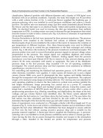
Space closure and canine retraction in continuous arch wire techniques require sufficient force to overcome frictional
forces between the bracket attached to the tooth and the arch wire (Fig. 16). Excessive wire/bracket friction may result in
loss of anchorage or binding accompanied by little tooth movement. Factors that may influence friction are engagement
of the arch wire in brackets that are out of alignment, ligatures pressing the arch wire against the base of the slot, active
torque in rectangular wires, and bodily tooth movement in which the tipping tendency is resisted by two-point contact
between the bracket and arch wire (Ref 235).
Fig. 16 Forces acting on a tooth/bracket system during translation. Source: Ref 235
Simulation Studies
Arch wire/bracket friction is affected by wire size, angulation, ligation force, bracket size, and wire type (Ref 236, 237,
238). Kinetic coefficients of friction of stainless steel, beta-titanium, nickel-titanium, and cobalt-chromium arch wires
sliding on stainless steel or Teflon polymer increase with normal load (Table 16) and increase in the presence of artificial
saliva as compared to dry conditions (Ref 239). Stainless steel and beta-titanium wires show wear tracks after sliding
under low normal loads (40 to 80 N).
Table 16 Coefficient of friction of orthodontic wire/material combinati
ons for a normal load of 40 and 80 N
under wet conditions
Material Coefficient of
friction, 40 N
Coefficient of
friction, 80 N
Stainless steel/stainless steel
0.035 0.085
Beta-titanium/stainless steel
0.025 0.122
Cobalt-chromium/stainless steel
0.041 0.122
Nickel-titanium/stainless steel
0.044 0.115
Stainless steel/Teflon
0.003 0.013
Source: Ref 239
There appears to be agreement that nickel-titanium wires have higher wire/bracket friction than stainless steel wires in
zero torque/zero angulated brackets (Ref 240). At bracket angulations >3°, nickel-titanium wires have lower wire/bracket
friction than stainless steel (Ref 238, 241). Under conditions of two-point contact, friction is inversely proportional to
bracket width and is less for stainless steel wires than for nickel-titanium or beta-titanium wires (Ref 235). Surface
roughness of nickel-titanium or beta-titanium wires may explain their relatively high friction (Ref 242).