INFLAMMATORY BOWEL DISEASE - PART 4 ppt
Bạn đang xem bản rút gọn của tài liệu. Xem và tải ngay bản đầy đủ của tài liệu tại đây (293.3 KB, 38 trang )
104 MacEneaney and Gasparaitis
SUMMARY
The vast array of imaging techniques available to today’s physi-
cians allows for “customization” of radiographic imaging for particular
inflammatory bowel patients. Although standard radiographs are used
to provide an initial “gestalt” in the evaluation of a patient with unknown
or acute disease, contrast-based studies are invaluable in providing more
details, especially in the evaluation of strictures, and of the small bowel
(an area “out of the reach” of traditional endoscopy). More in-depth
studies of the bowel wall and its environs or areas involved with fistulas
by CT, MRI, or US provide a unique perspective, and radionucleotide
studies may be helpful in locating areas of inflammation not seen by
other approaches. New techniques combining technologies with CT or
MRI and enteroclysis have provided stunning new insights into the
evaluation of these elusive diseases, and will likely become more readily
practiced and available in the future.
REFERENCES
1. Goldberg HI, Margulis AR. Gastrointestinal radiology in the United States: An
overview of the past 50 years. Radiology 2000;216:1–7.
2. Bartram C, Laufer I. Inflammatory bowel disease., in: Double Contrast Intestinal
Radiology. 2nd ed. Raven, New York, 1992;580–645.
3. Steinberg DM, Cooke WT, Alexander-Williams J. Abscess and fistulae in Crohn’s
disease. Gut 1973;14:865–869.
4. Wulfeck D, Williams T, Amin A, Huang TY. Crohn’s disease with unusual
enterouterine fistula in pregnancy. J Ky Med Assoc 1994;92:267–269.
5. Rowell DL, Longstreth GF. Colosplenic fistula and splenic abscess complicating
Crohn’s colitis. J Clin Gastroenterol 1995;21:74–75.
6. Mera A, Sugimoto M, Fukuda K, Tanaka F, Imamura F, Matsuda M, et al. Crohn’s
disease associated with colo-bronchial fistula. Intern Med 1996;35:957–960.
7. Karmy-Jones R, Chagpar A, Vallieres E, Hamilton S. Colobronchial fistula due
to Crohn’s disease. Ann Thorac Surg 1995;60:446–448.
8. Senay E, Sachar DB, Keohane M, Greenstein AJ. Small bowel carcinoma in Crohn’s
disease. Distinguishing features and risk factors. Cancer 1989;63:360–363.
9. Laufer I. The radiologic demonstration of early changes in ulcerative colitis by
double contrast technique. J Can Assoc Radiol 1975;26:116–121.
10. Scotiniotis I, Rubesin SE, Ginsberg GG. Imaging modalities in inflammatory
bowel disease. Gastroenterol Clin North Am 1999;28:391–421,ix.
11. Ekberg O. Crohn’s disease of the small bowel examined by double contrast tech-
nique: a comparison with oral technique. Gastrointest Radiol 1977;1:355–359.
12. Steinhardt HJ, Loeschke K, Kasper H, Holtermuller KH, Schafer H. European
Cooperative Crohn’s Disease Study (ECCDS): clinical features and natural his-
tory. Digestion 1985;31:97–108.
13. Herlinger H, Caroline DF, Crohns disease of the small bowel, in: Textbook of Gas-
trointestinal Radiology. 2nd ed., W B Saunders, Philadelphia, PA, 2000; pp. 726–745.
14. Glick SN. Crohn’s disease of the small intestine. Radiol Clin North Am 1987;
25:25–45.
Chapter 6 / Radiological Findings in IBD 105
15. Bender GN, Timmons JH, Williard WC, Carter J. Computed tomographic
enteroclysis: one methodology. Invest Radiol 1996;31:43–49.
16. Bender GN, Maglinte DD, Kloppel VR, Timmons JH. CT enteroclysis: a super-
fluous diagnostic procedure or valuable when investigating small–bowel disease?
AJR Am J Roentgenol 1999;172:373–378.
17. Raptopoulos V, Schwartz RK, McNicholas MM, Movson J, Pearlman J, Joffe N.
Multiplanar helical CT enterography in patients with Crohn’s disease. AJR Am
J Roentgenol 1997;169:1545–1550.
18. Kelvin FM, Helinger H. Crohn’s Disease, in: Clinical Imaging of the Small Intes-
tine. 2nd ed. Springer-Verlag, New York, 1999; pp. 259–289.
19. Lomas DJ, Graves MJ. Small bowel MRI using water as a contrast medium. Br J
Radiol 1999;72:994–997.
20. Low RN, Francis IR. MR imaging of the gastrointestinal tract with i.v., gado-
linium and diluted barium oral contrast media compared with unenhanced MR
imaging and CT. AJR Am J Roentgenol 1997;169:1051–1059.
21. Rubin DL, Muller HH, Young SW. Formulation of radiographically detectable
gastrointestinal contrast agents for magnetic resonance imaging: effects of a
barium sulfate additive on MR contrast agent effectiveness. Magn Reson Med
1992;23:154–165.
22. Rieber A, Wruk D, Nussle K, Potthast S, Reinshagen M, Brambs HJ. [Current
imaging in Crohn’s disease: value of MRI compared with conventional proceed-
ings]. Rontgenpraxis 2000;52:378–383.
23. Maccioni F, Viscido A, Broglia L, Marrollo M, Masciangelo R, Caprilli R, Rossi
P. Evaluation of Crohn disease activity with magnetic resonance imaging. Abdom
Imaging 2000;25:219–228.
24. Lichtenstein GR, Schnall M, Herlinger H. MRI evaluation of Crohn disease
activity. Abdom Imaging 2000;25:229.
25. Umschaden HW, Szolar D, Gasser J, Umschaden M, Haselbach H. Small-bowel
disease: comparison of MR enteroclysis images with conventional enteroclysis
and surgical findings. Radiology 2000;215:717–725.
26. Maglinte DD, Siegelman ES, Kelvin FM. MR enteroclysis: the future of small-
bowel imaging? Radiology 2000;215:639–641.
27. Adamek HE, Breer H, Karschkes T, Albert J, Riemann JF. Magnetic resonance
imaging in gastroenterology: time to say good-bye to all that endoscopy? [In
Process Citation]. Endoscopy 2000;32:406–410.
28. O’Donovan AN, Somers S, Farrow R, Mernagh JR, Sridhar S. MR imaging of
anorectal Crohn disease: a pictorial essay. Radiographics 1997;17:101–107.
29. Outwater E, Schiebler ML. Pelvic fistulas: findings on MR images. AJR Am J
Roentgenol 1993;160:327–330.
30. Stoker J, Fa VE, Eijkemans MJ, Schouten WR, Lameris JS. Endoanal MRI of
perianal fistulas: the optimal imaging planes. Eur Radiol 1998;8:7.
31. Semelka RC, Hricak H, Kim B, Forstner R, Bis KG, Ascher SM, et al. Pelvic
fistulas: appearances on MR images. Abdom Imaging 1997;22:91–95.
32. Myhr GE, Myrvold HE, Nilsen G, Thoresen JE, Rinck PA. Perianal fistulas: use
of MR imaging for diagnosis. Radiology 1994;191:545–549.
33. Barker PG, Lunniss PJ, Armstrong P, Reznek RH, Cottam K, Phillips RK. Mag-
netic resonance imaging of fistula-in-ano: technique, interpretation and accuracy.
Clin Radiol 1994;49:7–13.
34. Rioux M, Sonography of the small bowel and related strutures, in Textbook of
Gastrointestinal Radiology. 2nd ed. W B Saunders, Philadelphia, PA, 2000; pp.
125–152.
106 MacEneaney and Gasparaitis
35. Maconi G, Ardizzone S, Parente F, Bianchi Porro G. Ultrasonography in the
evaluation of extension, activity, and follow-up of ulcerative colitis. Scand J
Gastroenterol 1999;34:1103–7.
36. Tio TL, Mulder CJ, Wijers OB, Sars PR, Tytgat GN. Endosonography of peri-anal
and peri-colorectal fistula and/or abscess in Crohn’s disease. Gastrointest Endosc
1990;36:331–6.
37. Giaffer MH. Labelled leucocyte scintigraphy in inflammatory bowel disease:
clinical applications. Gut 1996;38:1–5.
38. Giaffer MH, Tindale WB, Holdsworth D. Value of technetium-99m HMPAO-labelled
leucocyte scintigraphy as an initial screening test in patients suspected of having
inflammatory bowel disease. Eur J Gastroenterol Hepatol 1996;8:1195–2000.
39. Shah DB, Cosgrove M, Rees JI, Jenkins HR. The technetium white cell scan as an
initial imaging investigation for evaluating suspected childhood inflammatory
bowel disease. J Pediatr Gastroenterol Nutr 1997;25:524–528.
40. Papos M, Varkonyi A, Lang J, Buga K, Timar E, Polgar M, et al. HM-PAO-
labeled leukocyte scintigraphy in pediatric patients with inflammatory bowel
disease. J Pediatr Gastroenterol Nutr 1996;23:547–552.
41. Scholmerich J, Schmidt E, Schumichen C, Billmann P, Schmidt H, Gerok W.
Scintigraphic assessment of bowel involvement and disease activity in Crohn’s
disease using technetium 99m-hexamethyl propylene amine oxine as leukocyte
label. Gastroenterology 1988;95:1287–1293.
42. Skehan SJ, Issenman R, Mernagh J, Nahmias C, Jacobson K. 18F-fluorodeoxyglucose
positron tomography in diagnosis of paediatric inflammatory bowel disease . Lancet
1999;354:836–837.
Chapter 7 / IBD Markers 107
107
From: Clinical Gastroenterology:
Inflammatory Bowel Disease: Diagnosis and Therapeutics
Edited by: R. D. Cohen © Humana Press Inc., Totowa, NJ
7
Inflammatory Bowel Disease Markers
Marla C. Dubinsky, MD
and Stephan R. Targan, MD
CONTENTS
INTRODUCTION
DIFFERENTIATION OF IBD FROM NON-IBD PATIENTS
DISTINGUISHING IBD SUBTYPES: ULCERATIVE COLITIS
VS
CROHN’S DISEASE
MONITOR DISEASE ACTIVITY AND EFFECT OF TREATMENT
ASSESS NATURAL HISTORY
IDENTIFY AT-RISK INDIVIDUALS
THE FUTURE OF IBD DISEASE MARKERS
REFERENCES
INTRODUCTION
For certain diseases that can only be diagnosed clinically, physicians
rely heavily on the presence of disease markers to support or even at
times modify their clinical impression. Typically, these markers play an
important role in helping to establish a diagnosis and to evaluate the
activity of a chronic disease over time. The diagnosis of inflammatory
bowel disease (IBD), however, is not based solely on clinical grounds.
Invasive endoscopic and radiological as well as histopathological crite-
ria need to be met in order to make a correct diagnosis. The search for
a noninvasive diagnostic marker that accurately distinguishes a group
of patients with IBD from those unaffected by the disease has become
an important focus in IBD research. The challenge lies in finding one
marker or a combination thereof, that not only distinguishes IBD from
non-IBD, or identifies at risk populations, but can also help clinicians
distinguish between the IBD subtypes, ulcerative colitis (UC) or Crohn’s
108 Dubinsky and Targan
disease (CD). Such diagnostic dilemmas occur as part of every day
practice for clinicians caring for children and adults with suspicion of,
or a diagnosis of, IBD. Efforts have also been focused on finding ideal
evaluative markers that can be used to monitor disease activity and the
effect of treatment over time. This search has taken a very exciting turn
in the direction of finding markers that can assess the natural history and
perhaps predict the course of individual’s disease over time. This chap-
ter highlights the recent advances in the area of IBD markers, discusses
the utility and feasibility of these novel markers as well as provides a
review of those currently employed in clinical practice.
DIFFERENTIATION OF IBD FROM NON-IBD PATIENTS
The recognition of IBD and subsequent diagnostic evaluation, in
most cases, can be straightforward when the clinical presentation is
unambiguous. However, a diagnostic challenge arises in patients who
present with overlapping, nonspecific and indolent symptoms that are
characteristic of both organic and nonorganic disorders. In the face of
diagnostic uncertainly clinicians are often obligated to exclude IBD
using invasive diagnostic testing, in particular contrast radiography and
colonoscopy with biopsies. Suspicion of IBD commonly results in exten-
sive diagnostic investigations of patients who are ultimately found to
have a functional bowel disorder. In contrast, the diagnosis of IBD,
particularly CD, can be missed or delayed owing to the nonspecific
nature of both the intestinal and extraintestinal symptoms at presenta-
tion. Given these clinical challenges, the search has intensified for an
accurate noninvasive diagnostic marker to aid clinicians in the prompt
recognition of IBD and the differentiation of these disorders from
mimickers.
Serological Markers
ANTIBODIES
The search for an etiologic agent responsible for triggering the
immune mediated bowel injury characteristic of IBD, has lead to the
discovery of immune markers present specifically in the sera of patients
with Crohn’s disease and/or ulcerative colitis. Antineutrophil cytoplas-
mic antibody (ANCA) was originally reported in IBD in the early 1980s
(1). Research and technological advancements subsequently led to the
identification of a novel subset of ANCA, distinct from that observed in
patients with Wegener’s granulomatosis (WG) or systemic vasculitis
with glomerulonephritis (2). This IBD-specific ANCA displays a unique
perinuclear highlighting (pANCA) on immunoflourence staining and is
DNAse sensitive (3). Although it remains undefined, it has been sug-
Chapter 7 / IBD Markers 109
gested that the antigen to which pANCA is directed is a nuclear histone
(H1) (4). This antigen is clearly distinct from the proteinase 3 or the
myeloperoxidase reactivity observed in those patients with vasculitic
disorders. pANCA is likely an autoantibody that is representative of a
cross-reactivity with a luminal bacterial antigen (5–7). Despite epide-
miological and methodological differences, pANCA has been shown
repeatedly to be prevalent in the sera of approx 60% and 20% of UC and
CD patients, respectively (Table 1) (8–14). Typically, <5% of non-IBD
patients are pANCA positive.
Anti-Saccharomyces cerevisiae (S. cerevisiae) antibody (ASCA) was
discovered in the course of studies designed to search for a putative
dietary antigen involved in the pathogenesis of CD (15–17). IgA and
IgG antibodies are directed against a specific oligomannosidic epitope
present on the cell wall of the yeast saccharomyces (18). Studies in both
the adult and pediatric IBD population have demonstrated that ASCA
is expressed in the sera of approx 60% of CD, 10% of UC and <5% of
non-IBD patients. (Table 1) (12–14). It remains unclear whether the
presence of ASCA represents an immune response to the antigens on the
S. cerevisiae itself or to an unidentified antigen, perhaps on the cell wall
of a luminal bacteria, which cross reacts with the yeast antigens.
Advances in technology have lead to the development of two novel
serodiagnostic assays designed specifically to detect both pANCA and
ASCA in the serum of patients with IBD (Prometheus Laboratories,
5739 Pacific Center Blvd., San Diego, CA; phone: 888-428-5227; fax:
958-824-0896; www.prometheus-labs.com) (Table 2). The traditional
ASCA and pANCA assays are adjusted to maximize disease specificity
(>90% specific for IBD) and accurately confirm a diagnosis of IBD
when positive and differentiate UC from CD. However, these highly
specific traditional assays are insufficiently sensitive to serve as diag-
nostic tools for populations with a lower prevalence of disease.
Recently, assays have been modified to be more sensitive (>90% sen-
sitive) and less expensive than the traditional assays.
To be clinically useful, a diagnostic marker must be both disease
sensitive and specific in order to detect all patients with IBD and exclude
all others. Neither the modified nor the traditional serodiagnostic assays
are capable of achieving such high diagnostic standards on their own.
However, recent research has focused on sequencing the sensitive
modified assay with the specific traditional assay in order to improve the
diagnostic accuracy of these noninvasive markers (19,20). A similar
strategy is currently used for the evaluation of patients with suspected
systemic lupus erythromatosis (SLE), whereby first the sensitive anti-
nuclear antibody (ANA) detection assay is followed by a second spe-
110 Dubinsky and Targan
Table 1
Test Characteristics of pANCA and ASCA in Inflammatory Bowel Disease
Antibody Test Sensitivity Specificity
Study n Marker Population (%) (%)
Duerr, et al. (8) 209 pANCA UC vs CD 60 94
& controls
Proujansky, et al. (9) 122 pANCA UC vs CD 46 79
& controls
Winter, et al. (10) 215 pANCA UC vs CD 62 97
& controls
Oberstadt, et al. (11) 151 pANCA UC vs CD 68 93
& controls
Ruemmele, et al. (12) 209 ASCA CD vs UC 55 95
& controls
pANCA UC vs CD 57 92
& controls
Quinton, et al. (13) 391 ASCA CD vs UC 61 88
pANCA UC vs CD 65 85
Hoffenberg, 119 ASCA CD vs UC 60 88
et al. (14) pANCA UC vs CD 60 65
Table 2
Novel Serodiagnostic Assays
Clinical Assay
Assay Name Applications Methodology Characteristics
Modified Assay “Rule out” a ASCA ELISA; Titer cut-offs
diagnosis of IBD IgG & IgA maximized for
sensitivity (>90%)
“IBD First Step” Objective: Distinguish ANCA ELISA
IBD from non-IBD
patients
Traditional Assay 1. “Rule in” a diagnosis ASCA ELISA; Titer cut-offs
of IBD IgG & IgA maximized for
specificity (>90%)
“IBD Diagnostic Objective: differentiate ANCA ELISA
System” inflammatory colitis + Indirect
from other colitides Immunoflouresence
(infectious, ischemic) + DNase
confirmation
2. IBD subtyping
Objective: Distinguish
UC from CD
Chapter 7 / IBD Markers 111
cific confirmatory double-stranded DNA test. Studies in children
showed that these paired tests were accurate in 84% of cases presenting
with nonspecific symptoms suggestive of these IBD (20). Based on the
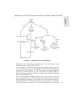
assay characteristics, a novel diagnostic strategy has been proposed to
facilitate clinical decision making when the diagnosis of IBD is initially
uncertain (Fig. 1). All patients undergo initial testing with the modified
assay in addition to other sensitive routine laboratory tests (CBC and
differential, ESR, CRP, and serum albumin). Only those patients with
a positive modified assay result would undergo sequential confirmatory
testing with the more specific traditional assay. Subsequently, only those
patients with a positive confirmatory traditional assay would undergo a
complete invasive work-up whereas all patients with a negative assay
result, either the initial modified or the subsequent traditional after a
positive modified assay, would be observed in follow-up.
Patients with false negative serology will likely return with symptoms
more compatible with IBD and should undergo a full work-up at that
time. Although the diagnosis of IBD will be delayed in a proportion of
patients, the advantage of this strategy really lies in its ability to avoid
unnecessary and invasive investigations in the majority of patients who
truly do not have IBD. Moreover, as new sensitive IBD markers are
identified, fewer patients will be missed by this sequential testing strat-
egy over time. Although promising, studies in larger adult and pediatric
cohorts are needed to validate these initial findings.
Fig. 1. Sensitive markers: ELISA-based ASCA and ANCA assays, hemoglo-
bin platelets, ESR, CRP, and albumin. Proposed diagnostic strategy for indi-
viduals suspected of having IBD (21).
112 Dubinsky and Targan
CLASSIC LABORATORY MARKERS
Attempts have been made to differentiate IBD from functional bowel
disorders using a panel of screening tools comprised of simple routine
blood tests (e.g., complete blood count, platelet count, erythrocyte
sedimentation rate [ESR], C reactive protein [CRP], and serum albu-
min) (2–24). It has been demonstrated in children that when all of the
results are normal, chronic inflammatory bowel disease is an unlikely
diagnosis. Therefore, these screening lab tests may select from among
patients with chronic gastrointestinal symptoms, those who require endo-
scopic assessment. These routine tests are very sensitive for inflamma-
tion, but lack the specificity for IBD. Thus, these tests need to be
combined with other diagnostic markers that are diagnostic of patients
with IBD and not other forms of inflammatory disorders.
Fecal Markers
Stool analysis has been proposed as a useful and inexpensive
noninvasive test to help clinicians delineate the potential causes of
chronic diarrhea. A simple latex agglutination test detecting the neutro-
phil protein, lactoferrin, has been shown to be potentially useful as a
marker of colonic inflammation (25). However, lactoferrin does not
necessarily distinguish between the different forms of inflammatory
colitis (e.g., ischemic vs microscopic vs ulcerative). Similarly, the dis-
tinction between inflammatory and infectious colitis may prove to be a
challenge given that both forms of colitis give rise to fecal leukocytes.
Thus, fecal markers may serve as an adjunct to the other noninvasive
markers used to distinguish IBD form non-IBD.
Genetic Markers
The search for susceptibility genes continues to be a major focus
among IBD researchers. The human major compatibility complex
(MHC) region located on chromosome 6 has been proposed to contain
potential candidate genes (MHC IBD-3 locus) (26–29). Previous stud-
ies have suggested that the susceptibility contributed by the HLA class
II genes to CD and UC are quite different. For epidemiological and
methodological reasons, conflicting and inconclusive results have been
reported regarding these genetic associations, particularly among the
CD population (30–33). A genome-wide search has led to the identifi-
cation of three other potential candidate loci situated on chromosome 16
(IBD-1 locus, recently identified as the Nod-2 gene), chromosome 12
(IBD-2 locus), and chromosome 14 (IBD-4 locus) (34–36). Given their
role as initiators and perpetuators of the inflammatory process charac-
teristic of IBD, genes involved in the regulation of cytokine production
Chapter 7 / IBD Markers 113
may be candidate loci for IBD susceptibility. The gene encoding the
interleukin 1 (IL-1) receptor antagonist (IL-1RA), a protein that modu-
lates the inflammatory response of IL-1, has also been suggested,
although not confirmed, as a susceptibility gene in UC (37). The multi-
factorial etiology of IBD likely precludes the use of these genetic mark-
ers alone as confirmatory diagnostic tools in IBD. However, the presence
of these candidate genes may identify at risk populations. As work goes
ahead in identifying these, it is likely that some of this will become part
of the diagnostic panel. As discussed below, candidate genes may regu-
late distinct immune processes, which, in turn, are manifested as specific
disease behaviors in patients with IBD.
DISTINGUISHING IBD SUBTYPES:
ULCERATIVE COLITIS VS CROHN’S DISEASE
Although UC and CD share may epidemiologic, immunologic, thera-
peutic and clinical features, they are currently considered to be two
distinct subtypes of IBD. Clinical, endoscopic, histopathologic and
radiographic criteria have been put forth to help clinicians differentiate
between these two diseases. However, despite published criteria, this
discrimination may still prove to be difficult in patients with disease
limited to the large bowel. This entity referred to as indeterminate colitis
(IC) occurs in approx 10–15% of IBD patients. Classically, this term
had applied to those patients whose diagnosis remained unknown even
after careful examination of resected surgical specimens. However, the
modern definition of IC refers to all patients pre or postcolectomy whose
categorization remains undefined. It must be emphasized that both sur-
gical options and medical treatment rely on a correct diagnosis. Not all
therapies, particularly the novel biologics, are indicated for both CD and
UC. Similarly, surgical procedures, such as the ileal pouch-anal anas-
tomosis, is intended specifically for patients with ulcerative colitis.
Serological Markers
ANTIBODIES
Given the CD-specificity of ASCA and the UC-specificity of
pANCA, the antibodies have become more widely accepted as useful
discriminatory markers that help clinicians differentiate UC from
Crohn’s colitis (Table 3). Recent reports have demonstrated that approx
2/3 of cases of IC were reclassified preoperatively as either UC or CD
based on the pANCA and ASCA profile (38). The presence of pANCA
in up to 25% of CD patients limits its ability to distinguish UC form CD
on its own. However, the discriminatory strength of these markers is
114 Dubinsky and Targan
amplified when they are evaluated in combination (13,14). (Table 4). A
pANCA+/ASCA– serological profile was shown to be 19 times more
likely to be present in the serum of a patient with UC than CD. Con-
versely, pANCA–/ASCA+ is 16 times more likely in CD than UC (39).
Although pANCA and ASCA have provided clinicians with an impor-
tant diagnostic tool, the search for the ideal serological profile that could
accurately discriminate CD for UC in all patients continues.
Pancreatic antibodies (PAB) have been shown to be present in approx
15–20% of CD patients (40–43). Although not particularly sensitive,
Table 3
Diagnostic Accuracy of ASCA and pANCA in Differentiating UC
from CD Colitis and Non-IBD Colitis in Children
Positive Negative
Sensitivity Specificity predictive predictive
Population Assay (%) (%) value value
CD colitis ASGA IgG 47 96 73 87
vs or IgA
UC & non-
IBD colitis
CD colitis ASCA 29 100 100 87
IgG & IgA
vs
UC & non-
IBD colitis
UC pANCA 57 97 91 79
vs
CD & non-
IBD colitis
Table 4
Diagnostic Accuracy of ASCA and pANCA
in Differentiating UC from CD in a European IBD Population
Ulcerative Crohn’s Positive
colitis disease Sensitivity Specificity predictive
Assay N = 101 N = 1000 (%) (%) value (%)
pANCA + 66 15 65 85 74
ASCA + 12 61 61 88 89
pANCA+/ASCA– 58 3 57 97 93 for UC
pANCA–/ASCA+ 3 49 49 97 96 for CD
Chapter 7 / IBD Markers 115
these antibodies are specific for CD and hence are very predictive of
CD. The antigen reacting with PAB as well as its pathogenic signifi-
cance in IBD remains unknown. Perhaps pancreatic antibodies will serve
as an additional noninvasive marker that clinicians can add to pANCA
and ASCA to help discriminate CD from UC. Assays to detect PAB are
not currently commercially available.
MONITOR DISEASE ACTIVITY AND EFFECT
OF TREATMENT
CD and UC-specific clinical activity indices have been developed as
indirect assessments of a patient’s overall condition. Although used in
clinical trials, indices such as the Crohn’s disease activity index (CDAI)
or the Truelove and Witt criteria are typically not used in everyday
practice. Therapeutic efficacy criteria are typically based on appropri-
ate changes in disease activity scores. Because of the subjective nature
of these indices and inability to accurately assess inflammatory activity,
their use for research purposes are at times questioned. Researchers
continue to search for a simple noninvasive test that provides clinicians
with an objective measure of intestinal function for the monitoring of
disease activity and effect of treatment. Table 5 provides a list of poten-
tial markers if disease activity.
Table 5
Potential Markers of Disease Activity, Disease Relapse
and Effects of Therapy
Serological markers Fecal markers Pereability ratios
Cytokines
TNF (63,64)
IL-6 (64)
IL-1(65)
IL-1RA (65)
Neutrophil products
Myeloperoxidase (66)
Calprotectin (67)
Enteric proteins
α-1-antitrypsin (70)
Antibodies
ASCA & pANCA
(9–13,42–44)
Classic laboratory tests
ESR (50)
CRP (46)
Orosomucoid (50)
Platelet count (48)
Cytokines
TNFα (63)
TNFα receptor (46)
IL-1RA (54)
IL-2R (55)
IL-6 (56-58)
Urinary Assays
Lactulose/L-rhamnose
(72–74)
Lactulose/mannitol (75)
116 Dubinsky and Targan
Serological Markers
ANTIBODIES
To date, an association between the presence and titer level of pANCA
or ASCA and disease activity, IBD duration, patient gender or treatment
has not been consistently demonstrated (9,12,13,44,45). The presence
of these markers has been shown to correlate with disease location in
patients with CD. The presence of pANCA was associated with colonic
involvement (11,46) and ASCA with small bowel involvement, alone or
in combination with large bowel (13,46). Unlike the ANCA associated
vasculitides, IBD-specific antibodies do not become undetectable after
initiating immunosuppressives nor does their persistence indicate fre-
quent relapses. Titer level may correlate with age of onset of CD, as a
recent study demonstrated that higher ASCA titers were observed in
patients with early age of disease onset and high pANCA levels in
patients with later age of onset (46). Prospective studies are needed to
determine if pANCA or ASCA expression changes in concordance with
disease activity and monitor the effect of treatment.
The effect of intestinal surgery on antibody expression remains
unknown. Future studies are needed to confirm whether pANCA persist
postcolectomy as suggested and to determine the fate of ASCA expres-
sion post resection (12,44).
C
LASSIC LABORATORY MARKERS
Acute phase protein concentrations have classically been used as
supplemental markers of clinical activity in IBD. The most common
are the ESR, CRP and serum orosomucoid (α 1-acid glycoprotein).
The latter protein has been shown to have similar sensitivity and speci-
ficity characteristics to the CRP. However, because of its long half-
life, its ability to indicate improvement in disease activity is limited
(47). CRP has a significantly shorter half-life and thus rapidly
decreases in response to a reduction in disease activity (48). A pro-
spective longitudinal study confirmed that CRP is also useful in moni-
toring the response to treatment (49). Platelet counts, yet not white
counts, have also been shown to be a useful measure of disease activity
(50). Few tools are available to help clinicians identify those IBD
patients at risk for a disease relapse. This knowledge may be of value
for determining which patients in remission would benefit from treat-
ment. In prospective longitudinal studies, both the ESR and CRP have
proven to be accurate in identifying those patients at high risk of dis-
ease relapse (49,51,52). These markers may be of particular use in
children with mild nonspecific symptoms in whom a diagnosis as well
as an assessment of disease activity is difficult. Further studies are
Chapter 7 / IBD Markers 117
needed to determine which combination of classic laboratory markers
forms the ideal prognostic index.
Cytokines. Concentrations of proinflammatory cytokines are
increased in the intestinal mucosa in patients with active CD. The evalu-
ation of serum concentrations on intestinally produced cytokines as
surrogate markers of bowel inflammation has yielded inconsistent
results. The theory being that in comparison to the classic laboratory
markers, cytokines more accurately reflect the underlying immuno-
pathogenic process. Although tumour necrosis factor-α (TNFα) pro-
duction is increased in the mucosa of patients with active CD, serum
levels of TNFα have not been consistently useful as markers of disease
activity in these patients (48,53–55). However, an association between
disease activity and serum levels of soluble TNFα receptors in both CD
and UC has been described (48). Further studies are needed to confirm
this association.
It has been suggested that the production of IL-1 receptor antagonist
(IL-1RA) may be a manner by which the host down regulates the inflam-
matory process perhaps amplified by IL-1. IL-1RA levels were found to
be increased in patients with active CD and UC as compared to patients
with inactive and infectious colitis (56). Interestingly, these IL-1RA
concentrations correlated with CRP and orosomucoid levels. IL-2
receptor is shed into the circulation by activated T cells along with IL-2.
Levels of IL-2R have been suggested as a new measure of disease activity
(57). IL-6 possesses both anti- and proinflammatory properties. It has
also been suggested that serum IL-6 levels correlate with disease activ-
ity in both CD and UC patients (58–60). Interestingly, in the Reinish
study, patients with primarily luminal inflammation displayed higher
IL-6 levels than CD patients with fibrostenosis or those with extensive
bowel resection. Vascular endothelial growth factor (VEGF) is a
cytokine released by cells that potentiate vascular permeability and
neovascularization. Significantly increased levels of VGEF have been
observed in the serum of patients with active CD and UC (61,62). The
potential pathogenic role of VEGF induced vascular permeability in
IBD remains unknown.
The role of cytokines as predictors of clinical relapse has also been
a focus of investigation. In one study, serum IL-6 level proved to be the
greatest predictor of time to relapse with a 17-fold risk over a 12-mo
period when levels reached a specific cut point (20 pg/mL). Of the other
variable tested, soluble TNF receptor, IL-2R and serum orosomucoid
levels also appeared to be useful for predicting the course of disease in
patients with quiescent CD (63). Thus, the combination of classic bio-
logical markers with the novel cytokine markers may prove to be the
118 Dubinsky and Targan
ideal serological prognostic index. A recent study suggested that the
capacity of intestinal lamina propria cells to secrete TNF and IL-1 may
identify patients at risk of a relapse and that may benefit from appropri-
ate anti-inflammatory therapy (64). The practicality and feasibility of a
tissue assay is limited compared to that of the serologic analysis.
Cytokine profiles exhibit interindividual variability and thus no one
cytokine will necessarily correlate with disease activity in all IBD
patients. Future association studies are needed to address this variability.
Fecal Markers
Inflammation of the gut is associated with leakage of cells, cellular
products, such as cytokines, and serum proteins. Given that circulating
cytokines may not accurately reflect mucosal production, researchers
have questioned whether stool cytokine levels may more closely mirror
the mucosal inflammation. Preliminary studies demonstrated that stool
TNFα and IL-6 concentrations were elevated in children with active CD
and UC (65,66). A more recent study observed a significant correlation
between stool concentrations of IL-1 and IL-1RA and disease activity
in CD and UC patients (67). TNFα was not increased in patients with
active disease. As observed with serum cytokine levels, the associations
are inconsistent and further studies are needed.
Leukocytes play an important role in initiating and amplifying the
mucosal immune process characteristic of IBD. Myeloperoxidase, a
constituent of neutrophil granules, has been shown to reflect the number
of neutrophils. Stool levels of myeloperoxidase were found to be
elevated in patients with active IBD and correlated well with classic
laboratory parameters and endoscopic indices of inflammation.
Although the presence of stool leukocytes is a sensitive marker of
inflammation, these results suggest that the stool levels of myelo-
peroxidase may be more specific for IBD (68). Levels of fecal
calprotectin, a calcium-binding protein found in neutrophil granulo-
cytes, have been shown to correlate with histologic and endoscopic
assessment of disease activity in UC patients (69). Additionally, its
ability to predict clinical relapse has been recently demonstrated among
CD and UC patients alike (70). Enteric protein loss in the face of inflam-
mation may be reflected in the measurement of serum proteins in the
stool. Fecal α 1-antitrypsin has been shown to be a sensitive, yet non-
specific marker of enteric inflammation (71). It has been shown to re-
flect disease activity in patients with CD, but not UC likely owing to the
small bowel involvement (72).
The results of these small studies bring forth the notion that fecal
markers, like serological markers, may prove to be useful surrogate
Chapter 7 / IBD Markers 119
markers of gut inflammation and helpful in differentiating active from
inactive disease. The true utility of these markers as predictors of relapse
needs to be evaluated prospectively so to determine if early treatment
based on subclinical marker changes will avert relapse and perhaps alter
a patients clinical course. As with tissue markers, the practicality of
fecal analysis is questionable and larger validation studies are needed.
Markers of Intestinal Permeability
Increased intestinal permeability is a well documented feature of CD.
It is unclear whether this increased permeability plays a primary patho-
genic role in IBD, perhaps genetically determined, or is secondary to the
inflammation. A number of permeability tests have been evaluated
over time. The urinary lactulose/L-rhamnose permeability ratio was
increased in the majority of children with active CD irrespective of
disease location (73). However, among UC patients, only those with
extensive colitis had similar abnormal changes in permeability. Despite
these findings, the utility of permeability assays is typically limited for
use in only CD patients whose disease is confined to the small bowel
(74–76). These ratios appeared to normalize in patients who completed
an initial course of steroids suggesting perhaps a role for permeability
assays for monitoring the effect of treatment. More attention has been
given to the potential role of sugar permeability assays in predicting
disease relapse. In one study, serial testing using a lactulose/mannitol
permeability assay was predictive of CD relapse over the short term
(77). Over a 12-mo period, approx 50% of 5-ASA treated IBD patients
relapsed after a disease-free interval of at least 1 mo. Intestinal perme-
ability was significantly increased in the CD patients who relapsed. The
sensitivity of these assays are high (>80%) but the specificity for disease
relapse is sub-optimal (<65%) (70). These assays are not widely used in
clinical practice. Although interesting, further longitudinal studies are
needed to determine whether prophylactic treatment in asymptomatic
patients, with the goal of averting disease relapse, will significantly alter
patient outcomes.
ASSESS NATURAL HISTORY
The more classic categorization of IBD into UC and CD was based
partially on the differences in the expected natural history of these two
perceived separate disease entities. However, over time, we have come
to realize that within each subtype patients behave very differently and
that the natural history has now become more difficult to predict at the
time of diagnosis. This observed clinical heterogeneity has lead to the
120 Dubinsky and Targan
development of certain classification systems in order to help character-
ize patients as specific disease phenotypes. In other words, attempting
to classify patients into more homogeneous subgroups. For UC, typi-
cally patients are classified based on disease location and according to
the most recent Vienna Classification, patients with CD are to be catego-
rized based on disease location, behavior, and age of disease onset.
Immunologic and genetic contributions play critical roles in defining
this clinical heterogeneity.
Serological Markers
ANTIBODIES
ASCA and pANCA may serve as surrogate markers for the genetic
contributions to the clinical heterogeneity observed among IBD patients.
The expression of these markers has been shown to reflect specific
disease phenotypes. The first clinical phenotype was described for the
pANCA positive subgroup of UC patients. A strong association was
observed between pANCA and the occurrence of chronic pouchitis after
Ileal Pouch Anal Anastomosis (IPAA) (78,79). To date, not all centers
have reproduced these findings (80–83). However, a recent prospective
study using second generation assays (Prometheus Laboratories)
reported that high levels of pANCA (>100 EU/mL) in UC patients pre-
dicted the development of chronic pouchitis with a high degree of cer-
tainty (84). Future studies are underway to determine if prophylactic
antibiotic treatment would decrease the incidence of chronic pouchitis
in at risk (high pANCA) individuals.
Standard treatment regimens are typically employed in all UC patients
at the time of diagnosis based on disease location and disease severity.
More aggressive immunosuppressive agents like 6-MP or azathioprine
are often reserved for patients who, over time, become unresponsive to
standard medical therapy. Unfortunately, an ideal tool to help clinicians
predict therapeutic response upon initial presentation has not been
developed. In a retrospective pilot study, however, the presence of
pANCA was found to be associated with resistance to standard medical
therapy in UC patients with left-sided colitis (85). Other studies have
suggested that pANCA expression is a possible marker of an aggressive
disease phenotype (86–88). These findings further suggest that pANCA
may serve as a surrogate for the genetic heterogeneity that determines
an individual’s clinical course.
Although pANCA has been established as a UC-specific marker,
approx 25% of all CD patients also express pANCA. These CD patients
have been found to have clinical features of left-sided colitis with endo-
scopic and/or histopathologic features of UC. These CD patients are
Chapter 7 / IBD Markers 121
described as “UC-like” (89). Vasiliauskas et al. established criteria to
define a “UC-like” state in patients with established CD. These patients
had to have had clinical features of left-sided colonic disease as well as
endoscopic and/or histopathologic criteria as outlined in Table 6 in
order to be defined as “UC-like”. Interestingly, none of the pANCA
positive CD patients had isolated small bowel disease and pANCA
expression was not related solely to the presence of colonic inflamma-
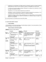
tion. Thus, the presence of pANCA may represent a distinct, genetically
conditioned mucosal inflammatory process common to both UC and
“UC-like” CD patients. The shared clinical features suggest that the
expression of pANCA may help restructure the categorization of UC
and CD into more homogeneous phenotypic subgroups (Fig. 2).
ASCA expression may reflect another unique inflammatory process
in patients with CD. The results of a recent study demonstrated that
higher ASCA levels were associated with earlier age of disease onset
and both fibrostenosing and internal penetrating disease behaviors (46).
In contrast, high pANCA levels were associated with the onset of
“UC-like” CD at an older age. Thus, the qualitative (expression) as well
as the quantitative (titer level) analysis of ASCA and pANCA may
represent select clinical phenotypes in CD patients.
If indeed IBD-specific antibodies are biological markers of distinct
disease behaviors, as suggested, these tools may help predict the natural
history of an individual’s disease. With this knowledge clinicians can
create and implement appropriate therapeutic management regimes based
on the aggressiveness of the IBD subtype so to alter and thus improve the
long-term prognosis. Prospective studies are needed to confirm the utility
of ascertaining ASCA and pANCA status at the time of diagnosis and to
evaluate the impact of this information on patient outcomes.
Genetic Markers
The clinical heterogeneity within UC and CD likely has a genetic basis.
The search for the genetic contributions to the various disease phenotypes
has been of great interest. Besides their possible implication in the determi-
nation of UC susceptibility, HLA class II genes may also influence the
pattern of disease behavior. Certain HLA haplotypes and alleles of IL-1RA
have emerged in various studies as potential markers of extensive UC,
perhaps even predicting the need for surgery in a select group of patients
(26,88). The link between genotype and clinical phenotype may indeed be
the expression of pANCA and ASCA. Although the results have not been
consistent, certain HLA class II genes have been linked to pANCA expres-
sion (91). Further association studies are needed to confirm genetic linkage
with disease susceptibility, disease behaviors and antibody expression.
122 Dubinsky and Targan
IDENTIFY AT-RISK INDIVIDUALS
Both genetic and antibody markers may aid in the identification of
individuals at risk of developing IBD in the future. The frequency of
both ASCA and pANCA expression has been shown to be higher in the
Table 6
“UC-Like” Crohn’s Disease
Clinical Features of Left-sided Folitis
Rectal bleeding
Urgency
Tenesmus
Treatment with topical therapies
Recommended or performed total or near-total colectomy
Endoscopic Appearance
Inflammation extending proximally from the rectum
Inflammation more severe distally than proximally
Continuous inflammation
Shallow ulceration/lack of deep ulcerations
Histopathological Features
Homogenous, continuous, predominantly superficial inflammation
Crypt abscesses
Lack of granulomas
Lack of “focality” in biopsy specimens
Fig. 2. Link between IBD genotypes and phenotypes based on pANCA expression.
Chapter 7 / IBD Markers 123
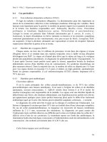
Fig. 3. Future of diagnostic and therapeutic management strategies for IBD patients.
nonaffected relatives of both CD and UC, respectively (92,93). Not all
centers have confirmed these findings. Studies in different population
groups, using different methodologies may explain the discrepancies
observed found between countries in Europe and North America (94).
It is unknown whether these patients will develop, or are at risk of
developing, IBD over time. However, given the multifactorial etiology
of IBD, the value of these antibodies alone as preclinical markers of
disease is questionable. As hypothesized with other autoimmune dis-
eases, the interaction between environmental and genetic factors is criti-
cal to the biologic onset of IBD. Prospective longitudinal studies are
needed to document the outcome of patients identified as “high risk”
based on the presence of subclinical markers, such as ASCA or pANCA.
If these patients do develop IBD, then perhaps subclinical disease mark-
ers could help establish an early diagnosis and appropriate therapeutic
decisions can then be made with the goal of improving patient outcomes
in those who have the target disorder.
THE FUTURE OF IBD DISEASE MARKERS
The clinical utility and importance of IBD-specific markers has been
reviewed in this chapter. Research and technological advancements
have fostered a novel approach to understanding the intricate relation-
ship between genetic and clinical expression of disease. Both genetic
and serum antibody markers hold the most promise in helping research-
ers better comprehend disease heterogeneity and natural history.
Although our current gold standard diagnostic tests do not possess this
capability, exciting preliminary research suggests IBD-specific genetic
and antibody markers may serve as predictors of an individual’s disease
course. Thus, the foundation has been laid upon which the discovery of
novel IBD-specific and IBD-sensitive markers will enable researchers
to identify at-risk individuals, as well as diagnose IBD and stratify
patients into homogeneous subtypes with certainty (Fig. 3). Clinicians
124 Dubinsky and Targan
can then create and implement individual treatment plans designed to
improve the long-term prognosis of these chronic diseases.
REFERENCES
1. Nielsen H, Wiik A, Elmgreen J. Granulocyte specific antinuclear antibodies in
ulcerative colitis. Aid in differential diagnosis of inflammatory bowel disease.
Acta Pathol Microbiol Immunol Scand [C] 1983;91:23–26.
2. Saxon A, Shanahan F, Landers C, Ganz T, Targan S. A distinct subset of
antineutrophil cytoplasmic antibodies is associated with inflammatory bowel
disease. J Allergy Clin Immunol 1990;86:202–210.
3. Vidrich A, Lee J, James E, Cobb L, Targan S. Segregation of pANCA antigenic
recognition by DNase treatment of neutrophils: ulcerative colitis, type 1 autoimmune
hepatitis, and primary sclerosing cholangitis. J Clin Immunol 1995;15:293–299.
4. Eggena M, Cohavy O, Parseghian MH, Hamkalo BA, Clemens D, Targan SR,
Gordon LK, Braun J. Identification of histone H1 as a cognate antigen of the ulcer-
ative colitis-associated marker antibody pANCA. J Autoimmun 2000;14:83–97.
5. Cohavy O, Harth G, Horwitz M, Eggena M, Landers C, Sutton C, et al. Identifi-
cation of a novel mycobacterial histone H1 homologue (HupB) as an antigenic
target of pANCA monoclonal antibody and serum immunoglobulin A from
patients with Crohn’s disease. Infect Immun 1999;67:6510–6517.
6. Cohavy O, Bruckner D, Gordon LK, Misra R, Wei B, Eggena ME, et al. Colonic
bacteria express an ulcerative colitis pANCA-related protein epitope. Infect
Immun 2000;68:1542–1548.
7. Seibold F, Brandwein S, Simpson S, Terhorst C, Elson CO. pANCA represents a
cross-reactivity to enteric bacterial antigens. J Clin Immunol 1998;18:153–160.
8. Duerr RH, Targan SR, Landers CJ, Sutherland LR, Shanahan F. Anti-neutrophil
cytoplasmic antibodies in ulcerative colitis. Comparison with other colitides/
diarrheal illnesses. Gastroenterology 1991;100:1590–1596.
9. Proujansky R, Fawcett PT, Gibney KM, Treem WR, Hyams JS. Examination of
anti-neutrophil cytoplasmic antibodies in childhood inflammatory bowel disease.
J Pediatr Gastroenterol Nutr 1993;17:193–197.
10. Winter HS, Landers CJ, Winkelstein A, Vidrich A, Targan SR. Anti-neutrophil
cytoplasmic antibodies in children with ulcerative colitis. J Pediatr 1994;125:707–711.
11. Oberstadt K, Schaedel W, Weber M, Classen M, Deusch K. P-ANCA as a differ-
ential diagnostic marker in inflammatory bowel disease. Adv Exp Med Biol
1995;371B:1313–1316.
12. Ruemmele FM, Targan SR, Levy G, Dubinsky M, Braun J, Seidman EG. Diag-
nostic accuracy of serological assays in pediatric inflammatory bowel disease
Gastroenterology 1998;115:822–829.
13. Quinton JF, Sendid B, Reumaux D, Duthilleul P, Cortot A, Grandbastien B, et al.
Anti-Saccharomyces cerevisiae mannan antibodies combined with antineutrophil
cytoplasmic autoantibodies in inflammatory bowel disease: prevalence and diag-
nostic role. Gut 1998;42:788–791.
14. Hoffenberg EJ, Fidanza S, Sauaia A. Serologic testing for inflammatory bowel
disease. J Pediatr 1999;134:447–452.
15. Main J, McKenzie H, Yeaman GR, Kerr MA, Robson D, Pennington CR, Parratt
D. Antibody to Saccharomyces cerevisiae (bakers’ yeast) in Crohn’s disease.
BMJ 1988;297:1105–1106.
16. McKenzie H, Main J, Pennington CR, Parratt D. Antibody to selected strains of
Saccharomyces cerevisiae (baker’s and brewer’s yeast) and Candida albicans in
Crohn’s disease. Gut 1990;31:536–538.
Chapter 7 / IBD Markers 125
17. Giaffer MH, Clark A, Holdsworth CD. Antibodies to Saccharomyces cerevisiae
in patients with Crohn’s disease and their possible pathogenic importance. Gut
1992;33:1071–1075.
18. Sendid B, Colombel JF, Jacquinot PM, Faille C, Fruit J, Cortot A, et al. Specific
antibody response to oligomannosidic epitopes in Crohn’s disease. Clin Diagn
Lab Immunol 1996;3:219–226.
19. Dubinsky MC, Targan S, Braun J, Seidman EG. Predictive Value of Screening
Tests in Pediatric Inflammatory Bowel Disease: A Prospective Comparative
Study. AJG 1998;93:A585.
20. Dubinsky MC, Ofman J, Targan SR, FM Ruemmele, Seidman EG. ASCA and
ANCA Testing: Important tools for clinical decision making in pediatric IBD.
Gastroenterology 1999;114:A702
21. Seidman EG, Dubinsky M, Patriquin H, Marx G, Theoret Y. Recent developments
in the diagnosis and Management of pediatric inflammatory bowel disease. In:
Inflammatory bowel disease therapy 1999. Kluwer Academic Publishers, The
Netherlands, 2000;87–95.
22. Shine B, Berghouse L, Jones JE, Landon J. C-reactive protein as an aid in the
differentiation of functional and inflammatory bowel disorders. Clin Chim Acta
1985;148:105–109.
23. Thomas DW, Sinatra FR. Screening laboratory tests for Crohn’s disease. West J
Med 1989;150:163–164.
24. Beattie RM, Walker-Smith JA, Murch SH. Indications for investigation of chronic
gastrointestinal symptoms. Arch Dis Child 1995;73:354–355.
25. Fine KD, Ogunji F, George J, Niehaus MD, Guerrant RL. Utility of a rapid fecal
latex agglutination test detecting the neutrophil protein, lactoferrin, for diagnosing
inflammatory causes of chronic diarrhea. Am J Gastroenterol 1998;93:1300–1305.
26. Toyoda H, Wang SJ, Yang HY, Redford A, Magalong D, Tyan D, et al. Distinct
associations of HLA class II genes with inflammatory bowel disease. Gastroen-
terology 1993;104:741–748.
27. Satsangi J, Welsh KI, Bunce M, Julier C, Farrant JM, Bell JI, Jewell DP. Contri-
bution of genes of the major histocompatibility complex to susceptibility and
disease phenotype in inflammatory bowel disease. Lancet 1996;347:1212–1217.
28. Hampe J, Schreiber S, Shaw SH, Lau KF, Bridger S, MacPherson AJ, et al. A
genomewide analysis provides evidence for novel linkages in inflammatory bowel
disease in a large European cohort. Am J Hum Genet 1999;64:808–816.
29. Yang H, Plevy SE, Taylor K, Tyan D, Fischel-Ghodsian N, McElree C, et al.
Linkage of Crohn’s disease to the major histocompatibility complex region is
detected by multiple non-parametric analyses. Gut 1999;44:519–526.
30. Smolen JS, Gangl A, Polterauer P, Menzel EJ, Mayr WR. HLA antigens in inflam-
matory bowel disease. Gastroenterology 1982;82:34–38.
31. Asakura H, Tsuchiya M, Aiso S, Watanabe M, Kobayashi K, Hibi T, et al. Asso-
ciation of the human lymphocyte-DR2 antigen with Japanese ulcerative colitis.
Gastroenterology 1982;82:413–418.
32. Cottone M, Bunce M, Taylor CJ, Ting A, Jewell DP. Ulcerative colitis and HLA
phenotype. Gut 1985;26:952–954.
33. Duerr RH, Neigut DA. Molecularly defined HLA-DR2 alleles in ulcerative colitis
and an antineutrophil cytoplasmic antibody-positive subgroup. Gastroenterology
1995;108:423–442
34. Hugot JP, Laurent-Puig P, Gower-Rousseau C, Olson JM, Lee JC, Beaugerie L,
et al. Mapping of a susceptibility locus for Crohn’s disease on chromosome 16
Nature 1996;379:821–823.
126 Dubinsky and Targan
35. Satsangi J, Parkes M, Louis E, Hashimoto L, Kato N, Welsh K, et al. Two stage
genome-wide search in inflammatory bowel disease provides evidence for sus-
ceptibility loci on chromosomes 3, 7 and 12. Nat Genet 1996;14:199–202.
36. Ma Y, Ohmen JD, Li Z, Bentley LG, McElree C, Pressman S, et al. A genome-
wide search identifies potential new susceptibility loci for Crohn’s disease.
Inflamm Bowel Dis 1999;5:271–278.
37. Mansfield JC, Holden H, Tarlow JK, Di Giovine FS, McDowell TL, Wilson AG,
et al. Novel genetic association between ulcerative colitis and the anti- inflamma-
tory cytokine interleukin-1 receptor antagonist. Gastroenterology 1994;106:
637–642.
38. Schwarz S, Ammirati, M, Korelitz B, Gleim G. Identification of Indeterminate
Colitis using pANCA and ASCA. Gastroenterology 2000:118 (suppl 2):A1891
39. Panaccione R, Sandborn WJ. Is antibody testing for inflammatory bowel disease
clinically useful? Gastroenterology 1999;116:1001–1002.
40. Stocker W, Otte M, Ulrich S, Normann D, Finkbeiner H, Stocker K, et alC.
Autoimmunity to pancreatic juice in Crohn’s disease. Results of an autoantibody
screening in patients with chronic inflammatory bowel disease. Scand J
Gastroenterol Suppl 1987;139:41–52.
41. Seibold F, Mork H, Tanza S, Muller A, Holzhuter C, Weber P, Scheurlen M.
Pancreatic autoantibodies in Crohn’s disease: a family study. Gut 1997;40:
481–484.
42. Folwaczny C, Noehl N, Endres SP, Loeschke K, Fricke H. Antineutrophil and
pancreatic autoantibodies in first-degree relatives of patients with inflammatory
bowel disease. Scand J Gastroenterol 1998;33:523–528.
43. Sandborn WJ, Loftus EV, Colombel JF, Fleming K, Seibold F, Homburger HA,
et al. Utility of perinuclear anti-neutrophil cytoplasmic antibodies (pANCA),
anti-saccharomyces antibody (ASCA), and anti-pancreatic antibodies (APA) as
serologic markers in a population based cohort of patients with Crohn’s disease
(CD) and ulcerative colitis (UC). Gsatroeneterology 2000;118 (supplement
2):A696.
44. Pool MO, Ellerbroek PM, Ridwan BU, Goldschmeding R, von Blomberg BM,
Pena AS, et al. Serum antineutrophil cytoplasmic autoantibodies in inflammatory
bowel disease are mainly associated with ulcerative colitis. A correlation study
between perinuclear antineutrophil cytoplasmic autoantibodies and clinical
parameters, medical, and surgical treatment. Gut 1993;34:46–50.
45. Roozendaal C, Pogany K, Hummel EJ, Horst G, Dijkstra G, Nelis GF, et al. Titres
of anti-neutrophil cytoplasmic antibodies in inflammatory bowel disease are not
related to disease activity. QJM 1999;92:651–658.
46. Vasiliauskas EA, Kam LY, Karp LC, Gaiennie J, Yang H, Targan SR. Marker
antibody expression stratifies Crohn’s disease into immunologically homoge-
neous subgroups with distinct clinicial charracteristics. Gut 2000; 47(4):
487–496.
47. Chambers RE, Stross P, Barry RE, Whicher JT. Serum amyloid A protein com-
pared with C-reactive protein, alpha 1- antichymotrypsin and alpha 1-acid glyco-
protein as a monitor of inflammatory bowel disease. Eur J Clin Invest 1987;
17:460–467.
48. Nielsen OH, Vainer B, Madsen SM, Seidelin JB, Heegaard NH. Established and
emerging biological activity markers of inflammatory bowel disease. Am J
Gastroenterol 2000;95:359–367.
49. Boirivant M, Leoni M, Tariciotti D, Fais S, Squarcia O, Pallone F. The clinical
significance of serum C reactive protein levels in Crohn’s disease. Results of a
prospective longitudinal study. J Clin Gastroenterol 1988;10:401–405.
Chapter 7 / IBD Markers 127
50. Harries AD, Fitzsimons E, Fifield R, Dew MJ, Rhoades J. Platelet count: a simple
measure of activity in Crohn’s disease. Br Med J (Clin Res Ed) 1983;286:1476.
51. Campbell CA, Walker-Smith JA, Hindocha P, Adinolfi M. Acute phase proteins
in chronic inflammatory bowel disease in childhood. J Pediatr Gastroenterol Nutr
1982;1:193–200.
52. Brignola C, Campieri M, Bazzocchi G, Farruggia P, Tragnone A, Lanfranchi GA.
A laboratory index for predicting relapse in asymptomatic patients with Crohn’s
disease. Gastroenterology 1986;91:1490–1494.
53. Hyams JS, Treem WR, Eddy E, Wyzga N, Moore RE. Tumor necrosis factor-
alpha is not elevated in children with inflammatory bowel disease. J Pediatr
Gastroenterol Nutr 1991;12:233–236.
54. Murch SH, Lamkin VA, Savage MO, Walker-Smith JA, MacDonald TT. Serum
concentrations of tumour necrosis factor alpha in childhood chronic inflamma-
tory bowel disease. Gut 1991;32:913–917.
55. Gardiner KR, Halliday MI, Barclay GR, Milne L, Brown D, Stephens S, et al.
Significance of systemic endotoxaemia in inflammatory bowel disease. Gut
1995;36:897–901.
56. Propst A, Propst T, Herold M, Vogel W, Judmaier G. Interleukin-1 receptor
antagonist in differential diagnosis of inflammatory bowel diseases. Eur J
Gastroenterol Hepatol 1995;7:1031–1036.
57. Williams AJ, Symons JA, Watchet K, Duff GW. Soluble interleukin-2 receptor
and disease activity in Crohn’s disease. J Autoimmun 1992;5:251–259.
58. Mahida YR, Kurlac L, Gallagher A, Hawkey CJ. High circulating concentrations
of interleukin-6 in active Crohn’s disease but not ulcerative colitis. Gut
1991;32:1531–1534.
59. Holtkamp W, Stollberg T, Reis HE. Serum interleukin-6 is related to disease
activity but not disease specificity in inflammatory bowel disease. J Clin
Gastroenterol 1995;20:123–126
60. Reinisch W, Gasche C, Tillinger W, Wyatt J, Lichtenberger C, Willheim M, et al.
Clinical relevance of serum interleukin-6 in Crohn’s disease: single point mea-
surements, therapy monitoring, and prediction of clinical relapse. Am J
Gastroenterol 1999;94:2156–2164.
61. Griga T, Tromm A, Spranger J, May B. Increased serum levels of vascular endot-
helial growth factor in patients with inflammatory bowel disease. Scand J
Gastroenterol 1998;33:504–508.
62. Bousvaros A, Leichtner A, Zurakowski D, Kwon J, Law T, Keough K, Fishman
S. Elevated serum vascular endothelial growth factor in children and young adults
with Crohn’s disease. Dig Dis Sci 1999;44:424–430.
63. Louis E, Belaiche J, van Kemseke C, Franchimont D, de Groote D, Gueenen V,
Mary JY. A high serum concentration of interleukin-6 is predictive of relapse in
quiescent Crohn’s disease. Eur J Gastroenterol Hepatol 1997;9:939–944.
64. Schreiber S, Nikolaus S, Hampe J, Hamling J, Koop I, Groessner B, et al. Tumour
necrosis factor alpha and interleukin 1beta in relapse of Crohn’s disease. Lancet
1999;353:459–461.
65. Braegger CP, Nicholls S, Murch SH, Stephens S, MacDonald TT. Tumour necro-
sis factor alpha in stool as a marker of intestinal inflammation. Lancet 1992;
339:89–91.
66. Nicholls S, Stephens S, Braegger CP, Walker-Smith JA, MacDonald TT.
Cytokines in stools of children with inflammatory bowel disease or infective
diarrhoea. J Clin Pathol 1993;46:757–760.
67. Saiki T, Mitsuyama K, Toyonaga A, Ishida H, Tanikawa K. Detection of pro- and
anti-inflammatory cytokines in stools of patients with inflammatory bowel dis-
ease. Scand J Gastroenterol 1998;33:616–622.
128 Dubinsky and Targan
68. Saiki T. Myeloperoxidase concentrations in the stool as a new parameter of
inflammatory bowel disease. Kurume Med J 1998;45:69–73.
69. Roseth AG, Aadland E, Jahnsen J, Raknerud N. Assessment of disease activity in
ulcerative colitis by faecal calprotectin, a novel granulocyte marker protein.
Digestion 1997;58:176–180.
70. Tibble JA, Sigthorsson G, Bridger S, Fagerhol MK, Bjarnason I. Surrogate mark-
ers of intestinal inflammation are predictive of relapse in patients with inflamma-
tory bowel disease. Gastroenterology 2000;119:15–22.
71. Becker K, Frieling T, Haussinger D. Quantification of fecal alpha 1-antitrypsin
excretion for assessment of inflammatory bowel diseases. Eur J Med Res
1998;3:65–70.
72. Becker K, Berger M, Niederau C, Frieling T. Individual fecal alpha 1-antitrypsin
excretion reflects clinical activity in Crohn’s disease but not in ulcerative colitis.
Hepatogastroenterology 1999;46:2309–2314.
73. Miki K, Moore DJ, Butler RN, Southcott E, Couper RT, Davidson GP. The sugar
permeability test reflects disease activity in children and adolescents with inflam-
matory bowel disease. J Pediatr 1998;133:750–754.
74. Wyatt J, Vogelsang H, Hubl W, Waldhoer T, Lochs H. Intestinal permeability and
the prediction of relapse in Crohn’s disease. Lancet 1993;341:1437–1439.
75. Teahon K, Smethurst P, Levi AJ, Menzies IS, Bjarnason I. Intestinal permeability
in patients with Crohn’s disease and their first degree relatives. Gut 1992;33:
320–323.
76. Bjarnason I, MacPherson A, Hollander D. Intestinal permeability: an overview.
Gastroenterology 1995;108:1566–1581.
77. Hilsden RJ, Meddings JB, Hardin J, Gall DG, Sutherland LR. Intestinal perme-
ability and postheparin plasma diamine oxidase activity in the prediction of
Crohn’s disease relapse. Inflamm Bowel Dis 1999;5:85–91.
78. Sandborn WJ, Landers CJ, Tremaine WJ, Targan SR. Antineutrophil cytoplasmic
antibody correlates with chronic pouchitis after ileal pouch-anal anastomosis. Am
J Gastroenterol 1995;90:740–747.
79. Vecchi M, Gionchetti P, Bianchi MB, Belluzzi A, Meucci G, Campieri M, de
Franchis R. p-ANCA and development of pouchitis in ulcerative colitis patients
after proctocolectomy and ileoanal pouch anastomosis. Lancet 1994;344:
886–887.
80. Esteve M, Mallolas J, Klaassen J, Abad-Lacruz A, Gonzalez-Huix F, Cabre E, et
al. Antineutrophil cytoplasmic antibodies in sera from colectomised ulcerative
colitis patients and its relation to the presence of pouchitis. Gut 1996;38:894–898.
81. Yang P, Oresland T, Jarnerot G, Hulten L, Danielsson D. Perinuclear anti-
neutrophil cytoplasmic antibody in pouchitis after proctocolectomy with ileal
pouch-anal anastomosis for ulcerative colitis. Scand J Gastroenterol 1996;31:
594–598.
82. Kaditis AG, Perrault J, Sandborn WJ, Landers CJ, Zinsmeister AR, Targan SR.
Antineutrophil cytoplasmic antibody subtypes in children and adolescents after
ileal pouch-anal anastomosis for ulcerative colitis. J Pediatr Gastroenterol Nutr
1998;26:386–392.
83. Yasuda N, Thomas P, Ellis H, Herbst F, Nicholls J, Ciclitira P. Perinuclear
anti-neutrophil cytoplasmic antibodies in ulcerative colitis after restorative
protocolectomy do not correlate with the presence of pouchitis. Scand J
Gastroenterol 1998;33:509–513.
84. Fleshner PR, Vasiliauskas EA, Kam L, Abreu M, Targan SR. High level peri-
nuclear antineutrophil cytoplasmic antibody (pANCA) in ulcerative colitis