Candida infections detection and epidemiology - part 7 doc
Bạn đang xem bản rút gọn của tài liệu. Xem và tải ngay bản đầy đủ của tài liệu tại đây (370.01 KB, 15 trang )
AFLP as an identification method for Candida spp.
82
C. albicans
(contin.)
19A567 (SENTRY)
19A568 (SENTRY)
23D045 (SENTRY)
TY727 (VUMC)
TY728 (VUMC)
TY729 (VUMC)
TY732 (VUMC)
Lausanne, Switzerland
Lausanne, Switzerland
Ankara, Turkey
Amsterdam, the Netherlands
Amsterdam, the Netherlands
Amsterdam, the Netherlands
Amsterdam, the Netherlands
blood
blood
urinary tract
oral cavity
oral cavity
oral cavity
faeces (human)
C. dubliniensis CBS 7987
CBS 7988
CBS 8500
CBS 8501
02A038 (SENTRY)
05C118 (SENTRY)
05C121 (SENTRY)
18A221 (SENTRY)
20C149 (SENTRY)
23A137 (SENTRY)
Dublin, Ireland
Melbourne, Australia
Nijmegen, the Netherlands
Nijmegen, the Netherlands
Brussels, Belgium
Lyon, France
Lyon, France
Barcelona, Spain
London, UK
Ankara, Turkey
oral cavity of HIV-infected
patient
oral cavity of HIV-infected
patient
blood of 38-year-old woman
with chronic myelogenous
leukaemia
child with neutropeny induced
by chemotherapy
blood
pneumonia
pneumonia
blood
pneumonia
blood
C. glabrata CBS 138
ATCC 90030
TY714 (VUMC)
TY715 (VUMC)
TY716 (VUMC)
TY717 (VUMC)
TY718 (VUMC)
TY719 (VUMC)
TY731 (VUMC)
unknown
Iowa, USA
Amsterdam, the Netherlands
Amsterdam, the Netherlands
Amsterdam, the Netherlands
Amsterdam, the Netherlands
Amsterdam, the Netherlands
Amsterdam, the Netherlands
Amsterdam, the Netherlands
faeces (human)
blood
oral cavity
faeces (human)
oral cavity
oral cavity
oral cavity
oral cavity
oral cavity
C. guilliermondii CBS 566
CBS 2024
14A097 (SENTRY)
unknown
Berlin, Germany
Cracow, Poland
sputum (human)
ulcer on horse
blood
C. krusei CBS 573
TY722 (VUMC)
TY723 (VUMC)
TY726 (VUMC)
Colombo, Sri Lanka
Amsterdam, the Netherlands
Amsterdam, the Netherlands
Amsterdam, the Netherlands
sputum of bronchitic convict
oral cavity
oral cavity
faeces (human)
C. lusitaniae CBS 4413 Portugal caecum of pig
C. parapsilosis CBS 604
CBS 2195
ATCC 90018
07A212 (SENTRY)
10A120 (SENTRY)
10A311 (SENTRY)
14A161 (SENTRY)
TY735 (VUMC)
TY736 (VUMC)
Puerto Rico
Austria
Virginia, USA
Freiburg, Germany
Genoa, Italy
Genoa, Italy
Cracow, Poland
Amsterdam, the Netherlands
Amsterdam, the Netherlands
case of sprue (human)
infected nail (11-year-old boy)
blood
blood
blood
blood
blood
oral cavity
unknown
C. pseudotropicalis CBS 607 Sri Lanka bronchitic patient
C. tropicalis CBS 94
CBS 2310
11D028 (SENTRY)
TY737 (VUMC)
TY739 (VUMC)
unknown
unknown
Roma, Italy
Amsterdam, the Netherlands
Amsterdam, the Netherlands
bronchitic patient
unknown
urinary tract
oral cavity
oral cavity
1
Identification of SENTRY isolates based on AFLP patterns
Chapter 7
83
Extraction of DNA. DNA was extracted from approximately 10
7
cfu using the DNeasy
Tissue kit (Qiagen, West Sussex, England) according to the manufacturer (protocol for
isolation of genomic DNA from yeasts). DNA was eluted in 100 µl elution buffer (buffer AE
of the kit) and stored at -20°C.
AFLP. The sequences of the adapters and primers used for AFLP are depicted in Table 2.
DNA was extracted from approximately 10
7
cfu C. albicans as described above. Five µl of the
DNA samples were added to 5 µl restriction-ligation reaction mixture (1x T
4
DNA ligase
buffer; 0.05 M NaCl; 0.5 µg BSA; 2 pmol EcoRI-adapter; 20 pmol MseI-adapter; 80 U T
4
DNA ligase; 1 U EcoRI; 1 U MseI, and incubated over night at 37°C. All enzymes were
obtained from New England BioLabs (Beverly, USA). The mixture was diluted 1:5 with 0.1x
TE (5 mM Tris-HCl (pH 7.5); 1 mM EDTA). Pre-selective PCR was performed using the core
sequences, i.e. primers without extensions. The AFLP primers, core mix, and internal size
standard were supplied by Applied Biosystems (Nieuwerkerk a/d IJssel, the Netherlands). Four
µl of diluted restriction-ligation product was added to 15 µl of AFLP amplification core mix,
0.5 µl EcoRI core sequence and 0.5 µl MseI core sequence. The mixture was amplified in a
GeneAmp
®
PCR System 9700 machine under the following conditions: 2 min. at 72°C,
followed by 20 cycles of 20 sec. at 94°C, 30 sec. at 56°C and 2 min. at 72°C each. The PCR
product was diluted by adding 25 µl sterile double distilled water. In a second PCR reaction
more selective primers were used: EcoRI-AC (FAM-labeled) and MseI-C. The conditions
were: 2 min. at 94°C, followed by 10 cycles consisting of 20 sec. at 94°C, 30 sec. at 66°C
decreasing 1°C every step of the cycle, and 2 min. at 72°C, followed by 25 cycles consisting of
20 sec. at 94°C, 30 sec. at 56°C and 2 min. at 72°C. After a final incubation of 30 min. at
60°C the samples were prepared for capillary electrophoresis by adding 2 µl of the selective
PCR product to 24 µl of deionized formamide and 1 µl of GeneScan-500 (ROX-labeled) as an
internal size standard. They were run on the ABI 310 Genetic Analyzer for 30 min. each. Data
were analyzed with the BioNumerics software package, version 2.5 (Applied Maths, Sint-
Martens-Latem, Belgium) using the Pearson correlation as a similarity coefficient in
combination with Unweighted Pair Group Method with Arithmatic Mean (UPGMA) cluster
analysis.
Table 2
The adapter- and primer-sequences used for AFLP
Adapter Sequence
EcoRI 5'-CTCGTAGACTGCGTACC-3'
3'-CATCTGACGCATGGTTAA-5'
MseI 5'-GACGATGAGTCCTGAG-3'
3'-CTACTCAGGACTCAT-5'
Primer Sequence
1
EcoRI 5'-GACTGCGTACCAATTCAC-3'
MseI 5'-GATGAGTCCTGAGTAAC-3'
1
bold: selective nucleotides (used only in the second PCR reaction)
AFLP as an identification method for Candida spp.
84
R
ESULTS AND DISCUSSION
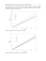
A dendogram representing all reference strains and clinical isolates is depicted in Figure 1.
The AFLP-patterns of the reference strains clearly show that each species forms a distinct
cluster, with the following cophenetic values: C. albicans: 78; C. dubliniensis: 92; C. glabrata:
99; C. krusei: 84; C. pseudotropicalis: 98; C. tropicalis: 85; C. parapsilosis: 91; C. lusitaniae:
98; C. guilliermondii: 94. These results were highly reproducible.
The C. albicans isolates show two main clusters. One cluster contains clinical isolates from
the VUMC and the SENTRY collection as well as reference strains from the CBS. The other
cluster only contains isolates from the SENTRY collection. There is no clear relation between
these clusters and the geographical origin or source of the isolates. North American C.
albicans isolates show a three-part division by several typing methods, such as RAPD,
multilocus enzyme electrophoresis (MLEE), and Southern blot hybridization with the
moderately repetitive C. albicans specific Ca3 probe. In South-Africa, an additional cluster is
found besides these same three clusters
4,18,28
. It will be interesting to investigate whether the
two AFLP clusters of C. albicans correspond with the North American or South-African
clusters.
The C. dubliniensis isolates also show two clusters, with remarkable high similarities (91%
and 98%) of the isolates within the clusters. One cluster contains all reference strains used and
one SENTRY clinical isolate, the other cluster is composed of SENTRY isolates only. Using
the C. dubliniensis-specific fingerprinting probe Cd25 on a panel of 98 isolates Gee et al. also
recognized two different clusters, one of which contained mainly isolates from HIV-infected
individuals while the other cluster contained mainly isolates derived from HIV-negative
individuals
9
. Strains CBS 7987 and CBS 7988, both part of the same AFLP cluster, are
isolated from an HIV-infected individual. However, data on the HIV-status of the patients of
which the other isolates were obtained (CBS 8500, CBS 8501, and SENTRY isolates) is
lacking. Further investigations are necessary to examine whether the AFLP clusters correspond
with the Cd25 clusters.
Another noteworthy finding is that all AFLP patterns for the C. glabrata isolates are very
similar (90%), except for the CBS reference strain (58%). This reference strain (CBS 138) was
isolated from human faeces in 1936. The fact that all other isolates studied were clinical
isolates which were isolated fairly recently may account for this difference.
The AFLP patterns of the 18 isolates from the VUMC all corresponded with the results of
the phenotypic identification (Germ-tube test and Vitek). The clinical isolates from the
European SENTRY collection were all originally identified on CHROMagar as being Candida
albicans. However, based on the AFLP patterns shown in Figure 1, some of these strains
presumably were misidentified and belong to different species. When the total collection of
isolates previously identified as C. albicans (n = 213) was screened with AFLP, a
misidentification rate of 6% was observed. Six strains are now identified as C. dubliniensis,
four as C. parapsilosis, one as C. tropicalis, and one as C. guilliermondii (results partly shown
in Figure 1).
CHROMagar identification of Candida species is based on differences in colony color. It
has been shown, that the reliability of this method depends on the incubation time and
temperature used
2,24,35
. However, even when optimum conditions are used, the method is
Chapter 7
85
Figure 1
Dendogram representing all reference strains and clinical isolates (see also Table 1)
10 0
90
80
70
60
50
40
30
20
10
CBS562
CBS1912
CBS1905
A
TCC9002
8
10A173
07C069
19A568
06A309
04A080
08E058
19A567
23D045
19A519
TY727
TY728
A
TCC9002
9
TY732
15A206
15A561
17A381
16C088
15A020
16A438
12E033
19A164
11C034
10C007
11A134
16A232
TY729
CBS8501
CBS7988
CBS8500
18A221
CBS7987
20C149
23A137
02A038
05C121
05C118
TY719
TY718
TY714
TY715
A
TCC9003
0
TY716
TY717
TY731
CBS138
TY723
TY722
TY726
CBS573
CBS 60 7
CBS94
CBS2310
11D028
TY739
TY737
TY736
A
TCC9001
8
TY735
07A212
CBS2195
10A120
14A161
10A311
CBS604
CBS4413
14A097
CBS2024
CBS566
C. albicans
C. dubliniensis
C. glabrata
C. krusei
C. pseudotropicalis
C. tropicalis
C. parapsilosis
C. lusitaniae
C. guilliermondii
AFLP as an identification method for Candida spp.
86
not ideal, and especially the differentiation between C. albicans and C. dubliniensis is
problematic. Kurzai et al. reported that only 81% of their C. dubliniensis isolates showed the
dark green color on CHROMagar, which is considered indicative for C. dubliniensis
17
.
Furthermore, 15.9% of their C. albicans isolates also showed a dark green coloration, instead
of the usual lighter green. Tintelnot et al. reported an even lower number of 57% of C.
dubliniensis isolates that showed the dark green coloration on CHROMagar, and only 48% of
the isolates of Kirkpatrick et al. showing the dark green coloration turned out to be C.
dubliniensis
15,31
.
Other commercial tests that allow (presumptive) identification of C. albicans as well as
non-albicans Candida species usually show high sensitivities and specificities for C. albicans,
but are less reliable or need further testing for the identification of other, less common,
species
3,5,8,12
. C. dubliniensis-specific PCR assays as well as generic PCR assays in
combination with species-specific probes have been developed
6,7,16,19,25
. The advantage of
AFLP, however, is that this method is based on ligation of known sequences (adapters) to
restriction fragments, which function as targets for the PCR primers. Therefore, the technique
is universally applicable. In the current assay we made use of two subsequent amplifications,
but similar results were obtained when only the second amplification was used (unpublished
observations). The use of an internal size standard with every sample for normalization
purposes greatly enhances the reproducibility between tests. Storing all patterns, including
those of the reference strains, in a general accessible database will provide a screening library
for identification of species.
Two other universally applicable methods for identification of Candida species have been
described: PCR fingerprinting and reference strand-mediated conformational analysis
(RSCA)
20,21
. However, whereas PCR fingerprinting uses mini- and microsatellite sequences as
targets for the primers and RSCA is based on 18S rRNA sequences, AFLP patterns are a
representation of the whole genome. Our results show very clear differences between
medically important Candida species. Therefore, AFLP might prove to be a reliable method
for the identification of medically important Candida species, including Candida dubliniensis.
A
CKNOWLEDGEMENTS
Annemarie Borst is supported by a grant from bioMérieux (formerly Organon Teknika).
R
EFERENCES
1. Abi Said, D., E. Anaissie, O. Uzun, I. Raad, H. Pinzcowski, and S. Vartivarian. 1997. The epidemiology
of hematogenous candidiasis caused by different Candida species. Infect. Dis. 24: 1122-1128
2. Baumgartner, C., A.M. Freydiere, and Y. Gille. 1996. Direct identification and recognition of yeast
species from clinical material by using albicans ID and CHROMagar Candida plates. J. Clin. Microbiol. 34:
454-456
Chapter 7
87
3. Bernal, S., M.E. Martin, M. Chavez, J. Coronilla, and A. Valverde. 1998. Evaluation of the new API
Candida system for identification of the most clinically important yeast species. Diagn. Microbiol. Infect.
Dis. 32: 217-221
4. Blignaut, E., C. Pujol, S. Lockhart, S. Joly, and D.R. Soll. 2002. Ca3 Fingerprinting of Candida albicans
Isolates from Human Immunodeficiency Virus-Positive and Healthy Individuals Reveals a New Clade in
South Africa. J. Clin. Microbiol. 40: 826-836
5. Campbell, C.K., K.G. Davey, A.D. Holmes, A. Szekely, and D.W. Warnock. 1999. Comparison of the
API Candida system with the AUXACOLOR system for identification of common yeast pathogens. J. Clin.
Microbiol. 37: 821-823
6. Donnelly, S.M., D.J. Sullivan, D.B. Shanley, and D.C. Coleman. 1999. Phylogenetic analysis and rapid
identification of Candida dubliniensis based on analysis of ACT1 intron and exon sequences. Microbiology
145 ( Pt 8): 1871-1882
7. Elie, C.M., T.J. Lott, E. Reiss, and C.J. Morrison. 1998. Rapid identification of Candida species with
species-specific DNA probes. J. Clin. Microbiol. 36: 3260-3265
8. Espinel-Ingroff, A., L. Stockman, G. Roberts, D. Pincus, J. Pollack, and J. Marler. 1998. Comparison of
RapID yeast plus system with API 20C system for identification of common, new, and emerging yeast
pathogens. J. Clin. Microbiol. 36: 883-886
9. Gee, S.F., S. Joly, D.R. Soll, J.F. Meis, P.E. Verweij, I. Polacheck, D.J. Sullivan, and D.C. Coleman.
2002. Identification of four distinct genotypes of Candida dubliniensis and detection of microevolution in
vitro and in vivo. J. Clin. Microbiol. 40: 556-574
10. Giamarellou, H. and A. Antoniadou. 1996. Epidemiology, diagnosis, and therapy of fungal infections in
surgery. Infect. Control Hosp. Epidemiol. 17: 558-564
11. Gumbo, T., C.M. Isada, G. Hall, M.T. Karafa, and S.M. Gordon. 1999. Candida glabrata Fungemia.
Clinical features of 139 patients. Medicine (Baltimore) 78: 220-227
12. Hoppe, J.E. and P. Frey. 1999. Evaluation of six commercial tests and the germ-tube test for presumptive
identification of Candida albicans. Eur. J. Clin. Microbiol. Infect. Dis. 18: 188-191
13. Jones, C.J., K.J. Edwards, S. Castaglione, M.O. Winfield, F. Sala, C. VandeWiel, G. Bredemeijer, B.
Vosman, M. Matthes, A. Daly, R. Brettschneider, P. Bettini, M. Buiatti, E. Maestri, A. Malcevschi, N.
Marmiroli, R. Aert, G. Volckaert, J. Rueda, R. Linacero, A. Vazquez, and A. Karp. 1997.
Reproducibility testing of RAPD, AFLP and SSR markers in plants by a network of European laboratories.
Molecular Breeding 3: 381-390
14. Kao, A.S., M.E. Brandt, W.R. Pruitt, L.A. Conn, B.A. Perkins, D.S. Stephens, W.S. Baughman, A.L.
Reingold, G.A. Rothrock, M.A. Pfaller, R.W. Pinner, and R.A. Hajjeh. 1999. The epidemiology of
candidemia in two United States cities: results of a population-based active surveillance. Clin. Infect. Dis. 29:
1164-1170
15. Kirkpatrick, W.R., S.G. Revankar, R.K. Mcatee, J.L. Lopez-Ribot, A.W. Fothergill, D.I. McCarthy,
S.E. Sanche, R.A. Cantu, M.G. Rinaldi, and T.F. Patterson. 1998. Detection of Candida dubliniensis in
oropharyngeal samples from human immunodeficiency virus-infected patients in North America by primary
CHROMagar candida screening and susceptibility testing of isolates. J. Clin. Microbiol. 36: 3007-3012
16. Kurzai, O., W.J. Heinz, D.J. Sullivan, D.C. Coleman, M. Frosch, and F.A. Muhlschlegel. 1999. Rapid
PCR test for discriminating between Candida albicans and Candida dubliniensis isolates using primers
derived from the pH-regulated PHR1 and PHR2 genes of C. albicans. J. Clin. Microbiol. 37: 1587-1590
17. Kurzai, O., H.C. Korting, D. Harmsen, W. Bautsch, M. Molitor, M. Frosch, and F.A. Muhlschlegel.
2000. Molecular and phenotypic identification of the yeast pathogen Candida dubliniensis. J. Mol. Med. 78:
AFLP as an identification method for Candida spp.
88
521-529
18. Lott, T.J. and M.M. Effat. 2001. Evidence for a more recently evolved clade within a Candida albicans
North American population. Microbiology 147: 1687-1692
19. Martin, C., D. Roberts, M. Van der Weide, R. Rossau, G. Jannes, T. Smith, and M. Maher. 2000.
Development of a PCR-based line probe assay for identification of fungal pathogens. J. Clin. Microbiol. 38:
3735-3742
20. McIlhatton, B.P., C. Keating, M.D. Curran, M.F. McMullin, J.G. Barr, J.A. Madrigal, and D.
Middleton. 2002. Identification of medically important pathogenic fungi by reference strand-mediated
conformational analysis (RSCA). J. Med. Microbiol. 51: 468-478
21. Meyer, W., K. Maszewska, and T.C. Sorrell. 2001. PCR fingerprinting: a convenient molecular tool to
distinguish between Candida dubliniensis and Candida albicans. Med. Mycol. 39: 185-193
22. Moran, G.P., D. Sanglard, S.M. Donnelly, D.B. Shanley, D.J. Sullivan, and D.C. Coleman. 1998.
Identification and expression of multidrug transporters responsible for fluconazole resistance in Candida
dubliniensis. Antimicrob. Agents Chemother. 42: 1819-1830
23. Moran, G.P., D.J. Sullivan, M.C. Henman, C.E. McCreary, B.J. Harrington, D.B. Shanley, and D.C.
Coleman. 1997. Antifungal drug susceptibilities of oral Candida dubliniensis isolates from human
immunodeficiency virus (HIV)-infected and non-HIV-infected subjects and generation of stable fluconazole-
resistant derivatives in vitro. Antimicrob. Agents Chemother. 41: 617-623
24. Odds, F.C. and A. Davidson. 2000. "Room temperature" use of CHROMagar Candida. Diagn. Microbiol.
Infect. Dis. 38: 147-150
25. Park, S., M. Wong, S.A. Marras, E.W. Cross, T.E. Kiehn, V. Chaturvedi, S. Tyagi, and D.S. Perlin.
2000. Rapid identification of Candida dubliniensis using a species-specific molecular beacon. J. Clin.
Microbiol. 38: 2829-2836
26. Pfaller, M.A., R.N. Jones, G.V. Doern, A.C. Fluit, J. Verhoef, H.S. Sader, S.A. Messer, A. Houston, S.
Coffman, and R.J. Hollis. 1999. International surveillance of blood stream infections due to Candida
species in the European SENTRY Program: species distribution and antifungal susceptibility including the
investigational triazole and echinocandin agents. SENTRY Participant Group (Europe). Diagn. Microbiol.
Infect. Dis. 35: 19-25
27. Pittet, D., N. Li, and R.P. Wenzel. 1993. Association of secondary and polymicrobial nosocomial
bloodstream infections with higher mortality. Eur. J. Clin. Microbiol. Infect. Dis. 12: 813-819
28. Pujol, C., S. Joly, S.R. Lockhart, S. Noel, M. Tibayrenc, and D.R. Soll. 1997. Parity among the randomly
amplified polymorphic DNA method, multilocus enzyme electrophoresis, and Southern blot hybridization
with the moderately repetitive DNA probe Ca3 for fingerprinting Candida albicans. J. Clin. Microbiol. 35:
2348-2358
29. Savelkoul, P.H., H.J. Aarts, J. de Haas, L. Dijkshoorn, B. Duim, M. Otsen, J.L. Rademaker, L.
Schouls, and J.A. Lenstra. 1999. Amplified-fragment length polymorphism analysis: the state of an art. J.
Clin. Microbiol. 37: 3083-3091
30. Sullivan, D. and D. Coleman. 1998. Candida dubliniensis: characteristics and identification. J. Clin.
Microbiol. 36: 329-334
31. Tintelnot, K., G. Haase, M. Seibold, F. Bergmann, M. Staemmler, T. Franz, and D. Naumann. 2000.
Evaluation of phenotypic markers for selection and identification of Candida dubliniensis. J. Clin. Microbiol.
38: 1599-1608
32. Vos, P., R. Hogers, M. Bleeker, M. Reijans, T. Van de Lee, M. Hornes, A. Frijters, J. Pot, J. Peleman,
M. Kuiper, and M. Zabeau. 1995. AFLP: a new technique for DNA fingerprinting. Nucleic Acids Res. 23:
Chapter 7
89
4407-4414
33. Wenzel, R.P. 1995. Nosocomial candidemia: risk factors and attributable mortality. Clin. Infect. Dis. 20:
1531-1534
34. Wey, S.B., M. Mori, M.A. Pfaller, R.F. Woolson, and R.P. Wenzel. 1988. Hospital-acquired candidemia.
The attributable mortality and excess length of stay. Arch. Intern. Med. 148: 2642-2645
35. Willinger, B., C. Hillowoth, B. Selitsch, and M. Manafi. 2001. Performance of Candida ID, a new
chromogenic medium for presumptive identification of Candida species, in comparison to CHROMagar
Candida. J. Clin. Microbiol. 39: 3793-3795
VIII: High levels of hydrolytic enzymes secreted by
Candida albicans isolates involved in pneumonia
A. Borst and A.C. Fluit
Eijkman-Winkler Center, University Medical Center, Utrecht, the Netherlands
Submitted for publication.
High levels of hydrolytic enzymes
92
A
BSTRACT
The differences in production of two putative virulence factors of Candida albicans,
(phospho)lipase and proteinase, were determined for a large (n = 186) panel of clinical C.
albicans isolates obtained from the European SENTRY program. Seventy-two percent of the
isolates produced detectable amounts of (phospho)lipase, 95% of the isolates produced
detectable amounts of proteinase. There was no clear correlation between the results of the
(phospho)lipase- and proteinase assays and the geographical distribution of the isolates.
However, isolates which originated from pneumonia produced significantly higher amounts of
(phospho)lipase than isolates obtained from blood, the urinary tract or wound/skin/soft tissue,
and also appeared to produce more proteinase. It is hypothesized that these virulent isolates
involved in pneumonia originate from the oral cavity. Whether these results are caused by
selection for these high virulent isolates remains to be solved.
I
NTRODUCTION
The opportunistic pathogen Candida albicans is considered to be the most virulent of the
Candida species. Several putative virulence factors of C. albicans have been described,
including phenotypic switching, host recognition biomolecules (adhesins), morphogenesis (the
reversible transition between unicellular yeast cells and filamentous, growth forms), and
secreted hydrolytic enzymes
1
. Two types of secreted enzymes have been described extensively:
phospholipases and secreted aspartyl proteinases.
C. albicans strains show phospholipase B as well as lysophospholipase-transacylase
activities. Both activities are performed by a single enzyme, C. albicans phospholipase B
(caPLB). Besides this secreted phospholipase, C. albicans also shows a phospholipase D
activity which appears to be membrane-associated
12
. The data on phospholipases in pathogenic
fungi has been reviewed by Ghannoum
6
.
Several researchers have found indications that phospholipases are virulence factors of C.
albicans. Ibrahim et al. compared the ability to produce phospholipases of clinical blood
isolates with oral strains from healthy volunteers. Significantly more phospholipase activity
was detected in the clinical isolates. Furthermore, in a mouse-model of haematogenously
disseminated candidiasis, a C. albicans strain which produced high amounts of phospholipases
was invasive whereas a low-producing strain was not, and phospholipase-activity was the only
putative virulence factor tested that predicted mortality
9
. In another animal pathogenicity
study, a significant correlation was found between phospholipase activity and the severity of
kidney-infections
10
. The ultimate proof was delivered by Leidich et al., who cloned and
disrupted a gene encoding for PLB and showed that the null mutant significantly attenuated
virulence in mice and dramatically reduced the ability of the yeast to penetrate host cells
11
.
Disruption of the gene did not affect adherence of the yeast cells to human endothelial or
epithelial cells. Thus, phospholipases most likely contribute to the pathogenicity of C. albicans
by damaging host cell membranes, aiding the fungus by invasion of host tissues. This role in
invasion is also implied by the finding that phospholipases are mainly concentrated at the tips
of the fungal hyphae
15
. Besides phospholipases, C. albicans strains also secrete lipases. It is
Chapter 8
93
likely that these enzymes, like phospholipases, are involved in virulence of C. albicans
5,8
.
The role of C. albicans secreted aspartyl proteinases in pathogenicity has recently been
reviewed by De Bernardis et al.
4
. These enzymes are encoded by a family of at least nine
genes, and are capable of degrading epithelial and mucosal barrier proteins like collagen,
keratin and mucin, as well as antibodies, complement and cytokines. Gene disruption
experiments showed altered adherence of yeast cells and attenuation of virulence in different
animal models
2,7,17,18
.
The expression of virulence factors may be associated with specific characteristics of
Candida isolates such as geographic origin or the type of infection. Knowledge of such
correlations may help to understand the epidemiology of these infections, which may result in
improved therapeutic regimens. Price et al. developed a simple egg yolk agar plate assay for
the detection of (phospho)lipase activity. Hydrolysis of lipid substrates present in the egg yolk
results in the formation of a calcium complex with the fatty acids released by the action of the
secreted enzymes. The diameter of this zone of precipitation around the colonies is very
constant for any given isolate, and correlates well with a biochemical assay for hydrolysis of
phosphatidylcholine
14
. Although this method does not detect (phospho)lipase activity in fungal
isolates that produce very low levels of phospholipase
6
, it is an excellent screening method for
large numbers of isolates. Therefore, we used this method to investigate the differences in
(phospho)lipase activity of a large collection of clinical C. albicans isolates obtained from 12
European countries, and the results were linked to data on the geographic origin of the isolates
and the site of infection. For the detection of proteinase activity we incorporated bovine serum
albumin (BSA) into YCB-agar plates and measured the clearing zone after staining with
Coomassie blue.
M
ATERIALS AND METHODS
Yeast strains. Candida albicans isolates were obtained from the European SENTRY
program. Only one isolate per patient was included. A total of 186 isolates derived from 19
medical centers in 12 European countries were studied (Table 1). One-hundred-and-thirty-one
isolates (70%) originated from infections from blood, 7 (4%) from wounds/skin/soft tissue, 25
(13%) from the urinary tract, and 23 (12%) from pneumonia. Most isolates were derived from
the intensive care (36%), internal medicine (15%), surgery (14%), pediatrics (12%) or
oncology ward (6%). The number of isolates derived from the most relevant hospital wards in
relation to the site of infection is depicted in Table 2. Identification of the isolates was
performed using CHROMagar plates (CHROMagar, Paris, France). The isolates were cultured
on Blood Agar and subcultured on Sabouraud Dextrose Agar (SDA) at 37°C.
(Phospho)lipase assay. SDA plates supplemented with 1 M NaCl, 5 mM CaCl
2
and 8%
sterile egg yolk (Oxoid, Basingstoke, England) were inoculated with 1 µl sterile saline
containing approximately 10
5
cfu C. albicans, and incubated at 37°C for three days. Each
isolate was tested in duplicate. The diameter of the colonies and the total diameter of the
colonies plus precipitation zones were measured. (Phospho)lipase activity was determined by
the ratio of the diameter of the colony plus precipitation zone to the diameter of the colony
alone, and scored as follows: - = no precipitation zone; +/- = ratio between 1.01 and 1.25; + =
High levels of hydrolytic enzymes
94
ratio between 1.26 and 1.50; ++ = ratio between 1.51 and 1.75; +++ = ratio between 1.76 and
2.00; ++++ = ratio between 2.01 and 2.25.
Proteinase assay. YCB-BSA plates (1.5% agar; 1.17% Yeast Carbon Base powder (Becton
Dickinson, Le Pont de Claix, France); 0.2% Bovine Serum Albumin (Instruchemie, Hilversum,
the Netherlands); 0.2% glucose; 100 µl/l Vitox solution (Oxoid)) were inoculated with 1 µl
sterile saline containing approximately 10
5
cfu C. albicans, and incubated at 25°C for three
weeks. Several isolates were tested twice or more. The plates were stained with Coomassie
brilliant blue (0.5% Coomassie brilliant blue R250 (Pierce, Rockford, USA); 10% v/v acetic
acid; 45% v/v ethanol) for 20 min. at room temperature, and destained three times with
destaining solution (10% v/v acetic acid; 45% v/v ethanol) for 20 min. at 37°C and one time
with water for 20 min. at 37°C. The diameter of the colonies was measured before Coomassie
staining, the diameter of the clear zones was measured after staining. Proteinase activity was
determined by the ratio of the diameter of the clear zone to the diameter of the colony, and
scored as follows: - = no clear zone; +/- = ratio < 0.9 (clear zone smaller than colony, limited
proteinase activity); + = ratio between 0.9 and 1.1 (clear zone and colony of similar size); ++ =
ratio > 1.1 (clear zone clearly larger than colony).
Table 1
The geographic origin of the isolates used in this study
Country Center No. of isolates (%)
Austria Linz 3 (2)
France Paris
Lille
6 (3)
7 (4)
Germany Freiburg
Dusseldorf
9 (5)
7 (4)
Greece Athens 3 (2)
Italy Genoa
Roma
24 (13)
17 (9)
the Netherlands Utrecht 6 (3)
Poland Warsaw
Cracow
1 (1)
2 (1)
Portugal Coimbra 23 (12)
Spain Sevilla
Madrid
Barcelona
21 (11)
4 (2)
1 (1)
Switzerland Lausanne 14 (8)
Turkey Ankara
Istanbul
20 (11)
6 (3)
United Kingdom London 12 (6)
Total: 186 (100)
Chapter 8
95
Table 2
The number of isolates derived from the different hospital wards in relation to the site of infection
No. of isolates (%)
Source IC Int. med. Surgery Pediatrics Oncology Other Total
blood 39 (30) 22 (17) 22 (17) 18 (14) 11 (8) 19 (15) 131 (100)
pneumonia 18 (78) 0 (0) 0 (0) 0 (0) (0) 5 (22) 23 (100)
urinary tract 8 (32) 4 (16) 3 (12) 4 (16) 0 (0) 6 (24) 25 (100)
wound/s/st 2 (29) 1 (14) 2 (29) 0 (0) 0 (0) 2 (29) 7 (100)
IC: intensive care
Int. med.: internal medicine
wound/s/st: isolates originating from wounds, skin or soft tissue
RESULTS
(Phospho)lipase assay. One-hundred-and-eighty-six isolates were tested in the
(phospho)lipase assay. The number of isolates and the different scores are depicted in Table 3.
No (phospho)lipase activity was detected in 28% of the isolates. Duplicate testing of the
isolates only showed minor differences (average difference between duplicate tests: 0.08).
There was no clear correlation between the results of the (phospho)lipase assay and the
geographical distribution of the isolates.
The results of the (phospho)lipase assay in relation with the site of infection are shown in
Table 4. Of all strains obtained from blood (n = 133), the urinary tract (n = 25), or
wounds/skin/soft tissue (n = 7) that were tested in the (phospho)lipase assay, most isolates
were either negative or produced only low amounts of (phospho)lipase (-, +/-, or +)(blood:
64%, urinary tract: 72%, wound/sst: 85%). However, 61% of the isolates obtained from
pneumonia (n = 23) produced high amounts of lipase (++, +++, or ++++). This difference was
statistically significant (p = 0.042; Pearson chi-square test (exact)).
Table 3
Results of the (phospho)lipase assay
Score - +/- + ++ +++ ++++
No. isolates (%) 53 (28) 13 (7) 51 (27) 33 (18) 28 (15) 8 (4)
Table 4
Results of the (phospho)lipase assay in relation with the site of infection
No. of isolates (%)
Source - +/- + ++ +++ ++++ Total
blood 38 (29) 10 (8) 36 (27) 22 (17) 19 (15) 6 (5) 131 (100)
pneumonia 3 (13) 1 (4) 5 (22) 8 (35) 5 (22) 1 (4) 23 (100)
urinary tract 7 (28) 2 (8) 9 (36) 3 (12) 3 (12) 1 (4) 25 (100)
wound/s/st 5 (71) 0 (0) 1 (14) 0 (0) 1 (14) 0 (0) 7 (100)
wound/s/st: isolates originating from wounds, skin or soft tissue