AIRWAY MANAGEMENT IN EMERGENCIES - PART 1 pot
Bạn đang xem bản rút gọn của tài liệu. Xem và tải ngay bản đầy đủ của tài liệu tại đây (361.45 KB, 24 trang )
AIRWAY MANAGEMENT
IN EMERGENCIES
᭤ NOTICE
Medicine is an ever-changing science. As new research and clinical experience broaden our
knowledge, changes in treatment and drug therapy are required. The authors and the publisher
of this work have checked with sources believed to be reliable in their efforts to provide infor-
mation that is complete and generally in accord with the standards accepted at the time of pub-
lication. However, in view of the possibility of human error and changes in medical sciences,
neither the editors nor the publisher nor any other party who has been involved in the prepara-
tion or publication of this work warrants that the information contained herein is in every respect
accurate or complete, and they disclaim all responsibility for any errors or omissions or for the
results obtained from use of the information contained in this work. Readers are encouraged to
confirm the information contained herein with other sources. For example and in particular, readers
are advised to check the product information sheet included in the package of each drug they
plan to administer to be certain that the information contained in this work is accurate and that
changes have not been made in the recommended dose or in the contraindications for adminis-
tration. This recommendation is of particular importance in connection with new or infrequently
used drugs.
AIRWAY MANAGEMENT
IN EMERGENCIES
GEORGE KOVACS, MD
Professor
Department of Emergency Medicine
Dalhousie University
Nova Scotia, Halifax, Canada
and
J. ADAM LAW, MD
Professor
Departments of Anesthesiology and Surgery
Dalhousie University
Nova Scotia, Halifax, Canada
New York Chicago San Francisco Lisbon London Madrid Mexico City
Milan New Delhi San Juan Seoul Singapore Sydney Toronto
Copyright © 2008 by The McGraw-Hill Companies, Inc. All rights reserved. Manufactured in the United States of America. Except
as permitted under the United States Copyright Act of 1976, no part of this publication may be reproduced or distributed in any form
or by any means, or stored in a database or retrieval system, without the prior written permission of the publisher.
0-07-159348-9
The material in this eBook also appears in the print version of this title: 0-07-147005-0.
All trademarks are trademarks of their respective owners. Rather than put a trademark symbol after every occurrence of a trademarked
name, we use names in an editorial fashion only, and to the benefit of the trademark owner, with no intention of infringement of the
trademark. Where such designations appear in this book, they have been printed with initial caps.
McGraw-Hill eBooks are available at special quantity discounts to use as premiums and sales promotions, or for use in corporate
training programs. For more information, please contact George Hoare, Special Sales, at or (212)
904-4069.
TERMS OF USE
This is a copyrighted work and The McGraw-Hill Companies, Inc. (“McGraw-Hill”) and its licensors reserve all rights in and to the
work. Use of this work is subject to these terms. Except as permitted under the Copyright Act of 1976 and the right to store and retrieve
one copy of the work, you may not decompile, disassemble, reverse engineer, reproduce, modify, create derivative works based upon,
transmit, distribute, disseminate, sell, publish or sublicense the work or any part of it without McGraw-Hill’s prior consent. You may
use the work for your own noncommercial and personal use; any other use of the work is strictly prohibited. Your right to use the work
may be terminated if you fail to comply with these terms.
THE WORK IS PROVIDED “AS IS.” McGRAW-HILL AND ITS LICENSORS MAKE NO GUARANTEES OR WARRANTIES
AS TO THE ACCURACY, ADEQUACY OR COMPLETENESS OF OR RESULTS TO BE OBTAINED FROM USING THE
WORK, INCLUDING ANY INFORMATION THAT CAN BE ACCESSED THROUGH THE WORK VIA HYPERLINK OR
OTHERWISE, AND EXPRESSLY DISCLAIM ANY WARRANTY, EXPRESS OR IMPLIED, INCLUDING BUT NOT LIMITED
TO IMPLIED WARRANTIES OF MERCHANTABILITY OR FITNESS FOR A PARTICULAR PURPOSE. McGraw-Hill and its
licensors do not warrant or guarantee that the functions contained in the work will meet your requirements or that its operation will be
uninterrupted or error free. Neither McGraw-Hill nor its licensors shall be liable to you or anyone else for any inaccuracy, error or
omission, regardless of cause, in the work or for any damages resulting therefrom. McGraw-Hill has no responsibility for the content
of any information accessed through the work. Under no circumstances shall McGraw-Hill and/or its licensors be liable for any
indirect, incidental, special, punitive, consequential or similar damages that result from the use of or inability to use the work, even if
any of them has been advised of the possibility of such damages. This limitation of liability shall apply to any claim or cause
whatsoever whether such claim or cause arises in contract, tort or otherwise.
DOI: 10.1036/0071470050
We hope you enjoy this
McGraw-Hill eBook! If
you’d like more information about this book,
its author, or related books and websites,
please click here.
Professional
Want to learn more?
From GK. To my partner in life, Sandra Kovacs, and my four children,
Hannah, Maya, Ben, and Aaron: thank you for your love, tolerance,
and support.
From JAL. For Trevor, Simon, and Julia—may your love of life and learning
always be with you—and my wife Kate, for her support and loyalty.
Thanks must also go to my parents, for laying the foundation,
and to fellow AIME contributors and instructors, for making a
difference.
This page intentionally left blank
This page intentionally left blank
Contents
Editors and Lead Authors/Contributing Authors ix
Illustrations/Photography x
Foreword xi
Preface xii
Acknowledgments xiii
1. Introduction 1
2. Definitive Airway Management: When Is It Time? 5
03. Airway Physiology and Anatomy 13
04. Oxygen Delivery Devices and Bag-Mask Ventilation 33
15. Tracheal Intubation by Direct Laryngoscopy 53
06. Alternative Intubation Techniques 93
07. Rescue Oxygenation 127
8. How to do Awake Tracheal Intubations—Oral and Nasal 151
9. Rapid Sequence Intubation—Why and How to do it 169
10. Postintubation Management 179
11. Approach to Tracheal Intubation 187
12. Response to an Encountered Difficult Airway 199
13. Airway Pharmacology 211
14. Central Nervous System Emergencies 237
15. Cardiovascular Emergencies 245
16. Respiratory Emergencies 251
17. The Critically Ill Patient 259
vii
For more information about this title, click here
18. The Very Young and the Very Old Patient 265
19. Prehospital Airway Management Considerations 275
20. Human Factors in Airway Management 283
Index 291
viii CONTENTS
This page intentionally left blank
This page intentionally left blank
laryngoscopy and intubation have estimated
that up to 50 intubations are required before a
predetermined level of proficiency is reached.
3–6
Although a prerequisite minimum number of
intubations alone will never guarantee compe-
tence or ensure safety, the message that volume
matters and practice improves skills cannot be
disputed.
Skills transfer from simulation to the live set-
ting is not perfect, and depends in part on the
degree of similarity between the two settings.
7,8
Although the airway equipment used in both
simulation and “live” airway management is
identical, the physical tissue interface used in
most simulators is still relatively immature com-
pared to the human patient. Imperfect as the
simulation setting may be, it does provide the
opportunity to attain the psychomotor skills
needed for many tools and techniques. In addi-
tion, instructors can manipulate the clinical con-
text to provide the learner with an opportunity
to address various cognitive and human factors
issues related to airway management.
Prior to this patient’s arrival (and during the
resuscitation) it is likely that the clinician will
have to acknowledge and deal with immediate
psychological (affective) issues. The ability to effec-
tively manage the patient in extremis requires more
than cognitive and psychomotor skill.
9
Excite-
ment, fear, and/or anxiety are all very real “gut”
emotions that even experienced clinicians will
feel on hearing the heads-up about this patient.
Professional athletes and actors acknowledge a
certain performance-enhancing effect associ-
ated with the stress of high-stakes events in their
respective areas of expertise. Unprepared, how-
ever, in times of extreme stress or near disas-
ter, it has been said that 10% of individuals will
naturally lead, 10% will be incapacitated, and
the remainder will neither lead nor flounder,
but are able to follow.
10
Successful resusci-
tative airway management requires effective
anticipation, communication, and leadership
skills in a team setting.
The major challenge in teaching and learn-
ing airway management for emergencies is to
create an integrated cognitive, psychomotor, and
affective network that promotes easy retrieval
and a rapid appropriate response to change.
Medical administrators, educators, and learners
all seem to have a natural affinity for line dia-
grams and algorithms. Rare is the medical text
that does not include such figures, and this book
is no exception (e.g., Fig. 11–3, and Fig. 12–1).
These algorithms support the three major
questions that must be addressed to manage
2 CHAPTER 1
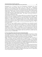
᭤ TABLE 1–1 AIRWAY MANAGEMENT CORE COMPETENCIES
Cognitive Psychomotor Affective
• Indications for advanced airway • Bag mask ventilation • Crisis Resource
management (including response to Management skills:
• Relevant airway anatomy and physiology difficulty) • Anticipation and
• Predictors of the difficult airway • Direct laryngoscopy and planning
• Approach to the difficult airway—whether intubation (including • Leadership and
predicted or not response to difficulty) communications
• Indications and contraindications for • Alternative intubation • Situation awareness
rapid sequence intubation and awake techniques • Team dynamics
intubation • Rescue oxygenation
• Airway pharmacology techniques, including
extraglottic devices and
cricothyrotomy
the airway, reinforced by experience, knowl-
edge, and skill:
A. Is the procedure indicated?
B. What is the safest and most efficacious way
to proceed when difficulty is anticipated
(see Fig. 11–3)?
C. How will you respond to difficulty once
encountered (see Fig. 12–1)?
Efforts to simplify airway management deci-
sion making have become somewhat clouded
in recent years by attempts to produce the
“Holy Grail” of airway tools. Such tools are
often marketed as requiring minimal skills and
possessing the potential to render the term
“difficult airway” obsolete. To this exploding
equipment industry can be added a growing
body of literature on managing the difficult air-
way. Is this devotion appropriate, or overkill?
In actual fact, we have not arrived at the point
where airway management decisions are black
and white, or our tools foolproof. Claims that
standard skills such as direct laryngoscopy are
soon to become procedures of the past are
likely premature. In addition, this direction car-
ries a significant risk of compromising the
acquisition and maintenance of competence in
needed core skills.
Successful airway management of the previ-
ously described case should not be defined sim-
ply by the correct placement of an endotracheal
tube. At the end of the day, success must be
measured by positive patient outcomes. To
improve these outcomes, the clinician must
work at enhancing the knowledge and skills
needed for successful airway management in
emergencies.
REFERENCES
1. Kovacs G, Croskerry P. Clinical decision making:
an emergency medicine perspective. Acad Emerg
Med. 1999;6(9):947–952.
2. Kovacs G, Bullock G, Ackroyd-Stolarz S, et al.
A randomized controlled trial on the effect of
educational interventions in promoting airway
management skill maintenance. Ann Emerg Med.
2000;36(4):301.
3. Charuluxananan S, Kyokong O, Somboonviboon
W, et al. Learning manual skills in spinal anesthe-
sia and orotracheal intubation: is there any recom-
mended number of cases for anesthesia residency
training program? J Med Assoc Thai. 2001;84 Suppl
1:S251–S5.
4. de Oliveira Filho GR. The construction of learning
curves for basic skills in anesthetic procedures: an
application for the cumulative sum method. Anesth
Analg. 2002;95(2):411–416.
5. Konrad C, Schupfer G, Wietlisbach M, et al. Learning
manual skills in anesthesiology: Is there a recom-
mended number of cases for anesthetic procedures?
Anesth Analg. 1998;86(3):635–639.
6. Mulcaster JT, Mills J, Hung OR, et al. Laryngoscopic
intubation: learning and performance. Anesthesiol-
ogy. 2003;98(1):23–27.
7. Issenberg SB, McGaghie WC, Hart IR, et al. Simu-
lation technology for health care professional
skills training and assessment. JAMA. 1999;282(9):
861–866.
8. Hall RE, Plant JR, Bands CJ, et al. Human patient
simulation is effective for teaching paramedic stu-
dents endotracheal intubation. Acad Emerg Med.
2005;12(9):850–855.
9. Schull MJ, Ferris LE, Tu JV, et al. Problems for clin-
ical judgement: 3. Thinking clearly in an emer-
gency. CMAJ. 2001;164(8):1170–1175.
10. Leach J. Why people ‘freeze’ in an emergency:
temporal and cognitive constraints on survival
responses. Aviat Space Environ Med. 2004;75(6):
539–542.
INTRODUCTION 3
This page intentionally left blank
This page intentionally left blank
The patients described in the above cases all
require urgent airway management. The ulti-
mate goal of resuscitation efforts and airway
management is gas exchange, with oxygen
delivery the priority. Although many clinicians
view endotracheal intubation as the definitive
intervention of airway management, other maneu-
vers, often perceived as basic, are all potentially
life saving. Recognition of the obstructed airway,
airway opening maneuvers, the administration
of high flow oxygen, and bag-mask ventilation
(BMV) are all crucial airway management skills.
In most cases it would be inappropriate to pro-
ceed with intubation before any of these basic
life support (BLS) interventions had first been
attempted.
Despite the importance of BLS maneuvers as
initial steps in correcting or maintaining oxy-
genation, many patients will go on to require
endotracheal intubation. A cuffed endotracheal
tube (ETT) placed below the cords provides both
airway protection and an efficient means of pro-
viding gas exchange. Although extraglottic devices
such as the Laryngeal Mask Airway (LMA) or
Esophageal-Tracheal Combitube (ETC) are also
very effective at providing gas exchange, place-
ment of an ETT remains a gold standard for airway
management in emergencies.
᭤ INDICATIONS FOR
ENDOTRACHEAL INTUBATION
There are four broad categories of indication for
endotracheal intubation in emergencies:
A. To obtain and maintain a patent airway
(e.g., in the face of an obstructed airway
from any cause).
B. To correct deficient gas exchange (i.e.,
hypoxia and/or hypercarbia).
C. To protect the airway (e.g., against aspira-
tion of gastric contents or blood).
D. To preempt predicted clinical deteriora-
tion (to one of the above three situations).
Obtain and Maintain
Airway obstruction can occur from functional,
pathologic, or mechanical causes. Functional
obstruction can occur in the patient with a
6 CHAPTER 2
᭤ Case 2.3
A 35-year-old female, 8 months pregnant,
was involved in a motor vehicle collision
(MVC). At the scene she was complaining of
right-sided chest discomfort and pain in what
appeared to be a broken right arm. She was
transported by ambulance to the ED “back-
boarded and collared.” She is now uncon-
scious, and has snoring respirations. Only
her systolic BP is obtainable at 50/, HR 140,
RR 35; SaO
2
is unobtainable on a 40% sim-
ple facemask, and her GCS is 7.
᭤ Case 2.4
A 55-year-old male was in a house fire. Although
his burns seemed limited, 6 hours after the
injury he started complaining of shortness
of breath, and subsequently developed stri-
dor on inspiration. His BP is 160/90, HR 90,
RR 30, SaO2 92% with NRFM, and his GCS
is 15.
᭤ Case 2.5
A 35-year-old female, well known in the
intensive care unit, has been receiving maxi-
mal medical therapy for an acute exacerbation
of asthma. She remains “tight” and is moving
very little air. Although her SaO
2
is 91% with
oxygen, she is visibly tired and getting drowsy.
Her BP is 170/100, HR 120, RR 30, SaO
2
91%
with NRFM, and her GCS is 14.
depressed level of consciousness, as loss of mus-
cular tone results in posterior relaxation of the
soft palate, tongue, and epiglottis toward the
posterior pharyngeal wall. Functional obstruc-
tion will most often be alleviated by BLS
maneuvers such as head tilt or chin lift (unless
contraindicated by C-spine precautions in the
trauma patient), or, more effectively, a jaw
thrust. If respiratory effort is still present, ade-
quate gas exchange can then resume, although
intubation may still be indicated to maintain
ongoing airway patency. In the apneic patient,
initial BLS maneuvers are still indicated to assess
and establish airway patency, but positive pres-
sure ventilation with BMV will be the next step
to reoxygenate the patient. Here again, unless
the cause of the apnea can be rapidly corrected,
intubation will be indicated to maintain a patent
airway.
Pathologic airway obstruction may result
from an intrinsic process such as edema,
hematoma, infection, or tumor, while mechanical
obstruction can occur from extrinsic processes
such as excessive application of cricoid pressure
or foreign body. Pathologic airway obstruction
is rarely quickly corrected and often requires
intubation to obtain and maintain a patent air-
way while the underlying cause of obstruction
is addressed.
Regardless of the nature of obstruction, it is
crucial that the signs and symptoms of obstruc-
tion (discussed in more detail in Chap. 4) be
recognized early and addressed promptly to
safely secure the airway.
Correction of Gas Exchange
Cellular metabolism and function depends on
the delivery and uptake of oxygen. Oxygen
delivery in turn depends on adequate lung func-
tion, sufficient hemoglobin levels, and an effec-
tive cardiac output. In return, carbon dioxide
(CO
2
) produced as a byproduct of cellular
metabolism must be delivered to the lungs for
removal by ventilation.
Respiratory failure is a clinical term describing
inadequate pulmonary gas exchange. Inade-
quate oxygenation (hypoxemia) can be quanti-
fied through the measurement of arterial blood
gases (ABGs) or estimated noninvasively using
pulse oximetry. Early clinical effects of hypox-
emia are not always readily apparent. Cyanosis
is a late clinical sign of hypoxemia and may be
absent in profoundly anemic patients or in those
with dark skin. Ventilation refers to the mechan-
ics of effective gas exchange, and is commonly
quantified using arterial Pco
2
. An acutely elevated
P
CO
2
is often clinically apparent as CO
2
narco-
sis with a diminished level of consciousness,
frequently combined with an inadequate respira-
tory effort.
Despite the fact that respiratory failure can
be determined by ABGs (i.e., Po
2
less than
60 mm Hg/Pco
2
greater than 60 mm Hg), the
decision to intervene with airway and venti-
latory support should be a clinical one, and
in most situations, precede ABG testing.
Although failure of oxygenation and ventila-
tion usually occur together, this is not always
the case. Critically ill asthmatics may be able to
maintain an SaO
2
above 90% with supplemen-
tal oxygen, but still require ventilatory support
as they fatigue. Furthermore, a patient in circu-
latory shock may have no ventilatory abnormal-
ities but may still require intubation to optimize
oxygen delivery.
Included in this category is the need for “pul-
monary toilet,” that is, the suctioning of secre-
tions from the lower airway of patients who
cannot adequately cough.
Protection
The awake patient with intact airway reflexes is
able to respond to secretions or other material
threatening the airway by swallowing and/or
coughing. Although the gag reflex is commonly
assessed as a measure of airway protection, its
utility has been questioned following findings
that up to a third of the general population
DEFINITIVE AIRWAY MANAGEMENT: WHEN IS IT TIME? 7
has an attenuated or absent gag reflex.
1,2
Fur-
thermore, testing for the gag reflex can itself be
hazardous, with the risk of provoking vomiting.
As with any reflex, intact and coordinated
sensory and motor pathways must exist through
a central connection.
3
Protective airway reflexes
become diminished as the patient’s level of con-
sciousness decreases. Rigid suction should always
be available during airway interventions and
the clinician should be prepared to rapidly suction
and safely reposition the patient.
The patient’s ability to swallow and cough
may be thought of as confirming intact protec-
tive reflexes. However, the effectiveness of these
reflexes in managing significant vomitus or
blood in a patient with a depressed level of con-
sciousness is always uncertain. The presence of
pooled secretions or fluid in the posterior phar-
ynx is strongly suggestive of impaired airway
protective reflexes, as is the ability to tolerate an
oropharyngeal airway.
The Glasgow Coma Scale (GCS) is often used
as a gross marker of a patient’s ability to protect
the airway.
4
The Advanced Trauma Life Support
(ATLS) program recommends that patients with a
GCS below 8 should be intubated, unless a rapid
improvement in level of consciousness occurs or
is anticipated.
5
Unfortunately, clinical application
of the GCS is fraught with difficulties as a prospec-
tive decision tool.
6–8
Rather than rigidly using a
certain GCS cutoff, the patient’s clinical ability to
handle secretions should be assessed in conjunc-
tion with level of consciousness (as measured by
GCS or otherwise).
᭤ Predicted Clinical Deterioration
The foregoing discussion refers to assessing the
patient’s immediate need for intubation. How-
ever, the clinician should always be thinking of
the patient’s expected clinical course. This
includes consideration of the patient’s present-
ing condition, potential for deterioration, and
other factors such as the need to facilitate emer-
gent investigations (e.g., computed tomography
[CT] scan) or transportation to another institu-
tion. In this population, intubation may be desir-
able in anticipation of the patient’s risk of dete-
riorating, which would require intubation in a
less favorable environment (where adequately
trained personnel or appropriate equipment may
be lacking), or at a time when intubating may be
significantly more difficult, for example, due to
progressive airway edema.
It must be appreciated that active airway
interventions such as intubation are not without
complications. Intubation for the indications of
obtaining and maintaining a patent airway and
correction of gas exchange may be mandated
urgently as part of the “ABCs” (airway, breath-
ing, circulation) of resuscitation. On the other
hand, intubation for the sole indication of air-
way protection or predicted clinical deteriora-
tion is somewhat different, especially in a patient
who is currently maintaining a patent airway
with adequate gas exchange. In this latter situ-
ation, risk/benefit analysis may point to defer-
ring the procedure until better conditions and
expert personnel are available.
᭤ CASE REVIEW
The five cases presented earlier will be reviewed
here, with reference to the four categories of
indication for intubation discussed above.
8 CHAPTER 2
᭤ Case 2.1
A 20-year-old male with a fracture/dislocation
of his ankle has had it reduced under “proce-
dural sedation”. Some time later, the spouse of
the patient in the adjoining bay comes to get
help. She reports that the 20-year old is blue
and doesn’t appear to be breathing. The blood
pressure (BP) is 170/90, heart rate (HR) is
100, respiratory rate (RR) 4, and the oxygen
saturation (SaO
2
) is 65% on room air.
With the relief of pain following reduction
of his fracture, the patient became bradyp-
neic, as he had lost much of the stimulus that
was competing with the respiratory depres-
sive effect of the sedative/analgesic combina-
tion. Visible cyanosis is a late sign of oxygen
desaturation. The patient should be briefly
assessed for airway patency and respiratory
effort. His airway should be opened with head
tilt/chin lift/jaw thrust, and if spontaneous
respirations do not resume, positive pressure
BMV with oxygen should be rapidly insti-
tuted. Naloxone administration (with or with-
out the benzodiazepine antagonist Flumaze-
nil) will probably result in a rapid return of
spontaneous respirations and consciousness,
and intubation will most likely not be needed.
Other clinical states which may be reversible
before intubation is required, include the fol-
lowing:
• Ventricular arrhythmias—may respond to
defibrillation.
• Hypoglycemia—may respond to glucose.
• Anaphylaxis—may respond to epinephrine.
Concomitant basic airway management may
well still be indicated in these scenarios, and
depending on the response to treatment, intu-
bation may also be required.
In assessing the ABCs in this patient, snor-
ing is likely to be indicative of functional airway
obstruction, due to the patient’s obtunded state.
Other signs of functional obstruction may be
present, such as supra- and intercostal indraw-
ing, and a “rocking” pattern of respiration,
whereby the chest falls and the abdomen rises
with attempted inspiration. The airway should
be opened with head tilt/jaw thrust. An oral air-
way can be inserted. If the airway is now patent,
oxygen by nonrebreathing face mask should be
administered. The patient will require intuba-
tion for a number of reasons: airway mainte-
nance, airway protection, and predicted clinical
course. The condition of this patient is too ten-
uous for her to be sent to the diagnostic imaging
department without having an airway secured by
intubation.
DEFINITIVE AIRWAY MANAGEMENT: WHEN IS IT TIME? 9
᭤ Case 2.2
A 45-year-old female presents to the emer-
gency department (ED). Shortly before, while
at home, she had complained of a sudden-
onset severe headache, then collapsed. She
was transported by ambulance. On arrival,
she is receiving oxygen, but is unresponsive
and has snoring respirations. The BP is
180/100, HR 55, RR 25, SaO
2
92% with non-
rebreathing face mask (NRFM), and the Glas-
gow Coma Scale (GCS) is 7.
᭤ Case 2.3
A 35-year-old female, 8 months pregnant,
was involved in a motor vehicle collision
(MVC). At the scene she was complaining of
right-sided chest discomfort and pain in what
appeared to be a broken right arm. She was
transported by ambulance to the ED “back-
boarded and collared.” She is now uncon-
scious, and has snoring respirations. Only
her systolic BP is obtainable at 50/, HR 140,
RR 35; SaO
2
is unobtainable on a 40% sim-
ple facemask, and her GCS is 7.
This patient also has an airway which is func-
tionally obstructed at initial presentation. As a
trauma victim with a depressed level of con-
sciousness, C-spine precautions are in effect.
However, the front of the cervical collar should
be removed and replaced with manual in-line
stabilization. A jaw thrust may be performed to
open the airway, but head tilt and chin lift
should be omitted. 100% oxygen should be
administered. Concomitantly, the patient must
be removed from the supine position, as a
supine hypotension syndrome due to the
gravid uterus causing aorto-caval compression
may be causing or contributing to the hypoten-
sion. A wedge should be placed under the right
side of the spine board. Fluid resuscitation
should be initiated, and vital signs reevaluated.
In this case, relief of caval compression helped
restore preload, and BP rapidly reached 100/70.
The patient regained consciousness, and now
maintaining her own airway, did not acutely
require intubation.
therapy and her condition will worsen as she
tires. Hypoxia will ensue and as her CO
2
nar-
cosis progresses, she may also be unable to pro-
tect her airway.
᭤ SUMMARY
In the foregoing cases, some patients required
immediate attention with basic airway opening
maneuvers and only temporary airway support,
while others went on to require intubation.
Either way, an initial assessment of airway patency
and effectiveness of gas exchange should always
be made. Noninvasive maneuvers to main-
tain oxygenation and ventilation should be
undertaken as needed, while a determina-
tion is made about the subsequent need for
intubation. Intubation may be needed to obtain
and maintain an airway, correct gas exchange,
protect the airway, or for an anticipated adverse
predicted clinical course.
REFERENCES
1. Bleach NR. The gag reflex and aspiration: a ret-
rospective analysis of 120 patients assessed by
videofluoroscopy. Clin Otolaryngol Allied Sci.
1993;18(4):303–307.
2. Davies AE, Kidd D, Stone SP, et al. Pharyngeal sen-
sation and gag reflex in healthy subjects. Lancet.
25, 1995;345(8948):487–488.
3. Altschuler SM. Laryngeal and respiratory protective
reflexes. Am J Med. 3, 2001;111 (Suppl 8A):90S–94S.
10 CHAPTER 2
᭤ Case 2.4
A 55-year-old male was in a house fire. Although
his burns seemed limited, 6 hours after the
injury he started complaining of shortness
of breath, and subsequently developed
stridor on inspiration. His BP is 160/90, HR
90, RR 30, SaO
2
92% with NRFM, and his
GCS is 15.
The patient with an inhalational thermal injury
can develop progressive airway edema which
may eventually threaten airway patency. Critical
narrowing at the laryngeal inlet is often heralded
by inspiratory stridor. Stridor should generally be
regarded as a sign of impending complete air-
way obstruction. Intubation is indicated in this
patient to obtain and maintain a patent airway
and for predicted clinical deterioration. While
making preparations for intubation, elevation of
the head of the bed and the administration of a
helium-oxygen mixture (if immediately available)
may help temporize the situation.
9
The patient described in Case 2.5 is main-
taining her airway at present, and requires no
basic airway intervention other than oxygen
administration. However, intubation is indicated
for impaired gas exchange (her Pco
2
is steadily
climbing), and predicted clinical deterioration.
The patient has received maximal medical
᭤ Case 2.5
A 35-year-old female, well known in the inten-
sive care unit, has been receiving maximal
medical therapy for an acute exacerbation of
asthma. She remains “tight” and is moving
very little air. Although her SaO
2
is 91% with
oxygen, she is visibly tiring and getting drowsy.
Her BP is 170/100, HR 120, RR 30, SaO
2
91%
with NRFM, and her GCS is 14.
4. Mackay LE, Morgan AS, Bernstein BA. Swallowing
disorders in severe brain injury: risk factors affect-
ing return to oral intake. Arch Phys Med Rehabil.
1999;80(4):365–371.
5. Advanced Trauma Life Support for Doctors.
American College of Surgeons; 2004; No. 46.
6. Gill M, Windemuth R, Steele R, et al. A comparison
of the Glasgow Coma Scale score to simplified
alternative scores for the prediction of traumatic
brain injury outcomes. Ann Emerg Med. 2005;45(1):
37–42.
7. Gill MR, Reiley DG, Green SM. Interrater reliabil-
ity of Glasgow Coma Scale scores in the emergency
department. Ann Emerg Med. 2004;43(2):215–223.
8. Al-Salamah MA, McDowell I, Stiell IG, et al. Initial
emergency department trauma scores from the
OPALS study: the case for the motor score in blunt
trauma. Acad Emerg Med. 2004;11(8):834–842.
9. Ho AM, Dion PW, Karmakar MK, et al. Use of heliox
in critical upper airway obstruction. Physical and
physiologic considerations in choosing the optimal
helium:oxygen mix. Resuscitation. 2002;52(3):
297–300.
DEFINITIVE AIRWAYMANAGEMENT:WHEN IS IT TIME?
11
This page intentionally left blank
This page intentionally left blank
Alveolar Ventilation
Oxygen in the atmosphere
∗
moves along a pres-
sure gradient, through the respiratory tract and
alveoli, via arterial blood and capillaries to tis-
sues and cells. In the alveoli, the partial pres-
sure of oxygen (PAO
2
) drops from 150 mm Hg
to around 100 mm Hg, due to the balance of
oxygen uptake by the pulmonary capillaries and
its supply by ventilation. The partial pressure of
deoxygenated blood in the pulmonary capillaries,
returned to the lungs via the pulmonary arteries,
is about 40 mm Hg. Oxygen thus diffuses from
the alveoli to the pulmonary capillaries along a
pressure gradient.
In a perfect lung, blood leaving the pul-
monary capillaries via the pulmonary veins
would be fully oxygenated, that is, with no
alveolar/arterial PO
2
difference. In practice,
this does not happen because of a number of
factors:
• Ventilation-perfusion mismatch: Ideally
all alveoli would receive an equal share of
alveolar ventilation and all pulmonary capil-
laries would receive an equal share of car-
diac output. In reality, some alveoli are rela-
tively overventilated, while some are relatively
overperfused.
• Shunt occurs when alveoli are perfused
but receive no ventilation (an extreme
form of ventilation-perfusion [VQ] mis-
match). Deoxygenated blood has no chance
to become oxygenated and returns to the
pulmonary veins still in a deoxygenated
state. While physiologic drainage of intrin-
sic cardiac (thebesian) and bronchial veins
into the pulmonary venous blood will always
create a small degree of shunt, other causes
include atelectasis, lung consolidation
with fluid-filled alveoli, or small airway
closure.
• Diffusion abnormalities: Generally, diffu-
sion of oxygen from alveolus to capillary
along the pressure gradient is complete by
the time blood has traveled only one-third of
the way along the capillary. Diffusion is gen-
erally completed even if cardiac output is
increased (e.g., in exercise). Thus, the con-
tribution of any impairment in diffusion to
an alveolar/arterial PO
2
difference will be
minimal in the absence of significant pul-
monary disease.
Oxygen Transport in the Blood
Following its entry by diffusion into blood, oxygen
is carried in two ways:
• In combination with hemoglobin:
Each gram of hemoglobin can chemically
combine with a maximum of 1.31 mL of
oxygen: this is termed the oxygen capacity.
Thus, with a blood hemoglobin concen-
tration of 15 g/dL (150 g/L), each liter of
blood can carry (150 g/L × 1.31 mL/g) =
197 mL of oxygen. The term oxygen sat-
uration (SaO
2
) describes the percentage
of hemoglobin which is combined with
oxygen.
• Dissolved in plasma and intracellular
fluid: At atmospheric pressure, 0.3 mL/dL
(or 3 mL/L) of oxygen are carried in phys-
ical solution. Although a very small pro-
portion, this amount increases to 20 mL/L
by breathing 100% oxygen, and can be
raised even further under hyperbaric
conditions.
The arterial oxygen content thus reflects
the hemoglobin concentration, its percent satura-
tion with oxygen, and the amount of dissolved
14 CHAPTER 3
*Air is composed of 21% oxygen, 78% nitrogen, and 1% other
gases, at a pressure of 760 mm Hg at sea level. The partial
pressure of oxygen when first inspired is therefore (760)
(.21) = 159 mm Hg. Inspired air is humidified in the upper
airway, and as the total pressure exerted by a mixture of
gases is equal to the partial pressure of each of the compo-
nent gases, the partial pressure of water vapor (47 mm Hg)
drops the partial pressure of inspired oxygen, thus: (760–47)
(.21) = 150 mm Hg.
oxygen. In a patient with a hemoglobin of 15
g/dL and an SaO
2
of 95%, the arterial oxygen
content will be (.95 × 150 g/L × 1.31 mL/g) +
3 mL/L dissolved O
2
= 190 mL/L.
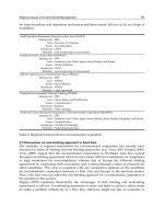
The relationship between the arterial partial
pressure of oxygen (PaO
2
) and SaO
2
is
described by the oxyhemoglobin dissociation
curve (Fig. 3–1). The flat upper portion of the
curve indicates that with an initial fall in PaO
2
,
the SaO
2
falls little, and the arterial oxygen con-
tent is little changed. However, as the PaO
2
continues to fall below 60 mm Hg, the slope of
the curve becomes steeper. While this steeper
part of the slope reflects easier offloading of
oxygen to the tissues, it also implies that once
oxygen desaturation begins, its progression is
quick. The linear portion of this curve can be
estimated by the 90–60, 60–30 rule of thumb,
whereby an SaO
2
of 90% corresponds roughly to
a PaO
2
of 60 and an SaO
2
of 60% corresponds
to a PaO
2
of 30.
The total quantity of oxygen available to
the tissues in one minute is termed oxygen
delivery (DO
2
), and equals the cardiac out-
put × arterial oxygen content. With a typical
cardiac output of 5 L/min, DO
2
is 5 L/min × 190
mL/L = approximately 1000 mL O
2
/min. In the
healthy, resting patient, VO
2
is 250 mL/min,
that is, 25% of available oxygen is consumed.
Thus, the hemoglobin in mixed venous
blood is 95% – 25% = 70% saturated. This
70% oxygen saturation of venous blood rep-
resents an important reserve from which
tissues can extract extra oxygen when com-
pensating for decreased DO
2
. Below a critical
value of DO
2
, however, compensation no
longer occurs and evidence of tissue hypoxia
occurs.
The foregoing discussion on DO
2
is clini-
cally relevant, as it points to areas which can
result in inadequate tissue oxygenation (i.e.,
tissue hypoxia):
A. Low cardiac output (stagnant or circula-
tory hypoxia). Even with a normal arterial
oxygen content, circulatory failure can result
AIRWAY PHYSIOLOGY AND ANATOMY 15
27 30 50 60 90 100
50
0
60
90
100
Pa
O
2
(mm Hg)
SaO
2
(%)
Good O
2
delivery to tissue
Right shift
Left shift
Poor O
2
delivery to tissue
pH
Temp
Pa
CO
2
pH
Temp
Pa
CO
2
Figure 3–1. The oxyhemoglobin dissociation curve.
in failure of tissue oxygenation, due to lack
of delivery of oxygen to the tissues. This
can happen globally, or regionally, with
inadequate blood flow to a particular organ.
Initially, tissues will compensate by increasing
oxygen extraction, but as perfusion wors-
ens, this becomes insufficient and tissue
hypoxia develops.
B. Low arterial oxygen saturation (hypoxic
hypoxia). This is defined as an inadequate
arterial PO
2
. This may result from many
causes, including decreased inspired par-
tial pressure of oxygen (e.g., at altitude);
hypoventilation from central (e.g., due to
sedative medications) or peripheral (e.g.,
functional airway obstruction) causes; or
from inadequate alveolar-capillary transfer
(e.g., from V-Q mismatch, shunt, or diffusion
abnormalities).
C. Low hemoglobin concentration (anemic
hypoxia). With profound anemia, oxygen
content will fall in proportion to the hemo-
globin concentration, even with a normal
PaO
2
. A compensatory increase in cardiac
output may occur, but if or when this can no
longer be sustained, tissue hypoxia occurs.
Alternatively, if hemoglobin is rendered inca-
pable of carrying oxygen, for example, by
carbon monoxide poisoning, a similar reduc-
tion in DO
2
can occur.
D. Histotoxic hypoxia. In spite of normal
delivery of oxygen to the tissues, cellular
metabolic processes utilizing oxygen can be
impaired, an example of which is cyanide
poisoning.
In the critically ill patient, VO
2
is often
increased, a factor over which we have little
control in the short term. Thus, in the early
phase of resuscitation, attention must be
directed to maximizing DO
2
, by avoiding oxygen
desaturation, as well as by maintaining or restor-
ing cardiac output and hemoglobin concentra-
tion. If tissue oxygenation demands are not
met, anaerobic metabolism occurs, leading to
lactic acid production and metabolic acidosis.
This in turn can affect the efficacy of pharma-
cologic and other therapy.
Oxygen Stores
Oxygen stores in the body are sufficiently lim-
ited that life cannot be sustained for more than
a few minutes once breathing stops. Oxygen is
stored mainly in the blood and lungs, with small
amounts bound or dissolved in tissues. Blood
stores depend on the blood volume and hemo-
globin concentration. Lung stores of oxygen
depend on the alveolar PO
2
and the lung volume
at end expiration (the functional residual
capacity [FRC], about 35 cc/kg or 2.5 L). This
volume of 2.5 L contains a reservoir of 2500 mL
x .21 (the FiO
2
) = 500 mL of oxygen. With threat-
ened hypoxemia, only part of the oxygen stored
in the blood (mainly bound to hemoglobin), is
released before a critical decrease in blood
PaO
2
has occurred (Fig. 3–1). The better reser-
voir for oxygen is the FRC of the lungs, partic-
ularly if preoxygenation has been undertaken
prior to apnea: this can increase the FRC
oxygen stores from 500 mL to 2500 mL (the
FRC of 2500 mL x 1.0 [the FiO
2
]), 80% of which
can be used before the PaO
2
falls below nor-
mal. Preoxygenation of a patient using 100%
oxygen, applied via a tightly fitting face mask,
prolongs the time to desaturation after onset of
apnea by many minutes, compared to a patient
breathing room air.
1
This is shown in Fig. 3–2,
using data derived from healthy elective surgi-
cal patients. Shown in the same graph is the
markedly shortened apnea time available in the
patient with an FRC decreased by obesity. Other
conditions that may lessen the effectiveness
of preoxygenation by limiting FRC include
advanced pregnancy and any process that limits
the patient’s ability to take a deep breath (e.g.,
rib fractures, pneumothorax, pulmonary con-
tusion). The critically ill patient has also been
shown to benefit less from preoxygenation,
2
as fever, trauma, and other physiological stres-
sors increase metabolic demands and the rate
of VO
2
.
16 CHAPTER 3