AIRWAY MANAGEMENT IN EMERGENCIES - PART 5 pot
Bạn đang xem bản rút gọn của tài liệu. Xem và tải ngay bản đầy đủ của tài liệu tại đây (1.06 MB, 32 trang )
with direct laryngoscopy. If a Cormack Grade 3
(epiglottis only) view persists despite “best look”
laryngoscopy, while retaining that view with
ongoing laryngoscopy, the tip of the scope/tube
assembly is placed, under direct vision, close
to, but slightly below and away from the tip of
the epiglottis (“tip-to-tip,” Fig. 6–25 A).
39
This
position can be retained by resting the tube gen-
tly against the upper teeth while the clinician
then transfers from direct vision to indirect
fiberoptic visualization through the scope eye-
piece. Once the glottic opening has been iden-
tified, the ETT/scope assembly is advanced
through the cords. During this advancement, to
conform to the axis of the trachea, the proximal
(eyepiece) end of the scope will have to be
gradually rotated downward. After the trachea
has been accessed, the laryngoscope can be
removed. While visualization through the eye-
piece is maintained, the left hand can now be
used to slide the ETT away from the tube holder
housing, and further on down the trachea. Alter-
natively, the laryngoscope can be maintained in
position while a briefed assistant advances the
ETT off the stylet. Once the ETT is placed, the
fiberoptic stylet is withdrawn from the tube by
forward rotation. After cuff inflation, the posi-
tion of the ETT is confirmed with a second objec-
tive method.
In the very rare situation in which a Cormack
Grade 4 (no identifiable structures) view is
obtained at direct laryngoscopy, the fiberoptic
stylet/tube assembly can be advanced along the
laryngoscope blade, using the blade as a guide
until the epiglottis is visualized through the
eyepiece. Appropriate maneuvers are then per-
formed to advance the tube beneath the epiglot-
tis and through the cords.
To attain and maintain skills with the device,
some clinicians have espoused the use of optical
stylets with every intubation attempt
39
: if the
cords are easily visualized with direct laryn-
goscopy, the tube can be advanced in regular
fashion with the fiberoptic stylet acting as a
ALTERNATIVE INTUBATION TECHNIQUES 113
A B
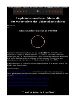
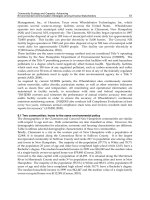
Figure 6–25. The Levitan FPS is placed under direct vision with aid of a laryngoscope. Once
the scope’s distal tip is positioned under the tip of the epiglottis (A), visualization of the glottic
inlet is sought through the eyepiece and the instrument then advanced through the cords (B).
standard malleable stylet, but if a Grade 3 or
worse view is obtained, the fiberoptic stylet/tube
assembly can be used to aid indirect visualization
of, and passage through the glottic opening, as
described above.
Stand-Alone Fiberoptic Stylet Use
Fiberoptic stylets can also be used on their own.
With such stand-alone use, the distal curvature
of semimalleable versions should be increased
for the midline approach, as mentioned above.
The scope should be antifogged and the patient’s
oropharynx suctioned. While performing a jaw
lift (Fig. 6–26) with the nondominant hand, the
scope is inserted either in the midline over the
tongue or via a more lateral approach, over the
molars. A midline insertion will involve the clin-
ician’s significantly bending over the patient to
access the scope’s eyepiece (Figure 6–27).
Anatomic landmarks are then sought as the
scope is advanced: uvula, base of tongue, then
epiglottis and cords with a midline approach, or
epiglottis then cords with an over-the-molar
approach. The tube/stylet assembly can be gen-
tly advanced through the cords, at which point
the tube is further advanced off the stylet down
the trachea.
Awake Intubation Using a Fiberoptic
Stylet
Fiberoptic stylets may be used in the sitting,
cooperative patient for an awake tracheal intu-
bation, using a face-to-face approach.
40
Follow-
ing appropriate application of topical airway
anesthesia (Chap. 8), gentle tongue traction is
applied by an assistant. The stylet with pre-
loaded ETT is guided through the mouth in the
midline, and advanced behind the tongue, until
its tip disappears. At this point, the long axis of
the scope will be parallel to the floor. The clin-
ician then looks through the proximal eye-
piece and seeks the anatomic landmark of the
epiglottis, leading to the glottic opening. In the
stand-alone manner described earlier, the distal
tip of the tube/ stylet assembly is navigated to
and through the glottic opening into the
proximal trachea. The tube is then further
advanced off the stylet, and the scope is
removed by forward rotation of the proximal end
back toward the patient’s chest.
114 CHAPTER 6
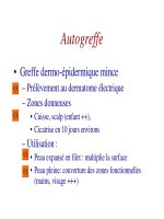
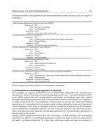
Figure 6–26. The Shikani SOS is inserted from the side of the mouth, advanced over the
molars, and then rotated upright.
Fiberoptic Stylet Troubleshooting
• Getting “lost.” It should be appreciated that
navigation of any fiberoptic instrument
through the airway is contingent on advancing
the device through a patent airway lumen.
While an awake patient will maintain air-
way patency, an obtunded or relaxed patient
(as during an RSI) must have a patent lumen
created by a laryngoscope blade, with a jaw
thrust, or gentle tongue traction during the
procedure. The stylet should not be blindly
advanced if no lumen is appreciable. In the
event that orientation is lost (often manifested
by “pink-out”), the scope should be partially
withdrawn until an anatomic landmark (e.g.,
uvula or epiglottis) can be reidentified and at
that point, advancement can resume.
• Fogging. If fogging is encountered once the
stylet is already in use, briefly holding the
stylet tip against the patient’s buccal mucosa
will help clear the view.
• Blood and secretions. It should be under-
stood that there is no integrated suction
mechanism with most of these instruments.
Blood, secretions, and vomitus will make use
of an indirect fiberoptic system difficult. For
this reason, fiberoptic scope use should
always be preceded by suctioning of the
oropharynx. Also, as blood and secretions
will pool posteriorly, the scope should be
kept anterior in the airway during navigation
toward the laryngeal inlet. The difficulty which
blood and secretions can cause with the use
of a fiberoptic scope points to the need for its
early use, before the airway has been trauma-
tized by multiple intubation attempts!
Fiberoptic Stylet Effectiveness
R
OUTINE AND
D
IFFICULT
A
IRWAY
M
ANAGEMENT
Shikani studied 120 patients, 74 of them chil-
dren, including 7 patients with Cormack Grade
3 or 4 views. All patients in the series, including
5 awake patients, were successfully intubated
with the scope, 88% on the first attempt. Five of
ALTERNATIVE INTUBATION TECHNIQUES 115
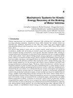
Figure 6–27. While maintaining a jaw lift, the user looks through the proximal eyepiece of the
SOS in an attempt to view the glottic structures.
the seven Grade 3 and 4 patients required con-
comitant direct laryngoscopy.
41
Bein et al.
42
studied use of the Bonfils fiberoptic stylet in
80 patients with predictors of difficult DL, com-
paring it to LMA Fastrach use. Thirty-nine of
40 patients randomized to the Bonfils were intu-
bated on the first attempt, in contrast to a 70%
first attempt success rate for the Fastrach.
A second study looked at Bonfils use after failed
DL. In 25 patients recruited following two failed DL
attempts, 88% were successfully intubated with
the Bonfils at the first attempt, and all but one
(96%) by the second attempt.
43
Evans and
coworkers compared the SOS to the bougie in a
manikin study with a fixed Grade 3 view. In this
model, the SOS resulted in faster intubation
times than the bougie, with significantly fewer
esophageal intubations.
44
A second manikin
study, this one comparing the bougie with
the Levitan FPS scope, showed the fiberoptic
scope to be significantly more successful than
the bougie in managing a simulated Grade 3B
view,
45
but did not demonstrate a significant
difference in intubation success in a simulated
Grade 3A view. The latter finding has been con-
firmed in a subsequent human study using sim-
ulated Grade 3 views in elective surgical
patients, in which the bougie was found to be
equally effective to the Levitan FPS scope.
46
Finally, a recently published case series has
documented successful use of the Bonfils in six
patients in whom difficulty had been encoun-
tered in the prehsopital setting.
47
S
KILLS
A
CQUISITION
These devices are relatively easy to learn on
manikins, as dealing with “pink out” is rarely
an issue, and the upper airway lumen is widely
patent. Skill transfer to the live setting is likely
to be more challenging. One study looking at
the learning curve of the Bonfils stylet suggested
that proficiency was attained after 20–25 intu-
bations.
48
Other studies with fiberoptic stylets
have reported that most of the failed intubations
occurred within the first 10 uses of the
device.
41,49
The fact that the fiberoptic stylet can
be used as an adjunct to the core skill of direct
laryngoscopy may contribute to an easier learn-
ing curve.
C-
SPINE
P
RECAUTIONS
In a study comparing intubation using Macin-
tosh blade direct laryngoscopy with the Bon-
fils stylet, Bullard laryngoscope, or LMA Fas-
trach, each of the Bonfils, Bullard, and Fastrach
resulted in significantly less C-spine movement
than Macintosh blade-facilitated intubation,
although Bonfils and Fastrach intubations took
significantly longer than those using the Mac-
intosh and Bullard blades.
50
A second study,
also using fluoroscopy to assess C-spine
movement, found that Bonfils intubations
caused significantly less extension of the
upper C-spine than Macintosh laryngoscope-
aided intubations.
51
᭤ VIDEOLARYNGOSCOPY
Displaying the view obtained at laryngoscopy
on a video monitor has a number of advantages:
• Display of an enlarged, panoramic viewing
field.
52
• In those devices using integrated video tech-
nology on rigid blades, as the camera is
located toward the distal end of the blade,
an improved view may be obtained com-
pared to direct laryngoscopy.
• Aids in teaching.
• Assisting personnel can see the results of their
manipulations, for example, external laryn-
geal manipulation (ELM).
• The procedure can be digitally stored for
documentation, teaching, or research pur-
poses.
• The user is at a greater distance away from
the patient’s face, decreasing the chance of
exposure to potentially infectious respiratory
secretions and spray.
Video technology can be applied in two
ways: (a) using an adapter, a video camera
116 CHAPTER 6
can be attached to the eyepiece of conven-
tional fiberoptic devices such as the Shikani,
Levitan FPS, Bullard, or flexible fiberoptic
bronchoscopes, or (b) integrated video is used
as the primary viewing mechanism (e.g., the
Glidescope).
The Glidescope
Commercially introduced in 2002, the
Glidescope
®
(GVL
®
) is a video laryngoscope
which has become increasingly available in
and out of the OR, as an alternative intubation
device. The one-piece blade and handle is
made of a durable medical-grade plastic. The
blade has a vertical profile of 14.5 mm, a 60°
bend midblade, and distally, houses a minia-
ture video camera and light-emitting diode
(LED) light source. The image obtained by the
camera is projected by cable to a liquid-crystal
display (LCD) color monitor. A heating element
covering the camera provides effective antifog-
g device has been turned on for
10–30 seconds. The reusable blades are avail-
able in large (patients 30 kg and up), midsize
(10 kg and up), and small (1.5 kg and up)
sizes, and can be sterilized. More recently
introduced versions of the GVL include the
GVL Ranger, which is a compact, battery-
based unit, and the GVL Cobalt, which fea-
tures a reusable internal video baton for
placement within large or small-sized dispos-
able blades.
The GVL is inserted orally in the midline. As
the scope is advanced, the uvula, base of tongue
and then epiglottis will be visualized on the
screen, helping to retain orientation to the mid-
line. Although the blade is designed to be placed
above the epiglottis in the vallecula, in contrast
to direct laryngoscopy, the blade tip need not
be advanced completely into the glossoepiglot-
tic fold: a more proximal tip location allows a
wider field of view and more room for ETT
manipulation (Fig. 6–28). A styletted ETT is
inserted immediately on the right side of the
blade and is navigated to the laryngeal inlet
under indirect visualization on the LCD screen.
An accompanying nonmalleable, reusable stylet
has been made available by the manufacturer to
facilitate tube passage (Fig. 6–29), or a regular
malleable stylet can be used, angled at about 60°
ALTERNATIVE INTUBATION TECHNIQUES 117
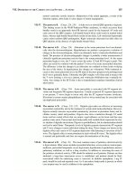
Figure 6–28. Glidescope video system use.
just proximal to the cuff.
53,54
Once the tip of the
ETT has been passed through the cords, the
stylet should be withdrawn 2 inches (4 cm),
whereupon the tube can be further advanced
off the stylet down the trachea.
55
There is a growing literature on the use of
this device, primarily in the OR setting. It is
clear that the GVL does provide good and often
superior views of the glottic opening when
compared with conventional laryngoscopy,
including a high rate of conversion of Cormack
Grade 3 (epiglottis only) views to Grade 2 or
better.
53,54,56,57
However, somewhat longer intu-
bation times have been reported with the GVL
compared to DL, even in the setting of Grade 1
views by DL, possibly related to user inexperi-
ence with tube delivery.
54,57–59
The GVL has been successfully used for
awake intubations in adults.
60
C-spine motion
during GVL use has been compared, using flu-
oroscopy, to that incurred with Macintosh blade
DL. Motion with GVL use was less than that
incurred by Macintosh laryngoscopy at only one
(C2-5) of 4 neck levels studied.
37
There are some recent reports of upper
airway trauma during GVL use.
55,61,62
This sug-
gests that especially in the patient with a smaller
oral cavity, awareness of the ETT tip location
must be maintained as it is advanced, ideally by
direct vision of the ETT until it has passed the
palatoglossal arch. Thereafter, the clinician’s
vision can be transferred to the screen and indi-
rect, videoscopic ETT navigation can occur to
and through the cords. Alternatively, some clin-
icians prefer to place the ETT into the patient’s
pharynx prior to insertion of the GVL blade.
55
The Berci-Kaplan DCI Video
Laryngoscope
The Berci-Kaplan DCI video laryngoscope (Karl
Storz Endoscopy, Culver City, CA) is a hybrid of
fiberoptic and video technology: an image-light
bundle in a laryngoscope blade delivers an
image to a video camera located in the handle
of what otherwise looks like a regular direct
laryngoscope. A cable attaches the device to a
cart-based camera-control unit, and also delivers
light from the remote light source. The image
obtained is displayed on a video monitor.
Macintosh # 3, Mac 4, adult- and pediatric-sized
Miller, and Dörges blades are available for use
with the system. This system offers the advan-
tage of being a familiar intubation technique
and may deliver a superior view of the laryn-
geal inlet compared to that obtained with direct
laryngoscopy.
52
The LMA CTrach
The LMA CTrach (LMA North America Inc, San
Diego CA) is a version of the previously discussed
LMA Fastrach
TM
which adds video-guidance
capability (Fig. 6–30). Looking otherwise like
the LMA Fastrach
TM
, the CTrach mask contains
fiberoptic bundles for light and image transmis-
sion, emerging at the distal end of the airway
barrel. In addition, a removable viewing moni-
tor (the CTrach Viewer) attaches to the CTrach
handle by way of a magnetic latch connector.
The battery-powered viewer is rechargeable,
and provides controls for focusing and image
adjustment. For use, the CTrach Viewer is
detached, and the mask is deflated, lubricated
posteriorly, and antifogged with application of
an appropriate solution to the fiberoptic lenses.
Mask insertion is identical to the technique used
118 CHAPTER 6
Figure 6–29. Dedicated rigid stylet (below)
for use with the Glidescope.
for the LMA Fastrach
TM
, with the head and neck
in a neutral position. Once seated, the mask is
inflated and the patient ventilated. The CTrach
viewer is then turned on and attached to the
magnetic latch connector on the mask, while
firmly holding the CTrach handle. The mask is
then manipulated as needed to attain a clear
image of the glottic opening. For intubation,
while lifting vertically on the CTrach handle
(i.e., the Chandy maneuver, as described for
LMA Fastrach
TM
intubation), the dedicated sili-
cone-based ETT is advanced through the cords
under indirect vision. The ETT cuff is inflated,
and tube position confirmed. The viewer is then
detached, whereupon the CTrach mask can be
removed in identical fashion to the Fastrach,
leaving the ETT in situ.
At the time of writing, early published clini-
cal experience with the CTrach suggests a high
rate of successful mask insertion and patient
ventilation, as with the LMA-Fastrach
TM
.
63,64
Although a view of the cords is not always easily
attained, even after manipulation.
63,64
a num-
ber of corrective maneuvers will help to attain
or improve the view of the laryngeal inlet.
65–68
As with the LMA Fastrach
TM
, the “up-down”
(withdrawing the inflated mask 6 cm, then
readvancing it) will often help release a down-
folded epiglottis.
65,66,68
If only the posterior
cartilages are visualized, withdrawing the
mask 1-cm and lifting will improve the view.
65
The need for medial-lateral corrections of the
mask can also be visualized on the screen.
66
Once a good view is attained, intubation usu-
ally succeeds, and even with poor visualiza-
tion, successful intubation follows in some
cases.
63–65
In published series, CTrach use has
permitted visualization of the larynx and suc-
cessful intubation in most patients presenting
Grade 3 or worse views at direct laryn-
goscopy.
63, 64, 68
Other case reports and series
have detailed successful CTrach intubation in
very difficult situations,
68
even when the LMA
Fastrach
TM
had failed.
69
ALTERNATIVE INTUBATION TECHNIQUES 119
Figure 6–30. The LMA CTrach. (With permission from LMA
North America).
The McGrath Video Laryngoscope
The McGrath video laryngoscope Series 5 (LMA
North America, San Diego, CA) is an additional
example of a video-based device (Fig. 6–31).
The scope features a rubberized handle with an
attached 1.7-inch video screen. The screen tilts
and rotates on the handle to optimize the view-
ing angle for the clinician. The blade is some-
what adjustable in length for different patients,
and is designed for use with a single-use
disposable plastic sleeve. The entire unit is
portable, and operates using a single AA bat-
tery. As with the Glidescope, once the laryn-
geal inlet has been indirectly visualized, the
clinician guides a styletted tube toward and
through the cords. Early experience suggests
easy McGrath blade insertion and a good view
of the larynx, even in patients with predictors
of difficult direct laryngoscopy.
70
As with the
Glidescope, tube passage to and through the
larynx can be challenging until the learning
curve is ascended.
70
A similar intubation tech-
nique to that described above for the
Glidescope should be successful.
᭤ OTHER RIGID AND FLEXIBLE
FIBEROPTIC AND OPTICAL
INSTRUMENTS
Rigid Fiberoptic Devices
Other rigid fiberoptic scopes exist. Some have
attained a small but loyal following, mainly in
the OR setting, however due to expense or unfa-
vorable learning curves, as a group, they are
rarely used in out-of-OR settings. One such is
the Bullard laryngoscope (Fig. 6–32), an
L-shaped rigid fiberoptic laryngoscope. The
Bullard has a blade enabling good tongue con-
trol, and a choice of two dedicated attached
stylets to facilitate tube passage. With or without
the attached stylet, tube passage can be difficult,
however, and this fact has limited its popular-
ity over the years. The Bullard has been shown
to result in less cervical spine movement than
that caused by Macintosh or Miller laryn-
goscopy,
71
although the clinical significance of
this finding is unclear. Similar J- or L-shaped
rigid fiberoptic scopes include the UpsherScope
Ultra and the WuScope System.
120 CHAPTER 6
Figure 6–31. The McGrath Video laryngo-
scope Series 5.
ALTERNATIVE INTUBATION TECHNIQUES 121
Figure 6–32. Bullard laryngoscope.
Rigid Optical Device: The Airtraq
The Airtraq optical laryngoscope (King Sys-
tems Corp., Noblesville, IN) is a single-use, L-
shaped device which uses a series of mirrors to
deliver an image of the laryngeal inlet to a prox-
imal eyepiece (Fig. 6–33). Insertion of the
device begins with the handle parallel to the
patient’s chest. As the blade is advanced into
the oropharynx, it is rotated down and around
the tongue, with the clinician looking through the
eyepiece to visualize airway structures. The blade
tip is placed into the vallecula and the cords
centered in the viewfinder, whereupon the pre-
loaded ETT is advanced into the trachea via a
built-in tube delivery channel. The ETT is then
separated from the delivery channel to the side,
and while holding the tube in place, the scope
is rotated back out of the patient. At the time of
writing, the Airtraq was available in two sizes:
“Regular,” accommodating tube sizes 7.0–8.5 mm
ID, and “Small Adult”, appropriate for use with
ETTs of size 6.0–7.5 mm ID.
Early manikin studies comparing the Airtraq
to Macintosh direct laryngoscopy have shown a
favorable learning curve for novice
72
and inex-
perienced
73
clinicians. With “difficult airway”
simulator features activated, tracheal intubation
has required less time and fewer attempts by
experienced clinicians using the Airtraq, com-
pared to Macintosh laryngoscopy.
74
In elective
surgical patients with no predictors of difficult
laryngoscopy, performance of the Airtraq was
comparable to Macintosh DL.
75
With known dif-
ficult laryngoscopy, however, the Airtraq was
successful in providing a view and enabling
intubation in a series of 8 elective surgical
patients in whom a Cormack Lehane Grade 4
laryngoscopy had been encountered.
76
Flexible Fiberoptic and Video
Devices
Flexible fiberoptic or video-based broncho-
scopes have been the mainstay of difficult
airway management in the OR. Most awake
intubations are performed with flexible fiberop-
tic bronchoscopes in this setting, although many
of the other techniques and devices described
in this and other chapters (including direct
laryngoscopy) can also be used on the awake
patient. Unfortunately, flexible fiberoptic- or
videobronchoscopes are expensive to attain
and maintain, and skills acquisition is also an
issue, resulting in these instruments rarely being
used for intubation by non-anesthesia clini-
cians. Having said this, flexible fiberoptic scopes
can be used in various capacities, including
nasopharyngoscopic upper airway assess-
ment, or flexible fiberoptic guided intubation
through the LMA Fastrach
TM
or AirQ extra-
glottic devices. With time, flexible fiberoptic
intubation may become a more commonly
used technique for awake intubation of the
difficult airway patient in out-of-OR locations,
by non-anesthesia personnel. For more details
on the technique, the reader is referred to
reviews
77
in other publications.
᭤ PEDIATRIC ALTERNATIVE
INTUBATION OPTIONS
For those departments or environments having
care of pediatric patients in their mandate,
when choosing equipment, consideration
should be directed toward whether it is avail-
able in pediatric sizes. It must be emphasized
that most children without congenital dys-
morphisms can be successfully intubated
with direct laryngoscopy and can almost
always be easily bag-mask ventilated. How-
ever, in the event that difficulty is encoun-
tered with direct laryngoscopy, the follow-
ing is a summary of the availability of
pediatric versions of the devices discussed
above:
• LMA Fastrach
TM
. At the time of writing, the
smallest reusable or disposable Fastrach avail-
able is the adult #3, appropriate for use in
patients weighing 30–50 kg.
122 CHAPTER 6
Figure 6–33. Airtraq optical laryngoscope (single-use).
• AirQ. The smallest size is the 1.5, for use in
patients weighing 10–20 kg.
• Trachlight. The Trachlight is available in two
pediatric sizes (child and infant). Clinicians
experienced with Trachlight use in children
have commented that, while effective, the
thin necks of the very young make it difficult
to distinguish the glow of a tube correctly
placed in the trachea from incorrect
esophageal placement. This is particularly
problematic in infants.
• Fiberoptic Stylets. The SOS is available in a
pediatric size, 27 cm in length and accepting
tubes down to 2.5 mm ID. One small case
series has described its successful use in four
children with various dysmorphisms.
78
The
Bonfils Retromolar Intubation Endoscope in
the pediatric/ small adult size will accept tube
sizes from 4.0 to 5.5 mm ID, while the Bram-
brinck Intubation Endoscope (both marketed
by Karl Storz) will accept a minimum tube
size of 2.5 mm ID.
• Video laryngoscopes. The mid-size (mini-
mum patient weight, 10 kg) and small (patient
weight, 1.5 kg) Glidescope blades are
appropriate for pediatric use. Pediatric and
neonatal blades are available for use with
the Berci-Kaplan DCI Video Laryngoscope.
• Other devices. The Bullard laryngoscope is
available in child and neonatal blade sizes,
while flexible fiberoptic bronchoscopes are
available in an array of pediatric sizes, com-
patible with flexible fiberoptic intubation of
even infants.
᭤ SUMMARY
Many alternative intubation devices are avail-
able. They differ in their degree of history, pub-
lished evidence of their effectiveness, cost, and
whether they are blind techniques or allow
indirect vision. Most are probably similar in their
learning curve and success rates in difficult sit-
uations. Unfortunately, many clinical trials of
these devices have been performed in compar-
ison to conventional DL, leaving unanswered
the question of how they compare to best look
DL (i.e., using head lift, ELM, and adjuncts such
as the bougie). However, case reports, case
series, and studies of patients with actual dif-
ficult airways do suggest their utility in diffi-
cult situations (although often, in the hands
of expert users). Certainly, moving on to an
alternative intubation device after a best look
laryngoscopy has failed is preferable to multi-
ple futile attempts at direct laryngoscopic intu-
bation. Which alternative intubation device
or devices the clinician chooses to become
familiar with will depend on individual or
institutional preference. However, no matter
which device, the clinician must make the effort
to gain experience by using it in lower-acuity
or routine situations until competence and
confidence in its use are attained.
REFERENCES
1. Crosby ET, Cooper RM, Douglas MJ, et al. The unan-
ticipated difficult airway with recommendations for
management. Can J Anaesth. 1998;45(8):757–776.
2. Mort TC. Emergency tracheal intubation: complica-
tions associated with repeated laryngoscopic
attempts. Anesth Analg. 2004;99(2):607–613, table
of contents.
3. Hung OR, Pytka S, Morris I, Murphy M, Stewart RD.
Lightwand intubation: II—Clinical trial of a new
lightwand for tracheal intubation in patients
with difficult airways. Can J Anaesth. 1995;42(9):
826–830.
4. Brimacombe JR. Intubating LMA for airway intuba-
tion. Laryngeal Mask Anesthesia: Principles and
Practice. Second ed. Philadelphia: Saunders;
2005:469–504.
5. Reardon RF, Martel M. The intubating laryngeal mask
airway: suggestions for use in the emergency depart-
ment. Acad Emerg Med. 2001;8(8):833–838.
6. Lu PP, Yang CH, Ho AC, Shyr MH. The intubating
LMA: a comparison of insertion techniques with
conventional tracheal tubes. Can J Anaesth.
2000;47(9):849–853.
7. Joo H, Rose K. Fastrach—a new intubating laryn-
geal mask airway: successful use in patients
with difficult airways. Can J Anaesth. 1998;45(3):
253–256.
ALTERNATIVE INTUBATION TECHNIQUES 123
8. Kihara S, Yaguchi Y, Brimacombe J, Watanabe S,
Taguchi N, Hosoya N. Intubating laryngeal mask
airway size selection: a randomized triple crossover
study in paralyzed, anesthetized male and female
adult patients. Anesth Analg. 2002;94(4):1023–1027.
9. Fan KH, Hung OR, Agro F. A comparative study of
tracheal intubation using an intubating laryn-
geal mask (Fastrach) alone or together with a
lightwand (Trachlight). J Clin Anesth. 2000;12(8):
581–585.
10. Martel M, Reardon RF, Cochrane J. Initial experience
of emergency physicians using the intubating laryn-
geal mask airway: a case series. Acad Emerg Med.
2001;8(8):815–822.
11. Rosenblatt WH, Murphy M. The intubating laryngeal
mask: use of a new ventilating-intubating device
in the emergency department. Ann Emerg Med.
1999;33(2):234–238.
12. Watson NC, Hokanson M, Maltby JR, Todesco JM.
The intubating laryngeal mask airway in failed
fibreoptic intubation. Can J Anaesth. 1999;46(4):
376–378.
13. Ferson DZ, Rosenblatt WH, Johansen MJ, Osborn
I, Ovassapian A. Use of the intubating LMA-Fastrach
in 254 patients with difficult-to-manage airways.
Anesthesiology. 2001;95(5):1175–1181.
14. Shung J, Avidan MS, Ing R, Klein DC, Pott L. Awake
intubation of the difficult airway with the
intubating laryngeal mask airway. Anaesthesia.
1998;53(7):645–649.
15. Frappier J, Guenoun T, Journois D, et al. Airway
management using the intubating laryngeal mask
airway for the morbidly obese patient. Anesth Analg.
2003;96(5):1510–1515.
16. Choyce A, Avidan MS, Shariff A, Del Aguila M,
Radcliffe JJ, Chan T. A comparison of the intubat-
ing and standard laryngeal mask airways for
airway management by inexperienced personnel.
Anaesthesia. 2001;56(4):357–360.
17. Baskett PJ, Parr MJ, Nolan JP. The intubating
laryngeal mask. Results of a multicentre trial with
experience of 500 cases. Anaesthesia. 1998;53(12):
1174–1179.
18. Agro F, Hung OR, Cataldo R, Carassiti M, Gherardi
S. Lightwand intubation using the Trachlight: a
brief review of current knowledge. Can J Anaesth.
2001; 48(6):592–599.
19. Hung OR, Stewart RD. Lightwand intubation: I—a
new lightwand device. Can J Anaesth. 1995;42
(9):820–825.
20. Stewart RD, LaRosee A, Kaplan RM, Ilkhanipour K.
Correct positioning of an endotracheal tube using
a flexible lighted stylet. Crit Care Med. 1990;
18(1):97–99.
21. Locker GJ, Staudinger T, Knapp S, et al. Assess-
ment of the proper depth of endotracheal
tube placement with the Trachlight. J Clin Anesth.
1998;10(5):389–393.
22. Hung OR, Tibbet JS, Cheng R, Law JA. Proper
preparation of the Trachlight and endotracheal tube
to facilitate intubation. Can J Anaesth. 2006;53(1):
107–108.
23. Hung OR, Pytka S, Morris I, et al. Clinical trial of
a new lightwand device (Trachlight) to intubate
the trachea. Anesthesiology. 1995;83(3):509–514.
24. Iwama H, Ohmori S, Kaneko T, Watanabe K.
Ambient light requirements for successful intuba-
tion with the Trachlight in adults. Anaesthesia.
1997;52(8):801.
25. Hodgson RE, Gopalan PD, Burrows RC, Zuma K.
Effect of cricoid pressure on the success of endo-
tracheal intubation with a lightwand. Anesthesiology.
2001;94(2):259–262.
26. Masso E, Sabate S, Hinojosa M, Vila P, Canet J,
Langeron O. Lightwand tracheal intubation with
and without muscle relaxation. Anesthesiology.
2006;104(2):249–254.
27. Agro F, Brimacombe J, Carassiti M, Morelli A,
Giampalmo M, Cataldo R. Use of a lighted stylet
for intubation via the laryngeal mask airway.
Can J Anaesth. 1998;45(6):556–560.
28. Agro F, Benumof JL, Carassiti M, Cataldo R, Gher-
ardi S, Barzoi G. Efficacy of a combined technique
using the Trachlight together with direct laryn-
goscopy under simulated difficult airway conditions
in 350 anesthetized patients. Can J Anaesth.
2002;49(5):525–526.
29. Wada H, Nakamura K, Nishiike S, Seki S, Tsuchida
H. [The combined use of laryngoscope and Trach-
light: another option for endotracheal intubation
in patients with large epiglottic cysts]. Masui.
2006;55(4):468–470.
30. Agro F, Brimacombe J, Marchionni L, Carassiti M,
Cataldo R. Nasal intubation with the Trachlight.
Can J Anaesth. 1999;46(9):907–908.
31. Favaro R, Tordiglione P, Di Lascio F, et al. Effective
nasotracheal intubation using a modified transillumi-
nation technique. Can J Anaesth. 2002;49(1):91–95.
32. Margolis GS, Menegazzi J, Abdlehak M, Delbridge
TR. The efficacy of a standard training program for
124 CHAPTER 6
transillumination-guided endotracheal intubation.
Acad Emerg Med. 1996;3(4):371–377.
33. Wik L, Naess AC, Steen PA. Intubation with laryn-
goscope versus transillumination performed by
paramedic students on manikins and cadavers.
Resuscitation. 1997;33(3):215–218.
34. Soh CR, Kong CF, Kong CS, Ip–Yam PC, Chin E,
Goh MH. Tracheal intubation by novice staff: the
direct vision laryngoscope or the lighted stylet
(Trachlight)? Emerg Med J. 2002;19(4):292–294.
35. Yamamoto T, Aoyama K, Takenaka I, Kadoya T,
Uehara H. [Light-guided tracheal intubation using a
Trachlight: causes of difficulty and skill acquisi-
tion]. Masui. 1999;48(6):672–677.
36. Konishi A, Kikuchi K, Sasui M. [Cervival spine
movement during light-guided orotracheal intuba-
tion with lightwand stylet (Trachlight)]. Masui.
1998;47(1):94–97.
37. Turkstra TP, Craen RA, Pelz DM, Gelb AW. Cervical
spine motion: a fluoroscopic comparison during
intubation with lighted stylet, GlideScope, and Mac-
intosh laryngoscope. Anesth Analg. 2005;101(3):
910–915.
38. Inoue Y, Koga K, Shigematsu A. A comparison of
two tracheal intubation techniques with Trachlight
and Fastrach in patients with cervical spine disor-
ders. Anesth Analg. 2002;94(3):667–671.
39. Levitan RM. Design rationale and intended use of
a short optical stylet for routine fiberoptic aug-
mentation of emergency laryngoscopy. Am J Emerg
Med. 2006;24(4):490–495.
40. Kovacs G, Law AJ, Petrie D. Awake fiberoptic intu-
bation using an optical stylet in an anticipated diffi-
cult airway. Ann Emerg Med. 2007;49(1):81–83.
41. Shikani AH. New “seeing” stylet-scope and
method for the management of the difficult airway.
Otolaryngol Head Neck Surg. 1999;120(1):113–116.
42. Bein B, Worthmann F, Scholz J, et al. A comparison
of the intubating laryngeal mask airway and the
Bonfils intubation fibrescope in patients with pre-
dicted difficult airways. Anaesthesia. 2004;59(7):
668–674.
43. Bein B, Yan M, Tonner PH, Scholz J, Steinfath M,
Dorges V. Tracheal intubation using the Bonfils
intubation fibrescope after failed direct laryn-
goscopy. Anaesthesia. 2004;59(12):1207–1209.
44. Evans A, Morris S, Petterson J, Hall JE. A compari-
son of the Seeing Optical Stylet and the gum elas-
tic bougie in simulated difficult tracheal intubation:
a manikin study. Anaesthesia. 2006;61(5):478–481.
45. Kovacs G, Law JA, McCrossin C. A comparison of
a fiberoptic stylet and a bougie as adjuncts to direct
laryngoscopy in a manikin simulated difficult
airway. Ann Emerg Med. 2007 [E pub ahead of
print].
46. Greenland KB, Liu G, Tan H, Edwards M, Irwin
MG. Comparison of the Levitan FPS Scope and
the single-use bougie for simulated difficult
intubation in anaesthetised patients. Anaesthesia.
2007;62(5):509–515.
47. Byhahn C, Meininger D, Walcher F, Hofstetter C,
Zwissler B. Prehospital emergency endotracheal
intubation using the Bonfils intubation fiberscope.
Eur J Emerg Med. 2007;14(1):43–46.
48. Halligan M, Charters P. Learning curve for the Bon-
fils intubation fibrescope. British Journal of Anaes-
thesia. 2003;90:826P.
49. Halligan M, Charters P. A clinical evaluation of
the Bonfils Intubation Fibrescope. Anaesthesia.
2003;58(11):1087–1091.
50. Wahlen BM, Gercek E. Three-dimensional cervi-
cal spine movement during intubation using the
Macintosh and Bullard laryngoscopes, the bon-
fils fibrescope and the intubating laryngeal
mask airway. Eur J Anaesthesiol. 2004;21(11):
907–913.
51. Rudolph C, Schneider JP, Wallenborn J, Schaffrani-
etz L. Movement of the upper cervical spine during
laryngoscopy: a comparison of the Bonfils intu-
bation fibrescope and the Macintosh laryngoscope.
Anaesthesia. 2005;60(7):668–672.
52. Kaplan MB, Ward D, Hagberg CA, Berci G, Hagiike
M. Seeing is believing: the importance of video laryn-
goscopy in teaching and in managing the difficult
airway. Surg Endosc. 2006;20 Suppl 2:S479–483.
53. Agro F, Barzoi G, Montecchia F. Tracheal intuba-
tion using a Macintosh laryngoscope or a GlideScope
in 15 patients with cervical spine immobilization.
Br J Anaesth. 2003;90(5):705–706.
54. Cooper RM, Pacey JA, Bishop MJ, McCluskey SA.
Early clinical experience with a new videolaryngo-
scope (GlideScope) in 728 patients. Can J Anaesth.
2005;52(2):191–198.
55. Cooper RM. Complications associated with the use
of the GlideScope videolaryngoscope. Can J Anaesth.
2007;54(1):54–57.
56. Hsiao WT, Lin YH, Wu HS, Chen CL. Does a new
videolaryngoscope (glidescope) provide better
glottic exposure? Acta Anaesthesiol Taiwan.
2005;43(3):147–151.
ALTERNATIVE INTUBATION TECHNIQUES 125
126 CHAPTER 6
57. Sun DA, Warriner CB, Parsons DG, Klein R,
Umedaly HS, Moult M. The GlideScope Video
Laryngoscope: randomized clinical trial in
200 patients. Br J Anaesth. 2005;94(3):381–384.
58. Rai MR, Dering A, Verghese C. The Glidescope
system: a clinical assessment of performance.
Anaesthesia. 2005;60(1):60–64.
59. Cooper RM. The GlideScope videolaryngoscope.
Anaesthesia. 2005;60(10):1042.
60. Doyle DJ. Awake intubation using the GlideScope
video laryngoscope: initial experience in four cases.
Can J Anaesth. 2004;51(5):520–521.
61. Malik AM, Frogel JK. Anterior tonsillar pillar perfo-
ration during GlideScope video laryngoscopy.
Anesth Analg. 2007;104(6):1610–1611; discussion
1611.
62. Hsu WT, Hsu SC, Lee YL, Huang JS, Chen CL. Pen-
etrating injury of the soft palate during GlideScope
intubation. Anesth Analg. 2007;104(6): 1609–1610;
discussion 1611.
63. Timmermann A, Russo S, Graf BM. Evaluation of
the CTrach—an intubating LMA with inte-
grated fibreoptic system. Br J Anaesth. 2006;96(4):
516–521.
64. Liu EH, Goy RW, Chen FG. The LMA CTrach, a
new laryngeal mask airway for endotracheal intu-
bation under vision: evaluation in 100 patients.
Br J Anaesth. 2006;96(3):396–400.
65. Liu EH, Goy RW, Chen FG. An evaluation of poor
LMA CTrach views with a fibreoptic laryngoscope
and the effectiveness of corrective measures.
Br J Anaesth. 2006;97(6):878–882.
66. Dhonneur G, Ndoko SK, Yavchitz A, et al. Tracheal
intubation of morbidly obese patients: LMA CTrach
vs direct laryngoscopy. Br J Anaesth. 2006;97(5):
742–745.
67. Timmermann A, Russo S, Natge U, Heuer J, Graf
BM. [LMA CTrachtrade mark : initial experiences
in patients with difficult-to-manage airways.].
Anaesthesist. 2006;55(5):528–534.
68. Goldman AJ, Rosenblatt WH. The LMA CTrach in
airway resuscitation: six case reports. Anaesthesia.
2006;61(10):975–977.
69. Goldman AJ, Rosenblatt WH. Use of the fibreoptic
intubating LMA-CTrach in two patients with diffi-
cult airways. Anaesthesia. 2006;61(6): 601–603.
70. Shippey B, Ray D, McKeown D. Case series: the
McGrath videolaryngoscope—an initial clinical
evaluation. Can J Anaesth. 2007;54(4):307–313.
71. Watts AD, Gelb AW, Bach DB, Pelz DM. Compari-
son of the Bullard and Macintosh laryngoscopes
for endotracheal intubation of patients with a
potential cervical spine injury. Anesthesiology.
1997;87(6):1335–1342.
72. Maharaj CH, Costello JF, Higgins BD, Harte BH,
Laffey JG. Learning and performance of tracheal
intubation by novice personnel: a comparison of
the Airtraq and Macintosh laryngoscope. Anaesthesia.
2006;61(7):671–677.
73. Maharaj CH, Ni Chonghaile M, Higgins BD, Harte
BH, Laffey JG. Tracheal intubation by inexperienced
medical residents using the Airtraq and Macintosh
laryngoscopes—a manikin study. Am J Emerg Med.
2006;24(7):769–774.
74. Maharaj CH, Higgins BD, Harte BH, Laffey JG.
Evaluation of intubation using the Airtraq or
Macintosh laryngoscope by anaesthetists in
easy and simulated difficult laryngoscopy—a
manikin study. Anaesthesia. 2006;61(5):469–477.
75. Maharaj CH, O’Croinin D, Curley G, Harte BH, Laffey
JG. A comparison of tracheal intubation using
the Airtraq or the Macintosh laryngoscope in
routine airway management: a randomised, con-
trolled clinical trial. Anaesthesia. 2006;61(11):
1093–1099.
76. Maharaj CH, Costello JF, McDonnell JG, Harte BH,
Laffey JG. The Airtraq as a rescue airway device
following failed direct laryngoscopy: a case series.
Anaesthesia. 2007;62(6):598–601.
77. Morris IR. Fibreoptic intubation. Can J Anaesth.
1994;41(10):996–1007; discussion 1007–1008.
78. Shukry M, Hanson RD, Koveleskie JR, Ramadhyani
U. Management of the difficult pediatric airway
with Shikani Optical Stylet. Paediatr Anaesth.
2005;15(4):342–345.
Chapter 7
Rescue Oxygenation
127
᭤ INTRODUCTION TO RESCUE
OXYGENATION
Following a failed intubation attempt using
direct laryngoscopy or an alternative intubating
technique, ease of bag mask ventilation (BMV)
should be assessed, and the patient reoxygenated
as needed. As discussed in more detail in Chap.
12, as long as oxygenation with BMV is non-
problematic, additional attempts at tracheal
intubation can then be made. However, total
attempts at intubation should be limited in this
setting, as patient morbidity and mortality climbs
with three or more attempts.
1
After three attempts
at intubation, unless a more experienced clini-
cian has arrived or additional equipment is
obtained, oxygenation should revert to BMV or
may proceed with the placement of an extra-
glottic device (EGD) while plans are made for
definitive care. In the more ominous situation
where tracheal intubation has failed and the
patient can’t be oxygenated with BMV, the so-
called “can’t intubate/can’t oxygenate” scenario,
preparations should be made to rapidly pro-
ceed with a cricothyrotomy. However, even
in this scenario, a quick trial of EGD placement
is usually warranted before proceeding with the
cricothyrotomy, as reoxygenation of the patient
often results. Indeed, since their introduction,
EGDs have enabled rescue oxygenation in many
᭤ KEY POINTS
• Failed oxygenation may be defined as the
inability to tracheally intubate the patient
in conjunction with failure to maintain
oxygen saturation above 90% with bag
mask ventilation.
• Failed oxygenation implies the immediate
need to proceed with cricothyrotomy,
although a brief attempt at extraglottic
device (EGD) placement should occur
first.
• By successfully enabling rescue oxygenation,
EGD use will often preclude the need for a
cricothyrotomy
• Widespread clinical experience and a sig-
nificant body of literature support the use
of EGDs such as the Laryngeal Mask
Airway or Combitube as a primary and
rescue airway.
• The LMA Fastrach
TM
is an effective rescue
EGD that also provides a means of facili-
tating blind endotracheal intubation.
• Extraglottic devices will not necessarily
work if obstructing pathology exists at or
below the cords.
• As an alternative to an open surgical
cricothyrotomy, kits are available containing
cuffed cannulae for percutaneous, needle-
guided insertion.
Copyright © 2008 by The McGraw-Hill Companies, Inc. Click here for terms of use.
failed airway situations, returning the patient to
a “you have time” scenario whereby cricothyro-
tomy can be avoided. Additional expertise or
equipment can then be obtained for successful
oral or nasal intubation, or if tracheostomy is
elected, it can be performed under more con-
trolled conditions.
Extraglottic devices (alternatively termed
supraglottic devices), well-known examples of
which include the Laryngeal Mask Airway
(LMA; LMA North America Inc, San Diego, CA)
and the Esophageal-Tracheal Combitube
(ETC; Tyco-Kendall-Sheridan, Mansfield, MA)
are so-named as they enable ventilation from
outside (i.e., above) the cords. Unlike bag mask
ventilation, however, these devices sit distal to
where a relaxed soft palate and tongue may fall
back to obstruct the airway, and as such are more
likely to result in successful patient oxygenation
and ventilation. Equally, their extraglottic loca-
tion also represents a potential limitation to EGD
effectiveness, when obstructing pathology is
present at or below the cords. Thus, while wide-
spread availability and use of EGDs may have
diminished the need for cricothyrotomy, any
clinician with airway management responsibili-
ties should still be prepared to rapidly perform
a cricothyrotomy to access the airway below the
cords.
This chapter will describe equipment and
techniques for rescue oxygenation using
EGDs and cricothyrotomy. More information
on decision-making about when to use these
techniques appears in Chap. 12.
᭤ THE LARYNGEAL MASK AIRWAY
(LMA)
Available for clinical use since the late 1980s,
the LMA has an established place as a device
to provide a hands-free airway in routine oper-
ating room (OR) cases. It has also been suc-
cessfully used on many occasions in airway
emergencies, both in and out of the OR. It is
reasonably easy to insert, even by unskilled
128 CHAPTER 7
personnel,
2–4
and most versions require no addi-
tional tools for placement. Now available in a
number of reusable and disposable formats,
the LMA consists of a plastic airway tube
attached distally to a cuffed, inflatable mask.
When properly seated in the pharynx, the
inflated cuff forms a seal around the laryngeal
inlet, enabling ventilation from immediately
above the cords, while bypassing more proxi-
mal sources of obstruction.
Relative contraindications to LMA use
include a high risk of passive regurgitation of
gastric contents; the need for high airway ven-
tilation pressures, and pathology that would
prevent, or be aggravated by its insertion.
5
However, none of these conditions preclude an
attempt at LMA use as a rescue device in an
emergency, failed oxygenation situation.
LMA Devices: Description
LMA Classic
TM
and Unique
The original LMA was introduced in 1988 and
has now been used well over 100 million times.
It remains in widespread use in its reusable for-
mat, the LMA Classic
TM
(Fig. 7–1), and a more
recently introduced disposable version, the LMA
Unique. Both versions are latex-free and consist
of a large-bore airway tube with proximal stan-
dard 15-mm connector, and a bowl-shaped dis-
tal cuff which is inflated via a valve on an infla-
tion line. With the opening of its lumen facing
the laryngeal inlet, the mask conforms to the
shape of the pharynx. Both the LMA Classic
TM
and Unique are available in a full range of sizes,
from neonatal to large adult.
LMA ProSeal
The LMA ProSeal (Fig. 7–2) was introduced in
2000. This version of the LMA includes a drain
tube, which originates from an orifice in the
distal tip of the mask cuff and travels proxi-
mally alongside the airway lumen. The drain
tube is designed to accept a catheter which can
be used for suctioning esophageal contents.
The cuff of the LMA ProSeal has also under-
gone modifications, including the addition of a
posterior component (only in the adult sizes) to
better conform to the shape of the pharynx.
These cuff modifications allow for an airway
seal pressure up to 10 cm H
2
O higher than that
of the LMA Classic
TM
.
5
A built-in proximal bite
block has also been added. With its improved
seal and provision for gastric tube placement,
the ProSeal offers more protection of the airway
against aspiration of gastric contents, and allows
ventilation at higher airway pressures, perhaps
making it a better choice than the LMA Clas-
sic
TM
or Unique for use in emergencies.
RESCUE OXYGENATION 129
Figure 7–1. The LMA Classic.
Figure 7–2. The LMA ProSeal.
LMA Supreme
The single-use LMA Supreme (Fig. 7–3) is a
recent addition to the LMA family. It features an
L-shaped airway tube, a modified cuff to enable
ventilation at higher airway pressures, a second
lumen for esophageal drainage, and a proximal
bite block. At the time of writing, no published
literature was available on this device. However,
designed for easy insertion (L-shaped tube), airway
protection (presence of esophageal drainage
lumen), and ventilation at higher airway pres-
sures (cuff design), this device has the potential
to become a good choice of single-use EGD for
the emergency patient.
LMA Fastrach
TM
and CTrach
The L-shaped LMA Fastrach
TM
and its similarly
shaped video-based sibling, the LMA CTrach,
were designed to enable blind or video-aided
intubation, respectively. However, both are also
effective as rescue oxygenation and ventilation
devices, with a high first attempt insertion and
successful ventilation rate. The LMA Fastrach
TM
has been shown to have an oropharyngeal leak
pressure 5–10 cm H
2
O higher than the LMA
Classic
TM
.
5
As they were designed to also facili-
tate intubation, these devices have been dis-
cussed in more detail in Chap. 6.
LMA Devices: Preparation for Use
In general, the largest-sized LMA compatible
with insertion should be selected. Studies in
adults indicate that the use of larger sizes sig-
nificantly improves seal efficacy with no
increased insertion difficulty.
5
As a general
rule, a size 5 mask should be chosen for an
average adult male, and size 4 for an average
adult female. Appropriate pediatric sizing can
be estimated by patient weight. The classic
TM
insertion technique recommended by the
manufacturer is to insert the mask with the
cuff fully deflated (Fig. 7–4). For cuff defla-
tion, while aspirating air via a syringe attached
to the inflation line, the mask should be
pressed down against a flat surface, as this
will help maintain the appropriate cuff shape.
Prior to insertion, the posterior surface of the
mask should be lubricated with a water-soluble
lubricant. The LMA ProSeal is supplied with
an L-shaped insertion tool: if this is used, the
distal end of the tool should be introduced
into the strap at the junction of cuff and tube.
The airway and drain tubes are then bent
around its convex surface, and proximally,
the airway tube is snapped into a matching
slot
5
(Fig. 7–5).
130 CHAPTER 7
Figure 7–3. The LMA Supreme.
LMA Devices: Insertion Techniques
LMA Classic
TM
and Unique
To ease LMA placement, the head should be
extended, when not contraindicated. Through an
opened mouth, the mask is inserted midline, with
the operator’s forefinger at the junction of the
tube with the inflatable cuff. Once the tip of the
inserting forefinger has passed the upper teeth,
pushing cephalad on the mask with this digit
(Figs. 7–6 to 7–8) during further advancement
will encourage the LMA to take on the curve of
the hard palate, increasing the ease of negotiat-
ing the turn down into the pharynx. The LMA is
then advanced gently until resistance is encoun-
tered. Once placed, the LMA cuff is inflated using
the recommended cuff volume printed on the
side of the LMA barrel, or simply using the cuff
volume formula “(LMA size–1) × 10.” Note that
some authorities prefer to initially inflate the cuff
with only 2/3 of this recommended volume, with
additional inflation used only to overcome a poor
seal.
5
However, for use in emergency, failed
airway situations, the full recommended vol-
ume should be used primarily. The proximal
connector of the LMA is attached to a manual
resuscitator, and ventilation attempted: appropri-
ate chest rise and bag compliance with positive
pressure ventilation suggest correct placement.
Many modified insertion techniques have
been suggested for the LMA Classic
TM
, reflecting
the fact that placement does not always succeed
on the first attempt. One study found similar
RESCUE OXYGENATION 131
Figure 7–4. The LMA is prepared for use by
fully deflating the cuff of air while pressing the
mask against a flat surface.
Figure 7–5. An L-shaped insertion tool can be used to facilitate correct placement of the LMA
ProSeal.
success rates when placing the single-use LMA
Unique with or without intraoral finger use.
6
In
general, however, the manufacturer’s recom-
mended technique for the LMA Classic
TM
and
Unique is the most reliable and should be used
for the initial insertion attempt.
LMA Supreme
The LMA Supreme can be inserted without intra-
oral finger use. Held at its proximal end, while
applying a jaw lift, it is simply rotated into the
patient, down and around the tongue, following
the curve of the hard palate.
LMA ProSeal
A few insertion techniques have been described
for the LMA ProSeal:
• Mask insertion with intraoral finger use, iden-
tical to that described above for the LMA
Classic
TM
;
• Insertion with the supplied rigid insertion tool.
When the ProSeal is loaded on the insertion
tool, it can be inserted in similar fashion to the
LMA Fastrach
TM
or CTrach (see next section);
• Bougie-guided: the non-coudé-tip end of a
bougie is passed through the drainage tube
of the LMA ProSeal. Laryngoscopy is per-
formed, and the bougie is passed deliberately
into the upper esophagus. The bougie then
acts as a guide during subsequent ProSeal
insertion, to help correctly situate its tip in the
upper esophagus
LMA Fastrach
TM
and CTrach
Both these devices, as well as the ProSeal
when using the rigid insertion tool, can be
inserted while holding the external guiding
132 CHAPTER 7
Figure 7–6. LMA Classic insertion begins by
inserting the mask tip behind the upper teeth
(With permission, LMA North America).
Figure 7–7. As the LMA is advanced, the
index finger pushes the mask cephalad
against the hard palate (With permission, LMA
North America).
Figure 7–8. Once in place, the LMA cuff is
inflated (With permission, LMA North America).
handle. A jaw lift is performed, the mask tip is
inserted behind the upper teeth, whereupon the
mask is rotated down into the pharynx, follow-
ing and maintaining pressure against the palate.
Cricoid pressure impedes successful place-
ment of an LMA,
5
and should be at least tran-
siently released during LMA placement. After
the LMA is correctly situated, cricoid pressure
can be reapplied, but should only be maintained
if it does not impede ventilation.
LMA Devices: Troubleshooting
Difficulty is occasionally encountered in negoti-
ating the turn into the pharynx, particularly with
attempted LMA Classic
TM
placement. The fol-
lowing strategies can be used in response:
• Lateral approach: Advancing the LMA from
the side of the oral cavity, aiming toward the
midline, sometimes results in successful
passage into the pharynx;
• Cuff partially inflated: Partially inflating the
cuff may result in a softer leading edge to the
advancing LMA, potentially helping naviga-
tion “around the corner” into the pharynx;
5
• Laryngoscope-aided: If difficulty is still
encountered, use of the direct laryngoscope
to control soft tissues enables the LMA to be
directly placed into the pharynx.
LMA Devices: Clinical Effectiveness
R
OUTINE AND
D
IFFICULT
A
IRWAY
M
ANAGEMENT
Data for the LMA Classic
TM
derived largely from
an OR population, using the standard insertion
technique, suggests first-attempt and overall
success rates of 87% and 98%, respectively. In
the difficult airway population, ease of LMA
insertion is independent of both Mallampati and
Cormack-Lehane scoring.
5
Both the LMA Clas-
sic
TM
and LMA Fastrach
TM
have high success rates
in achieving ventilation in patients with predicted
and unanticipated difficult airways, including
patients who could not be intubated, or could
not be intubated or ventilated.
5
In this latter
scenario, one analysis of 21 case reports
5
and a
descriptive study of 17 cases
7
reported success
in establishing ventilation using the LMA in 92%
and 94% of cases, respectively. Similar efficacy
of LMA devices in the difficult airway has been
reported in the pediatric population.
5
S
KILLS
A
CQUISITION
Good success rates have been achieved by
novices with LMA placement in human patients
after appropriate manikin training.
6
However,
as common sense would suggest, there is evi-
dence that with more experience, success rates
increase.
5
C-
SPINE
P
RECAUTIONS
In a study of various airway devices using a
cadaver with a posteriorly destabilized C3 ver-
tebra, LMA Classic
TM
insertion and LMA Fastrach
TM
insertion with subsequent intubation resulted in
movement comparable to both laryngoscopic
intubation and facemask ventilation.
8
LMA inser-
tion would also be expected to be more difficult
in situations where head extension is con-
traindicated. Some movement of the intact upper
C-spine has been shown with LMA Fastrach
TM
insertion and intubation
8,9
although this is of
uncertain clinical significance and does not pre-
clude use of this or other EGD for rescue oxy-
genation, if other techniques have failed.
᭤ THE ESOPHAGEAL-TRACHEAL
COMBITUBE (ETC)
The Combitube (Fig. 7–9) is another EGD with
an extensive history of use, primarily in the pre-
hospital resuscitation setting. It has also been
used in-hospital as a rescue ventilation device,
both in and out of the OR. As with the LMA, it
is easily used by inexperienced personnel. Its
strength lies in the ability to achieve patient ven-
tilation irrespective of its location: esophagus or
trachea. The Combitube may be placed blindly
or using a laryngoscope for soft tissue control.
With blind placement, esophageal placement of
the Combitube will occur in over 90% of cases.
5
RESCUE OXYGENATION 133
The ETC is available in two sizes, the Com-
bitube (41 French) and the Combitube SA
(37 French). Manufacturer recommendations
are for use of the larger Combitube in patients
over 5 ft (152 cm), although a number of authors
have observed that the smaller Combitube SA
works well in patients from 4–6 ft (122–183 cm)
in height.
10,11
At the time of writing, there was
no pediatric version.
As with other blind techniques, Combitube
use may be relatively contraindicated in the pres-
ence of airway pathology. Reports of esophageal
perforation with its use exist,
12, 13
possibly in
the context of an excessive volume of air
having been injected into the distal, esophageal
cuff. Finally, it is important to recognize that as
with the LMA, the Combitube ventilates from an
extraglottic position when located in the esoph-
agus, so will not necessarily work if obstructing
pathology exists at or below the cords.
Combitube Description
Designed for blind insertion, the Combitube
consists of a double-lumened tube, with a distal
and more proximal cuff. With the more likely
esophageal placement, the distal cuff seals the
upper esophagus, and the more proximal and
larger pharyngeal cuff seals the oro- and
nasopharynx. Applied ventilation through the
blind-ending esophageal lumen (labeled No. 1
and blue in color) exits through multiple fenes-
trations between the inflated distal and proxi-
mal cuffs and travels through the cords into the
trachea (Fig. 7–10). With tracheal placement,
ventilation would occur distally, through the
other lumen (labeled No. 2, Fig. 7–11) as with a
regular endotracheal tube.
When situated in the esophagus, the inflated
distal cuff helps protect the hypopharynx from
gastric contents,
11
and the open tracheal lumen
can be suctioned for liquid matter. Equally, the
more proximal pharyngeal cuff also provides
reasonable protection from tracheal soiling by
oral cavity contents (e.g., blood).
14
Oropharyn-
geal leak pressure is 25–40 cm H
2
O.
Combitube Preparation for Use
The device is removed from its packaging and
both cuffs are checked, then fully deflated. Some
clinicians elect to bend the Combitube anteriorly
to 90° or more for a few seconds prior to
insertion, (the “Lipp maneuver”) to augment
134 CHAPTER 7
Figure 7–9. The Esophageal-Tracheal Combitube (ETC).
the curve and help it to better conform to the
shape of the oropharyngeal curve.
Combitube Insertion
The Combitube is ideally inserted in conjunction
with direct laryngoscopy (Fig. 7–12), to help
control the tongue and improve the angle of
insertion. However, for blind placement, slight
head extension and a jaw lift will help (Fig. 7–13).
The Combitube is advanced gently through the
mouth in a curved, downward motion. Once in
the posterior pharynx, further advancement
should ideally be with the distal end of the device
parallel to the patient’s anterior chest wall, and
not angled further posteriorly. Once inserted,
two transverse lines appearing proximally on the
Combitube should be adjacent to the upper
teeth or alveolar ridges. During emergency use,
once placed, the two cuffs are inflated: first the
proximal (pharyngeal) occluding cuff (Com-
bitube SA 85 mL; Combitube 100 mL) using the
blue pilot balloon (labeled “No. 1”). The distal
(esophageal) cuff is then inflated (Combitube
SA 5–12 mL; Combitube 5–15 mL) using the
RESCUE OXYGENATION 135
Figure 7–10. Ventilation pathway when the
Combitube is located in the esophagus,
through lumen No. 1.
Figure 7–11. Ventilation is through lumen
No. 2 when the Combitube is located in the
trachea.
Figure 7–12. A laryngoscope may help with
Combitube placement.
white pilot balloon (labeled “No. 2”). Particu-
larly for the distal cuff, overinflation should be
avoided, as esophageal rupture can otherwise
occur.
12
Once a seal has been achieved and
the correct lumen identified, many clinicians
remove air from the proximal cuff until the
“minimum leak” volume is found, to help avoid
danger of mucosal damage.
Ventilation through the Combitube should
first be attempted through the blue lumen,
labeled “No. 1”, which will allow ventilation from
an esophageal location. End-tidal CO
2
detection
will help confirm the correct lumen, as will the
clinical signs of chest rise, breath sounds with
positive pressure, and manual resuscitator bag
compliance. If this is judged not to be the correct
lumen, ventilation should be attempted through
the other, clear lumen (labeled “No. 2”). This
will be the correct lumen on the rare occasion
that tracheal placement has occurred. In this
case, the Combitube will act as a regular ETT,
and the proximal cuff can be deflated.
Combitube Troubleshooting
If suboptimal ventilation is obtained through
both lumens, most often the Combitube is
located too far distally, and the pharyngeal cuff
is occluding the laryngeal inlet. In this situa-
tion, the Combitube should be pulled back in
small (1 cm) increments, up to a total of 3 cm,
until ventilation succeeds through the blue,
esophageal lumen.
Combitube Clinical Effectiveness
R
OUTINE AND
D
IFFICULT
A
IRWAY
M
ANAGEMENT
Published success rates for Combitube inser-
tion and ventilation are 97–99% for in-hospital
populations.
5
Slightly higher success rates in
the surgical population occur with laryngoscope-
guided placement. In the prehospital setting,
rescue ventilation with the Combitube fol-
lowing failed laryngoscopic intubation has
been reported to be successful in 75–100% of
cases.
15–18
C-
SPINE
P
RECAUTIONS
Combitube insertion has been shown to have a
lower success rate in patients wearing a rigid cer-
vical collar
19
although most failures could be cor-
rected using adjunctive laryngoscopy. However,
once placed, the presence of a C-collar does not
impede ventilation through a Combitube.
20
In a
cadaver model with a destabilized C3 segment,
Combitube insertion caused movement compa-
rable to oral intubation with direct laryngoscopy,
and exceeded that caused by LMA Fastrach
TM
intubation or LMA Classic
TM
placement.
8
᭤ NEWER EXTRAGLOTTIC DEVICES
In recent years, numerous new extraglottic
devices have been introduced. Many, but not
136 CHAPTER 7
Figures 7–13. A jaw lift and head extension
are performed to aid blind Combitube insertion.
all, are single-use items. Some have more
accompanying narrative in the literature than
others, however early experience looks promis-
ing for many in terms of ease of insertion and
effectiveness. As many hospitals are trending
toward the use of disposable equipment, the
clinician should be prepared to be presented
with an unfamiliar device from time to time!
The disposable Portex Soft-Seal laryngeal
mask (Smiths Medical, Inc., St. Paul, MN) is
similar in shape to the LMA Unique, but with a
blunter distal cuff, a deeper bowl, wider airway
tube, and no mask aperture bars.
21
Compared
to the LMA Unique or Classic
TM
, the Soft-Seal
has similar reported insertion success rates,
oropharyngeal leak pressure,
22,23
and ease of
ventilation.
24
The Soft-Seal (Fig. 7–14) is avail-
able in adult and pediatric sizes.
Ambu (Ambu Inc., Glen Burnie, MD) also
markets an extraglottic airway in both reusable
(the Aura40) and disposable (the AuraOnce)
formats (Fig. 7–15). It differs from the LMA
Classic
TM
/Unique and Portex Soft-Seal in having
a premolded L-shaped airway tube proximal to
the distal cuff. The cuff is manufactured from a
soft material and has a reinforced tip to resist
bending during insertion. For insertion, the cuff
is deflated, and, holding the device proximally,
the tip is inserted behind the upper teeth. Fol-
lowing the hard and soft palate, it is then rotated
down into the pharynx. The Aura extraglottic
airways are available in adult and pediatric
(Table 7–1) sizes. Early data suggests a good
first attempt success rate, and an oropharyngeal
leak pressure of 18–25 cm H
2
O.
24,25
The King Laryngeal Tube (LT; King Sys-
tems Corporation, Noblesville, IN, Fig. 7–16)
consists of an airway tube with two cuffs: one
distal, to seal the esophagus, and one proximal
midway up the tube, to seal the oro- and
nasopharynx. Between the two cuffs are multi-
ple ventilation apertures. As with the Com-
bitube, ventilation emerges from these apertures,
between the proximal pharyngeal and distal
esophageal cuffs. Unlike the Combitube, infla-
tion of both cuffs occurs through a single pilot
line. The LT is available in adult and pediatric
sizes, in reusable (LT) and disposable (LT-D)
versions. A disposable version with a separate
gastric drainage channel (LTS-D) is also avail-
able. Insertion is begun with the head and neck
in the ‘sniffing’ position, with concomitant
jaw lift. The lubricated LT is inserted through
the mouth and advanced behind the base of
RESCUE OXYGENATION 137
Figure 7–14. The Portex Soft Seal laryngeal.