Báo cáo y học: "A case-series study to explore the efficacy of foot orthoses in treating first metatarsophalangeal joint pain" pot
Bạn đang xem bản rút gọn của tài liệu. Xem và tải ngay bản đầy đủ của tài liệu tại đây (730.86 KB, 9 trang )
RESEA R C H Open Access
A case-series study to explore the efficacy of foot
orthoses in treating first metatarsophalangeal
joint pain
Brian J Welsh
1*
, Anthony C Redmond
2
, Nachiappan Chockalingam
3
, Anne-Maree Keenan
2
Abstract
Background: First metatarsophalangeal (MTP) joint pain is a common foot complaint which is often considered to
be a consequence of altered mechanics. Foot orthoses are often prescribed to reduce 1
st
MTP joint pain with the
aim of altering dorsiflexion at propulsion. This study explores changes in 1
st
MTP joint pain and kinematics
following the use of foot orthoses.
Methods: The effect of modified, pre-fabricated foot orthoses (X-line®) were ev aluated in thirty-two patients with
1
st
MTP joint pain of mechanical origin. The primary outcome was pain measured at baseline and 24 weeks using
the pain subscale of the foot function index (FFI). In a small sub-group of patients (n = 9), the relationship
between pain and kinematic variables was explored with and without their orthoses, using an electromagnetic
motion tracking (EMT) system.
Results: A significant reduction in pain was observed betw een baseline (median = 48 mm) and the 24 week
endpoint (median = 14.50 mm, z = -4.88, p < 0.001). In the sub-group analysis, we found no relationship between
pain reduction and 1
st
MTP joint motion, and no significant differences were found between the 1
st
MTP joint
maximum dorsiflexion or ankle/subtalar complex maximum eversion, with and without the orthoses.
Conclusions: This observational study demonstrated a significant decrease in 1
st
MTP joint pain associated with
the use of foot orthoses. Change in pain was not shown to be associated with 1
st
MTP joint dorsiflexion nor with
altered ankle/subtalar complex eversion. Further research into the effect of foot orthoses on foot function is
indicated.
Background
First metatarsophalangeal (MTP) joint pain is a common
foot complaint, often associat ed with osteoarthritis
(OA): with more than 20% of people over the age of 40
reporting 1
st
MTP joint OA pain [1]. Altered kinematics
at the foot and ankle have been suggested to be asso-
ciated with such 1
st
MTP joint pain which, in turn, may
act as a precursor to osteoarthritic changes at the 1
st
MTP joint [2,3]. Authors have proposed that failure of
the first metatarsal to achieve sufficient plantarflexion,
prior to the propulsive phase of the gait cycle, may pre-
vent the posterior glide of the metatarsal head along its
sesamoid apparatus [4,5]. This is thought to result in
abnormal hallux dorsiflexion, which terminates with
impingement between the dorsal a rticular surfaces of
the 1
st
metatarsal and the proximal phalanx, with result-
ing pain and inflammation within the joint capsule [6,7].
It is also suggested that this functional loss of hallux
dorsiflexion at the 1
st
MTP joint can occur despite the
fact that adequate dorsiflexion is available when the
joint is assessed in a non-weightbearing condition. This
functional, as opposed to structural, blockade has been
termed functional hallux limitus (FHL). Laird [8] defined
this concept as non-weightbearing dorsiflexion greater
than 50° at the 1
st
MTP joint, with less than 14° 1
st
MTP joint dorsiflexion at terminal stance. Others have
given descriptions, investigated FHL and offered their
own interpretation of the condition [9-14].
While 1
st
MTP joint pain is often associated with OA,
this is not the only cause of pain. The term “mechanical
* Correspondence:
1
Musculoskeletal and Rehabilitation Services, NHS Leeds Community
Healthcare, St Mary’s Hospital, Leeds, LS12 3QE, UK
Full list of author information is available at the end of the article
Welsh et al. Journal of Foot and Ankle Research 2010, 3:17
/>JOURNAL OF FOOT
AND ANKLE RESEARCH
© 2010 Welsh et al; licensee BioMed C entral Ltd. This is an Open Access article distributed unde r the terms of the Creative Comm ons
Attribution License ( whi ch permits unrestricted use, distribution, and reproduction in
any medium, provided the origina l work is prop erly cited.
joint pa in” is used commonly w ithin the rheumatology
literature and refers to pain that is mechanica l in origin
or influence when there is a p attern of increased symp-
toms with weightbearing activities and when ot her
potential systematic causeshavebeenexcluded[15].
More recently, this term has been extended to include
foot pain and given the importance placed on 1
st
MTP
joint function, it readily extends itself to 1
st
MTP joint
pain.
Foot orthoses are thought to decrease mechanically
induced 1
st
MTP joint pain by allowing the 1
st
metatar-
sal to achieve suffici ent plantarflexion in preparation for
propulsion [16]. Whilst the relationship between ankle/
subtalar joint pronation and 1
st
MTP joint pain is
unclear, clinicians commonly prescribe foot orthoses
with medial posting to alter the degree and timing of
ankle/subtalar complex pronation in the treatment of 1
st
MTP joint pain. First ray cut outs and forefoot postings
are further orthotic modifications that have been
employed to improve 1
st
ray function and reduce pain.
There are, however, limitations in the evidence to sup-
port such approaches. Despite sound reasoning and the-
oretical principles, the approach is largely based on
subjective justification [16] or single case design
[4,17,18]. The existing literature has also focused pri-
marily on normal or asymptomatic participants [19-25].
Furthermore, the relationship between pain and f oot
function has not been explored.
The aim of this study was to investigate change in 1
st
MTP joint pain levels when foot orthoses are prescribed
with the rationale of increasing 1
st
MTP joint dorsiflex-
ion. Relationships between changes in 1
st
MTP joint
pain levels and changes in the mechanical effects of foot
orthoses on 1
st
MTP joint and ankle/subtalar complex
kinematics were also explored in a small number of
participants.
Methods
This two part study was undertaken at St James’ and
Chapel Allerton Hospitals, Leeds, United Kingdom. Ethi-
cal approval was granted from the Faculty of Health and
Sciences Independent Peer Review Panel, Sta ffordshire
University and Leeds West Local Research Ethics Com-
mittee. The two parts of this study were (i) an investiga-
tion of the clinical effects of foot orthoses on
mechanical joint pain at the 1
st
MTP joint; and (ii) an
exploration of the mechanical effects of the foot
orthoses in a small sub-group of the same participants.
Participants
Thirty five participants (mean age, 42 years; range 21-
63 years) were recruited from primary care referrals
received within Musculoskeletal and Rehabilitation Ser-
vices a nd the Community Podiatry Service, Leeds,
United Kingdom. Participants were recruited who had
mechanically induced 1
st
MTP joint pain which was
required to be of at least 4 weeks duration and at a level
of at least 40 mm on a 100 mm visual analogue pain
scale (VAPS) as previously described [26], which was
considered an appropriate pain level to warrant inter-
vention [27].
As the foot orthoses being tested in the study were to
be modified to offer a tailored level of pronatory control,
participants were required to demonstrate a Foot Pos-
ture Index (FPI-6) score of greater than 4/12 [28].
Participants were excluded if they had establish ed hal-
lux valgus, a previous history of foot and ankle trauma,
fracture or surgery, or an existing diagnosis of inflam-
matory, metabolic, neurological or vascular disease. Indi-
viduals who exhibited less than 40° of available 1
st
MTP
joint dorsiflexion, measured by a non-weightbearing
technique previously described by Buell et al [29], were
also excluded from the study as this has been reported
to be the range of 1
st
MTP joint dorsiflexion used dur-
ing normal propulsion [10,21,22] and would therefore
have indicated structural limitation at the joint. Where
participants reported bilateral 1
st
MTP joint pain, the
joint which gave the most pain was selected for the pur-
poses of the study.
ThesamplesizewasbasedonPitman’ s Asymptotic
Relative Efficiency [30]. A calculation was performed
using a sta ndard deviation of 17.8, based on previous
work that used the same outcome measure as this study
[31]. A sample size of 32 participants provided 80%
power to detect a 10 mm differe nce at an alpha level of
0.05.Thiswasincreasedto35toallowforapproxi-
mately 10% drop-out [32]. A reduction of 10 mm on the
pain subscale of the FFI was chosen apriorias a clini-
cally relevant change, based on previous data [33,34].
Foot orthoses
All patients were prescribed pre-fabricated, foot orthoses
(X-line®, Healthystep, Mossley, UK). Sagittal and frontal
plane pronatory control was increased using high den-
sity (400 kg/m
3
) ethyl-vinyl acetate wedged posting,
adhered to t he medial underside of the foot orthoses.
The posting was tailored to each individual’srequire-
ments as determin ed by a standard clinical evaluation
by an experienced musculoskeletal specialist podiatrist
(BJW). Adequacy of pronatory control provided by the
foot orthos es was assessed through a reduction in FPI-6
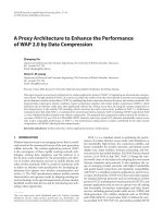
score of at least 2 points. All foot orthoses were cut to
the level of the toe sulci. The first metatarsal head
region of the foot o rthoses was cut out and a forefoot
extension of 3 mm open cell polyurethane foam was
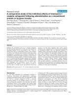
added as in Figure 1. It is acknowledged that a prag-
matic prescription protocol increases variability in
device prescription, but the t ailoring of prescriptio ns to
Welsh et al. Journal of Foot and Ankle Research 2010, 3:17
/>Page 2 of 9
the patient ’s specific needs reflects a more realistic ther-
apeutic approach than the use of artificially standardised
prescriptions.
Clinical Outcome Measures
The primary outcome for this study was pain measured
using a modification of the pain subscale of the Foot
Function Index (FFI) [35] , with an endpoint of 24
weeks. This instrum ent has demonstrated good test-ret -
est reliability, internal consistency and both construct
and criterion validity [35]. While the FFI was developed
to assess the effectiveness of foot orthoses on foot
pathology in people with rheumatoid arthritis (RA), it
has been widely used to investigate non-RA populations
[31,36-38] and was deemed suitable for this study.
Secondary outcomes were 1
st
MTP joint and ankle/
subtalar complex motions derived from an electromag-
netic motion tracking (EMT) system, as described pre-
viously by Halstead et al [22] and Longworth et al [39].
Clinical protocol
At initial c ontact, a clinical assessme nt was conducted
and baseline outcomes were captured. Parti cipants were
provided with guidance on how to complete the FFI
pain subscale, in relation to their 1
st
MTP joint pain,
prior to data capture. The pre-fabricated foot orthoses
used in the study were modified as described above and
fitted into the participant’ sshoes.Footorthoseswere
issued only if footwear was appropriate, determined by
an assessment tool devised by Menz and Sherrington
[40]. In addition to the footwear assessment tool, heel
counter height was also assessed which was considered
an important factor in the provisio n of foot orthoses.
Verbal and written advice was issued to each partici-
pant, detailing important information about the wearing
of foot orthoses.
While the primary endpoint was 24 weeks, participants
underwent interim clinical review after 8 weeks, at which
time they were asked to complete again the pain subscale
of the FFI. Further postal FFI pain subscale questionnaires
were administered, and telephone reviews were conducted,
at 12 weeks as well as at the 24 week endpoint.
Gait Analysis
At the 8 wee k review, 1 0 participants were invited to
participate in the 2
nd
stage of the study exploring the
Figure 1 Type of foot ort hoses used in the s tudy. This shows the pre-fabricated foot orthoses that were used in the study, following
individually tailored modification.
Welsh et al. Journal of Foot and Ankle Research 2010, 3:17
/>Page 3 of 9
relationship between pain and kinematic outcomes.
Individuals were invited on the basis of the extent of
any benefit that had been gained from the wearing of
the foot orthoses at this stage of the trial. A sample was
constructed to include a range of participants, from
those who had gained much pain relief, to those who
had gained the least benefit. The intention was to
explore the relationship between change in pain and
kinematic response for the indexed (or painful) foot.
Joint kinematic data for the 1
st
MTP joint and the
ankle/subtalar complex was collected using a Fastrak™
EMT system with a long-range transmitter (Polhemus
Inc., Colchester, VT). The long-range configuration pro-
duces a low frequency electromagnetic field, with a
radius of approximately two metres from the transmit-
ter, which was centred along a walkway. The four metre
walkway was raised from the floor of the gait laboratory
to prevent metallic interference. Four sensors were used,
capturing at 30 Hz. S ensors were attached to the hallux
and 1
st
metatarsal to derive sagittal plane motion data
for the 1
st
MTP joint. Sensors were also attached to the
posterior calcaneus and medial tibia to derive ankle/sub-
talar complex motion data. The 1
st
metatarsal sensor
was attached using a Velcro strap in accordance with a
protocol devised by Longworth et al [39]. This reduces
potential error due to extensor hallucis longus contrac-
tion during hallux dorsiflexion, as previously described
by Umberger et al [41]. The other sensors were attached
with double-sid ed tape, and secured with Hypafix™ tape
over the sensors, to an atomi cal sites with minimal over-
lying soft tissue to reduce possible sensor movement
during walking. All cables were secured with straps to
the limb and waist with a belt. The 6 D Research™ soft-
ware package ( Skill Technologies, Phoenix, AZ, USA)
was used to post-process the data fro m the EMT sen-
sors as described previously [42].
Angular rotation between the anatomically mounted
sensors was determined using a previously described
joint co-ordinate system [1 0,42]. The joint motions
investigated were: x-axis maximum stance phase dorsi-
flexion at the 1
st
MTP joint and y-axis maximum stance
phase eversion at the ankle/subtalar complex. 6DNorm
software (M.Cornwall, Northern Arizona University,
Flagstaff, AZ, USA) was used to generate motion-time
curves, normalized to 100% of the gait cyc le, for each
axis of rotation.
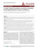
EMT sensors were secured as indicated in Figure 2a.
Participants wore Velcro fastening, neoprene boot, of
appropriate size, similar to a previously used protocol
[31]. The flexibility of the boots minimised the con-
founding effects of structured footwear and allowed win-
dows to be cut into the footwear so that the hallux, 1
st
metatarsal and calcaneal placed sensors were not dis-
turbed or displaced during data capture (figure 3b). For
calibration or ‘boresighting’ participants stood near the
centre of the electromagnetic field, with the calcaneus
vertical and talar head palpable equally on both medial
and lateral sides to e stablish a reference position [43].
This ‘boresighting’ procedure has been described pre-
viously [42].
When comfortable with the set-up, participants
initiated gait for one metre at a self-selected speed,
before enteri ng the 1.5 metre calibrated capture volume
and continued walking for a further 1.5 metres once
through the volume. Three trials were completed for
each of the experimental conditions ‘orthoses’ and ‘ no-
orthoses’ . Care was taken when inserting the foot
orthoses into, and removing them from, the boots, so
that the sensors were not disturbed or displaced. This
was enabled by the Velcro fastening. The order of data
collection was randomized. Data from the three trials of
the indexed limb was normalised to percentiles of the
gait cycle and averaged to maximise within-subject con-
sistency. The original dataset of 10 was reduced to 9
due to technical problems.
Data analysis
All an alyses were conducted using Microsoft Excel™ and
SPSS version 15 for Windows. The analysis strategy was
divided into two phases, an efficacy phase and an
explorato ry phase. For the efficacy analysis, comparisons
of pain scores between baseline and primary endpoint
(24 weeks) were explored descriptively and using a Wil-
coxon’ s Signed Rank test. The exploratory analysis
investigated a subset of patients (n = 9) who attended
the gait laboratory for d etailed kinematic studies. F or
the exploratory analysis, data were explored descriptively
and using Wilcoxon’ s Signed Rank test. Relationships
between variables were explored g raphically and using
Spearman’s Rho. Additionally, a difference in response
between 1
st
MTP joint dorsiflexion and ankle/subtalar
complex maximum eversion was explored using descrip-
tive statistics and then using a Mann-Whitn ey U test.
P values < 0.05 were considered significant.
Results
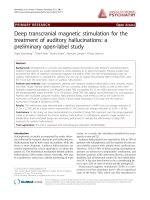
Table 1 shows the participant characteristics at base-
line. Complete data were obtained from 32 participants
in the efficacy phase (6 male:26 female) and from nine
in the exploratory phase, as described in the partici-
pant flow diagram, Figure 3. Data was obtained for the
left index limb for 14 participants and for the right
index limb in 18 participants. At baseline, there were
no significant correlations between reported pain and
the following: age (r = -0.231, p = 0.203), body m ass
index (r = -0.155, p = 0.397), 1
st
MTP joint dorsif lex-
ion (r = 0.24, p = 0.895) or FPI-6 (r = 0.273,
p = 0.130).
Welsh et al. Journal of Foot and Ankle Research 2010, 3:17
/>Page 4 of 9
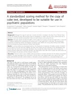
Efficacy analysis
Following the introduction of the treatment foot
orthoses, there was a significant reduction in median
pain score from baseline (48 mm) to 24 weeks (14.5
mm, z = -4.88, p < 0.001). Figure 4 illustrates the sys-
tematic reduction in pain scores at baseline, eight weeks
(29 mm), 12 weeks (20.5 mm) and the 24 week
endpoint.
Exploratory analysis
The exploratory kinematic analysis indicated that the
pain reduction reported by participants between pre-
intervention baseline scores and post-intervention scores
was not accompanied by any systematic change in 1
st
MTP joint dorsiflexion or ankle/subtalar complex pro-
nation (eversion). After wearing foot orthoses for 8
weeks, there was no systematic change in maximum 1
st
Figure 2 2a. Sensor placement. 2b. With neoprene boot. This shows the position of the EMT sensors attached at the anatomical landmarks
and with the Velcro fastening, neoprene boot secured, which was used during the capture of kinematic data.
Welsh et al. Journal of Foot and Ankle Research 2010, 3:17
/>Page 5 of 9
MTP joint dorsiflexion during the walking cycle (no-
orthoses median = 8°, IQR = 22.1 vs orthoses median =
7°, IQR = 23.3, p = 0.954). Similarly, there was no con-
sistent effect of the foot orthoses on maximum ankle/
subtalar complex eversion (no-orthoses median = 1°,
IQR=8.9vsorthosesmedian=1°,IQR=6.4,p=
0.672).
Discussion
The aim of this study was to explore the efficacy of
orthotic treatment for mechanically induced 1
st
MTP
joint pain and to investigate whether any change in pain
correlated with changes in foot and ankle kinematic
values, as predicted by a previously proposed mechan-
ism of function [16]. From the efficacy analysis, the 33.5
mm difference between baseline FFI(pain) scores (48
mm) and endpoint (14.5 mm) was in excess of the 10
mm identified apriorifor this study as clinically rele-
vant [33]. Previous studies that have explored the level
of pain red uction required on a 0-100 mm VAPS, to
offer individuals an adequate analgesic response to
treatment, have concluded that this requires a 30 mm
reduction [27] and a 32 mm reduction [44] in pain
score. The 33.5 mm reduction in pain score achieved in
this study is therefore in excess of what has been advo-
cated previously as an adequate analgesic response to
treatment.
Figure 3 Participant flow diagram. This shows the journey for all participants through the study protocol including both efficacy analysis,
which all participants took part in, and the exploratory analysis, which a selected cohort took part in.
Table 1 Participant characteristics at baseline
N = 35 Mean Std
Dev.
Age (years) 42.2 ± 11.5
Body mass index 24.4 ± 3.8
Duration of symptoms (months) 26.3 ± 30.8
1
st
metatarsophalangeal joint range of motion: non-
weightbearing
63.5° ± 15.2°
Foot Posture Index-6 left: baseline 7.3 ± 2.0
Foot Posture Index-6 right: baseline 7.0 ± 2.2
Welsh et al. Journal of Foot and Ankle Research 2010, 3:17
/>Page 6 of 9
With pain reduction sustained, and even continuing to
improve, over the twenty-four weeks, this study indi-
cates that foot orthoses may have treatment effects over
clinically relevant periods of time. This is in agreement
with other authors who have explored the efficacy of
variously produced foot o rthoses for other indications
[45,46]. Contrary to the hypothesised mode of action
[16], the exploratory analysis revealed that the reduction
in 1
st
MTP joint pain, following orthotic intervention,
was not accompanied by change in 1
st
MTP joint dorsi-
flexion, nor did orthoses induc e significant ankle/subta-
lar complex frontal plane change. It remains unclear
precisely how foot orthoses may influence 1
st
MTP joint
pain.
In this study, the maximum dorsiflexion values
obtained at the 1
st
MTP joint (median = 8.34°, mini-
mum = 1.81°, maximum = 31.07°) were lower than
reported following previous investigations using similar
EMT systems to measure 1
st
MTP joint motion: 37°
[22]; 42° [10]; and 38° - 40° [21]. Although our partici-
pants were selected on the basis that they possessed at
least 40° non-weightbearing dorsiflexion, the lower
values in our study are consistent with a cohort of parti-
cipants specifically selected for 1
st
MTP joint pain of
mechanical origin and a potential for functional block-
ade of 1
st
MTP joint dorsiflexion.
This study demonstrated a large kinematic variability
in both the no-orthoses and the orthoses conditions.
While large variation is expected with such a small sam-
ple size, this is a finding mirrored by a previo us study
that used intracortical pins to assess kinematic effects of
foot orthoses [47], where a similar subject-specific and
unsystematic effect was reported. Nester [48], from a
review of recent dynamic cadaver and invasive kinematic
research approaches, concluded that there was a simi-
larly high and normal variation of foot kinematics
between individuals.
The modified, prefabricated orthotic device used in
this study is of a type that is being increasingly favoured
over more expensive, casted devices due to evidence
that there may be little functional difference between
the two types of orthotic device [49].
In the absence of a control group and a randomisation
protocol, we recognise that this study provides only
minimal further support for the therapeutic effect of
foot orthoses. It is possible that the pain reduction
gained by the participants could have been related to
reasons other than the therapeutic effect of the foot
Figure 4 Distribution of the reduction in FFI (pain) scores from baseline to week 24 (0 = no pain and 100 = worst pain imaginable).
This shows the systematic reduction in pain scores over the treatment period.
Welsh et al. Journal of Foot and Ankle Research 2010, 3:17
/>Page 7 of 9
orthoses such as the placebo effect, a change in footwear
required for t he accommodation of the foot orthoses,
the participant incorrectly reporting lower pain levels to
please the clinician or through natural resolution of
symptoms over time. We also acknowledge that the
small data set for the ki nematic analysis may not have
been sufficient to detect the effect of orthoses on joint
motion. Furthermore the results can be applied only to
the specific orthotic device tested and it is no t known if
the results obtained would have been different for a
device manufactured by a different method, and/or from
different materials. Future research should employ gold
standard methods and should extend the scope of the
study to investigate a variety of different manufacturing
and prescription methods that are commonly employed.
In the longitudinal efficacy study, participants wore
their own footwear foll owing assessment for suitability.
There was however a level of variability amongst partici-
pants’ footwear that may have influenced the therapeutic
effect of the devices as there is a known orthotic effect
of footwear alone [50,51]. For the kinematic exploratory
phase, participants wore a standardised neoprene boot
in the laboratory settin g. We no te that foot orthoses are
typically worn in structured footwear though we wanted
to minimise the potential confounding effect of foot-
wear, focusing on the functional changes associated with
the orthotic device alone.
Potential participant s with a known traumatic aetio-
logy or systemic disease, that could have contributed to
their 1
st
MTP joint pain, were exclud ed from the study.
There was an assumption that those included in t he
study had therefore mechanically induced pain. The
authors acc ept that there may possibly have been other
unknown factors th at may have contributed to the onset
of symptoms.
The study focused on ki nematic changes at the foot in
the attempt to determine if a correlation could be
drawn between changes in pain and changes in kine-
matics. It is acknowledged that the therapeutic effect
may be due to other factors such as kinetic or temporal
changes. Further research should include analysis of
these variables.
Finally, the authors appreciate that the findings of this
cohort study are at a hypo thesis generating level and
can only suggest certain trends which would inform a
more robust analysis in the form of a future rando-
mised, controlled trial. To our knowledge however, this
is the first study to date that has investigated the effi-
cacy of foot orthoses on individuals with mechanically
induced 1
st
MTP joint pain prospectively, as well as
looking at associated changes in foot and ankle
kinematics.
Conclusions
The results of this study suggest that a c ommonly used
orthotic design can offer a reduction in mechanically
induc ed pain at the 1
st
MTP joint to a level t hat is con-
sidered an adequate analgesic response to treatment.
The hypothesised mode of action was not confirmed
however, as pain relief was not associated with increased
dorsiflexion at the 1
st
MTP joint or reduced eversion
(pronation) at the ankle/subtalar complex. Further study
is required to determine definitively the efficacy of foot
orthoses in the management of 1
st
MTP joint pain and
to explore the mechanism of action.
Acknowledgements
The authors are grateful for the assistance of colleagues Bob Longworth, Lee
Short, Carl Ferguson, Jo Mugan, Lesley Spencer and Helen Keen for their
assistance with patient recruitment.
Author details
1
Musculoskeletal and Rehabilitation Services, NHS Leeds Community
Healthcare, St Mary’s Hospital, Leeds, LS12 3QE, UK.
2
NIHR Leeds
Musculoskeletal Biomedical Research Unit and Section of Musculoskeletal
Disease, University of Leeds, 2nd Floor, Chapel Allerton Hospital, Leeds LS7
4SA, UK.
3
Faculty of Health, Staffordshire University, Stoke on Trent ST4 2DF,
UK.
Authors’ contributions
BJW conceived the study design, undertook the clinical investigations and
contributed to the data analysis and writing of the manuscript. AMK
contributed to the study design, clinical and laboratory investigations and to
the data analysis and writing of the manuscript. ACR contributed to the
laboratory investigations and contributed to the data analysis and writing of
the manuscript. NC contributed to the study design and writing of the
manuscript. All authors read and approved the final manuscript.
Competing interests
The study was supported in part through an unrestricted grant from Healthy
Step (Sensograph) who distribute X-line® foot orthoses in the UK.
Received: 6 November 2009 Accepted: 27 August 2010
Published: 27 August 2010
References
1. Wilder FV, Barrett JP, Farina EJ: The association of radiographic foot
osteoarthritis and radiographic osteoarthritis at other sites. Osteoarthr
Cartilage 2005, 13(3):211-215.
2. Drago JJ, Oloff L, Jacobs AM: A comprehensive review of hallux limitus. J
Foot Surg 1984, 23(3):213-220.
3. Piscoya JL, Fermor B, Kraus VB, Stabler TV, Guilak F: The influence of
mechanical compression on the induction of osteoarthritis-related
biomarkers in articular cartilage explants. Osteoarthr Cartilage 2005,
13(12):1092-1099.
4. Michaud TC: Pathomechanics and treatment of hallux limitus: A case
report. Chiropr Sport Med 1988, 2(2):55-60.
5. Shereff MJ, Bejjani FJ, Kummer FJ: Kinematics of the first
metatarsophalangeal joint. J Bone Joint Surg Am 1986, 68(3):392-398.
6. Lichniak JE: Hallux limitus in the athlete. Clin Podiatr Med Surg 1997,
14(3):407-426.
7. Shereff MJ, Baumhauer JF: Hallux rigidus and osteoarthrosis of the first
metatarsophalangeal joint. J Bone Joint Surg Am 1998, 80(6):898-908.
8. Laird PO: Functional hallux limitus. Illinois Podiatrists 1972, 9(4).
9. Dananberg HJ: Functional hallux limitus and its relationship to gait
efficiency. J Am Podiatr Med Assoc 1986, 76(11):648-652.
Welsh et al. Journal of Foot and Ankle Research 2010, 3:17
/>Page 8 of 9
10. Nawoczenski DA: Nonoperative and operative intervention for hallux
rigidus. J Orthop Sports Phys Ther 1999, 29(12):727-735.
11. Chapman C: Functional hallux limitus: The essentials. Br J Pod 1999, 2:40.
12. Payne C, Chuter V, Miller K: Sensitivity and specificity of the functional
hallux limitus test to predict foot function. J Am Podiatr Med Assoc 2002,
92(5):269-271.
13. Van Gheluwe B, Dananberg HJ, Hagman F, Vanstaen K: Effects of hallux
limitus on plantar foot pressure and foot kinematics during walking. J
Am Podiatr Med Assoc 2006, 96(5):428-436.
14. Nawoczenski DA, Baumhauer JF, Umberger BR: Relationship between
clinical measurements and motion of the first metatarsophalangeal joint
during gait. J Bone Joint Surg AM 1999, 81:370-376.
15. Isenberg D, Maddison P, Woo P, Glass D, Breedveld F: Oxford handbook of
rheumatology. USA: Oxford University Press 2004.
16. Dananberg HJ, Philips AJ, Blaakman HE: A rationale approach to the
nonsurgical treatment of hallux limitus. In Advances in Podiatric Medicine
and Surgery. Edited by: Kominsky SJ, May RM, Silvani SH, S.L.T, M.J.T. Mosby;
1996:.
17. Shrader JA, Siegel KL: Nonoperative management of functional hallux
limitus in a patient with rheumatoid arthritis. Phys Ther 2003,
83(9):831-843.
18. Michaud TC, Nawoczenski DA: The influence of two different types of
foot orthoses on first metatarsophalangeal joint kinematics during gait
in a single subject. J Manipulative Physiol Ther 2006, 29(1):60-65.
19. Harradine PD, Bevan LS: The effect of rearfoot eversion on maximal
hallux dorsiflexion. A preliminary study. J Am Podiatr Med Assoc 2000,
90(8):390-393.
20. Smith C, Spooner SK, Fletton JA: The effect of 5-degree valgus and varus
rearfoot wedging on peak hallux dorsiflexion during gait. J Am Podiatr
Med Assoc 2004, 94(6):558-564.
21. Nawoczenski DA, Ludewig PM: The effect of forefoot and arch posting
orthotic designs on first metatarsophalangeal joint kinematics during
gait. J Orthop Sports Phys Ther 2004, 34(6):317-327.
22. Halstead J, Turner DE, Redmond AC: The relationship between hallux
dorsiflexion and ankle joint complex frontal plane kinematics: a
preliminary study. Clin Biomech (Bristol, Avon) 2005, 20(5):526-531.
23. Scherer PR, Sanders J, Eldredge DE, Duffy SJ, Lee RY: Effect of functional
foot orthoses on first metatarsophalangeal joint dorsiflexion in stance
and gait. J Am Podiatr Med Assoc 2006, 96(6):474-481.
24. Munuera PV, Dominguez G, Palomo IC, Lafuente G: Effects of rearfoot-
controlling orthotic treatment on dorsiflexion of the hallux in feet with
abnormal subtalar pronation: a preliminary report. J Am Podiatr Med
Assoc 2006, 96(4):283-289.
25. Munteanu SE, Bassed AD: Effect of foot posture and inverted foot
orthoses on hallux dorsiflexion. J Am Podiatr Med Assoc 2006, 96(1):32-37.
26. Price DD, McGrath PA, Rafii A, Buckingham B: The validation of visual
analogue scales as ratio scale measures for chronic and experimental
pain. Pain 1983, 17(1):45-56.
27. Lee JS, Hobden E, Stiell IG, Wells GA: Clinically important change in the
visual analog scale after adequate pain control. Acad Emerg Med 2003,
10(10):1128-1130.
28. Redmond AC, Crosbie J, Ouvrier RA: Development and validation of a
novel rating system for scoring standing foot posture: the Foot Posture
Index. Clin Biomech (Bristol, Avon) 2006, 21(1):89-98.
29. Buell T, Green DR, Risser J: Measurement of the first metatarsophalangeal
joint range of motion. J Am Podiatr Med Assoc 1988, 78(9):439-448.
30. Pitman EJG: Unpublished lecture notes. 1949.
31. Redmond AC, Helliwell PS, Bird HA, Davys HJ, Turner DE, Emery P: Pain and
health status in people with hypermobility syndrome are associated
with overall joint mobility and selected local mechanical factors.
(Proceedings of the British Society of Rheumatology Meeting, Glasgow 2-5
May). Rheumatology 2006, 45(1):i108.
32. Landorf KB, Keenan AM, Herbert RD: Effectiveness of foot orthoses to
treat plantar fasciitis: a randomized trial. Arch Intern Med 2006,
166(12):1305-1310.
33. Landorf KB, Radford JA: Minimal important difference: Values for the Foot
Health Status Questionnaire, Foot Function Index and Visual Analogue
Scale. The Foot 2008, 18(1):15-19.
34. Kelly AM: Does the clinically significant difference in visual analog scale
pain scores vary with gender, age, or cause of pain? Acad Emerg Med
1998, 5(11):1086-1090.
35. Budiman-Mak E, Conrad KJ, Roach KE: The Foot Function Index: a measure
of foot pain and disability. J Clin Epidemiol 1991, 44(6):561-570.
36. Caselli MA, Levitz SJ, Clark N, Lazarus S, Velez Z, Venegas L: Comparison of
Viscoped and PORON for painful submetatarsal hyperkeratotic lesions. J
Am Podiatr Med Assoc 1997, 87(1):6-10.
37. Pfeffer G, Bacchetti P, Deland J, Lewis A, Anderson R, Davis W, Alvarez R,
Brodsky J, Cooper P, Frey C, et al: Comparison of custom and
prefabricated orthoses in the initial treatment of proximal plantar
fasciitis. Foot Ankle Int 1999, 20(4):214-221.
38. Landorf KB, Keenan AM, Herbert RD: Effectiveness of different types of
foot orthoses for the treatment of plantar fasciitis. J Am Podiatr Med
Assoc 2004, 94(6):542-549.
39. Longworth R, Chockalingam N, Redmond AC: An enhanced protocol to
reduce error in electromagnetic tracking of first metatarsophalangeal
joint motions. Gait Posture 2006, 23(3):391-394.
40. Menz HB, Sherrington C: The Footwear Assessment Form: a reliable
clinical tool to assess footwear characteristics of relevance to postural
stability in older adults. Clin Rehabil 2000, 14(6):657-664.
41. Umberger BR, Nawoczenski DA, Baumhauer JF: Reliability and validity of
first metatarsophalangeal joint orientation measured with an
electromagnetic tracking device. Clin Biomech (Bristol, Avon) 1999,
14(1):74-76.
42. Woodburn J, Turner DE, Helliwell PS, Barker S: A preliminary study
determining the feasibility of electromagnetic tracking for kinematics at
the ankle joint complex. Rheumatology (Oxford) 1999, 38(12):1260-1268.
43. Elveru RA, Rothstein JM, Lamb RL, Riddle DL: Methods for taking subtalar
joint measurements. A clinical report. Phys Ther 1988, 68(5):678-682.
44. Lock BG, Carrier ER, Greenwald PW: The Minimum Acceptable Reduction
in Pain on a Visual Analog Scale. Acad Emerg Med 2003, 10(5):482.
45. Landorf KB, Keenan AM: Efficacy of foot orthoses. What does the
literature tell us? J Am Podiatr Med Assoc 2000, 90(3):149-158.
46. Mundermann A, Nigg BM, Humble RN, Stefanyshyn DJ: Foot orthotics
affect lower extremity kinematics and kinetics during running. Clin
Biomech (Bristol, Avon) 2003, 18(3):254-262.
47. Stacoff A, Reinschmidt C, Nigg BM, van den Bogert AJ, Lundberg A,
Denoth J, Stussi E: Effects of foot orthoses on skeletal motion during
running. Clin Biomech (Bristol, Avon) 2000, 15(1):54-64.
48. Nester CJ: Lessons from dynamic cadaver and invasive bone pin studies:
do we know how the foot really moves during gait? J Foot Ankle Res
2009, 2:18.
49. Redmond AC, Landorf KB, Keenan AM: Contoured, prefabricated foot
orthoses demonstrate comparable mechanical properties to contoured,
customised foot orthoses: a plantar pressure study. J Foot Ankle Res 2009,
2:20.
50. Cornwall MW, McPoil TG: Footwear and foot orthotic effectiveness
research: a new approach. J Orthop Sports Phys Ther 1995, 21(6):337-344.
51. Woodburn J, Barker S, Helliwell PS: A randomized controlled trial of foot
orthoses in rheumatoid arthritis. J Rheumatol 2002, 29(7):1377-1383.
doi:10.1186/1757-1146-3-17
Cite this article as: Welsh et al.: A case-series study to explore the
efficacy of foot orthoses in treating first metatarsophalangeal joint pain.
Journal of Foot and Ankle Research 2010 3:17.
Submit your next manuscript to BioMed Central
and take full advantage of:
• Convenient online submission
• Thorough peer review
• No space constraints or color figure charges
• Immediate publication on acceptance
• Inclusion in PubMed, CAS, Scopus and Google Scholar
• Research which is freely available for redistribution
Submit your manuscript at
www.biomedcentral.com/submit
Welsh et al. Journal of Foot and Ankle Research 2010, 3:17
/>Page 9 of 9