Báo cáo y học: "Metastatic breast carcinoma in the mandible presenting as a periodontal abscess: a case report" pps
Bạn đang xem bản rút gọn của tài liệu. Xem và tải ngay bản đầy đủ của tài liệu tại đây (2.45 MB, 5 trang )
CAS E REP O R T Open Access
Metastatic breast carcinoma in the mandible
presenting as a periodontal abscess: a case report
Evmenios Poulias
1*
, Ioannis Melakopoulos
2
and Konstantinos Tosios
3
Abstract
Introduction: Tumors can metastasize to the oral cavity and affect the jaws, soft tissue and salivary glands. Oral
cavity metastases are considered rare and represent approximately 1% of all oral malignancies. Because of their
rarity and atypical clinical and radiographic appearance, metastatic lesions are considered a diagnostic challenge.
The purpose of this report is to present a rare case of a metastatic breast carcinoma mimicking a periodontal
abscess in the mandible.
Case presentation: A 55-year-old Caucasian woman was referred to our clinic for evaluation of bisphosphonate-
induced jaw osteonecrosis. She had undergone modified radical mastectomy with axillary lymph node dissection
for invasive ductal carcinoma of the left breast. Her clinical examination showed diffuse swelling and a periodontal
pocket of 6 mm exhibiting suppuration in the posterior right mandible. Moreover, paresthesia of the lower right lip
and chin was noted. There were no significant radiographic findings other than alveolar bone loss due to her
periodontal disease. Although the lesion resembled a periodontal abscess, metastatic carcinoma of the breast was
suspected on the basis of the patient ’s medical history. The area was biopsied, and histological analysis confirmed
the final diagnosis of metastatic breast carcinoma.
Conclusion: The general dentist or dental specialist should maintain a high level of suspicion while evaluating
patients with a history of cancer. Paresthesias of the lower lip and the chin should be considered ominous signs of
metastatic disease. This case highlights the importance of the value of a detailed medical history and thorough
clinical examination for the early detection of metastatic tumors in the oral cavity.
Introduction
Metastases in the oral cavity are rare and comprise
approximately 1% of all oral malignancies [1]. They
usually involve the jaws but may also be found in the
soft tissues and salivary glands. The most common
metastatic malignancies in women are from primary
cancers in t he breasts, kidneys, colorectal region, genital
organs and thyroid glands, and in men they arise from
the lungs, prostate, kidneys and colorectal region [2,3].
The mandible is affected more frequently than the max-
illa, with a predilection for the areas distal to the
canines, including the body and ramus [1,2,4]. These
sites are consi dered vulnerable to the deposition of neo-
plastic cells because of the presence of hematopoietic
bone marrow, branching of the local blood vessels and
slowing of blood flow [4].
A wide range of clinical signs and symptoms may be
seen in association with metastatic tumors of the oral
cavity, with the most common being pain, swelling,
altered sensation, halitosis, gum irr itation, tooth loosen-
ing and mobility, exophytic masses of the soft tissues,
trismus a nd, rarely, pathologic fractures [1,2,4]. Numb-
ness or paresthesia of the lower lip and chin is consid-
ered an important sign of metastatic disease [5].
Metastatic tumors of the oral cavity do not exhibit a
pathognomonic radiographic appearance; therefore,
radiographic examination is rarely considered diagnosti-
cally important. Osteolytic radiolucent lesions with ill-
defined and irregu lar margins may be seen, while osteo-
blastic lesions with a pure radiopaque or a mixed radio-
paque-radiolucent appearance are typically associated
with prostate cancer [2,4,6]. Early detection of jaw
metastasis can be challenging. In the initial stages of the
* Correspondence:
1
Department of Periodontics, University of Louisville School of Dentistry,
Louisville, KY, USA
Full list of author information is available at the end of the article
Poulias et al. Journal of Medical Case Reports 2011, 5:265
/>JOURNAL OF MEDICAL
CASE REPORTS
© 2011 Poulias et al; licensee BioMed Central Ltd. This is an Open Access article distributed under the terms of the Creative Commons
Attribution License ( which permits unrestricted use, distribution, an d reproduction in
any medium, provided the original work is properly cited.
disease, the lesion may not produce a radiographic
appearance. In an analysis of 390 cases of metastatic
tumors of the jaw, Hirshberg et al. [6] found that 5.4%
of them did n ot show any i mportant radiographic
change.
The purpose of this report is to describe a rare case of
a metastatic breast carcinoma in the mandibular gingival
tissue that mimicked a periodontal abscess.
Case presentation
A 55-year-old Caucasian woman with subtle pain and
tenderness in the area surrounding the right third man-
dibular molar was referred to our clinic by her oncolo-
gist with the provisional diagnosis of bisph osphonate-
induced jaw osteo necrosis. Her medical history revealed
a modified radical mastectomy with axillary lymph node
dissection for invasive ductal carcinoma of the left
breast. The tumor was positive for estrogen receptors
and cerbB2, but negative for progesterone receptors;
thus she received adjuvant hormone therapy with
tamoxifen. Moreover, bisphosphonate treatment was
initiated with 4 mg of intravenous ibandronic acid admi-
nistered every three weeks.
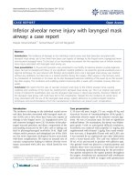
An intra-oral examination revealed diffuse swelling o f
the buccal gingiva surrounding the second and third
molar teeth t hat was soft and tender on palpation, with
signs of inflammation (Figure 1). The involved teeth
showed slight mobility, moderate plaque and calculus
deposits, bled upon probing and reacted positively in
repeated vitality tests. The patient’s periodontal exami-
nation revealed severe generalize d chronic periodontitis,
with pockets in the posterio r area of the ri ght mandibu-
lar quadrant ranging from 3 mm to 7 mm in depth. A 6
mm periodontal po cket with sup puration was d etected
in the mesial buccal aspect of the third molar. An exam-
ination of the intra-oral area innervated by the mental
nerve also revealed altered sensation, and the patient
admitted paresthesia of the lower lip and chin during an
extra-oral examination. Regional lymph nodes were not
palpable.
A panoramic radiograph showed generalized horizontal
bone loss throughout the patient’s dentition (Figure 2). A
peri-apical radiograph of theinvolvedarearevealed
alveolar bone loss attributable to the periodontal disease.
Axial and serial cross-sectional 1 mm-thick cone beam
computed tomography (CBCT) showed small radiolucent
areas in close proximity to the third molar (Figures 3 and
4) that were not diagnostic of metastases.
On the basis of the patient’smedicalhistoryandpar-
esthesia of the lower lip and chin, metastatic disease was
highly suspected. The differential diagnosis included
acute or chronic periodontal abscess, acute alveolar
abscess, bisphosphonate-induced jaw osteonecrosis and
osteomyelitis.
The swelli ng of the bucca l gingiva was biopsied. Five-
micron-thick, formalin-fixed, paraffin-embedded tissue
sections stained with hematoxylin and eosin showed a
fragment of parakeratinized oral mucosa infiltrated by
solid and cribriform nests of neoplasti c cells in a vascu-
lar and myxofibromatous stroma (Figure 5). The neo-
plastic cells contained abundant eosinophilic cytoplasm
and large, pleomorphic, darkly staine d nuclei (Figure 6).
Several mitoses were observed, including atypical forms,
as well as minimal lymphoplasmacytoid inflammatory
infiltration of the stroma. The diagnosis was consistent
with metastatic carcinoma of breast origin. Slides from
the primary breast lesion were not available for compar-
ison with the metastatic focus.
The patient was referred back to her oncologist. A full
body scan did not reveal additional metastases, and a
technetium-99 m-methylene diphosphonate bone scan
Figure 1 Intra-oral view showing a diffuse swelling located
over the buccal gingiva of the mandibular molar region and
drainage of purulent exudate.
Figure 2 Panoramic radiograph showing generalized bone loss
throughout the dentition.
Poulias et al. Journal of Medical Case Reports 2011, 5:265
/>Page 2 of 5
located a region of increased radioisotope uptake ("hot
spot”) on the posterior right side of the mandible.
The bisphosphonate treatment was continued, and
local irradiation of the right posterior mandible was
administered as palliative treatment. Although extraction
of the involved teeth prior to radiotherapy was feasible,
it was decided to preserve them and re-evaluate their
prognosis during the foll ow-up appointments. The
patient underwent radiation therapy with a cumulative
dose of 3000 cGy fractionated over two weeks, w hich
resulted in complete relief of her symptoms and remis-
sion of the disease (Figure 7). Follow-up examinatio ns
were performed every two weeks for the first two
months and bimonthly over the next two years. At the
time of this writing, there is no evidence of recurrence.
Discussion
The diagnosis of metastasis to the oral cavity is a signifi-
cant challenge to the clinician because of the lack of
pathognomonic signs and symptoms. To the best of our
knowledge, this is the fi rst reported case of a metastati c
breast cancer mimicking a periodontal abscess. Pre-
viously described cases of metastases to the periodontal
tissues were associated with extensive osteolytic destruc-
tion of the alveolar bone and root apex resorption [7-9],
even in cases in which an exophytic mass was seen
[10,11].
Our patient was referred to our clinic by her oncolo-
gist for the evaluation of possible osteonecrosis of the
jaw caused by bisphosphonate treatment. The patient’s
oral cavity was carefully examined, but no signs of
exposed avascular necrotic bone were found in the
mandible. The existence of exposed necrotic bone over
a period of eight weeks with past or recent use of
bisphosphonates is an esse ntial element for r endering
the diagnosis of osteonecrosis associated with bispho-
sphonates, along with the absence of previous radiation
therapy to the jaws [12]. Therefore, on the basis of the
Figure 3 Small radiolucent areas in close proximity with the
third molar on an axial cone beam computed tomographic
(CBCT) image of the mandible.
Figure 4 Small radiolucent areas in close proximity to the third
molar on serial cross-sectional CBCT images of the mandible.
Figure 5 Solid and cribriform nests of neoplastic cells in
vascular, myxofibromatous stroma (hematoxylin and eosin
stain; original magnification, × 200).
Figure 6 Neoplastic cells with abundant eosinophilic cytoplasm
and large, pleomorphic, darkly stained nuclei (hematoxylin and
eosin stain; original magnification, × 400).
Poulias et al. Journal of Medical Case Reports 2011, 5:265
/>Page 3 of 5
clinical characteristics of our patient, this type of lesion
was excluded.
The local inflammation of the soft tissues that sur-
rounded the area and the periodontal pocket exhibiting
suppuration were signs of possible inflammatory reac-
tions such as an acute or chronic periodontal a bscess,
an acute alveolar abscess or a combined endodontic-per-
iodontic lesion. However, the relatively healthy condi-
tion of the patient’ s teeth, the existence of vital pulp
after several diagnostic tests and the lack of radiographic
signs eliminated the possibili ty of an endodontic-related
lesion.
In the c ase presented herein, the location of the swel-
ling, spontaneous intra-pocket suppuration and the exis-
tence of typical signs of periodontal disease were
suggestive of a periodontal abscess. Periodontal
abscesses are most often associated with a pre-existing
periodontal pocket and present as an ovoid elevation of
the gingival tissue along the late ral side of the root [13].
Depending on their location, a small or a diffuse swel-
ling may be seen. They may also appear as erythema
when they are located deep in the periodontium. The
most common symptoms reported are pain and tender-
ness of the affected area. Dr aina ge of puru lent exudate
from the periodontal pocket itself or from a fistula in
the oral cavity i s often noted. Other findings include
increased tooth mob ility, increased sensitivity to percus-
sion, as well as, occasionally, lymphadenopathy and ele-
vated body temperature. Radiographic examination o f
the periodontal abscess can vary significantly, and the
findings can range from widening of the periodontal
ligament to pronounced bone loss along the root of the
infected tooth. Furthermore, in many cases, the radio-
grap hic examination may reveal a normal appearance of
the inter-dental bone, especially when the abscess is
located on the facial or lingual surfaces of the tooth
[13,14]. In our patient, we decided to perform a biopsy
because of the history of malignant disease and the exis-
tence of lip and chin paresthesia.
Paresthesia of the lower lip and chin is the maj or
symptom suggestive of metastatic disease. It is descr ibed
in the lite rature as m ental nerve neuropathy or numb
chin syndrome (NCS) [5,15]. The nerves associated with
the NCS are the inferior alveolar nerve and its terminal
branch, the mental nerve, which are branches of the
third (mandibular) division of the trigeminal nerve. In
addition to the chin and lip paresthesia, numbness of
the teeth and mucosa may occur. Although NCS may be
iatrogenic and is often caused by dental anesthesia or
inferior alveolar nerve injury after improper placement
of dental implants, it may also occur as the result of a
benign or malignant neoplasm that disrupts the function
of the nerve. Neoplas ms that are most commonly asso-
ciated with NCS are lymphomas and metastatic carcino-
mas of the mandible [15,16]. Our patient did not report
paresthesia as the chief complai nt, but c arefu l intra-oral
and extra-oral e xaminations revealed altered sensation
to the lip and chin. Therefore, the existence of NCS
should always alert the dentist or the physician to inves-
tigate the presence of a primary or recurrent malignant
neoplasm, especially in cases that involve a significant
medical history.
The management of metastatic breast carcinomas of
the oral cavity is primarily palliative and may include
radiotherapy, chemotherapy, hormone therapy and,
rarely, surgical intervention. Pain relief and avoidance of
possible infections, fractures or hemorrhage should be
the major goals [17]. Local radiotherapy is almost always
the trea tment of choice as it relieves pain, prevents loss
of function and arrests growth of the tumor [18,19]. A
combination of surgical excision and radiation therapy
is used in most cases of soft-tissue metastases [19].
The prognosis for patients with metastatic lesions of
the oral cavity is generally poor, primarily because of
the delay in the detection of the lesions. The average
survival time for patients with metastatic tumors in the
oral cavity is six to seven months, with approximately
70% of patients dying within one year of diagnosis
[6,19,20]. Most patients with oral metastases have
already developed gener aliz ed metastases by the time of
diagnosis; however, in many cases, a solitary mandibular
metastasis can be the initial manifestation of the pri-
mary tumor.
Conclusion
In conclusion, this case illustrates the importance of
suspecting a metastatic lesion in the jaw, despite the
lack of clinical or radiographic evidence. The general
dentist or dental specialist should obtain the patient’ s
complete medical history and carefully evaluate unusual
clinical and radiographic findings such as lip and chin
Figure 7 One-year follow-up intra-oral view of the buccal
gingiva of the mandibular right molar region. No inflammatory
signs were noted, and remission of the disease was achieved.
Poulias et al. Journal of Medical Case Reports 2011, 5:265
/>Page 4 of 5
paresthesias to differentiate metas tatic lesions from
clinically similar entities. As th ese lesions are associat ed
with a poor prognosis, early detection is of extreme
importance.
Consent
Written informed consent was obtained from the patient
for publicatio n of this case report and any accompany-
ing images. A copy of the written consent is available
for review by the Editor-in-Chief of this journal.
Author details
1
Department of Periodontics, University of Louisville School of Dentistry,
Louisville, KY, USA.
2
Private Practice, Athens, Greece.
3
Department of Oral
Pathology and Surgery, School of Dentistry, National and Kapodestrian
University of Athens, Athens, Greece.
Authors’ contributions
PE and MI analyzed and interpreted the patient data. TK performed the
histological examination of the biopsy specimen and was involved in the
manuscript editing and review. PE was involved in the literature review as
well as manuscript preparation, editing and submission. MI was involved in
the manuscript editing and review. All authors read and approved the final
manuscript.
Competing interests
The authors declare that they have no competing interests.
Received: 1 February 2011 Accepted: 1 July 2011 Published: 1 July 2011
References
1. Dib LL, Soares AL, Sandoval RL, Nannmark U: Breast metastasis around
dental implants: a case report. Clin Implant Dent Relat Res 2007, 9:112-115.
2. D’Silva NJ, Summerlin DJ, Cordell KG, Abdelsayed RA, Tomich CE, Hanks CT,
Fear D, Meyrowitz S: Metastatic tumors in the jaws: a retrospective study
of 114 cases. J Am Dent Assoc 2006, 137:1667-1672.
3. Friedrich RE, Abadi M: Distant metastases and malignant cellular
neoplasms encountered in the oral and maxillofacial region: analysis of
92 patients treated at a single institution. Anticancer Res 2010,
30:1843-1848.
4. Akinbami BO: Metastatic carcinoma of the jaws: a review of literature.
Niger J Med 2009, 18:139-142.
5. Ryba F, Rice S, Hutchison IL: Numb chin syndrome: an ominous clinical
sign. Br Dent J 2010, 208:283-285.
6. Hirshberg A, Leibovich P, Buchner A: Metastatic tumors to the jawbones:
analysis of 390 cases. J Oral Pathol Med 1994, 23:337-341.
7. Lu SY, Chen L: Mandible metastasis as the initial manifestation of breast
carcinoma: report of a case. Zhonghua Ya Yi Xue Hui Za Zhi 1991,
10:98-103.
8. Ogütcen-Toller M, Metin M, Yildiz L: Metastatic breast carcinoma
mimicking periodontal disease on radiographs. J Clin Periodontol 2002,
29:269-271.
9. Rawal YB, Blakenship JA, Mincer HH, Parrish ML, Anderson KM: Metastatic
adenocarcinoma of the breast presenting as pulpal/periodontal disease.
J Tenn Dent Assoc 2007, 87:11-13.
10. Rajesh KS, Varma BR, Bhat KM: Metastasis to maxillary gingiva from
carcinoma of breast: a case report. Indian J Dent Res 1998, 9:23-27.
11. Alvarez-Alvarez C, Iglesias-Rodríguez B, Pazo-Irazu S, Delgado-Sánchez-
Gracián C: Colonic adenocarcinoma with metastasis to the gingiva. Med
Oral Patol Oral Cir Bucal 2006, 11:E85-E87.
12. Ruggiero SL, Dodson TB, Assael LA, Landesberg R, Marx RE, Mehrotra B:
American Association of Oral and Maxillofacial Surgeons position paper
on bisphosphonate-related osteonecrosis of the jaws: 2009 update. J
Oral Maxillofac Surg 2009, 67:2-12.
13. Herrera D, Roldán S, Sanz M: The periodontal abscess: a review. J Clin
Periodontol 2000, 27:377-386.
14. Dahlén G: Microbiology and treatment of dental abscesses and
periodontal-endodontic lesions. Periodontol 2000 2002, 28:206-239.
15. Lesnick JA, Zallen RD: Numb chin syndrome secondary to metastatic
breast disease. J Colo Dent Assoc 1999, 78:11-14.
16. Harris CP, Baringer JR: The numb chin in metastatic cancer. West J Med
1991, 155:528-531.
17. Stavropoulos MF, Ord RA: Lobular adenocarcinoma of breast metastatic
to the mandibular condyle: report of a case and review of the literature.
Oral Surg Oral Med Oral Pathol 1993, 75:575-578.
18. Khalili M, Mahboobi N, Shams J: Metastatic breast carcinoma initially
diagnosed as pulpal/periapical disease: a case report. J Endod 2010,
36:922-925.
19. van der Waal RI, Buter J, van der Waal I: Oral metastases: report of 24
cases. Br J Oral Maxillofac Surg 2003, 41:3-6.
20. Hirshberg A, Shnaiderman-Shapiro A, Kaplan I, Berger R: Metastatic
tumours to the oral cavity: pathogenesis and analysis of 673 cases. Oral
Oncol 2008, 44:743-752.
doi:10.1186/1752-1947-5-265
Cite this article as: Poulias et al.: Metastatic breast carcinoma in the
mandible presenting as a periodontal abscess: a case report. Journal of
Medical Case Reports 2011 5:265.
Submit your next manuscript to BioMed Central
and take full advantage of:
• Convenient online submission
• Thorough peer review
• No space constraints or color figure charges
• Immediate publication on acceptance
• Inclusion in PubMed, CAS, Scopus and Google Scholar
• Research which is freely available for redistribution
Submit your manuscript at
www.biomedcentral.com/submit
Poulias et al. Journal of Medical Case Reports 2011, 5:265
/>Page 5 of 5