Advanced therapy in thoracic surgery - part 7 pps
Bạn đang xem bản rút gọn của tài liệu. Xem và tải ngay bản đầy đủ của tài liệu tại đây (843.09 KB, 52 trang )
tice, the allograft is gently reinflated before reperfusion
and ventilated with an FiO
2
of 0.5, PEEP of 5 cm H
2
O,
and a pressure-control ventilation limiting the peak
airway pressures to 25 cm H
2
O.
100,101
Gene Therapy
The utilization of gene therapy in the transplantation
setting is advantageous because immunosuppressive ther-
apy may potentially allow repeated transfection with the
same viral vector without developing immunization.
102,103
Multiple strategies have been used experimentally to trans-
fect donor lungs with variable success. Genes have been
administered to the donor before lung retrieval, on the
back table during the cold ischemic time, and to the recipi-
ent after reperfusion. They have been delivered intravascu-
larly, intramuscularly, and transtracheally as naked
deoxyribonucleic acid (DNA) or with the help of a vector,
either viral or nonviral, such as cationic liposomes.
102–108
We have demonstrated that transfection of the donor
lung is possible through the transtracheal route using a
second-generation adenoviral vector without contami-
nating other organs such as the heart, liver, or kidneys.
104
Since the transfection rate is significantly decreased at
cold temperatures, this mode of administration is useful
in that it allows for efficient transfection before retrieving
and cooling the lungs. We have shown that the transtra-
cheal administration of the gene coding for the anti-
inflammatory cytokine (human interleukin-10) to the
donor 12 and 24 hours prior to lung retrieval reduces
ischemia-reperfusion injury and improves lung function
in a rat single lung transplant model.
108
A high dose of
steroids given before the administration of the adenoviral
vector can reduce the inflammation induced by the aden-
oviral vector and allow the transfection time to be
reduced to 6 hours before retrieving the lungs. We are
currently performing similar experiments in a large
animal study. Once similar results can be reproduced,
human lung protection from reperfusion injury by gene
therapy may be possible.
Mechanisms of Ischemia-Reperfusion
Lung Injury
Calcium Overload
Hypothermic storage alters calcium metabolism in cells
both by release of calcium from intracellular depots and
by pathological influx through the plasma membrane.
The alteration of pH and intracellular calcium concen-
tration disrupts many intracellular functions causing
cellular damage, leading to the activation of phospholi-
pase A
2
and to the production of free radicals by
macrophages. Elevated cytosolic calcium can also
enhance the conversion of xanthine dehydrogenase to
xanthine oxidase and potentiate the damaging effect of
free radicals on mitochondria.
Ve rapamil, a calcium channel blocker, was found to
protect the lung from warm- and cold-preservation
injury.
109,110
If the drug is administered just before reper-
fusion or immediately after reperfusion, arterial oxygena-
tion may not be improved, although the lung water
content has been found to be significantly lower in all
groups receiving verapamil. In an isolated rabbit lung
perfusion model, Yokomise and colleagues observed that
verapamil had the most dramatic effect when it was
administered to the donor before lung retrieval.
110
The
administration of verapamil to the donor can reduce
lipid peroxidation during the ischemic time and prevent
endothelial damage after reperfusion.
111,112
In the long
term,however, the administration of the drug to the
donor and to the recipient did not seem to improve
survival.
112
Similar results have been observed with other
calcium blockers such as nifedipine and diltiazem.
113
Oxidative Stress
Oxidative stress is characterized by the formation of reac-
tive oxygen species such as superoxide anion (O2
-
•
∑
),
H
2
O
2
, and hydroxyl radical (HO
•
∑
).
114
These molecules, in
particular the hydroxyl radical, are highly unstable and
react with the first structure they encounter, usually the
lipid component of the cell membrane. Cell injury
produced by lipid peroxidation can range from increased
permeability to cell lysis. The generation of intracellular
oxygen species has been found to predominate in
endothelial cells, type II cells, Clara cells, ciliated cells,
and in macrophages.
115
Commonly, ischemia-reperfusion corresponds to
anoxia–reoxygenation. However, the lung has to be
considered differently because it contains oxygen in the
alveoli during ischemia. Alveolar oxygen helps maintain
aerobic metabolism and prevents hypoxia.
84,116
Hence, in
the lung, the oxidative stress resulting from ischemia
should be distinguished from the oxidative stress result-
ing from hypoxia.
Hypoxia and, ultimately, anoxia results in a sharp
decrease of ATP and a corresponding increase in the
ATP-degradation product hypoxanthine, which generates
superoxide when oxygen is reintroduced with reperfu-
sion or ventilation. This phenomenon can occur in the
lung when alveolar oxygen tension drops below 7 mm Hg
during ischemia.
117
The mechanism can be blocked by
inhibitors of the xanthine oxidase such as allopurinol but
not by inhibitors of the reduced form of nicotinamide
adenine dinucleotide phosphate (NADPH) oxidase.
118–120
Ischemia is characterized by the absence of blood flow
into the lung and can cause lipid peroxidation and
330
/ Advanced Therapy in Thoracic Surgery
oxidant injury despite the absence of hypoxia.
84,120
The
mechanism of oxidative stress is different from that
occurring during anoxia–reoxygenation, because it is not
associated with ATP depletion and it can occur during
the storage period.
84,116,120
The endothelium appears to be the predominant
source of oxidants during nonhypoxic lung ischemia.
121
Endothelial cells are highly sensitive to the physical forces
resulting from blood flow variation and are able to trans-
form these mechanical forces into electrical and biochem-
ical signals (mechanotransduction).
122,123
The absence of
the mechanical component of flow during lung ischemia
stimulates membrane depolarization of endothelial cells
with the activation of NADPH oxidase, nuclear factor
kappa-B (NF-B), and calcium/calmodulin-dependent
nitric oxide synthase (NOS).
121,124,125
Other cells such as
macrophages and marginated polymorphonuclear leuko-
cytes, which are known to have high NADPH oxidase
activity, could also contribute to the lung oxidant burden
that takes place during storage.
126,127
Several antioxidants and free radical scavengers have
been developed and incorporated into preservation solu-
tions to minimize lung injury from the oxidative stress
that takes place during ischemia-reperfusion. These
include xanthine oxidase inhibitors such as lodoxamide
and allopurinol, superoxide dismutase, catalase,
glutathione, dimethylsulfoxide, and alpha toco-
pherol.
119,128,129
While experimental evidence supporting
their use is strong, they have not made a major clinical
impact on reperfusion injury.
Pulmonary Surfactant Dysfunction
Surfactant dysfunction has been shown to occur during
ischemia-reperfusion injury of the lung.
130–134
Ultrastructural analyses have shown an increase in the
small to large surfactant aggregate ratio, an increase in
sphingomyelin, and a decrease in phosphatidylglycerol
and phosphatidylcholine, which correlated with detri-
mental changes in pulmonary compliance and lung
oxygenation.
130–132,135
These changes were also associated
with a deficit in surfactant adsorption and a decrease in
surfactant protein A (SP-A).
131,134,136
Alveolar surfactant
dysfunction may occur despite the absence of plasma
protein leakage or changes in lamellar bodies of type II
pneumocytes.
130,137
The dysfunction is most likely the
result of numerous insults occurring during lung storage
such as production of phospholipase A
2
,mechanical
distorsion, altered phospholipid metabolism, reduced
production of SP-A, and accumulation of C-reactive
protein.
132,134,138
Although some alterations in surfactant
can be observed immediately after pulmonary artery
flushing, most of the alterations have been shown to
progressively increase during ischemic storage and to be
significantly less with extracelullar-type preservation
solutions.
132,133,135,136
Experimental studies and anecdotal clinical observa-
tions have found that exogenous surfactant therapy can
improve pulmonary function after lung transplanta-
tion.
139–142
The administration of exogenous surfactant is
associated with a higher amount of total surfactant phos-
pholipids, a higher percentage of the heavy subtype of
surfactant, a normalized percentage of phosphatidyl-
choline, and a higher amount of endogenous SP-A—
which has been shown to improve oxygenation and
compliance of the transplanted lung.
140
Exogenous surfac-
tant has also been shown to enhance immediate recovery
from transplantation injury and to be persistently benefi-
cial for endogenous surfactant metabolism for up to 1
week after transplantation.
143
Exogenous surfactant given
to the donor before retrieval has been associated with
better and more reliable results than when it was adminis-
tered just before or immediately after reperfusion.
141,144
Since 1995, Struber and coworkers have successfully used
a nebulized synthetic surfactant in several patients with
reperfusion injury after lung transplantation.
142,145
They
observed a rapid improvement in pulmonary compliance
and in alveolar–arterial oxygen difference (A-aDO
2
), lead-
ing to extubation within a few days after surgery.
142
In the
future, these promising results need to be confirmed with
a prospective, randomized trial.
Cell Death
In human lung transplantation, we have observed that
lungs with excellent function and good clinical outcome
have up to 30% of their cells undergoing apoptosis after 2
hours of reperfusion.
146
Similar findings have been
observed experimentally after 6 and 12 hours of cold
ischemic time in rats, whereas longer ischemic times were
associated with a preponderance of necrotic cell death in
lung tissue.
147
In contrast to necrosis, which may occur
prior to reperfusion, apoptosis appears after reoxygena-
tion, peaks rapidly after reperfusion, and does not corre-
late with lung function.
146,147
Whether apoptotic cells have a deleterious impact on
organ function remains controversial. Some authors have
demonstrated that ischemia-reperfusion injury of
kidneys and hearts is reduced when antiapoptotic agents
are injected prior to reperfusion in mice models of warm
ischemia.
148
However, other investigators have argued that
by blocking the apoptotic molecular cascade after a
period of brain ischemia, injured cells may not be able to
recover but may instead continue to release proinflam-
matory agents and subsequently die by necrosis, a mode
of cell death more injurious to surrounding tissue.
149
We
have observed that for a similar amount of dead cells in
the transplanted lung, the presence of apoptotic cells was
Lung Preservation for Transplantation
/
331
332
/ Advanced Therapy in Thoracic Surgery
associated with better lung function than if the cells had
died by necrosis. Clearly agents and techniques that
prevent cell death in the transplanted lung will play an
important role in future strategies for lung preservation.
The Cytokine Network
Experimental studies have shown that ischemia-
reperfusion of the lung
150–152
induces a rapid release of
proinflammatory cytokines including tumor necrosis
factor (TNF)-␣, interferon (IFN)-␥,IL-1,IL-6,
membrane cofactor protein (MCP)-1, and IL-8
(Table 26-2). In human lung transplantation, we have
demonstrated a striking relationship between IL-8 levels
and graft function after lung transplantation. IL-8, which
is a potent chemokine promoting neutrophil migration
and activation, is rapidly released following reperfusion,
and levels in lung tissue 2 hours after reperfusion corre-
lated with lung function assessed by the PaO
2
/FiO
2
ratio,
the mean airway pressure, and the acute physiology and
chronic health evaluation (APACHE) score during the
first 24 postoperative hours. The potential importance of
IL-8 has also been demonstrated in patients with acute
respiratory distress syndrome and in human liver trans-
plantation. In addition, Sekido and colleagues have
shown that the intravenous administration of anti-IL-8
antibody at the beginning of the reperfusion period
markedly reduced lung injury and neutrophil infiltration
3 hours after reperfusion in a rabbit model of warm lung
ischemia.
153
In contrast to liver transplantation, we did not find a
significant release of the anti-inflammatory cytokine IL-
10 after reperfusion in lung transplantation.
154
However,
we did observe a significant decline in the release of IL-
10 in lung tissue after reperfusion in older donors.
Interestingly, the release of IL-10 has also been shown to
be decreased in older mice subjected to the stressful event
of trauma-hemorrhage.
155
This finding may thus, in part,
explain why lungs from older donors are more suscepti-
ble to ischemic injury and are associated with a higher
mortality rate than lungs from younger donors.
10
Lentsch and colleagues
156
and Daemen and colleagues
157
have recently shown in a murine model of warm ischemia
that IL-12 and IL-18 cytokines play a significant role in
ischemia-reperfusion injury of the liver and kidney by
inducing the release of TNF-␣ and IFN-␥ and by enhanc-
ing the expression of MHC class I and II. In human lung
transplantation, we observed that both IL-12 and IL-18
were significantly higher during the ischemic time than
after reperfusion. In addition, IL-18 was the only
cytokine that correlated with the length of ischemic time
in our study. Since longer ischemic times have been
shown to induce the expression of MHC class II, our
finding suggests that long ischemic times may influence
acute rejection and subsequent chronic allograft dysfunc-
tion through the release of IL-18. Clearly, cytokine-medi-
ated injury can have important early and late effects on
the lung and further study is ongoing in this area.
Lipid Mediated Network
Cell injury is accompanied by a rapid remodeling of
membrane lipids with the generation of bioactive lipids
that can serve as both intra- and extracellular media-
tors.
158
Phospholipases such as phospholipase A
2
have a
pivotal role in the generation of these lipid mediators.
Phospholipase A
2
has been detected in a wide variety of
inflammatory conditions such as ischemia-reperfusion.
The activation of phospholipase A
2
induces the produc-
tion of platelet-activating factor (PAF), an extraordinarily
potent mediator of inflammation, and mobilizes arachi-
donic acid from the membrane lipid pool, which is then
degraded by two major pathways into eicosanoids. The
potent vaso- and bronchoconstrictor thromboxane A
2
(TXA
2
) and various prostaglandins (PGs), such as PGD
2
,
PGE
2
,PGF
2
, and PGI
2
,are produced via the cyclooxygenase
pathway. The lipoxygenase pathway, on the other hand,
catalyzes leukotrienes (LTs) such as LTB
4
,LTC
4
,LTD
4
, and
LT E
4
,which can increase capillary permeability.
To date, only a few studies have analyzed the effect of
phospholipase A
2
inhibitors in lung ischemia-reperfusion
injury. Shen and colleagues found that mepacrine
TABLE 26-2. Source and Function of Cytokines Potentially Involved in Ischemia-Reperfusion Injury
Cytokine Main Cell Source Function
Tumor necrosis factor-␣ Macrophages, lymphocytes Cell activation
Interferon-␥ Lymphocytes Cell activation
Macrophage chemoattractant protein-1 Immune cells, lung epithelial cells Macrophage chemotaxis
Interleukin-1 Macrophages, fibroblasts Cell activation
Interleukin-2 Lymphocytes Lymphocyte proliferation
Interleukin-6 Macrophages, endothelial cells, epithelial cells Cell activation
Interleukin-8 Immune cells, lung epithelial cells, fibroblasts Neutrophil chemotaxis
Interleukin-10 Macrophages, lymphocytes Anti-inflammatory
Interleukin-12 Macrophages Proinflammatory
Interleukin-18 Macrophages Proinflammatory
reduces lung injury after hypoxia–reoxygenation of the
lung, and Nagahiro and colleagues observed that the
administration of EPC-K1 in the flush and preservation
solution can enhance lung function after reperfusion.
159,160
PAF can be released by a wide variety of cells includ-
ing macrophages, platelets, endothelial cells, mast cells,
and neutrophils.
158
It exerts its biological effects by acti-
vating the PAF receptors, which consequently activates
leukocytes, stimulates platelet aggregation, induces the
release of cytokines and the expression of cell adhesion
molecules.
161
PAF has been shown to play a critical role in
initiating lung injury. The most direct evidence was
published by Nagase and colleagues, who demonstrated
that PAF receptor knockout mice developed a mild form
of acute lung injury after acid aspiration whereas the
overexpression of PAF receptor in transgenic mice exag-
gerated the acute lung injury when compared with
control mice.
162
A number of studies have demonstrated
that the administration of PAF antagonists during the
ischemic storage and after reperfusion reduces ischemia-
reperfusion injury and improves lung function.
163–166
Similar results have been observed when PAF acetylhy-
drolase was administered to the flush solution and after
reperfusion to increase the rate of degradation of PAF.
167
Wittwer and colleagues have recently reported their
clinical experience with a PAF antagonist in 24 patients
randomly assigned to a high dose of PAF antagonist in
the flush solution and after reperfusion (n = 8), a low
dose of PAF antagonist in the flush solution and after
reperfusion (n = 8), and a control group (n = 8).
168
They
observed a trend towards better A-aDO
2
within the first
32 hours after reperfusion and better chest radiograph
score. However, the postoperative ventilation time did
not show any significant difference between groups. In
clinical kidney transplantation, a randomized, double-
blind single center trial with 29 recipients showed a
significant reduction in the incidence of primary graft
failure after transplantation in the group of patients
receiving the PAF antagonist.
169
These interesting results
from single centers will hopefully stimulate large multi-
center trials.
Arachidonic acid metabolites such as leukotrienes and
thromboxanes have been shown to increase in the lung
during ischemia-reperfusion in a dog model of warm
ischemia. Thromboxanes may contribute to reperfusion
injury and exacerbate lung edema; however, their role in
the development of pulmonary hypertension after reper-
fusion remains controversial. Zamora and colleagues
observed in an isolated perfused rabbit lung model that a
TXA
2
receptor antagonist administered before ischemia
and after reperfusion attenuated the degree of lung
edema.
170
Similar results have been observed with the
simultaneous administration of cyclooxygenase
inhibitors before and after ischemia in different models
of warm ischemia-reperfusion of the lung.
171,172
However,
Ljungman and colleagues and Kukkonen and colleagues
found that the administration of cyclooxygenase or
thromboxane inhibitors after reperfusion only did not
prevent the development of pulmonary hyperten-
sion.
171,173
Hence, thromboxane inhibitors may reduce the
degree of reperfusion injury when given during storage,
but do not appear to affect pulmonary artery pressure
when administered after reperfusion only.
Leukotrienes have not been systematically studied
during ischemia-reperfusion of the lung. However, mast
cells, which are known to release large amounts of
leukotrienes and histamine, are increased in number
after lung ischemia and reperfusion.
174
In addition, the
administration of mast cell membrane–stabilizing agents
before cold or warm ischemia has been shown to
improve lung function.
175
The effect was associated with a
decreased expression of adhesion molecules and an
increased expression of NOS-2 and tissue cyclic guano-
sine monophosphate (cGMP) levels.
Adhesion Molecules
Adhesion molecules can be upregulated on endothelial
cells in the lung during the ischemic period. Several
experiments have shown a reduction in lung ischemia-
reperfusion injury by alternatively blocking selectins,
intracellular adhesion molecule (ICAM) 1, and CD18
before initiating reperfusion.
Moore and colleagues demonstrated that blockade of
P-selectin, ICAM-1, and the integrin CD18 using mono-
clonal antibodies can reduce lung reperfusion injury as
determined by the coefficient of filtration in an in vivo
model of warm ischemia.
176
The role of P-selectin in the
early phase of reperfusion has been confirmed by other
studies using monoclonal antibodies and knockout mice
deleted for the P-selectin gene.
177
In contrast to P-selectin,
E-selectin and L-selectin may have little influence in the
early phase of reperfusion, while having an established
role in late reperfusion.
176
This effect may relate to the
predominant role of neutrophils in the second phase of
reperfusion. The use of biostable analogs of the oligosac-
charides Lewis X and Lewis A, which are potent ligands
for selectin adhesion molecules, has also been shown to
reduce ischemia-reperfusion injury and to improve lung
function when given before reperfusion in several
studies.
178–180
ICAM-1 blockade by monoclonal antibody adminis-
tered in the flush solution or immediately prior to reper-
fusion has been shown to reduce leukocyte sequestration
and to improve lung function.
181
Similar results have been
observed with an antisense oligodeoxyribonucleotide,
which selectively prevented the synthesis of ICAM-1
Lung Preservation for Transplantation
/
333
during lung preservation.
182
Blockade of CD18 with
monoclonal antibody also improved lung function with
an increasing effect after a prolonged period of reperfu-
sion.
183
A phase I clinical trial of immunosuppression
with anti-ICAM-1 monoclonal antibody in 18 renal allo-
graft recipients showed that the drug could be used safely
and that an adequate serum level of antibody was associ-
ated with significantly less graft dysfunction and less
acute rejection in the postoperative period.
184
No clinical
trials have been performed in lung transplantation yet.
Metals and Metalloenzymes
Although iron is an essential element for all living cells, it
can be highly toxic under pathophysiologic or stress
conditions because of its ability to participate in the
generation of powerful oxidants. Free iron can be
released from the ferritin core and from cytochrome P-
450 during ischemia by a number of factors such as
acidosis, proteolysis, and superoxide. In addition to tissue
oxidation, iron can be released into the circulation and
potentially activate platelet aggregation.
120
The importance of iron in promoting injury during
ischemia-reperfusion has been demonstrated by the
increased injury observed in iron-supplemented tissue
and conversely, by the protection offered with the iron
chelator deferoxamine. Recently, a novel iron chelator
(desferriexochelin 772SM) has been shown to enhance
the effect of a P-selectin antagonist in preventing
ischemia-reperfusion injury in a rat liver model. Laz-
aroids, which are aminosteroids that inhibit iron-
dependent lipid peroxidation, have also shown good
results in protecting the lung from ischemia-reperfusion
injury in all but one study.
185–187
Metals other than iron have been less extensively stud-
ied in the setting of ischemia-reperfusion injury. Zinc has
been shown to have a protective effect on the lungs
during hyperbaric oxygenation and on the kidneys after a
period of ischemia. The protective effect may be medi-
ated through the induction of metallothionein or
through its interaction with free iron and copper.
188
Zinc
and copper are both constituents of copper/zinc-
superoxide dismutase–an antioxidant enzyme that has
been shown to be important in ischemia-reperfusion of
the gut and brain. Copper may also be involved in the
production of the protective antioxidant enzyme heme
oxygenase 1 (HO-1).
189
Selenium is involved in the
glutathione antioxidant system, and some authors have
shown that its addition to the preservation solution can
be beneficial in ischemia-reperfusion of the lung.
190
Prothrombotic and Antifibrinolytic Agents
Hypoxia can induce endothelial cells and macrophages to
develop procoagulant properties, which may contribute
to the formation of microvascular thrombosis and
impede the return of blood flow after reperfusion. In
vitro studies have shown that endothelial cells subjected
to hypoxia can suppress their production of the anticoag-
ulant cofactor thrombomodulin and increase their
production of a membrane-associated factor X activa-
tor.
191
Tissue factor has also been shown to be upregu-
lated on endothelial cells and macrophages by hypoxia
and to play a significant role in modulating ischemia-
reperfusion injury in a model of liver warm ischemia.
192
The administration of C1-esterase inhibitor, which
inhibits the classical pathway of the complement system
as well as the contact phase and the intrinsic pathway of
the coagulation system, has been shown to improve early
lung function and to reduce ischemia-reperfusion injury
in a dog lung transplantation model.
193
C1-esterase
inhibitor has also been used successfully to treat lung
graft failure in two patients, but further clinical studies
are required to prove its efficacy.
194
Recent experiments have demonstrated that mice
placed in a hypoxic environment suppressed their fibri-
nolytic axis by increasing macrophage release of plas-
minogen activator inhibitor 1 (PAI-1) and decreasing
macrophage release of tissue plasminogen activator (t-
PA) and urinary plasminogen activator (u-PA).
Additional studies in mice have shown that the beneficial
effects of HO-1, carbon monoxide, and IL-10 during
lung ischemia are partially mediated by their ability to
potentiate the fibrinolytic axis.
195,196
Recombinant tissue
plasminogen activator (rt-PA) has also been shown to
improve early lung function in a canine model of lung
transplantation from a non–heart-beating donor.
197
Further studies should determine more precisely the role
of fibrinolytic agents in ischemia-reperfusion of the lung.
Role of Vasomodulators
Under hypoxic or ischemic conditions, in addition to the
release of mediators, endothelial cell dysfunction can lead
to an imbalance between vasodilatator and vasoconstric-
tor agents that may have severe consequences for the
microcirculation. Endothelin is a potent vasoconstrictor
that has been shown to be upregulated during ischemia
and after reperfusion, whereas vasodilatators such as NO
and cyclic adenosine monophosphate (cAMP) have been
shown to be down-regulated.
Endothelins (ETs) are powerful vasoconstrictors—10
times more active than angiotensin II or vasopressin.
198
Three isoforms have been described in human and other
mammals, ET-1, ET-2, and ET-3, among which ET-1 has
been most extensively studied because it is released by
endothelial cells and smooth muscle cells and its expres-
sion is predominant in the lung. In addition to being a
334
/ Advanced Therapy in Thoracic Surgery
potent vasoconstrictor, ET-1 can stimulate the produc-
tion of cytokines by monocytes and promote the reten-
tion of leukocytes in the lung.
Studies in human liver transplantation have shown
that ET-1 accumulates in the vascular space during
harvesting and cold storage. Similar findings have been
observed in lung transplantation with ET-1 levels being
elevated in lavage fluid of transplanted allografts or in
plasma during the first few hours after reperfusion when
compared with preischemic values.
199–201
The role of ET-1
in ischemia-reperfusion injury is supported by the
improvement in lung function when endothelin receptor
antagonists were administered before or during reperfu-
sion.
202,203
The administration of ET-1 receptor antagonist
is associated with a reduction in the expression of
inducible NOS (iNOS) and with a lower proportion of
apoptotic cells in the lung.
204
Paradoxically, in vitro studies with pulmonary
endothelial cells have shown that hypoxia and oxidant
stress can decrease the production of ET-1.
205
This finding
suggests that the production of ET-1 in vivo could result
from stimuli other than hypoxia or oxidant stress and
could be related to, for instance, the absence of blood
flow into the vascular bed during ischemia.
NO is a messenger gas molecule with many physio-
logic effects, including potent vasoregulatory and
immunomodulatory properties.
206
It is produced by a
family of enzymes—the NOSs, which catalyze the
conversion of l-arginine to l-citrulline with the help of
five cofactors.
Endogenous NO has been found to be decreased after
ischemia and reperfusion of the lung in human and
animal studies.
207
The fall in detectable endogenous NO
may be due to an accelerated destruction of NO by
oxygen free radicals or the presence of NOS inhibitors
that may be produced during ischemia-reperfusion of the
lung.
207,208
Multiple strategies have been developed to compen-
sate for the fall in endogenous NO during lung trans-
plantation. These strategies have been applied in the
donor and in the recipient and have targeted each step of
the pathway described above, including the administra-
tion of the upstream molecule l-arginine,
209
the incre-
ment of the downstream molecule cGMP,
207
or the
administration of exogenous NO. Exogenous NO has
been given directly by inhalation (inhaled NO),
210,211
or
indirectly by infusion of an NO-donating agent (NO
donor), such as FK409,
212
nitroprusside,
213,214
glyceryl
trinitrate,
215
nitroglycerin,
216,217
or SIN-1.
218
Other strate-
gies have been directed at increasing the activity of the
NOS enzyme by the addition of one of its cofactors
(tetrahydrobiopterin) to the preservation solution,
219
or
by transfecting the donor with an adenovirus containing
endothelial derived NOS (eNOS) before lung retrieval.
107
These experimental strategies have been shown to be
effective and to have a prolonged effect if they are initi-
ated before the occurrence of reperfusion injury.
However, NO can react with superoxide anion and form
peroxynitrous acid (ONOOH), which is a highly reactive
oxidant that can induce the release of ET-1, damage alve-
olar type II cells even after a short period of ischemia,
and cause structural and functional alterations of surfac-
tant.
220
Hence, this reaction may explain some of the
conflicting reports in the literature, where some authors
have shown that NO administered during ischemia or
early reperfusion may be ineffective or even harmful, in
particular when it is given with a high fraction of
inspired oxygen at the time of reperfusion.
210,221,222
Inhaled NO has been extremely useful clinically to
treat ischemia-reperfusion injury of the lung because it
can improve ventilation-perfusion mismatch and
decrease pulmonary artery pressures without affecting
systemic pressures.
223
However, the role of inhaled NO in
preventing ischemia-reperfusion injury during clinical
lung transplantation remains controversial. Ardehali and
colleagues have shown that the application of inhaled
NO to 28 consecutive recipients after lung transplanta-
tion did not prevent the occurrence of reperfusion
injury.
224
We have recently completed a randomized and
blinded placebo-controlled trial of inhaled NO adminis-
tered to lung transplant recipients, starting 10 minutes
after reperfusion for a minimum of 6 hours.
225
We
observed no significant differences in the immediate
oxygenation, time to extubation, and length of stay in the
intensive care unit (ICU) or 30-day mortality. In conclu-
sion, while our clinical experience indicates that inhaled
NO therapy appears to be useful in improving gas
exchange in cases of established reperfusion injury, the
role for NO in the prevention of ischemia-reperfusion
injury remains unproven in clinical lung transplantation.
Prostaglandins
PGE
1
has been shown to be beneficial when added to
intracellular preservation solutions such as EC and
UW.
87,226
The beneficial effect of PGE
1
was initially attrib-
uted to its vasodilatative property that may lead to a
better distribution of the preservation solution and to the
stimulation of cyclic-3Ј,5Јadenosine monophosphate
(cAMP)-dependent protein kinase during the cold
ischemic time, which may reduce endothelial permeabil-
ity, neutrophil adhesion and platelet aggregation upon
reperfusion.
226
However, its association with the already
improved LPD solution has not been shown to further
enhance lung preservation.
85
The continuous intravenous administration of PGE
1
to the recipient during the early phase of reperfusion has
Lung Preservation for Transplantation
/
335
been shown to reduce ischemia-reperfusion injury of the
lung.
227
Although this effect can be partially attributed to
the vasodilatative property of PGE
1
during the initial 10
minutes of reperfusion,
228
after a longer period of reper-
fusion PGE
1
achieved significantly better lung function
than other vasodilatative agents such as prostacyclin and
nitroprusside.
229
Hence, the continuous infusion of PGE
1
clearly has a beneficial role on ischemia-reperfusion
injury, some of which can be attributable to its beneficial
action on pro- and anti-inflammatory cytokines.
230,231
We
have recently demonstrated that the continuous adminis-
tration of PGE
1
during reperfusion is associated with a
shift from proinflammatory cytokines such as TNF-␣,
IFN-␥, and IL-12 to anti-inflammatory cytokines such as
IL-10 in a rat lung transplant model. Other effects of
PGE
1
,such as its antiaggregant action on platelets,
232
have
not been specifically explored in the setting of lung trans-
plantation but may also potentially contribute to its
beneficial role.
Although experimental studies suggest a beneficial
effect of PGE
1
after reperfusion, no randomized clinical
trial has yet been reported in lung transplantation to
demonstrate that it prevents ischemia-reperfusion injury.
In human liver transplantation, two randomized trials
have shown a significant reduction in the duration of
ICU stay, although no difference in the incidence of
primary graft dysfunction was detected.
233,234
Studies in
clinical lung transplantation are required to determine
whether PGE
1
has a beneficial effect in the postoperative
course. Such studies should probably use the newly
developed aerosolized form of PGE
1
,which has been
shown experimentally to reduce ischemia-reperfusion
injury of the lung without having the systemic side
effects of intravenous PGE
1
.
235
Macrophages
Alveolar macrophages have been shown to produce a
large number of cytokines, cell surface receptors, and
procoagulant agents in vitro in response to oxidative
stress or hypoxia. In an in vivo model of warm ischemia,
Eppinger and colleagues demonstrated the importance of
TNF-␣, IFN-␥, and MCP-1 in the early phase of reperfu-
sion and suggested that alveolar macrophages could play
an important role during that period.
236
Fiser and
colleagues recently confirmed this hypothesis by specifi-
cally inhibiting pulmonary passenger macrophages with
gadolinium chloride before a period of cold ischemia,
showing significant improvement in lung function
immediately after reperfusion.
237,238
The Complement System
Complement is a collective term used to designate a group
of plasma and cell membrane proteins that play a key role
in the cell defense process. Studies in ischemia-reperfusion
of the lung have shown an activation of the complement
system after reperfusion that may lead to cellular injury
through direct and indirect mechanisms.
239,240
Products of
complement activation cause smooth muscle contraction
and increase vascular permeability as well as degranulation
of phagocytic cells, mast cells, and basophils. The activated
complement product C5a is also capable of amplifying the
inflammatory response via its chemoattractant properties,
its induction of granule secretion from phagocytes, and its
ability to induce neutrophil and monocyte or macrophage
generation of toxic oxygen metabolites. Activation of C3
and C5 via their respective convertases is essential for acti-
vation of the complement cascade and generation of the
membrane attack complex, which leads to direct cell lysis.
241
Complement receptor 1 is a natural complement
antagonist present on erythrocytes and leukocytes. This
protein was cloned and the transmembrane portion was
removed to obtain a soluble form of CR1 (sCR1). sCR1
suppresses complement activation in vivo by inhibiting
C3 and C5 convertases, which prevent the activation of
both the classical and alternative pathways. In a swine
single lung transplant model, we and others have shown
that the administration of sCR1 to the recipient before
reperfusion reduced lung edema as well as the accumula-
tion of neutrophils in BAL and improved oxygena-
tion.
242,243
Similar findings have been observed in a rat
single lung transplant model.
239
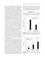
Following these results, a
multicenter randomized, double-blinded, placebo-
controlled trial with 59 lung transplant recipients was
carried out.
244
Among 29 patients receiving a dose of
sCR1 before reperfusion, 14 (48%) were extubated
within 24 hours, which was significantly better than in
the control arm, with only 6 patients of a total of 30
(20%). In addition, the overall duration of mechanical
ventilation and length of ICU stay tended to be shorter in
the group receiving sCR1, but the PaO
2
/FiO
2
ratio was
not different between groups. Recently, Stammberger and
colleagues have demonstrated that the administration of
a molecule combining sCR1 with sialyl Lewis X (a
selectin receptor antagonist), can achieve significantly
better results than the adminsitration of sCR1 alone.
245
Neutrophils
Neutrophils progressively infiltrate the transplanted lung
during the initial 24 hours of reperfusion. Although they
certainly play an important role in perpetuating reperfu-
sion injury, their function in the early phase of reperfu-
sion remains more controversial. Several experiments
have been performed with the use of a leukocyte filter to
deplete the blood at the time of reperfusion, demonstrat-
ing a beneficial effect of leukocyte depletion even after
short periods of reperfusion.
246,247
However, few studies
336
/ Advanced Therapy in Thoracic Surgery
have examined the specific role of neutrophils.
Using an isolated rat lung perfusion model, Deeb and
colleagues demonstrated that the addition of neutrophils
to the perfusion system was not necessary for the induc-
tion of reperfusion injury after a period of warm
ischemia.
248
With an antineutrophil antibody, the same
group went on to demonstrate that reperfusion injury
exhibited a bimodal pattern, consisting of neutrophil-
independent events during the early phase of reperfusion
and of neutrophil-mediated events in the late phase of
reperfusion.
249
Other studies with specific antibodies
against neutrophils confirm these findings and show that
other leukocytes such as macrophages have a more
important role in the early phase of reperfusion.
238,250,251
Clinical Lung Preservation at the
University of Toronto
When a potential lung donor is identified, 1 g of intra-
venous Solumedrol is administered. After the lungs have
been assessed and the other procurement teams have
finished their dissection, the donor is fully heparinized,
and the main pulmonary artery is cannulated with a 20
French cannula. Prostaglandin PGE
1
(Prostin VR,
UpJohn) 500 µg is added to the preservation solution
(Perfadex), and 500 µg is injected directly into the main
pulmonary artery just prior to flushing the lungs. The
lungs are recruited with 25 cm H
2
O prior to flushing to
remove atelectasis. After inflow occlusion, the left atrial
appendage is transected for drainage and the lungs are
flushed antegrade with 50 mL/kg of Perfadex solution at
4°C, with the bag hung approximately 30 cm above the
heart. The lungs are ventilated throughout the flush with
a tidal volume of 10 mL/kg, a PEEP of 5 cm H
2
O, and an
FiO
2
of 50%. A retrograde flush is then performed in situ
with ventilation being continued (250 mL Perfadex into
each pulmonary vein orifice). After completion of the
flush, the heart and then the lungs are extracted. We
inflate the lungs with a pressure of approximately 20 cm
H
2
O before tracheal cross-clamping to obtain lung
expansion but avoid overdistension. The lungs are then
packaged floating in 2 L of flush solution and stored on
ice for transport (Table 26-3).
At the beginning of the recipient operation we admin-
ister 500 mg of Solumedrol. The donor lung is kept cool
with a cooling jacket in the chest during implantation.
After implantation, the lung is gently recruited and venti-
lated: FiO
2
= 0.5, PEEP = 5 cm H
2
O, and pressure control
ventilation limiting the peak airway pressure to a maxi-
mum of 25 cm H
2
O. The lung is then reperfused slowly
over a 10-minute period by gradually removing the
pulmonary artery clamp or by allowing the right heart to
eject in a controlled fashion if on cardiopulmonary
bypass. We give no other routine pharmacologic therapy
following reperfusion—nitric oxide or PGE
1
are used
only for clinical indications of reperfusion injury.
Summary
It is now 20 years since the first successful single lung
transplant. Considerable progress has been made in lung
preservation since that time. The development of a
specific lung preservation solution has been an impor-
tant advance and the clinical introduction of the low-
potassium dextran solution has been a long time coming.
In general the lung transplant community has been
slow to translate the findings from animal experimental
work to the bedside, but this is changing. Ischemia-
reperfusion injury is still a significant clinical problem,
and our goals for the future are to be able to better assess
the degree of injury, to predict the degree of dysfunction,
and hopefully to develop strategies to treat or prevent the
injury in the first place. Ultimately, we strive towards
repairing or modifying a donor lung, allowing time for
repair of the injuries, and then testing the lungs ex vivo to
ensure good function before transplanting the organ into
the recipient.
References
1. Hosenpud JD, Bennett LE, Keck BM, et al. The registry of
the international society for heart and lung transplanta-
tion: seventeenth official report-2000. J Heart Lung
Tr ansplant 2000;19:909–31.
2. Anderson DC, Glazer HS, Semenkovich JW, et al. Lung
transplant edema: chest radiography after lung transplan-
tation—the first 10 days. Radiology 1995;195:275–81.
3. Kundu S, Herman SJ, Winton TL. Reperfusion edema
after lung transplantation: radiographic manifestations.
Radiology 1998;206:75–80.
4. King RC, Binns OA, Rodriguez F, et al. Reperfusion injury
significantly impacts clinical outcome after pulmonary
transplantation. Ann Thorac Surg 2000;69:1681–5.
5. Meyers CH, Purut CM, D’Amico TA, et al. Pulmonary
arterial impedance after single lung transplantation. J
Surg Res 1992;52:459–65.
Lung Preservation for Transplantation
/
337
TABLE 26-3. Current Recommendations for Lung
Preservation
Volume of flush solution 50 mL/kg
Pressure during flush solution 10–15 mm Hg
Temperature of flush solution 4°C–8°C
Lung ventilation 10 mL/kg
Lung inflation (airway pressure) 20 cm H
2
O
Oxygenation ≤ 50% FiO
2
Storage temperature 4°C–8°C
6. Qayumi AK, Nikbakht-Sangari MN, Godin DV, et al. The
relationship of ischemia-reperfusion injury of transplant-
ed lung and the up-regulation of major histocompatibility
complex II on host peripheral lymphocytes. J Thorac
Cardiovasc Surg 1998;115:978–89.
7. Toronto Lung Transplant Group. Unilateral lung trans-
plantation for pulmonary fibrosis. N Engl J Med
1986;314:1140–5.
8. Sommers KE, Griffith BP, Hardesty RL, Keenan RJ. Early
lung allograft function in twin recipients from the same
donor: risk factor analysis. Ann Thorac Surg
1996;62:784–90.
9. Madill J, Gutierrez C, Grossman J, et al. Nutritional assess-
ment of the lung transplant patient: body mass index as a
predictor of 90-day mortality following transplantation. J
Heart Lung Transplant 2001;20:288–96.
10. Meyer DM, Bennett LE, Novick RJ, Hosenpud JD. Effect of
donor age and ischemic time on intermediate survival and
morbidity after lung transplantation. Chest
2000;118:1255–62.
11. Pierson RN, Milstone AP, Loyd JE, et al. Lung allocation in
the United States, 1995–1997: an analysis of equity and
utility. J Heart Lung Transplant 2000;19:846–51.
12. deMeester J, Smits JM, Persijn GG, Haverich A. Lung
transplant waiting list: differential outcome of type of
end-stage lung disease, one year after registration. J Heart
Lung Transplant 1999;18:563–71.
13. Cohen RG, Starnes VA. Living donor lung transplantation.
World J Surg 2001;25:244–50.
14. Steen S, Sjoberg T, Pierre L, et al. Transplantation of lungs
from a non-heart-beating donor. Lancet 2001;357:825–9.
15. Pierre AF, Sekine Y, Hutcheon M, et al. Evaluation of
extended donor and recipient criteria for lung transplan-
tation. J Heart Lung Transplant 2001;20:256.
16. Sundaresan S, Trachiotis GD, Aoe M, et al. Donor lung
procurement: assessment and operative technique. Ann
Thorac Surg 1993;56:1409–13.
17. Gabbay E, Williams TJ, Griffiths AP, et al. Maximizing the
utilization of donor organs offered for lung transplanta-
tion. Am J Respir Crit Care Med 1999;160:265–71.
18. Sundaresan S, Semenkovich J, Ochoa L, et al. Successful
outcome of lung transplantation is not compromised by
the use of marginal donor lungs. J Thorac Cardiovasc Surg
1995;109:1075–9.
19. Bhorade SM, Vigneswaran W, McCabe MA, Garrity ER.
Liberalization of donor criteria may expand the donor
pool without adverse consequence in lung transplanta-
tion. J Heart Lung Transplant 2000;19:1199–204.
20. Bittner HB, Kendall SW, Chen EP, et al. The effects of brain
death on cardiopulmonary hemodynamics and pulmonary
blood flow characteristics. Chest 1995;108:1358–63.
21. Follette DM, Rudich SM, Babcock WD. Improved oxy-
genation and increased lung donor recovery with high-
dose steroid administration after brain death. J Heart Lung
Tr ansplant 1998;17:423–9.
22. Takada M, Nadeau KC, Hancock WW, et al. Effects of
explosive brain death on cytokine activation of peripheral
organs in the rat. Transplantation 1998;65:1533–42.
23. Pratschke J, Wilhelm MJ, Kusaka M, et al. Accelerated
rejection of renal allografts from brain-dead donors. Ann
Surg 2000;232:263–71.
24. DerHoeven JA, TerHorst GJ, Molema G, et al. Effects of
brain death and hemodynamic status on function and
immunologic activation of the potential donor liver in the
rat. Ann Surg 2000;232:804–13.
25. Koo DD, Welsh KI, McLaren AJ, et al. Cadaver versus living
donor kidneys: impact of donor factors on antigen induc-
tion before transplantation. Kidney Int 1999;56:1551–9.
26. Schwarz C, Regele H, Steininger R, et al. The contribution
of adhesion molecule expression in donor kidney biopsies
to early allograft dysfunction. Transplantation
2001;71:1666–70.
27. Fisher AJ, Donnelly SC, Hirani N, et al. Elevated levels of
interleukin-8 in donor lungs is associated with early graft
failure after lung transplantation. Am J Respir Crit Care
Med 2001;163:259–65.
28. Hopkinson DN, Bhabra MS, Hooper TL. Pulmonary graft
preservation: a worldwide survey of current clinical prac-
tice. J Heart Lung Transplant 1998;17:525–31.
29. Fujimura S, Handa M, Kondo T, et al. Successful 48-hour
simple hypothermic preservation of canine lung trans-
plants. Transplant Proc 1987;19:1334–6.
30. Keshavjee SH, Yamazaki F, Cardoso PF, et al. A method for
safe twelve-hour pulmonary preservation. J Thorac
Cardiovasc Surg 1989;98:529–34.
31. Yamazaki F, Yokomise H, Keshavjee SH, et al. The superior-
ity of an extracellular fluid solution over Euro-Collins’
solution for pulmonary preservation. Transplantation
1990;49:690–4.
32. Keshavjee SH, Yamazaki F, Yokomise H, et al. The role of
dextran 40 and potassium in extended hypothermic lung
preservation for transplantation. J Thorac Cardiovasc
Surg 1992;103:314–25.
33. Date H, Matsumura A, Manchester JK, et al. Evaluation of
lung metabolism during successful twenty-four hour
canine lung preservation. J Thorac Cardiovasc Surg
1993;105:480–91.
34. Steen S, Kimbald PO, Sjoberg T, et al. Safe lung preserva-
tion for twenty-four hours with Perfadex. Ann Thorac
Surg 1994;57:336–41.
35. Date H, Izumi S, Miyade Y, et al. Successful canine bilater-
al single-lung transplantation after 21-hour lung preserva-
tion. Ann Thorac Surg 1995;59:336–41.
338
/ Advanced Therapy in Thoracic Surgery
36. Spaggiari L, Bobbio P. Dextran 40 at 2% versus 5% in low-
potassium solutions: which is best? Ann Thorac Surg
1994;58:1784–6.
37. Miyoshi S, Shimokawa S, Schreinemakers HH, et al.
Comparision of the University of Wisconsin perservation
solution and other crystalloid perfusates in a 30-hour rab-
bit lung preservation model. J Thorac Cardiovasc Surg
1992;103:27–32.
38. Wagner FM, Jamieson SW, Fung J, et al. A new concept for
successful long-term pulmonary preservation in a dog
model. Transplantation 1995;59:1530–6.
39. Chien S, Zhang F, Niu W, et al. Comparison of university
of wisconsin, euro-collins, low-potassium dextran, and
krebs-henseleit solutions for hypothermic lung preserva-
tion. J Thorac Cardiovasc Surg 2000;119:921–30.
40. Roberts RF, Nishanian GP, Carey JN, et al. A comparison
of the new preservation solution Celsior to Euro-Collins
and University of Wisconsin solutions in lung reperfusion
injury. Transplantation 1999;67:152–5.
41. Wittwer T, Wahlers T, Fehrenbach A, et al. Improvement of
pulmonary preservation with Celsior and Perfadex:
impact of storage time on early post-ischemic lung func-
tion. J Heart Lung Transplant 1999;18:1198–201.
42. Keshavjee SH, McRitchie DI, Vittorini T, et al. Improved
lung preservation with dextran 40 is not mediated by a
superoxide radical scavenging mechanism. J Thorac
Cardiovasc Surg 1992;103:326–8.
43. Kimbald PO, Sjoberg T, Massa G, et al. High potassim con-
tents in organ preservation solutions cause strong pul-
monary vasocontraction. Ann Thorac Surg
1991;52:523–8.
44. Fukuse T, Albes JM, Wilhelm A, et al. Influence of dextrans
on lung preservation: is the molecular weight important? J
Heart Lung Transplant 1996;15:903–10.
45. Sakamaki F, Goffmann H, Munzing S, et al. Effects of lung
preservation solutions on PMN activation in vitro.
Tr ansplant Int 1999;12:113–21.
46. Maccherini M, Keshavjee SH, Slutsky AS, et al. The effect
of low-potassium-dextran versus Euro-Collins solution
for preservation of isolated type II pneumocytes.
Tr ansplantation 1991;52:621–6.
47. Suzuki S, Inoue K, Sugita M, et al. Effects of EP4 solution
and LPD solution vs Euro-Collins solution on
Na(+)/K(+)-ATPase activity in rat alveolar type II cells
and human alveolar epithelial cell line A549 cells. J Heart
Lung Transplant 2000;19:887–93.
48. Struber M, Hohlfeld JM, Fraund S, et al. Low-potassium
dextran solution ameliorates reperfusion injury of the
lung and protects surfactant function. J Thorac Cardiovasc
Surg 2000;120:566–72.
49. Sakamaki F, Hoffmann H, Muller C, et al. Reduced lipid
peroxidation and ischemia-reperfusion injury after lung
transplantation using low-potassium dextran solution for
lung preservation. Am J Respir Crit Care Med
1997;156:1073–81.
50. Hopkinson DN, Odom JJ, Bridgewater BJ, Hooper TL.
University of Wisconsin solution for lung graft preserva-
tion: which components are important? J Heart Lung
Tr ansplant 1994;13:990–7.
51. Fischer S, Hopkinson D, Liu M, Keshavjee SH. Raffinose
improves the function of rat pulmonary grafts stored for
twenty-four hours in low-potassium dextran solution. J
Thorac Cardiovasc Surg 2000;119:488–92.
52. Fischer S, Hopkinson D, Liu M, et al. Raffinose improves
24-hour lung preservation in low potassium dextran glu-
cose solution: a histologic and ultrastructural analysis.
Ann Thorac Surg 2001;71:1140–5.
53. Muller C, Furst H, Reichenspurner H, et al. Lung procure-
ment by low-potassium dextran and the effect on preserva-
tion injury. Munich Lung Transplant Group.
Tr ansplantation 1999;68:1139–43.
54. Struber M, Wilhelmi M, Harringer W, et al. Flush perfu-
sion with low potassium dextran solution improves early
graft function in clinical lung transplantation. Eur J
Cardiothorac Surg 2001;19:190–4.
55. Fischer S, Matte-Martyn A, DePerrot M, et al. Low-
potassium dextran preservation solution improves lung
function after human lung transplantation. J Thorac
Cardiovasc Surg 2001;121:594–6.
56. Haverich A, Aziz S, Scott WC, et al. Improved lung preser-
vation using Euro-Collins solution for flush-perfusion.
Thorac Cardiovasc Surg 1986;34:368–76.
57. Sasaki M, Muraoka R, Chiba Y, Hiramatu Y. Influence of
pulmonary arterial pressure during flushing on lung
preservation. Transplantation 1996;61:22–7.
58. Tanaka H, Chiba Y, Sasaki M, et al. Relationship between
flushing pressure and nitric oxide production in preserved
lungs. Transplantation 1998;65:460–4.
59. Andrade RS, Wangensteen OD, Jo JK, et al. Effect of
hypothermic pulmonary artery flushing on capillary fil-
tration coefficient. Transplantation 2000;70:267–71.
60. Kimbald PO, Sjoberg T, Steen S. Pulmonary vascular resis-
tance related to endothelial function after lung transplan-
tation. Ann Thorac Surg 1994;58:416–20.
61. Wang LS, Nakamoto K, Hsieh CM, et al. Influence of tem-
perature of flushing solution on lung preservation. Ann
Thorac Surg 1993;55:711–5.
62. Albes JM, Fischer F, Bando T, et al. Influence of the per-
fusate temperature on lung preservation: is there an opti-
mum? Eur Surg Res 1997;29:5–11.
63. Steen S, Ingemansson R, Budrikis A, et al. Successful trans-
plantation of lungs topically cooled in the non-heart-beat-
ing donor for 6 hours. Ann Thorac Surg 1997;63:345–51.
64. VanRaemdonck DE, Jannis NC, Rega FR, et al.
External cooling of warm ischemic rabbit lungs after
death. Ann Thorac Surg 1996;62:331–7.
65. Hall SM, Odom N, McGregor CG, Haworth SG. Transient
ultrastructural injury and repair of pulmonary capillaries
in transplanted rat lung: effect of preservation and reper-
fusion. Am J Respir Cell Mol Biol 1992;7:49–57.
Lung Preservation for Transplantation
/
339
66. Steen S, Sjoberg T, Ingemansson R, Lindberg L. Efficacy of
topical cooling in lung preservation: is a reappraisal due?
Ann Thorac Surg 1994;58:1657–63.
67. Toronto Lung Transplant Group. Experience with single-
lung transplantation for pulmonary fibrosis. JAMA
1988;259:2258–62.
68. Sakuma T, Takahashi K, Ohya N, et al. Ischemia-
reperfusion injury in rabbits: mechanisms of injury and
protection. Am J Physiol 1999;276:L137–45.
69. VanRaemdonch DE, Jannis NC, Rega FR, et al. Extended
preservation of ischemic pulmonary graft by postmortem
alveolar expansion. Ann Thorac Surg 1997;64:801–8.
70. Veith FJ, Sinha SB, Graves JS, et al. Ischemic tolerance of
the lung. The effect of ventilation and inflation. J Thorac
Cardiovasc Surg 1971;61:804–10.
71. Kuang JQ, VanRaemdonck DE, Jannis NC, et al.
Pulmonary cell death in warm ischemic rabbit lung is
related to the alveolar oxygen reserve. J Heart Lung
Tr ansplant 1998;17:406–14.
72. Date H, Matsumura A, Manchester JK, et al. Changes in
alveolar oxygen and carbon dioxide concentration and
oxygen consumption during lung preservation. The main-
tenance of aerobic metabolism during lung preservation. J
Thorac Cardiovasc Surg 1993;105:492–501.
73. Fukuse T, Hirata T, Nnakamura T, et al. Influence of
deflated and anaerobic conditions during cold storage on
rat lungs. Am J Respir Crit Care Med 1999;160:621–7.
74. DeCampos KN, Keshavjee S, Liu M, Slutsky AS. Optimal
inflation volume for hypothermic preservation of rat
lungs. J Heart Lung Transplant 1998;17:599–607.
75. Sakuma T, Tsukano C, Ishigaki M, et al. Lung deflation
impairs alveolar epithelial fluid transport in ischemic rab-
bit and rat lungs. Transplantation 2000;69:1785–93.
76. Puskas JD, Hirai T, Christie N, et al. Reliable thirty-hour
lung preservation by donor lung hyperinflation. J Thorac
Cardiovasc Surg 1992;104:1075–83.
77. Baretti R, Bitu-Morsdorf J, Beyersdorf F, et al. Distribution
of lung preservation solutions in parenchyma and airways:
influence of atelectasis and route of delivery. J Heart Lung
Tr ansplant 1995;14:80–91.
78. Aoe M, Okabayashi K, Cooper JD, Patterson GA.
Hyperinflation of canine lung allografts during storage
increases reperfusion pulmonary edema. J Thorac
Cardiovasc Surg 1996;112:94–102.
79. Haniuda M, Hasegawa S, Shiraishi T, et al. Effects of infla-
tion volume during lung preservation on pulmonary cap-
illary permeability. J Thorac Cardiovasc Surg
1996;112:85–93.
80. Kayano K, Toda K, Naka Y, Pinsky DJ. Identification of
optimal conditions for lung graft storage with Euro-
Collins solution by use of a rat orthotopic lung transplant
model. Circulation 1999;100:II257–61.
81. We der W, Harper B, Shimokawa S, et al. Influence of
intraalveolar oxygen concentration on lung preservation in
a rabbit model. J Thorac Cardiovasc Surg
1991;101:1037–43.
82. Fukuse T, Hirata T, Hitomi S, Wada H. Influence of alveo-
lar gas during pulmonary preservation on reperfusion
injury. Transplant Proc 2000;32:334–5.
83. Haniuda M, Dresler CM, Mizuata T, et al. Free radical-
mediated vascular injury in lungs preserved at moderate
hypothermia. Ann Thorac Surg 1995;60:1376–81.
84. Fisher AB, Dodia C, Tan ZT, et al. Oxygen-dependent lipid
peroxidation during lung ischemia. J Clin Invest
1991;88:674–9.
85. Ueno T, Yokomise H, Oka T, et al. The effect of PGE1 and
temperature on lung function following preservation.
Tr ansplantation 1991;52:626–30.
86. Date H, Lima O, Matsumura A, et al. In a canine model,
lung preservation at 10 degrees C is superior to that at 4
degrees C. A comparision of two preservation tempera-
tures on lung function and on adenosine triphosphate
level measured by phosphorus 31-nuclear magnetic reso-
nance. J Thorac Cardiovasc Surg 1992;103:773–80.
87. Mayer E, Puskas JD, Cardoso PF, et al. Reliable eighteen-
hour lung preservation at 4 degrees and 10 degrees C by
pulmonary artery flush after high-dose prostaglandin E1
administration. J Thorac Cardiovasc Surg
1992;103:1136–42.
88. Varela A, Montero C, Cordoba M, et al. Clinical experience
with retrograde lung preservation. Transplant Int
1996;9:S296–8.
89. Wittwer T, Fehrenbach A, Meyer D, et al. Retrograde flush
perfusion with low-potassium solutions for improvement
of experimental pulmonary preservation. J Heart Lung
Tr ansplant 2000;19:976–83.
90. Va rela A, Montero CG, Cordoba M, et al. Improved distrib-
ution of pulmonary flush solution to the tracheobronchial
wall in pulmonary transplantation. Eur Surg Res
1997;29:1–4.
91. Chen CZ, Gallagher RC, Ardery P, et al. Retrograde flush
and cold storage for twenty-two to twenty-five hours lung
preservation with and without prostaglandin E1. J Heart
Lung Transplant 1997;16:658–66.
92. Parrott NR, Forsythe JL, Matthews JN, et al. Late perfu-
sion. A simple remedy for renal allograft primary non-
function. Transplantation 1990;49:913–5.
93. Serrick CJ, Jamjoum A, Reis A, et al. Amelioration of pul-
monary allograft injury by administering a second rinse
solution. J Thorac Cardiovasc Surg 1996;112:1010–6.
94. Venuta F, Rendina EA, Bufi M, et al. Preimplantation ret-
rograde pneumoplegia in clinical lung transplantation. J
Thorac Cardiovasc Surg 1999;118:107–14.
340
/ Advanced Therapy in Thoracic Surgery
95. Bhabra MS, Hopkinson DN, Shaw TE, et al. Controlled
reperfusion protects lung grafts during a transient early
increase in permeability. Ann Thorac Surg
1998;65:187–92.
96. Clark SC, Sudarshan C, Khanna R, et al. Controlled reper-
fusion and pentoxifylline modulate reperfusion injury
after single lung transplantation. J Thorac Cardiovasc Surg
1998;115:1335–41.
97. Pierre AF, DeCampos KN, Liu M, et al. Rapid reperfusion
causes stress failure in ischemic rat lungs. J Thorac
Cardiovasc Surg 1998;116:932–42.
98. Bhabra MS, Hopkinson DN, Shaw TE, Hooper TL. Critical
importance of the first 10 minutes of lung graft reperfu-
sion after hypothermic storage. Ann Thorac Surg
1996;61:1631–5.
99. DosSantos CC, Slutsky AS. Invited review: Mechanisms of
ventilator-induced lung injury: a perspective. J Appl
Physiol 2000;89:1645–55.
100. McRae K. Con: lung transplantation should not be rou-
tinely performed with cardiopulmonary bypass. J
Cardiothorac Vasc Anesth 2000;14:746–50.
101. DeCampos KN, Keshavjee S, Slutsky AS, Liu M. Alveolar
recruitment prevents rapid-reperfusion-induced injury of
lung transplants. J Heart Lung Transplant
1999;18:1096–102.
102. Cassivi SD, Liu M, Boehler A, et al. Transgene expression
after adenovirus-mediated retransfection of rat lungs is
increased and prolonged by transplant immunosuppres-
sion. J Thorac Cardiovasc Surg 1999;117:1–7.
103. Cassivi SD, Liu M, Boehler A, et al. Transplant immuno-
suppression increases and prolongs transgene expression
following adenoviral-mediated transfection of rat lungs. J
Heart Lung Transplant 2000;19:984–94.
104. Cassivi SD, Cardella JA, Fischer S, et al. Transtracheal gene
transfection of donor lungs prior to organ procurement
increases transgene levels at reperfusion and following
transplantation. J Heart Lung Transplant 1999;18:1181–8.
105. Yano M, Hiratsuka M, Mora BN, et al. Transfection of pul-
monary artery segments in lung isografts during storage.
Ann Thorac Surg 1999;68:1810–4.
106. Yano M, Mora BN, Ritter JM, et al. Ex vivo transfection of
transforming growth factor-beta1 gene to pulmonary
artery segments in lung grafts. J Thorac Cardiovasc Surg
1999;117:705–13.
107. Suda T, Mora BN, D’Ovidio F, et al. In vivo adenovirus-
mediated endothelial nitric oxide synthase gene transfer
ameliorates lung allograft ischemia-reperfusion injury. J
Thorac Cardiovasc Surg 2000;119:297–304.
108. Fischer S, Liu M, MacLean AA, et al. In vivo transtracheal
adenovirus-mediated transfer of human interleukin-10
gene to donor lungs ameliorates ischemia-reperfusion
injury and improves early posttransplant graft function in
the rat. Hum Gene Ther 2001;12:1513–26.
109. Wickersham NE, Johnson JJ, Meyrick BO, et al. Lung
ischemia-reperfusion injury in awake sheep: protection
with verapamil. J Appl Physiol 1991;71:1554–62.
110. Yokomise H, Ueno T,Yamazaki F, et al. The effect and opti-
mal time of administration of verapamil on lung preser-
vation. Transplantation 1990;49:1039–43.
111. Pickford MA, Gower JD, Dore C, et al. Lipid peroxidation
and ultrastructural changes in rat lung isografts after sin-
gle-passage organ flush and 48-hour cold storage with and
without one-hour reperfusion in vivo. Transplantation
1990;50:210–8.
112. Pickford MA, Gower JD, Simpkin S, et al. Function of sin-
gle rat lung isografts after 48-hour cold storage. The effect
of treatment with free radical antagonists and prostacyclin
PGI2. Transplantation 1991;51:733–49.
113. Haverich A, Karck M. Role of calcium channel blockers in
postischemic lungs. Ann N Y Acad Sci 1994;723:51–8.
114. McCord JM. Oxygen-derived free radicals in postischemic
tissue injury. N Engl J Med 1985;312:159–63.
115. AlMehdi AB, Shuman H, Fisher AB. Intracellular genera-
tion of reactive oxygen species during nonhypoxic lung
ischemia. Am J Physiol 1997;272:L294–300.
116. Eckenhoff RG, Dodia C, Tan Z, Fisher AB. Oxygen-
dependent reperfusion injury in the isolated rat lung. J
Appl Physiol 1992;72:1454–60.
117. Fisher AB, Dodia C. Lung as a model for evaluation of crit-
ical intracellular PO
2
and PCO. Am J Physiol
1981;241:E47–50.
118. Adkins WK, Taylor AE. Role of xanthine oxidase and neu-
trophils in ischemia-reperfusion injury in rabbit lung. J
Appl Physiol 1990;69:2012–8.
119. Kennedy TP, Rao NV, Hopkins C, et al. Role of reactive
oxygen species in reperfusion injury of the rabbit lung. J
Clin Invest 1989;83:1326–35.
120. Zhao G, AlMehdi AB, Fisher AB. Anoxia-reoxygenation
versus ischemia in isolated rat lungs. Am J Physiol
1997;273:L1112–7.
121. AlMehdi AB, Zhao G, Dodia C, et al. Endothelial NADPH
oxidase as the source of oxidants in lungs exposed to
ischemia or high K+. Circ Res 1998;83:730–7.
122. Davies PF, Tripathi SC. Mechanical stress mechanisms and
the cell. An endothelial paradigm. Circ Res 1993;72:239–45.
123. Lansman JB. Endothelial mechanosensors. Going with the
flow. Nature 1988;331:481–2.
124. AlMehdi AB, Zhao G, Fisher AB. ATP-independent mem-
brane depolarization with ischemia in the oxygen-venti-
lated isolated rat lung. Am J Respir Cell Mol Biol
1998;18:653–61.
125. Wei Z, Costa K, AlMehdi AB, et al. Simulated ischemia in
flow-adapted endothelial cells leads to generation of reactive
oxygen species and cell signaling. Circ Res 1999;85:682–9.
Lung Preservation for Transplantation
/
341
126. Henderson LM, Chappell JB, Jones OT. Superoxide gener-
ation by the electrogenic NADPH oxidase of human neu-
trophils is limited by the movement of compensating
charge. Biochem J 1988;255:285–90.
127. Kitagawa S, Johnston RB. Relationship between mem-
brane potential changes and superoxide-releasing capacity
in resident and activated mouse peritoneal macrophages. J
Immunol 1985;135:3417–23.
128. Kelly RF. Current strategies in lung preservation. J Lab
Clin Med 2000;136:427–40.
129. Baker CJ, Longoria J, Gade PV, et al. Addition of a water-sol-
uble alpha-tocopherol analogue to University of Wisconsin
solution improves endothelial viability and decreases lung
reperfusion injury. J Surg Res 1999;86:145–9.
130. Ochs M, Nenadic I, Fehrenbach A, et al. Ultrastructural
alterations in intraalveolar surfactant subtypes after exper-
imental ischemia and reperfusion. Am J Respir Crit Care
Med 1999;160:718–24.
131. Veldhuizen RA, Lee J, Sandler D, et al. Alterations in pul-
monary surfactant composition and activity after experi-
mental lung transplantation. Am Rev Respir Dis
1993;148:208–15.
132. Erasmus ME, Petersen AH, Oetomo SB, Prop J. The func-
tion of surfactant is impaired during the reimplantation
response in rat lung transplants. J Heart Lung Transplant
1994;13:791–802.
133. Andrade RS, Solien EE, Wangensteen OD, et al. Surfactant
dysfunctionin lung preservation. Transplantation
1995;60:536–41.
134. Casals C, Varela A, Ruano ML, et al. Increase of C-reactive
protein and decrease of surfactant protein A in surfactant
after lung transplantation. Am J Respir Crit Care Med
1998;157:43–9.
135. Ochs M, Fehrenbach H, Nenadic I, et al. Preservation of
intraalveolar surfactant in a rat lung ischaemia/reperfu-
sion injury model. Eur Respir J 2000;15:526–31.
136. Fehrenbach A, Ochs M, Warnecke T, et al. Beneficial effect
of lung preservation is related to ultrastructural integrity
of tubular myelin after experimental ischemia and reper-
fusion. Am J Respir Crit Care Med 2000;161:2058–65.
137. Ochs M, Fehrenbach H, Richter J. Ultrastructure of canine
type II pneumocytes during hypothermic ischemia of the
lung: a study by means of conventional and energy filter-
ing transmission electron microscopy and stereology. Anat
Rec 2001;263:118–26.
138. Klepetko W, Lohninger A, Wisser W, et al. Pulmonary sur-
factant in bronchoalveolar lavage after canine lung trans-
plantation: effect of L-carnitine application. J Thorac
Cardiovasc Surg 1990;99:1048–58.
139. Buchanan SA, Mauney MC, Parekh VI, et al. Intratracheal
surfactant administration preserves airway compliance
during lung reperfusion. Ann Thorac Surg
1996;62:1617–21.
140. Erasmus ME, Petersen AH, Hofstede G, et al. Surfactant
treatment before reperfusion improves the immediate
function of lung transplants in rats. Am J Respir Crit Care
Med 1996;153:665–70.
141. Hohlfeld JM, Struber M, Ahlf K, et al. Exogenous surfac-
tant improves survival and surfactant function in
ischaemia-reperfusion injury in minipigs. Eur Respir J
1999;13:1037–43.
142. Struber M, Hirt SW, Cremer J, et al. Surfactant replace-
ment in reperfusion injury after clinical lung transplanta-
tion. Intensive Care Med 1999;25:862–4.
143. Erasmus ME, Hofstede GJ, Petersen AH, et al. Effects of
early surfactant treatment persisting for one week after
lung transplantation in rats. Am J Respir Crit Care Med
1997;156:567–72.
144. Novick RJ, Veldhuizen RA, Possmayer F, et al. Exogenous
surfactant therapy in thirty-eight hour lung graft preser-
vation for transplantation. J Thorac Cardiovasc Surg
1994;108:259–68.
145. Struber M, Cremer J, Harringer W, et al. Nebulized syn-
thetic surfactant in reperfusion injury after single lung
transplantation. J Thorac Cardiovasc Surg
1995;110:563–4.
146. Fischer S, Cassivi SD, Xavier AM, et al. Cell death in
human lung transplantation: apoptosis induction in
human lungs during ischemia and after transplantation.
Ann Surg 2000;231:424–31.
147. Fischer S, Maclean AA, Liu M, et al. Dynamic changes in
apoptotic and necrotic cell death correlate with severity of
ischemia-reperfusion injury in lung transplantation. Am J
Respir Crit Care Med 2000;162:1932–9.
148. Yaoita H, Ogawa K, Maehara K, Maruyama Y. Attenuation
of ischemia/reperfusion injury in rats by a caspase
inhibitor. Circulation 1998;97:276–81.
149. Hartmann A. Antiapoptotic agents in brain ischemia. N
Engl J Med 2000;342:823.
150. Serrick C, Adoumie R, Giaid A, Shennib H. The early
release of interleukin-2, tumor necrosis factor-alpha and
interferon-gamma after ischemia reperfusion injury in the
lung allograft. Transplantation 1994;58:1158–62.
151. Chang DM, Hsu K, Ding YA, Chiang CH. Interleukin-1 in
ischemia-reperfusion acute lung injury. Am J Respir Crit
Care Med 1997;156:1230–4.
152. Khimenko PL, Bagby GJ, Fuseler J, Taylor AE. Tumor
necrosis factor-alpha in ischemia and reperfusion injury in
rat lungs. J Appl Physiol 1998;85:2005–11.
153. Sekido N, Mukaida N, Harada A, et al. Prevention of lung
reperfusion injury in rabbits by a monoclonal antibody
against interleukin-8. Nature 1993;365:654–7.
154. LeMoine O, Marchant A, Durand F, et al. Systemic release
of interleukin-10 during orthotopic liver transplantation.
Hepatology 1994;20:889–92.
342
/ Advanced Therapy in Thoracic Surgery
Lung Preservation for Transplantation
/
343
155. Kahlke V, Angele MK, Ayala A, et al. Immune dysfunction
following trauma-haemorrhage: influence of gender and
age. Cytokine 2000;12:69–77.
156. Lentsch AB, Yoshidome H, Kato A, et al. Requirement for
interleukin-12 in the pathogenesis of warm hepatic
ischemia/reperfusion injury in mice. Hepatology
1999;30:1448–53.
157. Daemen MA, van’t Veer C, Wolfs TG, Buurman WA.
Ischemia/reperfusion induced IFN-gamma up-regulation:
involvement of IL-12 and IL-18. J Immunol
1999;162:5506–10.
158. Heller A, Koch T, Schmeck J, vanAckern K. Lipid media-
tors in inflammatory disorders. Drugs 1998;55:487–96.
159. Shen CY, Wang D, Chang ML, Hsu K. Protective effect of
mepacrine on hypoxia-reoxygenation-induced acute lung
injury in rats. J Appl Physiol 1995;78:225–31.
160. Nagahiro I, Aoe M, Yamashita M, et al. EPC-K1 is effective
in lung preservation in an ex vivo rabbit lung perfusion
model. Ann Thorac Surg 1997;63:954–9.
161. Miotla JM, Jeffery PK, Hellewell PG. Platelet-activating
factor plays a pivotal role in the induction of experimental
lung injury. Am J Respir Cell Mol Biol 1998;18:197–204.
162. Nagase T, Ishii S, Kume K, et al. Platelet-activating factor
mediates acid-induced lung injury in genetically engi-
neered mice. J Clin Invest 1999;104:1071–6.
163. Kawahara K, Tagawa T, Takahashi T, et al. The effect of the
platelet-activating factor inhibitor TCV-309 on reperfu-
sion injury in a canine model of ischemic lung.
Tr ansplantation 1993;55:1438–9.
164. Wahlers T, Hirt SW, Haverich A, et al. Future horizons of
lung preservation by application of a platelet-activating
factor antagonist compared with current clinical stan-
dards. Euro-Collins flush perfusion versus donor core
cooling. J Thorac Cardiovasc Surg 1992;103:200–4; discus-
sion 5.
165. Qayumi AK, English JE, Duncan S, et al. Extended lung
preservation with platelet-activating factor-antagonist
TCV-309 in combination with prostaglandin E1. J Heart
Lung Transplant 1997;16:946–55.
166. Qayumi AK, Jamieson WR, Poostizadeh A. Effects of
platelet-activating factor antagonist CV-3988 in preserva-
tion of heart and lung for transplantation. Ann Thorac
Surg 1991;52:1026–32.
167. Kim JD, Baker CJ, Roberts RF, et al. Platelet activating fac-
tor acetylhydrolase decreases lung reperfusion injury. Ann
Thorac Surg 2000;70:423–8.
168. Wittwer T, Grote M, Oppelt P, et al. Impact of PAF antag-
onist BN 52021 (Ginkolide B) on post-ischemic graft
function in clinical lung transplantation. J Heart Lung
Tr ansplant 2001;20:358–63.
169. Grino JM. BN 52021: a platelet activating factor antagonist
for preventing post-transplant renal failure. A double-
blind, randomized study. Ann Intern Med
1994;121:345–7.
170. Zamora CA, Baron DA, Heffner JE. Thromboxane con-
tributes to pulmonary hypertension in ischemia-reperfu-
sion lung injury. J Appl Physiol 1993;74:224–9.
171. Ljungman AG, Grum CM, Deeb GM, et al. Inhibition of
cyclooxygenase metabolite production attenuates ischemia-
reperfusion lung injury. Am Rev Respir Dis 1991;143:610–7.
172. Segiet W, Krieter H, Stieber C, et al. Effect of cyclooxyge-
nase inhibition in a canine model of unilateral pulmonary
occlusion and reperfusion. Intensive Care Med
1995;21:817–25.
173. Kukkonen S, Heikkila L, Verkkala K, et al. Thromboxane
receptor blockade does not attenuate pulmonary pressor
response in porcine single lung transplantation. J Heart
Lung Transplant 1996;15:409–14.
174. Su M, Chi EY, Bishop JJ, Henderson WR. Lung mast cells
increase in number and degranulate during pulmonary
artery occlusion/reperfusion injury in dogs. Am Rev
Respir Dis 1993;147:448–56.
175. Vural KM, Liao H, Oz MC, Pinsky DJ. Effects of mast cell
membrane stabilizing agents in a rat lung ischemia-
reperfusion model. Ann Thorac Surg 2000;69:228–32.
176. Moore TM, Khimenko P, Adkins WK, et al. Adhesion mol-
ecules contribute to ischemia and reperfusion-induced
injury in the isolated rat lung. J Appl Physiol
1995;78:2245–52.
177. Naka Y, Toda K, Kayano K, et al. Failure to express the P-
selectin gene or P-selectin blockade confers early pul-
monary protection after lung ischemia or transplantation.
Proc Natl Acad Sci U S A 1997;94:757–61.
178. Reignier J, Sellak H, Lemoine R, et al. Prevention of
ischemia-reperfusion lung injury by sulfated Lewis(a)
pentasaccharide. The Paris-Sud University Lung
Tr ansplantation Group. J Appl Physiol 1997;82:1058–63.
179. Schmid RA, Yamashita M, Boasquevisque CH, et al.
Carbohydrate selectin inhibitor CY-1503 reduces neu-
trophil migration and reperfusion injury in canine pul-
monary allografts. J Heart Lung Transplant
1997;16:1054–61.
180. Brandt M, Boeke K, Phillips ML, et al. Effect of oligosac-
charides on rejection and reperfusion injury after lung
transplantation. J Heart Lung Transplant 1997;16:352–9.
181. Horgan MJ, Ge M, Gu J, et al. Role of ICAM-1 in neu-
trophil mediated lung vascular injury after occlusion and
reperfusion. Am J Physiol 1991;261:H1578–84.
182. Toda K, Kayano K, Karimova A, et al. Antisense intercellu-
lar adhesion moleule-1 (ICAM-1) oligodeoxyribonu-
cleotide delivered during organ preservation inhibits post-
transplant ICAM-1 expression and reduces primary lung
isograft failure. Circ Res 2000;86:166–74.
183. Kapelanski DP, Iguchi A, Niles SD, Mao HZ. Lung reperfu-
sion injury is reduced by inhibiting a CD18-dependent
mechanism. J Heart Lung Transplant 1993;12:294–306;
discussion 7.
184. Haug CE, Colvin RB, DelMonico FL, et al. A phase I trial of
immunosuppression with anti-ICAM-1 (CD54) mAb in
renal allograft recipients. Transplantation 1993;55:766–72.
185. Qayumi AK, Jamieson WR, Poostizadeh A, et al.
Comparison of new iron chelating agents in the preven-
tion of ischemia/reperfusion injury: a swine model of
heart-lung transplantation. J Invest Surg 1992;5:115–27.
186. Hillinger S, Schmid RA, Stammberger U, et al. Donor and
recipient treatment with the Lazaroid U-74006F do not
improve post-transplant lung function in swine. Eur J
Cardiothorac Surg 1999;15:475–80.
187. Kuwaki K, Komatsu K, Sohma H, Abe T. The effect of var-
ious doses of lazaroid U74389G on lung ischemia reperfu-
sion injury. Thorac Cardiovasc Surg 1999;47:67–72.
188. Ogawa T, Mimura Y. Antioxidant effect of zinc on acute
renal failure induced by ischemia-reperfusion injury in
rats. Am J Nephrol 1999;19:609–14.
189. Vanacore RM, Eskew JD, Morales PJ, et al. Role for copper
in transient oxidation and nuclear translocation of MTF-
1, but not of NF-kappa B, by the heme-hemopexin trans-
port system. Antioxid Redox Signal 2000;2:739–52.
190. Soncul H, Kaptanoglu M, Oz E, et al. The role of selenium
added to pulmonary preservation solutions in isolated
guinea pig lungs. J Thorac Cardiovasc Surg
1994;108:922–7.
191. Ogawa S, Gerlach H, Esposito C, et al. Hypoxia modulates
the barrier and coagulant function of cultured bovine
endothelium. Increased monolayer permeability and
induction of procoagulant properties. J Clin Invest
1990;85:1090–8.
192. Yoshimura N, Kobyashi Y, Nakamura K, et al. The effect of
tissue factor pathway inhibitor on hepatic ischemic reper-
fusion injury of the rat. Transplantation 1999;67:45–53.
193. Salvatierra A, Velasco F, Rodriguez M, et al. C1-esterase
inhibitor prevents early pulmonary dysfunction after lung
transplantation in the dog. Am J Respir Crit Care Med
1997;155:1147–54.
194. Struber M, Hagl C, Hirt SW, et al. C1-esterase inhibitor in
graft failure after lung transplantation. Intensive Care Med
1999;25:1315–8.
195. Fujita T, Toda K, Karimova A, et al. Paradoxical rescue
from ischemic lung injury by inhaled carbon monoxide
driven by derepression of fibrinolysis. Nat Med
2001;7:598–604.
196. Okada K, Fujita T, Minamoto K, et al. Potentiation of
endogenous fibrinolysis and rescue from lung
ischemia/reperfusion injury in interleukin (IL)-10 recon-
stituted IL-10 null mice. J Biol Chem 2000;275:21468–76.
197. Akasaka S, Nishi H, Aoe M, et al. The effects of recombi-
nant tissue-type plasminogen activator (rt-PA) on canine
cadaver lung transplantation. Surg Today 1999;29:747–54.
198. Boscoe MJ, Goodwin AT, Amrani M, Yacoub MH.
Endothelins and the lung. Int J Biochem Cell Biol
2000;32:41–62.
199. Shennib H, Serrick C, Saleh D, et al. Plasma endothelin-1
levels in human lung transplant recipients. J Cardiovasc
Pharmacol 1995;26:S516–8.
200. Shennib H, Serrick C, Saleh D, et al. Alterations in bron-
choalveolar lavage and plasma endothelin-1 levels early
after lung transplantation. Transplantation 1995;59:994–8.
201. Okada M, Yamashita C, Okada M, Okada K. Contribution
of endothelin-1 to warm ischemia/reperfusion injury of
the rat lung. Am J Respir Crit Care Med
1995;152:2105–10.
202. Shennib H, Lee AG, Kuang JQ, et al. Efficacy of adminis-
tering an endothelin-receptor antagonist (SB209670) in
ameliorating ischemia-reperfusion injury in lung allo-
grafts. Am J Respir Crit Care Med 1998;157:1975–81.
203. Mizutani H, Minamoto K, Aoe M, et al. Expression of
endothelin-1 and effects of an endothelin receptor antag-
onist, TAK-044, at reperfusion after cold preservation in a
canine lung transplantation model. J Heart Lung
Tr ansplant 1998;17:835–45.
204. Shaw MJ, Shennib H, Bousette N, et al. Effect of endothe-
lin receptor antagonist on lung allograft apoptosis and
NOSII expression. Ann Thorac Surg 2001;72:386–90.
205. Markewitz BA, Kohan DE, Michael JR. Hypoxia decreases
endothelin-1 synthesis by rat lung endothelial cells. Am J
Physiol 1995;269:L215–20.
206. Meyer KC, Love RB, Zimmerman JJ. The therapeutic
potential of nitric oxide in lung transplantation. Chest
1998;113:1360–71.
207. Pinsky DJ, Naka Y, Chowdhury NC, et al. The nitric
oxide/cyclic GMP pathway in organ transplantation: criti-
cal role in successful lung preservation. Proc Natl Acad Sci
USA 1994;91:12086–90.
208. Liu M, Tremblay L, Cassivi SD, et al. Alterations of nitric
oxide synthase expression and activity during rat lung
transplantation. Am J Physiol Lung Cell Mol Physiol
2000;278:L1071–81.
209. Vainikka T, Heikkila L, Kukkonen S, Toivonen HJ. L-
Arginine in lung graft preservation and reperfusion. J
Heart Lung Transplant 2001;20:559–67.
210. Murakami S, Bacha EA, Mazmanian GM, et al. Effects of
various timings and concentrations of inhaled nitric oxide
in lung ischemia-reperfusion. The Paris-Sud University
Lung Transplantation Group. Am J Respir Crit Care Med
1997;156:454–8.
211. Bhabra MS, Hopkinson DN, Shaw TE, Hooper TL. Low-
dose nitric oxide inhalation during initial reperfusion
enhances rat lung graft function. Ann Thorac Surg
1997;63:339–44.
344
/ Advanced Therapy in Thoracic Surgery
212. Takeyoshi I, Otani Y, Yoshinari D, et al. Beneficial effects of
novel nitric oxide donor (FK409) on pulmonary ischemia-
reperfusion injury in rats. J Heart Lung Transplant
2000;19:185–92.
213. Yamashita M, Schmid RA, Ando K, et al. Nitroprusside
ameliorates lung allograft reperfusion injury. Ann Thorac
Surg 1996;62:791–6; discussion 6–7.
214. Fujino S, Nagahiro I, Yamashita M, et al. Preharvest nitro-
prusside flush improves posttransplantation lung func-
tion. J Heart Lung Transplant 1997;16:1073–80.
215. Bhabra MS, Hopkinson DN, Shaw TE, Hooper TL.
Attenuation of lung graft reperfusion injury by a nitric
oxide donor. J Thorac Cardiovasc Surg 1997;113:327–33;
discussion 33–4.
216. Kawashima M, Bando T, Nakamura T, et al.
Cytoprotective effects of nitroglycerin in ischemia-reper-
fusion-induced lung injury. Am J Respir Crit Care Med
2000;161:935–43.
217. Kayano K, Toda K, Naka Y, et al. Superior protection in
orthotopic rat lung transplantation with cyclic adenosine
monophosphate and nitroglycerin-containing preserva-
tion solutions. J Thorac Cardiovasc Surg
1999;118:135–44.
218. Clark SC, Sudarshan C, Roughan J, et al. Modulation of
reperfusion injury after single lung transplantation by
pentoxifylline, inositol polyanions, and sin-1. J Thorac
Cardiovasc Surg 1999;117:556–64.
219. Hillinger S, Sandera P, Carboni GL, et al. Survival and graft
function in a large animal lung transplant model after 30 h
preservation and substitution of the nitric oxide pathway.
Eur J Cardiothorac Surg 2001;20:508–13.
220. Eichert K, Hamacher J, Wunder MA, Wendel A. Intravasal
peroxynitrite generation causes dysfunction in the isolated
perfused rat lung via endothelin. J Pharmacol Exp Ther
2001;297:128–32.
221. Bhabra MS, Hopkinson DN, Shaw TE, Hooper TL.
Modulation of lung reperfusion injury by nitric oxide:
impact of inspired oxygen fraction. Transplantation
1999;68:1238–43.
222. Eppinger MJ, Ward PA, Jones ML, et al. Disparate effects of
nitric oxide on lung ischemia-reperfusion injury. Ann
Thorac Surg 1995;60:1169–75.
223. Date H, Triantafillou AN, Trulock EP, et al. Inhaled nitric
oxide reduces human lung allograft dysfunction. J Thorac
Cardiovasc Surg 1996;111:913–9.
224. Ardehali A, Laks H, Levine M, et al. A prospective trial of
inhaled nitric oxide in clinical lung transplantation.
Tr ansplantation 2001;72:112–5.
225. Meade M, Granton JT, Matte-Martyn A, et al. A random-
ized trial of inhaled nitric oxide to prevent reperfusion
injury following lung transplantation. J Heart Lung
Tr ansplant 2001;20:254–5.
226. Naka Y, Roy DK, Liao H, et al. cAMP-mediated vascular
protection in an orthotopic rat lung transplant model.
Insights into the mechanism of action of prostaglandin E1
to improve lung preservation. Circ Res 1996;79:773–83.
227. Aoe M, Trachiotis GD, Okabayashi K, et al. Administration
of prostaglandin E1 after lung transplantation improves
early graft function. Ann Thorac Surg 1994;58:655–61.
228. DeCampos KN, Keshavjee SH, Liu M, Slutsky AS.
Prevention of rapid reperfusion-induced lung injury with
prostaglandin E1 during the initial period of reperfusion.
J Heart Lung Transplant 1998;17:1121–8.
229. Matsuzaki Y, Waddell TK, Puskas JD, et al. Amelioration of
post-ischemic lung reperfusion injury by prostaglandin
E1. Am Rev Respir Dis 1993;148:882–9.
230. Renz H, Gong JH, Schmidt A, et al. Release of tumor
necrosis factor-alpha from macrophages. Enhancement
and suppression are dose-dependently regulated by
prostaglandin E2 and cyclic nucleotides. J Immunol
1988;141:2388–93.
231. Tannenbaum CS, Hamilton TA. Lipopolysaccharide-
induced gene expression in murine peritoneal
macrophages is selectively suppressed by agents that ele-
vate intracellular cAMP. J Immunol 1989;142:1274–80.
232. Himmelreich G, Hundt K, Neuhaus P, et al. Evidence that
intraoperative prostaglandin E1 infusion reduces impaired
platelet aggregation after reperfusion in orthotopic liver
transplantation. Transplantation 1993;55:819–26.
233. Henley KS, Lucey MR, Normolle DP, et al. A double-blind,
randomize, placebo-controlled trial of prostaglandin E1 in
liver transplantation. Hepatology 1995;21:366–72.
234. Klein AS, Cofer JB, Bruiett TL, et al. Prostaglanding E1
administration following orthotopic liver transplantation:
a randomized prospective multicenter trial.
Gastroenterology 1996;111:710–5.
235. Lockinger A, Schutte H, Walmrath D, et al. Protection
against gas exchange abnormalities by pre-aerosolized
PGE1, iloprost and nitroprusside in lung ischemia-
reperfusion. Transplantation 2001;71:185–93.
236. Eppinger MJ, Deeb GM, Bolling SF, Ward PA. Mediators of
ischemia-reperfusion injury of rat lung. Am J Pathol
1997;150:1773–84.
237. Fiser SM, Tribble CG, Long SM, et al. Pulmonary
macrophages are involved in reperfusion injury after lung
transplantation. Ann Thorac Surg 2001;71:1134–8; discus-
sion 8–9.
238. Fiser SM, Tribble CG, Long SM, et al. Lung transplant
reperfusion injury involves pulmonary macrophages and
circulating leukocytes in a biphasic response. J Thorac
Cardiovasc Surg 2001;121:1069–75.
239. Naka Y, Marsh HC, Scesney SM, et al. Complement activa-
tion as a cause for primary graft failure in an isogenic rat
model of hypothermic lung preservation and transplanta-
tion. Transplantation 1997;64:1248–55.
Lung Preservation for Transplantation
/
345
346
/ Advanced Therapy in Thoracic Surgery
240. Bishop MJ, Giclas PC, Guidotti SM, et al. Complement
activation is a secondary rather than a causative factor in
rabbit pulmonary artery ischemia/reperfusion injury. Am
Rev Respir Dis 1991;143:386–90.
241. Frank MM. Complement in the pathophysiology of
human disease. N Engl J Med 1987;316:1525–30.
242. Pierre AF, Xavier AM, Liu M, et al. Effect of complement
inhibition with soluble complement receptor 1 on pig
allotransplant lung function. Transplantation
1998;66:723–32.
243. Schmid RA, Zollinger A, Singer T, et al. Effect of soluble
complement receptor type 1 on reperfusion edema and
neutrophil migration after lung allotransplantation in
swine. J Thorac Cardiovasc Surg 1998;116:90–7.
244. Zamora MR, Davis RD, Keshavjee SH, et al. Complement
inhibition attenuates human lung transplant reperfusion
injury: a multicenter trial. Chest 1999;116:46S.
245. Stammberger U, Hamacher J, Hillinger S, Schmid RA.
sCR1sLe ameliorates ischemia/reperfusion injury in
experimental lung transplantation. J Thorac Cardiovasc
Surg 2000;120:1078–84.
246. Levine AJ, Parkes K, Rooney S, Bonser RS. Reduction of
endothelial injury after hypothermic lung preservation by
initial leukocyte-depleted reperfusion. J Thorac
Cardiovasc Surg 2000;120:47–54.
247. Ross SD, Tribble CG, Gaughen JR Jr, et al. Reduced neu-
trophil infiltration protects against lung reperfusion
injury after transplantation. Ann Thorac Surg
1999;67:1428–33; discussion 1434.
248. Deeb GM, Grum CM, Lynch MJ, et al. Neutrophils are not
necessary for induction of ischemia-reperfusion lung
injury. J Appl Physiol 1990;68:374–81.
249. Eppinger MJ, Jones ML, Deeb GM, et al. Pattern of injury
and the role of neutrophils in reperfuison injury of rat
lung. J Surg Res 1995;58:713–8.
250. Lu YT, Hellewell PG, Evans TW. Ischemia-reperfusion
lung injury: contribution of ischemia, neutrophils, and
hydrostatic pressure. Am J Physiol 1997;273:L46–54.
251. Steimle CN, Guynn TP, Morganroth ML, et al. Neutrophils
are not necessary for ischemia-reperfusion lung injury.
Ann Thorac Surg 1992; 53:64–72; discussion 73.
347
C
HAPTER 27
MODERN CONCEPTS OF
IMMUNOSUPPRESSION FOR
LUNG T
RANSPLANTATION
SANGEETA
M. BHORADE, MD
JAIME VILLANUEVA, MD
ASHBY JORDAN, MD
EDWARD R. GARRITY, MD
Immunosuppression for solid organ transplantation has
evolved over the past decade. Corticosteroids and
azathioprine were the initial primary immunosuppres-
sive agents that were used in solid organ transplantation
in the late 1950s and the early 1960s. However, it wasn’t
until the discovery of cyclosporine. A that success rates
after solid organ transplantation truly began to rise. Over
the past decade, further development of biologic agents
and newer immunosuppressive agents (tacrolimus,
mycophenolate mofetil, sirolimus) has continued to
improve outcomes after transplantation.
Cyclosporin A
Cyclosporin A (CsA) is a natural, highly aliphatic cyclic
peptide that was initially isolated from the fungus
Tolypocladium inflatum Gams in 1979.
1
Its immunosup-
pressive properties were subsequently discovered in 1972;
however, it was not until the early 1980s that CsA gained
widespread use and, ultimately, revolutionized the
success of renal transplantation. One-year renal graft
survival increased from approximately 50 to 90% with
the addition of CsA to the azathioprine and prednisone
based immunosuppressive regimen.
2
In addition, the
advent of CsA has enabled liver, heart, and lung trans-
plantation to become a reality. The unique structure of
CsA impacts upon its delivery system, absorptive proper-
ties, and dosing regimens.
Mechanism of Action
CsA is a potent inhibitor of T cell activation and prolifer-
ation. CsA enters lymphocytes by either passive diffusion
or at high concentrations, by active transport through the
low-density lipoprotein (LDL) cholesterol receptor. CsA
then binds to cyclophilin, a 17 kD immunophilin with
isomerase activity important for intracellular protein
folding. The cyclosporine–cyclophilin complex engages
and inhibits calcineurin, a calcium-dependent phos-
phatase. Calcineurin inhibition decreases activation of
several transcription factors including the nuclear factor
of activated T cells (NFAT). Therefore, CsA arrests the
lymphocyte cell cycle in the early phase of activation
(G0-G1 phase). Inhibition of NFAT blocks transcription
of other cytokine growth factors, including formation of
interleukin (IL)-2, IL-3, IL-4, IL-5, tumor necrosis factor
(TNF), and granulocyte macrophage colony stimulating
factor as well as costimulatory molecules including CD40
ligand. Decreased elaboration of cytokines and growth
factors subsequently leads to decreased antigen recogni-
tion and clonal expansion of lymphocytes.
2
However,
cytokines and growth factors may be elaborated by cells
other than T lymphocytes, which may account for refrac-
tory rejection episodes on CsA.
Pharmacology
The chemical structure of CsA, specifically, its aqueous
insolubility, has made reliable formulations and delivery
systems of this immunosuppressive medication more
complicated. Two formulations of CsA currently exist in
the marketplace. The initial oral formulation was an oil-
based formulation (Sandimmune) that resulted in vari-
able absorption due to dependence on bile flow and the
timing and nature of oral intake. In addition, certain
patient populations including cystic fibrosis patients,
African Americans, and diabetics tend to absorb this
agent erratically. More recently, a microemulsion formu-
lation of cyclosporine has been developed (Neoral). In
general, absorption of Neoral tends to be independent of
interactions with food and bile. It has reduced the intra-
and interpatient variability compared with Sandimmune.
Both Sandimmune and Neoral are available in gel and
liquid capsules.
The efficacy and safety profile of CsA correlate best
with the total drug exposure as measured by area under
the curve (AUC). However, because the technique of
obtaining AUC is cumbersome, most transplant
programs generally tend to dose CsA twice daily with
measurement of 12-hour trough levels. Cyclosporine
trough levels are measured by either specific monoclonal
antibody (mAb) or high-pressure liquid chromatogra-
phy. The latter is more cumbersome and is only
performed in specialized laboratories. AUC measure-
ments with Sandimmune reveal slow absorption with
low peak concentrations and an overall decreased
bioavailability. As a result, 12-hour trough levels for
Sandimmune tend to correlate poorly with drug expo-
sure measured by AUC (correlation coefficient r = 0.4).
On the other hand, Neoral has been shown to increase
peak concentration (C
max
) by more than 60% and
increase overall bioavailability by 30 to 50%.
3
Therefore,
12-hour trough levels for Neoral are more consistent
with AUC measurements (correlation coefficient r = 0.8).
Overall, Neoral has a more rapid, complete, and consis-
tent absorption compared with Sandimmune. Recently,
two-point sampling (0 and 2 hours) of cyclosporine
levels showed a correlation of 95% with AUC measure-
ments. This approach may be appropriate in those
patients with a greater heterogeneity of absorption of
cyclosporine.
4
In some programs, levels 2 hours postdose
are routinely measured, rather than the trough.
Dosage and Administration
Induction and maintenance immunosuppression of CsA is
generally between 4 and 5 mg/kg/d orally in divided doses.
If intravenous cyclosporine is necessary, the daily dose is
3 mg/kg/d via continuous infusion over 24 hours. The
trough target levels during the first month after lung trans-
plant should be maintained between 350 and 500 ng/mL
during the first month, between 300 and 350 ng/mL during
the first year and between 200 and 300 ng/mL thereafter.
Aerosolized CsA has been used in lung transplant recipients
with refractory acute rejection with the hope of increasing
drug delivery to the areas of rejection without increasing
overall systemic toxicity. Although initial results appear
promising, further larger randomized studies are necessary
to confirm these preliminary findings.
CsA is metabolized via the hepatic cytochrome P-450
system. Therefore any alteration of the P-450 system
either by medications or hepatic dysfunction will result
in variable CsA trough levels. In the presence of severe
hepatic dysfunction, CsA dosing should be withheld until
stabilization of hepatic function. Additionally, several
medications may interact with the P-450 system and
result in variability in CsA levels (Table 27-1). More care-
ful monitoring of CsA levels is warranted if any of these
medications are added to a patient’s regimen. CsA should
be dose adjusted for renal dysfunction.
There are several side effects and toxicities that are
associated with CsA. The most significant side effect is
nephrotoxicity. Nephrotoxicity is dose related to CsA and
has been best described in renal transplantation. In
general, there appear to be three forms of renal injury due
to CsA. The initial insult is intrarenal vasoconstriction
early after transplantation. The second form of injury is
endothelial injury and microangiopathic hemolytic
anemia, which usually occurs 2 to 3 weeks post-
transplantation. Occasionally, CsA-induced nephrotoxic-
ity may manifest as a form of hemolytic uremic
syndrome. Lastly, chronic renal dysfunction related to
CsA may be the result of chronic interstitial fibrosis and
arteriolar sclerosis associated with persistent deterioration
of renal function.
5
Other common side effects include hypertension,
gingival hyperplasia, hypertrichosis, hyperkalemia, hyper-
glycemia, hyperlipidemia, and elevated uric acid levels.
348
/ Advanced Therapy in Thoracic Surgery
TABLE 27-1A. Drugs That May Increase Cyclosporine A
Levels
Calcium Channel Blockers Antibiotics or Antifungals Other
Diltiazem Erythromycin Colchicine
Nicardipine Clarithromycin Cimetidine
Verapamil Doxycycline Tacrolimus
Fluconazole Tamoxifen
Itraconazole Metoclopramide
Ketoconazole
TABLE 27-1B. Drugs That May Decrease Cyclosporine A
Levels
Anticonvulsants Antibiotics Other
Carbamazepine Rifabutin Omeprazole
Phenobarbital Rifampin Sulfinpyrazone
Phenytoin Nafcillin
Neurological side effects are well-described, and range
from mild tremor to frank delirium and seizures.
Gastrointestinal complications include dyspepsia, nausea,
and diarrhea may also occur with CsA. Most side effects
are dose-related and improve with reduction of CsA dose.
Ta crolimus
Tacrolimus (FK506, Prograf) is a macrolide antibiotic
that was initially isolated from the soil microorganism
Streptomyces tsukubaensis in Northern Japan in 1984. Its
immunosuppressive properties were subsequently dis-
covered by Ochiai in 1985.
6
Further investigations at the
University of Pittsburgh and in Japan helped to define its
mechanism of action and its therapeutic benefit in solid
organ transplantation. Tacrolimus was initially evaluated
as salvage therapy for refractory acute rejection and as an
alternate for CsA-induced toxicity. Currently, tacrolimus
is being utilized as both a rescue agent and an alternative
to cyclosporine for primary immunosuppression after
solid organ transplantation.
Mechanism of Action
Ta crolimus is a potent inhibitor of T lymphocyte prolif-
eration. The mechanism of action for tacrolimus is very
similar to that of CsA. Tacrolimus binds intracellularly
with cytoplasmic immunophilin, FK binding protein
(FKBP). The tacrolimus–FKBP complex then engages
and inhibits calcineurin, a calcium-dependent phos-
phatase. Calcineurin inhibition prevents the dephospho-
rylation of NFAT; thereby, inhibiting the transcription of
several T cell growth cytokines. Tacrolimus is approxi-
mately 100 times more potent than CsA. However, when
administered to provide equivalent levels of calcineurin
inhibition, the efficacy of the two drugs is similar.
Clinical Trials Involving Tacrolimus
The majority of multicenter clinical trials involving
tacrolimus were performed in liver and kidney transplan-
tation in the early 1990s. In two large randomized trials
in liver transplantation, tacrolimus was found to be supe-
rior to CsA in decreasing the overall incidence of acute
rejection, the incidence of steroid resistant rejection and
the incidence of refractory rejection. However, there was
no difference in patient or graft survival at 1 year
between the two groups. Although there was no differ-
ence in the number of adverse events between tacrolimus
and CsA, the types of adverse events differed between the
two groups. Neurotoxicity and glucose intolerance
seemed to be more prevalent in patients who received
tacrolimus, while hypertension and hyperlipidemia were
more apparent in patients treated with CsA.
7,8
In addition, there have been several large multicenter
trials evaluating tacrolimus in renal and heart trans-
plantation. There have been similar findings of a reduc-
tion in the incidence of acute rejection with the use of
tacrolimus in renal transplantation. Again, there was no
demonstrable difference between tacrolimus and CsA in
patient or graft survival. Adverse events and infection
rates were comparable with the two immunosuppressive
agents.
9
Interestingly, neither of the two multicenter
heart transplant studies comparing tacrolimus to CsA
has shown a significant decrease in acute or chronic
rejection with the use of tacrolimus.
10
A limitation of
several of the trials is that tacrolimus was compared
with Sandimmune, a formulation of CsA that has vari-
able absorption compared with Neoral.
In lung transplantation, there has been only one
prospective randomized study comparing CsA and
tacrolimus. At the University of Pittsburgh 133 lung
transplant recipients (54 bilateral lung transplants and 79
single lung transplants) were randomized to receive
either CsA or tacrolimus. The study demonstrated a
decreased risk of obliterative bronchiolitis with a trend
towards decreased acute rejection with tacrolimus
compared with CsA. There was no difference in survival
rates at 1 or 2 years between the two groups. In addition,
there was a slightly higher incidence of bacterial infec-
tions in the CsA group and a higher incidence of fungal
infection in the tacrolimus group. There were more
patients in the CsA arm who required crossover to
tacrolimus because of persistent rejection rather than
vice versa.
11,12
Several small reports evaluate conversion from CsA to
tacrolimus in lung transplant recipients with refractory
acute rejection or chronic rejection. These reports
suggest that tacrolimus may be beneficial in decreasing
the number of acute rejection episodes and, possibly,
decreasing the rate of decline of pulmonary function in
obliterative bronchiolitis. These studies show promise for
the use of tacrolimus in lung transplantation. Currently,
approximately 20% of lung transplant programs use
tacrolimus as primary immunosuppression in their lung
transplant recipients.
13–15
Dosage and Administration
The suggested initial dosage for oral tacrolimus is 0.1 to
0.15 mg/kg/d administered in divided doses. Since
gastrointestinal absorption is bile-independent and is
generally not affected by food intake, intravenous
administration is rarely required. In certain situations,
including in patients who remain on mechanical venti-
lation in the early postoperative period and those who
have gastrointestinal difficulties, sublingual administra-
tion may be useful by opening the capsule and placing
the powder under the tongue. The sublingual dosing is
Modern Concepts of Immunosuppression for Lung Transplantation
/
349
similar to the oral dosing regimen. Preliminary studies
suggest similar absorption with sublingual administra-
tion compared with oral administration of tacrolimus.
If intravenous tacrolimus becomes necessary in certain
situations, the current recommended intravenous
dosage is 0.01 to 0.05 mg/kg via continuous infusion
over 24 hours.
16
Due to increased variability of absorption among
individuals, tacrolimus levels should be monitored care-
fully by measuring whole blood trough levels. Target
levels should be 10 to 25 ng/mL for the first 2 weeks post-
transplantation, followed by levels of 10 to 20 ng/mL for
the next 6 to 10 weeks and 10 to 15 ng/mL thereafter.
Appropriate dosing should be based upon evidence of
rejection, toxicity, and infection. Since tacrolimus bio-
availability depends upon hepatic metabolism, patients
who develop hepatic dysfunction should have their levels
monitored more closely and decreased appropriately. In
certain situations of sudden deterioration of liver func-
tion, tacrolimus should be withheld completely until
nontoxic levels have been reached. Importantly, there are
several drug interactions that may increase or decrease
tacrolimus levels that may require more intensive moni-
toring (Table 27-2).
The most frequent adverse events associated with
tacrolimus include neuropathy, glucose intolerance, and
nephropathy. Toxicity is clearly associated with higher
trough levels and may be treated with dose adjustments.
In general, the most common side effects tend to be
minor neurotoxicity, including tremor and paresthesias.
Other side effects include nausea, diarrhea, dyspepsia,
hypertension, hyperkalemia, hypomagnesemia, and more
severe neurological toxicities.
Azathioprine
Developed in the 1960s, azathioprine (AZA) in combina-
tion with steroids transformed organ transplantation
from an experimental science to an acceptable therapy
for end-stage organ disease. The combination of CsA,
AZA, and prednisone has now become the primary
immunosuppressive regimen in many lung transplant
centers.
Mechanism of Action
AZA is an imidazole derivative of 6-mercaptopurine. The
drug is well absorbed from the gastrointestinal tract and
is metabolized in vivo to mercaptopurine. AZA acts as a
purine analog to inhibit deoxyribonucleic acid (DNA)
replication. It also suppresses de novo purine synthesis.
AZA inhibits the proliferation of T and B lymphocytes
and reduces the number of circulating monocytes.
Dosing and Administration
The initial dose of AZA post-transplantation is 2 mg/kg/d
given as a single oral dose. For those unable to tolerate
oral intake, AZA can be given intravenously at the same
dose. The dose may be titrated as necessary to keep the
white blood cell count over 4,000/mm
3
.The main adverse
effects related to AZA are bone marrow suppression,
gastrointestinal distress, and hepatic dysfunction.
Mycophenolate Mofetil
Mycophenolate mofetil (MMF) is a prodrug that when
hydrolyzed by the liver produces the active compound
mycophenolic acid (MPA). Discovered in 1986, MPA did
not surface as an immunosuppressive agent until the early
1990s. Development of the drug was based on the princi-
ple that defects of the de novo purine biosynthesis lead to
immunosuppression without affecting other tissues.
Mechanism of Action
MPA is a noncompetitive inhibitor of inosine
monophosphate dehydrogenase (IMPDH). The inhibi-
tion of IMPDH blocks the conversion of inosine
monophosphate to xanthosine 5Ј- monophosphate, the
rate limiting enzyme in the de novo synthesis of guano-
sine monophosphate (GMP). Although resting lympho-
cytes and other proliferating tissues can rely on the
salvage pathway for purine biosynthesis alone, T and B
lymphocytes depend on both the salvage and the de novo
pathway for proliferation. Therefore, by blocking the de
novo pathway for GMP production, T and B lymphocyte
clonal expansion is selectively inhibited. Because of its
inhibition of both T and B lymphocytes, MMF has the
advantage that it inhibits cell-mediated immunity and
humoral immunity. Since humoral immunity has been
350
/ Advanced Therapy in Thoracic Surgery
TABLE 27-2A. Drugs That May Increase Tacrolimus
Blood Levels
Calcium Channel Blockers Antibiotics or Antifungals Other
Diltiazem Clotrimazole Bromocriptine
Nicardipine Erythromycin Cimetidine
Verapamil Clarithromycin Cyclosporine
Fluconazole Danazol
Itraconazole Metoclopramide
Ketoconazole Grapefruit juice
TABLE 27-2B. Drugs That May Decrease Tacrolimus Blood
Levels
Anticonvulsants Antibiotics Other
Carbamazepine Rifabutin Omeprazole
Phenobarbital Rifampin Sulfinpyrazone
Phenytoin
implicated in the development of chronic rejection, the
inhibition of B cell proliferation may be beneficial in the
prevention of bronchiolitis obliterans.
Pharmacokinetics
MMF has double the bioavailability when compared with
MPA. After conversion by the liver from MMF, MPA is
metabolized to the inactive metabolite mycophenolic
acid glucuronide, which is then excreted in the urine and
bile. There is some enterohepatic circulation; however, it
is unclear how much will be converted back to the active
drug. MPA is highly protein-bound and has a half-life of
approximately 16 to 18 hours. Renal impairment does
not affect the pharmacokinetics of MPA, but it does
increase the levels of MPAG in the blood. Dose adjust-
ment in renal failure has not been recommended.
Clinical Trials
The effectiveness of MMF as an immunosuppressant
agent has been validated in renal and cardiac transplanta-
tion. MMF in combination with a calcineurin inhibitor
(CI) has been shown to be effective for the prevention of
acute allograft rejection and for treatment of refractory
rejection in both of these groups. There have been few
studies investigating the use of MMF in lung transplanta-
tion. In a small non-randomized study, Ross and
colleagues compared MMF with azathioprine in combi-
nation with a CI and prednisone. Their findings showed
a reduction in the episodes of acute rejection and better
spirometric function in the MMF-treated group. There
was also a trend towards a decrease in the incidence of
bronchiolitis obliterans in the MMF-treated group.
17
These results were supported by data from Zuckermann
and colleagues and O’Hair and coworkers, who also
reported a decrease in the rate of acute rejection when
using MMF as part of the immunosuppressive regimen
in lung transplant recipients.
18,19
These data along with
the renal and cardiac literature on MMF, suggest that the
drug may be superior to AZA in lung transplantation. An
ongoing randomized multicenter trial will further eluci-
date the role of MMF in lung transplantation.
Dosing and Administration
Because of its specific effects on lymphocyte proliferation,
MMF was introduced as an immunosuppressive agent
with less toxicity than its predecessors. Indeed it has no
renal or liver toxicity, no effect on lipids, and minimal
drug interactions. The primary toxicities of MMF are
gastrointestinal and hematologic. The most common
adverse reactions reported in renal transplant recipients
were abdominal pain, diarrhea, and leukopenia. These
reactions appear to be dose-related. Patients receiving
3 g/d of MMF were more likely to develop these adverse
effects when compared with the 2 g/d dose. With regards
to infectious complications, MMF-treated patients may be
at increased risk for tissue invasive cytomegalovirus
(CMV) than those treated with AZA. It is unclear if this
risk is higher in patients treated with the 3 g/d than in
those treated with the 2 g/d. The incidence of malignancy
appears to be comparable between AZA and MMF.
20–22
The required immunosuppressive dose of MMF is
between 2 and 3 g/d in divided doses. Studies on the use
of MMF in lung and renal transplantation support the
use of the 2 g/d dose since it is effective and has less toxi-
city than MMF at 3 g/d. No dose adjustment is necessary
in renal failure; however, the dose should be kept under
2 g/d in patients with a glomerular filtration rate less
than 25 mL/min/1.73 m
2
.
Corticosteroids
Corticosteroids (CS) have been an integral aspect of
immunosuppression in solid organ transplantation since
the inception of renal transplantation in the late 1950s.
CS have been used as both induction and maintenance
immunosuppressive therapy in solid organ transplanta-
tion in conjunction with a combination of the CIs, AZA,
and mycophenolate mofetil. In addition, CS have been
utilized successfully as rescue therapy after episodes of
acute rejection. While CS remain a mainstay of immuno-
suppression in lung transplantation, several transplant
centers have minimized the dose of CS in order to atten-
uate the toxicities of steroid use. Currently, steroid with-
drawal is not advocated in lung transplantation because
of the high risk of developing acute or chronic rejection.
Mechanisms of Action
CS have both immunosuppressive and anti-inflammatory
properties. CS may affect the immune system by a myriad
of pathways; most remain to be elucidated. The major
effects of CS include suppression of T lymphocyte prolifer-
ation, suppression of macrophage function, inhibition of
cytokines, decrease in adhesion molecules and the induc-
tion of T cell apoptosis. The varied mechanism of action of
CS affects both leukocytes (lymphocytes, neutrophils, and
macrophages and monocytes) as well as endothelial cells.
CS freely diffuse across cell membranes into leuko-
cytes and bind to specific glucocorticoid receptors. The
CS–glucocorticoid receptor complex then translocates
into the nucleus and binds to glucocorticoid receptor
elements (GREs). This interaction may either suppress or
induce the transcription of target genes. In this way, CS
inhibit the action of transcription factors activator
protein 1 (AP-1) and nuclear factor kappa B (NFB).
Inhibition of AP-1 represses the transcription of various
cytokines and growth factors, subsequently inhibiting T
Modern Concepts of Immunosuppression for Lung Transplantation
/
351
cell and macrophage proliferation. NFB, an important
regulator of cytokines and cell adhesion molecules, is also
a key factor in the immunosuppressive properties of CS.
In addition, CS are potent anti-inflammatory agents as
manifested by inhibition of leukotrienes and
prostaglandins via a variety of different pathways.
23
Dosing and Administration
The most common steroid preparations in transplanta-
tion include oral prednisone, oral prednisolone, intra-
venous methylprednisolone, and intravenous
hydrocortisone. In general, many transplant centers use
steroid induction therapy (methylprednisolone 500 to
1000 mg intravenously) intraoperatively prior to implan-
tation. This dose is usually followed by a prednisone
taper. This steroid taper generally begins with prednisone
0.25 mg/kg twice daily while in the hospital followed by
prednisone 40 mg/d for 2 weeks. This dose is decreased
by 5 mg on a weekly basis to a final goal of prednisone
10 mg/d. However, the initial dose of steroids and length
of steroid taper varies by center and type of transplanted
organ. In lung transplantation, complete steroid with-
drawal is not recommended because of the high rates of
acute and chronic rejection.
CS continue to be the most important first-line agent
in the treatment of acute rejection. In general, once the
diagnosis of acute rejection is confirmed, typically intra-
venous methylprednisolone 500 to 1000 mg intravenously
is administered for 3 days. This dose is usually followed by
a rapid prednisone taper to the previous maintenance
dose of CS. In cases of milder rejection, high dose pred-
nisone (80 to 100 mg/d) may be considered for approxi-
mately 7 to 10 days followed by a rapid steroid taper.
The side effects of CS are numerous and are associated
with considerable morbidity. CS have been associated with
Cushingoid features (acne, moon facies, buffalo hump,
truncal obesity), weight gain, fluid retention, diabetes
mellitus, peptic ulcer disease hypertension, cataracts,
emotion lability, osteoporosis, poor wound healing, and
growth retardation in children. The side effects associated
with CS are clearly dose-related and may be attenuated by
decreasing the dose of CS whenever possible.
Sirolimus
Sirolimus, an inhibitor of T lymphocyte activation and
proliferation, has been used successfully to prevent allo-
graft rejection in renal transplantation. First discovered
in the mid-1970s, sirolimus was initially evaluated as an
antifungal medication. Because of its effects on lymphoid
tissue, further research into its antifungal properties was
abandoned.
24
It was not until 1989 that researchers real-
ized its potential as an immunosuppressive agent.
25,26
Mechanism of Action
Sirolimus is a macrocyclic lactone produced by the actin-
omycete Streptomyces hygroscopicus.It inhibits T cell acti-
vation and proliferation by a pathway distinct from other
immunosuppressive medications. Sirolimus binds to
FKBP-12 inside cells to form an immunosuppressive
complex. This complex then binds to and inhibits the
activation of the mammalian target of rapamycin, a regu-
latory kinase. This inhibition prevents T cell proliferation
by inhibiting cell-cycle progression from the G1 to the S
phase. In addition, sirolimus may also inhibit the prolif-
eration of mesenchymal and endothelial cells.
27–29
Sirolimus is metabolized in the liver by the cytochrome
P-450 system (CYP3A4). Data suggest that sirolimus may
have synergistic immunosuppressive effects when used in
combination with tacrolimus.
30
Clinical Trials
Studies on the use of sirolimus in lung transplantation
are limited. Animal models have shown it to be effective
at preventing lung allograft rejection
31
and recent
abstracts have described the successful conversion of a
small number of lung transplant recipients to siro-
limus
32,33
Nevertheless, studies in renal and liver trans-
plant recipients suggest that sirolimus can be an effective
agent in clinical lung transplantation. A study of
sirolimus in renal transplant recipients demonstrated
that the drug was safe, effective, and possibly superior in
preventing allograft rejection.
34
Similarly, in liver trans-
plant recipients, sirolimus was well tolerated and effica-
cious alone and in combination with CsA.
35
Due to its
synergistic effect with the CIs, sirolimus has allowed for a
reduction in the dose of the CI while maintaining
adequate immunosuppression.
36
Dosing and Administration
The recommended dose of sirolimus is 2 mg/d given
orally once daily. At our institution, we have opted not to
give a loading dose when converting patients from an
antimetabolite to sirolimus. Serum drug levels of
sirolimus are available and can help with dosing. At
present our target drug level is between 6 and 10 ng /mL
of blood with an associated reduction of the CI dose by
one-third.
The toxicities associated with sirolimus include
leukopenia, thrombocytopenia, rash, nausea, hyperlipi-
demia, and mouth ulcers. There have also been reports of
renal transplant recipients developing interstitial pneu-
monitis related to sirolimus. Because of its interaction
with CsA and tacrolimus, patients on sirolimus may expe-
rience adverse effects related to potentiation of the CI.
352
/ Advanced Therapy in Thoracic Surgery
Biologic Agents
The use of cytolytic therapy for immunosuppression
dates to the very beginnings of solid organ transplanta-
tion. It has been used for both induction agents and for
treatment of acute rejection with a great deal of success
in kidney, liver, and heart transplantation. These suc-
cesses, however, have not been as well demonstrated in
lung transplantation.
In renal and liver transplantation the use of induction
immunosuppression is well established. The incidence of
acute rejection has declined as antibody therapy has
evolved over the past two decades. The introduction of
directed therapy with anti-CD25 mAbs has increased the
safety of induction immunosuppression without sacrific-
ing efficacy. Similarly in heart transplant there is growing
support for biologic agents to prevent early acute rejection.
The use of antibody therapy in lung transplantation
is much more controversial. Lung transplantation
presents several unique problems not associated with
other solid organ transplants. Infection is both more
commonplace and more severe. CMV infection in
particular presents a more serious problem. Antibody
therapy induces profound immunosuppression. Patients
are more susceptible to Epstein-Barr virus and CMV,
which can cause pneumonia, acute rejection, chronic
rejection, and post-transplant lymphoproliferative
disease. Although these problems are present in other
solid organ transplants, they present a greater dilemma
in lung transplantation. Little literature about use in
lung transplantation exists and most knowledge is
derived from other solid organ transplants.
Polyclonal Antibodies
The first induction agent used was antilymphocyte
serum (ALS) created by immunizing animals with
human lymphoid cells. It was a very nonspecific agent
with low potency and significant toxicity. This was
refined into several purified antilymphocyte and
antithymocyte immunoglobulin (Ig) preparations: anti-
lymphocyte globulin (ALG); antithymocyte globulin
(ATGAM, horse); Thymoglobulin, rabbit); and
Minnesota antilymphoblast globulin (MALG). Currently
only ATGAM and Thymoglobulin are commercially
available in the United States.
Polyclonal antibodies act by inducing profound gener-
alized lymphocyte depletion. The action is nonspecific
and is directed against a wide range of lymphocyte
surface antigens. They have had considerable success in
the treatment of rejection, particularly steroid-resistant
rejection. Several studies show significant reduction in
the incidence of acute rejection in renal and cardiac allo-
grafts with induction therapy.
37–39
A double-blind
controlled trial comparing Thymoglobulin and ATGAM
in renal transplant recipients demonstrated the signifi-
cant superiority of Thymoglobulin over ATGAM.
39
There
were both a lower rate of acute rejection and increased
graft survival in the Thymoglobulin group. Palmer and
colleagues displayed a reduced incidence of acute rejec-
tion in lung transplant recipients treated with rabbit
antithymocyte globulin compared with patients treated
with standard triple therapy.
40
However, Wiebe and
colleagues found no difference in the incidence of
bronchiolitis obliterans (BO) in patients receiving induc-
tion versus those who did not.
41
These agents can be used for both induction and to
treat acute rejection. When used for induction, the first
dose is given intraoperatively before implantation of the
allograft. Both agents have half-lives of 2 to 7 days and are
given as daily doses. They must be infused through a
central venous catheter. Skin testing is needed before
using ATGAM because of potential cross-reactivity to the
horse sera, but is not needed for Thymoglobulin.
Following infusion, T-cell levels should be checked and
the dose increased if they are greater than 100/mL.
Patients should have routine monitoring of blood counts.
Treatment should be suspended if platelets fall below
50,000/mL or if the white blood count falls below
2,000/mL.
The most significant toxicity can be attributed to the
release of TNF-␣ ,IL-1, IL-6, and interferon (IFN)-␥
after the first dose. This “cytokine release syndrome”
causes fever, chills, diarrhea, nausea, and vomiting.
Increased vascular permeability can result in significant
fluid shifts causing pulmonary edema and hemodynamic
instability. This can be prevented by prophylaxing with
anti-inflammatory agents.
In up to 30% of cases the recipient can form an
immune response to the foreign antibodies. The most
common consequence is a partial or complete negation of
the beneficial effect depending on the strength of the
immune response. The formation of anti-rabbit or anti-
horse antibodies does not necessarily preclude continued
use. Serum sickness is relatively rare, which is likely a result
of the combination of steroids and other immunosuppres-
sive agents given with the sera. Serum sickness is treated by
discontinuing the agent and infusing high-dose steroids.
Anti-CD3 Monoclonal Antibodies
Kohler and Milstein created muromonab-CD3 by
producing a murine myeloma and human B cell
hybridoma that manufactured IgG2a mAb (OKT3). This
antibody targets the epsilon ()chain of the CD3 in the
CD3–T cell antigen receptor (TCR) complex. It activates a
large number of T cells, releasing massive amounts of
cytokines. There is a rapid depletion of T cells caused by
Modern Concepts of Immunosuppression for Lung Transplantation
/
353
cytolysis and sequestration in the reticuloendothelial
system. Binding to the CD3–TCR OK may also induce
apoptosis in activated T cells. Muromonab induces anti-
genic modulation by internalizing the CD3–TCR
complex. The reexpressed CD3–TCR molecule is
nonfunctional. The result of these actions is a profound
immunosuppression in patients receiving therapy.
The introduction of OKT3 created the ability to delay
CsA therapy, reducing the risk of nephrotoxicity in the
fresh transplant. A large multicenter trial comparing
OKT3 plus triple drug therapy with standard triple ther-
apy in renal transplants showed that the OKT3-treated
patients had delayed onset to first rejection episode, fewer
rejection episodes, and fewer patients with multiple rejec-
tion episodes.
42
It also was used successfully to treat acute
rejection and in particular steroid-resistant acute rejection.
These results have been repeated in liver and cardiac trans-
plants. It became a standard for both prophylaxis for and
treatment of acute rejection in the 1980s and early 1990s.
Early trials in lung transplantation were limited to
single-center retrospective analyses of OKT3 with and
without historical control subjects. Ross and colleagues
reported a longer latency to the development of BO with
OKT3 when compared with historical control subjects
using MALG or rabbit antithymocyte globulin (RATG).
43
Wain and colleagues reported a decreased incidence of
acute rejection in OKT3 treated recipients compared
with historical control subjects who received no induc-
tion therapy.
44
Like polyclonal preparations, OKT3 is given as a daily
infusion. Its peak response is within 2 to 3 hours of infu-
sion. Therapy should be monitored by measuring drug
levels or, more commonly, by measuring CD3 levels. The
goal is to achieve CD3 counts of 10 to 25 cells/mL. Like
polyclonal antibodies, patients should be pretreated with
steroids, antihistamines, and acetaminophen.
The toxicities of OKT3 are similar to polyclonal anti-
bodies. Cytokine release following the first dose can cause
hemodynamic insufficiency, pulmonary edema, renal fail-
ure, and encephalopathy. It can be more pronounced than
with polyclonal agents. Close attention should be paid to
fluid status in an effort to avoid pulmonary edema, and
patients frequently need treatment with diuretics.
Treatment includes discontinuation of the drug, high-
dose steroids, and occasionally anti-TNF mAbs.
Another complication is the formation of human
anti-mouse antibodies (HAMAs). The incidence is 30 to
50% but clinical effects are less frequent. Assays should
be drawn 3 to 4 weeks after treatment is initiated. The
main effect is the interference of the anti-OKT3 antibod-
ies with the drug. This response is attenuated, but not
eliminated, by the use of other immunosuppressants in
conjunction with OKT3. Retreatment of patients who are
HAMA-positive is typically ineffective, and patients
should be assayed prior to starting.
Anti-CD25 Monoclonal Antibodies
Great promise has more recently been shown in the use
of anti-CD25 mAbs for induction. They offer several
advantages over both polyclonal and OKT3 therapy. They
are more specific inhibitors of T cell proliferation and do
not interact with the entire T cell population. Two agents
are currently available, daclizumab (Zenapax), a
murine–human hybrid mAb, and basiliximab (Simulect),
a chimeric human mAb. They are currently the only
agents approved by the US Food and Drug
Administration for the induction of immunosuppression
in transplantation.
The T cell activating antigen serves as the primary
receptor for IL-2 to induce T cell activation and subse-
quent proliferation. It has three subunits, ␣, ,and ␥
expressed on the cell surface. IL-2 binds to ␣ and 
subunits and transforms it from a low-affinity to a high-
affinity receptor. Anti-CD25 mAbs bind to the ␣ subunit
and inhibit this transformation. The antigen-presenting
cell is thus inactivated, and T-cell proliferation is inhib-
ited.
Multiple studies have shown that daclizumab is effica-
cious in reducing the incidence of acute rejection in renal,
liver, and heart allografts with limited toxicity.
41–47
Langrehr and colleagues showed that daclizumab has
equal efficacy with both OKT3 and antithymocyte globu-
lin in reducing the incidence of acute rejection in liver
transplants with a safer side effect profile.
48
Data on the
use of anti-CD25 mAbs in lung transplant is sparse. Most
recently, Brock and colleagues prospectively compared
patients treated with a cyclosporine-based regimen and
either ATGAM, OKT3, or daclizumab. They demon-
strated equal efficacy in preventing acute rejection among
the three agents.
49
Garrity and colleagues reported a
significantly decreased incidence of acute rejection in lung
allografts receiving a tacrolimus-based regimen with
daclizumab when compared with historical control
subjects that received a tacrolimus-based regimen without
induction therapy.
50
There was no difference in the rate of
infection or post-transplant lymphoproliferative disease.
Both agents have relatively long half-lives and can be
dosed less frequently. Daclizumab can be given intraop-
eratively and then biweekly and basiliximab intraopera-
tively and then weekly. Because they only block a specific
segment of the immune cascade, they should be given as
an adjunct to standard triple-drug therapy. There are no
levels to monitor. Because both drugs are humanized
mAbs, there is no risk of cross-reactivity.
354
/ Advanced Therapy in Thoracic Surgery