báo cáo khoa học: "Right subclavian vein catheterism complication due to a ‘foreign body’: a case report" ppt
Bạn đang xem bản rút gọn của tài liệu. Xem và tải ngay bản đầy đủ của tài liệu tại đây (396.99 KB, 3 trang )
CAS E REP O R T Open Access
Right subclavian vein catheterism complication
due to a ‘foreign body’: a case report
Zacharoula Sidiropoulou
1*
, Pedro João
2
, Paula Vasconcelos
2*
, Cristiana Couceiro
2
Abstract
Introduction: Central venous access devices are widely used in hospital practice. Complications associated with
their use are well described and reviewed. In this paper, we report a former complication that in turn created a
new complication during a standardized procedure.
Case presentation: We report the case of an 81-year-old Caucasian woman requiring total parenteral nutrition
due to a high-debt enterocutaneous fistula. In a previous right subclavian catheterization a fragmentation of the tip
of the catheter, probably not recognized at the time, provoked an extrinsic compression of the vessel.
Conclusion: Fragmentation of a central venous catheter is a possible complication of catheterization and can be
missed. Control of a catheter is imperative after its removal, even if not always practiced.
Introduction
Central venous access devices are widely used for the
administration of antibiotics and chemotherapeutic
drugs, total parenteral nutrition, providing high-flow
access for hemodi alysis and plasmapheresis, and central
venous pressure monitoring. In many cases the same
patient will undergo this procedure on more than one
occasion, leading to an increase in the possibility of
complications.
Central venous catheterization has multiple advan-
tages, for example the reduction of irritation and throm-
bosis of smaller peripheral veins, the avoidance of
peripheral phlebitis and scarring, and a much better
patient tolerance. The immediate complications are
insertion site bleeding, pneumothorax and hemothorax,
arter ial puncture, displacement of the catheter and frag-
mentation of the catheter [1,2]. Late complications can
be catheter infection, surgical site infection, occlusion,
endocarditis, and valve embolism [3].
Case presentation
An 81-year-old Portuguese Cauc asian woman, in the
immediate post-operative period for incisional relapsing
hernia, developed an enterocutaneous, high debit fistula
requiring total parenteral nutrition.
Our patient presented no other major medical comor-
bidity. Her surgical history consisted of seven abdominal
operations. The first was a total hyst erectomy with bilat-
eral adnexectomy by a midline abdominal incision
(1979). Secondly, two years later she developed a n inci-
sional hernia and was submitted to a herniorraphy
(1981). Her third operation was a laparotomy for intest-
inal occlusion due to adhesions (1984). Then, three years
later she developed a new incisio nal hernia that was cor-
rected by hernioplasty (1991). Because of surgical site
infection, the prosthes is had to be removed and replaced
(1991). After a new episode of intestinal occlusion, this
time with necrosis, a segment al resection of ileum (2004)
was performed, af ter which she presented with a recur-
rence of the incisional hernia and was operated on again
by hernioplasty (2008). There was a new recurrence of
the incisional hernia one year later and an application of
biological prosthesis was completed (2009).
The surgical team had no data about the intestinal
occlusions and the following resection operations that
were reported later by one of the daughters of our
patient, who lived abroad. No detailed medical reports
hadbeenpresented,anditseemedthatsomeofthe
oper ations had been performed in another hospital dur-
ing occasional stays of our p atient at her daughter’ s
home abroad.
* Correspondence: ;
1
Surgery Department, Hospital N. S. Rosário, Av. Forças Armadas, Barreiro,
Portugal
2
Radiology Department, Hospital N. S. Rosário, Av. Forças Armadas, Barreiro,
Portugal
Full list of author information is available at the end of the article
Sidiropoulou et al. Journal of Medical Case Reports 2010, 4:327
/>JOURNAL OF MEDICAL
CASE REPORTS
© 2010 Sidiropoulou et al; licensee BioMed Central Ltd. This is an Open Access article distributed under the terms of the Creative
Commons Attribution License (http://creativecommons. org/licenses/by/ 2.0), which permits unrestricted use, distribution, and
reproduction in any medi um, provided the original work is properly cited.
The first approach to the central venous catheteriza-
tion was made through the right subclavian vein, follow-
ing the Seldinger technique. During the introduction o f
the wire, resistance was encountered, so the surgeon
extracted the guide and reattempted introduction. Dur-
ing this second attempt the arterial vessel was acciden-
tally punctured and l ocal compression was applied to
successfully stop the bleeding.
A new approach was attempted with the same cathe-
ter on the left subclavian vein, which was successful and
without complications. A control chest X-ray was
ordered and showed the correct positioning of the
catheter.
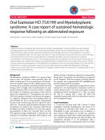
The following day, during a medical review, a hema-
toma on the neck of our patient located at the right
supraclavicular fossa was noted, so a computed tomo-
graphy (CT) contrast scan wa s performed. The results
of the CT scan showed a small right supraclavicular
fossa hematoma, with no active bleeding, and a triangu-
lar foreign body of metallic tomographic appearance,
approximately 5 mm in length, in the interstitial space
between right subclavian vein and artery. There was no
pneumothorax or hemothorax. The left subclavian vein
catheter was intact and well positioned (Figures 1, 2, 3).
We presumed that the foreign body detected was a
central venous catheter tip. This probably fragmented
during the extraction of the device placed in 2004 when
our patient underwent intestinal resection, and later
migrated to the interstitial space between the vessels.
However, we have no means to confirm our theory. The
only certainty we had is that it is not a complication of
our procedure since we did not change the catheter dur-
ing its replacement and our patient’ sCTscanresults
showed two catheter tips.
Discussion
We did not take any additional surgical or interventional
measures, since our patient was asym ptoma tic and th e
‘em bolus’ was fixed in the interstitial space. Neverthe-
less, our patient has continued taking enoxaparin 20 mg
subcutaneous daily in order to prevent any thromboem-
bolic complications [4].
Conclusions
Taking of a thorough medical history is extremely
important for the safe and successful management of a
patient, but it is not always possible to obtain. Central
venous catheterization complications can be misdiag-
nosed by the time they occur. When rare difficulties
during catheter placement occur, the possibility that
relevant data could be missing from a patient’s clinical
history with regard to previous complications should be
considered. It is also good practice t o check catheters
and perform microbiological cultures of the tip. An
ultrasound-guided catheter insertion could possibly have
Figure 1 Previous catheter tip.
Figure 2 Previous catheter tip localization between the two
right subclavian vessels.
Figure 3 Left subclavian catheter tip.
Sidiropoulou et al. Journal of Medical Case Reports 2010, 4:327
/>Page 2 of 3
detected, in real t ime, the extrinsic compression and
further manipulation could have been avoided.
Consent
Written informed consent was obtained from the patient
for publicatio n of this case report and any accompany-
ing images. A copy of the written consent is available
for review by the Editor-in-Chief of this journal.
Acknowledgements
We thank Pedro Gameiro (Surgery Chief), Ana Cecilia Sousa (Surgery
Assistant) and Afonso Janeiro (Surgery Department Director) for their help in
managing our patient.
Author details
1
Surgery Department, Hospital N. S. Rosário, Av. Forças Armadas, Barreiro,
Portugal.
2
Radiology Department, Hospital N. S. Rosário, Av. Forças Armadas,
Barreiro, Portugal.
Authors’ contributions
ZS analyzed and interpreted data from our patient regarding the surgical
procedure. PJ, PV and CC performed the radiological tests and oriented their
interpretation. All authors read and approved the final manuscript.
Competing interests
The authors declare that they have no competing interests.
Received: 22 October 2009 Accepted: 19 October 2010
Published: 19 October 2010
References
1. Marcondes C, Biojone C, Cherri J, Moryia T, Piccinato C: Early and late
complications in long term central venous access. Analysis of 66
implants [in Portugese]. Acta Cir Bras 2000, 15:73-75.
2. Seldinger technique. [ />7F880D3CB24B4A6586A771F17149822C].
3. Emedicine Health: Venous Access Devi ces. [ />venous_access_devices/page3_em.htm].
4. Randolph AG, Cook DJ, Gonzales CA, Andrew M: Benefit of heparin in
central venous and pulmonary artery catheters: a meta-analysis of
randomized controlled trials. Chest 1998, 113:165-167.
doi:10.1186/1752-1947-4-327
Cite this article as: Sidiropoulou et al.: Right subclavian vein catheterism
complication due to a ‘foreign body’: a case report. Journal of Medical
Case Reports 2010 4:327.
Submit your next manuscript to BioMed Central
and take full advantage of:
• Convenient online submission
• Thorough peer review
• No space constraints or color figure charges
• Immediate publication on acceptance
• Inclusion in PubMed, CAS, Scopus and Google Scholar
• Research which is freely available for redistribution
Submit your manuscript at
www.biomedcentral.com/submit
Sidiropoulou et al. Journal of Medical Case Reports 2010, 4:327
/>Page 3 of 3