báo cáo khoa học: " Acute thrombosis of the superior mesenteric artery in a 39-year-old woman with protein-S deficiency: a case report" pot
Bạn đang xem bản rút gọn của tài liệu. Xem và tải ngay bản đầy đủ của tài liệu tại đây (389.79 KB, 5 trang )
CAS E REP O R T Open Access
Acute thrombosis of the superior mesenteric
artery in a 39-year-old woman with protein-S
deficiency: a case report
Nicola Romano
*
, Valerio Prosperi, Giancarlo Basili, Luca Lorenzetti, Valerio Gentile, Remo Luceretti, Graziano Biondi,
Orlando Goletti
Abstract
Introduction: Acute thromboembolic occlusion of the superior mesenteric artery is a condition with an
unfavorable prognosis. Treatment of this condition is focused on early diagnosis, surgical or intravascular
restoration of blood flow to the ischemic intestine, surgical resection of the necrotic bowel and supportive
intensive care. In this report, we describe a case of a 39-year-old woman who developed a small bowel infarct
because of an acute thrombotic occl usion of the superior mesenteric artery, also involving the splenic artery.
Case presentation: A 39-year-old Caucasian woman presented with acute abdominal pain and signs of intestinal
occlusion. The patient was given an abdominal computed tomography scan and ultrasonography in association
with Doppler ultrasonography, highlighting a thrombosis of the celiac trunk, of the superior mesenteric artery, and
of the splenic artery. She immediately underwent an explorative laparotomy, and revascularization was performed
by thromboendarterectomy with a Fogarty catheter. In the following postoperative days, she was given a
scheduled second and third look, evidencing necrotic jejunal and ileal handles. During all the surgical procedures,
we performed intraoperative Doppler ultrasound of the superior mesenteric artery and celiac trunk to control the
arterial flow without evidence of a new thrombosis.
Conclusion: Acute mesenteric ischemia is a rare abdominal emergency that is characterized by a high mortality
rate. Generally, acute mesenteric ischemia is due to an impaired blood supply to the intestine caused by
thromboembolic phenomena. These phenomena may be associated with a variety of congenital pro thrombotic
disorders. A prompt diagnosis is a prerequisite for successful treatment. The treatment of choice remains
laparotomy and thromboendarterectomy, although some prefer an endovascular approach. A second-look
laparotomy could be required to evaluate viable intestinal handles. Some authors support a laparoscopic second-
look. The possibility of evaluating the arteriotomy, during a repeated laparotomy with a Doppler ultrasound, is
crucial to show a new thrombosis. Althoug h the prognosis of acute mesenteric ischemia due to an acute arterial
mesenteric thrombosis remains poor, a prompt diagnosis, aggressive surgical treatment and supportive intensive
care unit could improve the outcome for patients with this condition.
Introduction
Acute thromboembolic occlusion of the superior mesen-
teric artery (SMA) is a condition with a serious prognosis
[1]. Acute mesenteric ischemia (AMI) is an uncommon
occurrence and represents 0.1% of hospital admissions
[2]. Despite considerable advances in medical diagnosis
and treatments over the past f our decades, m esenteric
vascular occlusion still has a poor prognosis, with an in-
hospital mortality rate of 59 to 93% [3]. The high rate of
mortality can be explained b y the nonspecific signs and
symptoms that characterize AMI. The classic teaching of
“pain out of proportion to physical examination findings”
is often seen during the early stage of ischemia when the
abdomen is soft and not tender. Distention and severe
tenderness with rebound guarding appear as a conse-
quence of the bowel infarction [2]. The serologic markers
* Correspondence:
General Surgery Department, Health Unit Five, “F. Lotti” hospital Pontedera,
Pisa, Italy
Romano et al. Journal of Medical Case Reports 2011, 5:17
/>JOURNAL OF MEDICAL
CASE REPORTS
© 2011 Romano et al; licensee BioMed Central Ltd. This is an Open Access article distributed under the terms of the Creative
Commons Attribution License (http://c reativecommons.org/licenses/by/2.0), which permits unrestricted use, distribution, and
reproductio n in any medium, provided the original work is properly cite d.
cannot aid in the diagnostic process because they are
nonspecific (inorganic phosphate, lactic acid, aldolase,
creatinine kinase, and alkaline phosphate) [2]. An
elevated white blood cell (WBC) count (leukocytes mea-
suring over 15,000 cells) is a common, but unspecific,
findin g [2] . According to Kurland [4], another nonspeci-
fic sign is metabolic acidosis. Treatment of this condition
is focused on early diagnosis, surgical or intravascular
restoration of blood flow to the ischemic intestine, surgi-
cal resection of the necrotic bowel, and supportive inten-
sive care.
One aspect that influences survival is the cause of the
bowel ischemia, which can be classified as a non-thrombo-
tic or a thrombotic event [5]. Conditions that cause
nonthromboticmesentericischemia(NOMI)includea
low-flow state (for example, cardiogenic shock, pancreati-
tis, sepsis, hypovolemia), mechanical causes (for example,
strangulated hernia, adhesive bands, intussusceptions), and
colon ischemia after aortic aneurysm repair [5]. NOMI
represents 25% of the causes of the AMI [2]. The specific
thrombotic conditions include arterial embolization
(superior mesenteric artery embolization; SMAE), arterial
thrombosis (superior mesenteric artery thrombosis;
SMAT), and mesenteric venous thrombosis (acute mesen-
teric venous thrombosis; AMVT) [5]. The most common
cause of AMI is SMAE, which represents 50% of the
causes of AMI [2]. SMAT can be seen in 10% of the
patients after AMVT [2]. These thromboembolic phenom-
ena may be associated with prothrombotic disorders, such
as protein C, protein S, and antithrombin III (AT III) defi-
ciency [6]. In this report, we describe the case of a woman
with a thrombophilic state, in whom a small bowel infarct
developed because of an acute thrombotic occlusion of the
SMA, involving the splenic artery as well.
Case presentation
A 39-year-old Caucasian woman presented in our emer-
gency department with acute abdominal pain associated
with nausea, vomiting, and signs of intestinal occlusion.
The clini cal history of the patient highlighted two other
admissions for the same clinical signs. During the first
admission, she was given an abdominal computed tomo-
graphy (CT) scan that demonstrated only the presence
of free fluid localized in the pouch of Douglas and the
perihepatic region. In relation to the se signs, she was
given an emergen cy, explorative laparotomy, with lavage
of the abdomen. The laparotomy demonstrated only
hyp eremic jejunal and ileal handles. She was discharged
after nine days without any complications. Two weeks
after the patient was readmitted to the same hospital
with similar symptoms, and she was treated with corti-
costeroids, m esalazine, and metronidazole with a com-
plete resolution of the symptoms. Five days later, the
patient was admitted t o our unit. A t admission, she had
leukocytosis (WBC, 19.960 × 10
6
/L) and normal levels
of the coagulation parameters. She was given abdominal
ultrasonography in association with Doppler ultrasono-
graphy (Esaote Megas GPX 7.5-MHz convex probe),
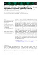
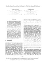
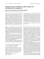
highlighting a thrombosis of the SMA. As a result of
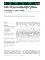
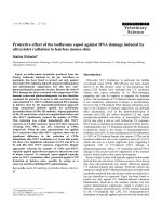
this clinical picture, she underwent an abdominal CT
scan (Figures 1 to 3), demonstrating the presence of a
partial thrombosis of the c eliac trunk, a thrombosis o f
the SMA for a 25- to 30-mm tract, and the lack of a
splenicartery.Sheimmediately underwent an explora-
tive laparotomy, showing ischemic, but viable handles,
and a tree revascularization by thromboendarterectomy
with a Fogar ty catheter was performed. In the following
postoperative days, she was given a scheduled second
and third look, showing necrotic handles (the first jeju-
nal handle, the last ileal handle, and about 20 cm of the
medium ileum) in the first procedure, and another
necrotic tract of small bowel (the other 10 cm of the
first jejunal tract) in the last procedure. During that sur-
gical procedure, we performed duodenojejunal and three
other laterolateral anastomoses to reestablish the bowel
continuity. A T-tube was inserted to protect the duode-
nojejunal anastomosis. A cholecystectomy and biliary
diversion were performed to reduce the biliary output.
In relation to the risk of dehiscence, we performed a
colonostomy in the right flank. During all the surgical
procedures, we perfo rmed intraoperatory Doppler ultra-
sound of the SMA and celiac tru nk to control the arter-
ial flow without evidence of a new thrombosis. The
patient stayed in the ICU for 27 days with total parent-
eral nutrition and antibiotics therapy. A coagulation
screening demonstrated a thrombophilic state for a pro-
tein-S (16%) deficiency wit h normal values of VIII, IX,
and XI factors. The search for antiphospholipid antibo-
dies w as negative, and the genetics test for factors II to
V and methylenetetrahydrofolate reductase (MTHFR;
Figure 1 Abdominal computed tomography scans.
Romano et al. Journal of Medical Case Reports 2011, 5:17
/>Page 2 of 5
the deficiency of this enzyme is associated with an
increased risk to develop massive thromboembolic
events) was negative (no mutations). She was discharged
from our unit after 37 days without any complications.
After three months, the patient had a surgical procedure
for restoring the bowel continuity. The patient was eval-
uated after one week, and one, three, and six months
after discharge with blood and coagulation examina-
tions, abdominal ultrasonography, Doppler ultrasound,
and abdominal CT scan. She was asymptomatic and
stayed well. At one year, we had successfully restored
the bowel continuity without complications.
Discussion
Acute mesenteric ischemia is a rare abdominal emer-
genc y that usually requires wide intestina l resec tion and
carriesahighmortalityrate(Table1[7-13])withthe
adverse effects of short-bowel syndrome in the surviving
patients [6]. A critical point that influences the survival
rate is prompt diagnosis in patients with AMI. Numer-
ous surgical reports indicated that acute intestinal ische-
mia (AII) is associated with a poor prognosis [13]. The
poor signs, symptoms, and nonspecific laboratory tests
are among the causes of the delay i n the diagnosis.
Other examinations that can be helpful in the diagnostic
process are angiography, computed tomography angio-
graphy (CTA), and magnetic resonance angiography
(MRA) [2]. When no clinical evidence is found for an
immediate surgical intervention, such as peritonitis or
gastrointestinal hemorrhage, angiography could be con-
sidered the treatment of choice in patients with sus-
pected AMI, because this investigation allows us to
distinguish between nonthrombotic and thrombotic
causes [14]. M oreover, angiography allows us to treat
the occlusion with a restoration of the blood flow by
using an endovascular approach, such as percutaneous
transluminal angioplasty and thrombolysis [5-14].
Simo et al. [14] reported a 90% success rate for lysis of
the embolus in patients with SMAE. However, although
the endovascular approach may rapidly restore the blood
flow to the bo wel, the time needed for thrombolysis is
variable, and the bowel viability cannot be assessed with
laparotomy [14]. This can result in a diagnostic delay
that can compromise other viable bowel tracts [5].
According to Kirkpatric [1], t he CTA h as shown a diag-
nostic sensitivity of 96% and a specificity of 94%. The
magnetic resonance angiography (MRA) is another
newer imaging technique that seems to be promising for
the diagnosis of AMI, although this technique cannot
help us to diagnose NOMI secondary to a low-flow state
or to identify distal embolic disease [2]. Generally, the
IMA is due to an impaired blood supply to the intestine
caused by thromboembolic phenomena. These phenom-
ena may be associated with a variety of congenital pro-
thrombotic disorders (PDs), such as protein-C and
protein-S deficiencies, AT III deficiencies (anti-phospho-
lipid antibodies), Factor V Leiden (FVL), Prothrombin
G20210A mutation, and C677T homozygous mutation of
the MTHFR gene. The prevalence of these mutations dif-
fers among geographic areas and ethnic groups [6]. In
our patient, we found deficiencies of the S protein,
although some studies demonstrated a prevalence of this
disorder in a Chinese population (59%) compared to a
Caucasian population (15%)[6]. The level of S protein is
higherinmenthaninwomen,butincreaseswithagein
women but not in men [16]. In women, the levels of an S
protein are lower before menopause, while taking
oral contraceptives, or while undergoing hormone-
replacement therapy, and during pregnancies [16].
The International Society of Thrombosis and Haemos-
tasis Standardization Subcommittee defined three
n-types of hereditary S-protein deficiencies [16]. Type
I is defined by low levels of free and total antigen with
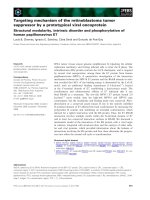
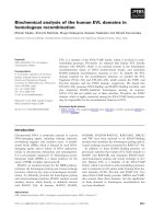
Figure 3 Abdominal computed tomography scans.
Figure 2 Abdominal computed tomography scans.
Romano et al. Journal of Medical Case Reports 2011, 5:17
/>Page 3 of 5
decreased APC cofactor activity [16]. Type II protein-S
deficiency is characterized by normal levels of a free and
total antigen, with low levels of APC cofactor activity
[16]. Type III protein-S deficiency is defined by normal
to low levels of total antigen, low free protein S, and an
elevated f raction of protein S bound to C4BP [16]. The
role of the protein S is based on an increase of the
anticoagulant action of p rotein C [16]. Protein C is a
proteinase that inactivates the coagulation factors V,
Leiden, and VIII, and protein S increases the action of
protein C [17]. The SMA normally serves as the primary
arterial supply of the jejunum, the ileum, and the colon
to the level of the splenic flexure [7].
Ottinger et al. [7] demonstrated a general correspon-
dencebetweenthesiteoftheocclusion,theextentof
the infarcted areas, and the prognosis [7]. To explain
this concept, we can divide the SMA into four
regions [7]. The first portion is the artery origin, and
the second tract is represented by the main trunk,
including the middle colic artery (MCA). Region three
corresponds to the main trunk beyond the origin of the
MCA, and the last region (IV) is the most peripheral
portion of the SMA and its b ranches [7]. The occlusion
of the SMA in the first region produces a more-exten-
sive infarction than that when the site of occlusion is
distal to the origin of some of its branches [7].
Another factor that influences the prognosis is the etio-
logic subsets [3]. We can grossly distinguish two different
origins, thrombotic and non-thrombotic. Non-occlusive
mesenteric ischemia, the more frequent non-thr ombotic
cause, is caused by low-flow states. The thrombotic condi-
tion includes arterial embolism, arterial thrombosis, and
mesenteric venous thrombosis. According to Schoots [3],
acute mesenteric ischemia due to a venous thrombosis has
a better prognosis compared with arterial causes of MIA.
In this case, the improved survival rate can be explained
by the segmental bowel infarction and the need for limited
intestinal resection. The poor prognosis of patients with
mesenteric arterial occlusions is most likely due to the
proximal location of the occlusion in the vessel tree; this
determines a more extensive bowel infarction and the
need for extended intestinal resection. A mesenteric arter-
ial embolism results in a different extension of the
infarcted areas because the emboli can occlude the vessel
tree to different levels. The prerequisite for success of a
revascularization is prompt diagnosis. The delay from the
first examination to laparotomy was significantly shorter
among the patients in whom the diagnosis was suspected;
however, early diagnosis did not improve survival [1].
Moreover, Giulini [18] demonstrated a correlation
between of prompt diagnosis of an AMI and survival.
However, for the non-specific symptoms, during the early
phase, the diagnosis is often delayed [19].
The second-look laparotomy remains the gold stan-
dard for the assessment of further bowel viability, and,
at the same time, it is the only way to remove infarcted
tracts of the bowel [20]. During t he surgical procedure,
the bowel viability can be assessed by the physical exam-
ination (inspection of bowel and palpation of the vessel)
or by ultrasound examination and intravenous fluores-
cein [20]. Although the second-look laparotomy is the
gold standard for the treatment of AMI, some authors
perform a second-look laparoscopy to decrease the
severe anesthesiologic and surgical trauma in these criti-
cally ill patients [20]. Levy et al. [20], in a series of 92
patients, underlined the beneficial role of the second-
look laparoscopy in patients’ survival.
Conclusion
Acute thrombosis of the SMA represents a rare emer-
gency in young female patients. Although in these
patients, mesenteric infarction has a low incidence,
acute thrombosis should be always suspected, especial ly
in young female patients receiving therapy with estro-
progestinic hormones and who show signs of an acute
abdomen. These cases should be investigated with CT-
angiography or, if feasi ble, with arteriography to exclude
an acute mesenteric infarction. If the CT-angiography or
the arteriography confirms this diagnosis, an early lapar-
otomy should be performed.
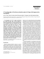
Table 1 Comparative death rates for thrombotic causes of acute intestinal ischemia
Arterial embolism Arterial thrombosis Venous thrombosis Overall deaths
Authors Year No. % No. % No. % No. %
Ottinger [7] 1967 22/29 76 21/22 95 8/10 80 51/61 83
Smith [8] 1976 6/7 86 9/10 90 3/3 100 18/20 90
Kairaluoma [9] 1977 10/11 91 19/21 90 - - 29/32 91
Hertzer [10] 1978 4/7 57 2/2 100 - - 6/9 67
Sachs [11] 1982 9/14 64 12/12 100 4/11 36 25/37 68
Bergan [12] 1987 5/6 83 6/8 75 - - 11/14 79
Klempnauer [13] 1997 16/21 76 22/27 81 11/30 37 49/78 62
Endan [5] 2000 13/22 59 13/21 62 2/15 13 28/58 48
Collated experience 85/117 74 104/123 86 28/69 53 217/309 73
Romano et al. Journal of Medical Case Reports 2011, 5:17
/>Page 4 of 5
In our case, we performed a second-look laparotomy
because this surgical procedure allowed us to conduct a
physical examinat ion of the bowel and artery (for exam-
ple, palpation of the vessels, inspection of the bowel,
and evaluation of the anastomosis). Moreover, the
second-look and other laparotomies suggest the perfor-
man ce of an intraoperato ry Doppler ultrasound to eval-
uate the artery flow. According to Ottinger [7], a new
thrombosis of the SMA can develop in the site of the
arteriotomy during the first 48 hours. The possibility of
evaluating the arteriotomy, during a repeated lapa rot-
omy with a Doppler ultrasound, is cruci al; an early
planned repeated laparoto my improv es the prog nosis of
the surgical approach. Although the prognosis of the
AMI due to an acute arterial mesenteric thrombosis
remains poor, a prompt diagnosis, aggressive surgical
treatment, and a supportive intensive care unit for a
patient with AMI could improve the prognosis.
Consent
Written informed consent was obtained from the patient
for publication of this case report and accompanying
images. A copy of the written consent is available for
review by the Editor-in-Chief of this journal.
Authors’ contributions
NR wrote the article. VP researched and retrieved the bibliography. GB was
the language supervisor. LL analyzed and interpreted the abdominal
ultrasound data. VG acquired and interpreted the Doppler ultrasound data.
RL contributed to writing the manuscript, controlling and correcting the
general surgery portion. GB interpreted the hematology. OG supervised and
was the chief of the team. All authors read and approved the final version
of the manuscript.
Competing interests
The authors declare that they have no competing interests.
Received: 11 October 2009 Accepted: 18 January 2011
Published: 18 January 2011
References
1. Björck M, Acosta S, Lindberg F, Troëng T, Bergqvist D: Revascularization of
the superior mesenteric artery after acute thromboembolic occlusion. Br
J Surg 2002, 89:923-927.
2. Kozuch PL, Brandt LJ: Review article: diagnosis and management of
mesenteric ischemia with an emphasis on pharmacotherapy. Aliment
Pharmacol Ther 2005, 23:201-215.
3. Schoots IG, Koffeman GI, Legemate DA, Levy M, Van Gulik TM: Systematic
review of survival after acute mesenteric ischemia according to disease
aetiology. Br J Surg 2004, 91:17-21.
4. Kurland B, Brandt LJ, Delany HM: Diagnostic test for intestinal ischemia.
Surg Clin North Am 1992, 72:85-105.
5. Endean ED, Barnes SL, Kwolek CJ, Minion DJ, Schwarcz TH, Metzer RM:
Surgical management of thrombotic acute intestinal ischemia. Ann Surg
2001, 6:801-808.
6. Ağaoğlu N, Türkyilmanz S, Ovah E, Uçar F, Ağaoğlu C: Prevalence of
prothrombotic abnormalities in patients with acute mesenteric ischemia.
World J Surg 2005, 29:1135-1138.
7. Ottinger LW: The surgical management of acute occlusion of the
superior mesenteric artery. Ann Surg 1978, 188:721-731.
8. Smith JS Jr, Patterson LT: Acute mesenteric infarction. Am Surg 1976,
42:562-567.
9. Kairaluoma MI, Karkola P, Heikkinene D, Huttunen R, Larmi TK: Mesenteric
infarction. Am J Surg 1977, 133:188-193.
10. Hertzer NR, Beven EG, Humphries AW: Acute intestinal Ischemia. Am Surg
1978, 44:744-749.
11. Sachs SM, Morton JH, Schwartz SI: Acute mesenteric ischemia. Surgery
1982, 92:646-653.
12. Bergan JJ, Mc Carthy WJ, Flinn WR, Yao JS: Nontraumatic mesenteric
vascular emergencies. J Vasc Surg 1987, 5:903-909.
13. Klempnauer J, Grothues F, Bektas H, Pichlmayr R: Long-term results after
surgery for acute mesenteric ischemia. Surgery 1997, 121:239-243.
14. Barakate MS, Cappe I, Curtin A, Angel KD, Li-Kim-Moy J, Poon MSF,
Sandeman MD: Management of acute superior mesenteric artery
occlusion. A NZ J Surg 2002,
72:25-29.
15. Kirkpatrick LD, Kroeker MA, Greenberg HM: Biphasic CT with mesenteric
CT angiography in the evaluation of acute mesenteric ischemia: initial
experience. Radiology 2003, 229 :91-98.
16. D’angelo A, D’angelo S: Protein-S deficiency. Haematology 2008,
93:498-501.
17. Fauci AS, Braunwald E, Isselbacher Kurt J, Wilson JD, et al: Harrison’s
Principles of Internal Medicine. 14 edition. New York: McGraw-Hill.
18. Giulini S, Bonardelli S, Cangiotti L, Floriani M, Cervi GC, Portolani N,
Tiberio G: Factors affecting prognosis in acute ischemia. Int Angiol 1987,
72:157-182.
19. Rush DS, Levy PJ, Haynes JL: Acute embolic and thrombotic mesenteric
ischemia. In Current Therapy in Vascular Surgery. Edited by: Ernst CB, Stanley
JC. St. Louis: Mosby; 1995:693-697.
20. Yanar H, Taviloglu K, Ertekin C, Ozcinar B, et al: Planned second-look
laparoscopy in the management of acute mesenteric ischemia. World J
Gastroenterol 2007, 13:3350-3353.
doi:10.1186/1752-1947-5-17
Cite this article as: Romano et al.: Acute thrombosis of the superior
mesenteric artery in a 39-year-old woman with protein-S deficiency: a
case report. Journal of Medical Case Reports 2011 5:17.
Submit your next manuscript to BioMed Central
and take full advantage of:
• Convenient online submission
• Thorough peer review
• No space constraints or color figure charges
• Immediate publication on acceptance
• Inclusion in PubMed, CAS, Scopus and Google Scholar
• Research which is freely available for redistribution
Submit your manuscript at
www.biomedcentral.com/submit
Romano et al. Journal of Medical Case Reports 2011, 5:17
/>Page 5 of 5