Báo cáo y học: "Maxillary sinus textiloma: a case report" pps
Bạn đang xem bản rút gọn của tài liệu. Xem và tải ngay bản đầy đủ của tài liệu tại đây (779.35 KB, 2 trang )
CAS E REP O R T Open Access
Maxillary sinus textiloma: a case report
Yoann Pons
*
, Thomas Schouman
Abstract
Introduction: Textilomas have been reported in many locations. We report the first case of textiloma located in
the maxillary sinus that mimicked a sinus cyst recurrence on computed tomography images.
Case presentation: A 60-year-old Caucasian man was referred for pers istent infection of the right maxillary sinus.
A maxillary sinus benign cyst had been removed three months before. Computed tomography showed a sinus
opacity evoking a cyst recurrence. A new operation was planned to remove the cyst by a Caldwell-Luc approach.
After excision of very thick fibrous tissue, a compress was discovered in the maxillary sinus. The patient did not
present with any sinus infection after the operation.
Conclusion: The surgeon should always take into account the possibility of textilomas in a patient with a history
of sinus surgery.
Introduction
Textiloma can be defined as a mass within the body
composed of cotton matrix, which usually refers to a
retained surgical sponge or compress, surrounded by a
foreign-body reaction [1].
Most cases of textiloma reported in the literature have
been connected to abdominal, orthopaedic and cardi-
othoracic surgery [1-3]. At the head level, few intra-cra-
nial cases have been reported [4,5]. No case, to date, has
been reported at the face level. The authors reported
the first case of textiloma located in the maxillary sinus.
Case presentation
A 60-year-old Caucasian man was referred to us for per-
sistent infection of the right maxillary sinus. He was
operated on three months ago for a benign cyst. A Cald-
well -Luc operation was performed. Since this operation,
the patient complained of having recurrent sinusalgia
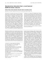
with purulent rhinorrhea. Computed tomography (CT)
showed a sinus opacity evocating a cyst recurrence (Fig-
ure 1). A new surgery was planned to remove the cyst
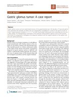
by a new Caldwell-Luc operation. After excision of very
thick fibrous tissue, a compress was discovered in the
maxillary sinus (Figure 2). The patient did not present
any sinus infection after the operation.
Conclusions
The main complication of a maxillary sinus textiloma is
a persistent infection. In this case, the sinusitis was lim-
ited (that is, no orbital or meningeal complications
occurred).
The erroneous diagnosis of a mass provoked by the
presence of a textiloma was frequently reported in the
literature in other regions [1-5]. In this case, both
the radiologist and the surgeon had suggested the diag-
nosis of cyst recurrence, given the CT-scan examination.
However, at a second viewing of the images, some radi-
ologic signs of textiloma were noticed: the mass was
heterogeneous with a rectilinear alternation of thin
bands (solid-band and air-band de nsities) that corre-
sponded to the meshing of the compress. Moreover, for-
eign bodies of the maxillary sinus are a common cause
of persistent infection. The diagnosis was finally cor-
rected by the surgery, which definitively cured the
patient.
The suspicion of textiloma should be raised when a
patient with a history of previous maxillary sinus surgery
presents with a history of chronic sinus infection asso-
ciated with a sinus mass on CT images, even though
textiloma is unlikely to be found in such a small cavity.
Consent
Written informed consent was obtained from the patient
for publication of this case report and accompanying
* Correspondence:
Maxillofacial Surgery Department, AP-HP - Pitié-Salpêtrière University
Hospital, University of Paris 6, France
Pons and Schouman Journal of Medical Case Reports 2010, 4:288
/>JOURNAL OF MEDICAL
CASE REPORTS
© 2010 Pons and Schouman; licensee BioMed Central Ltd. Thi s is an Open Access article distributed under the terms of the Creative
Commons Attribution License (http://creativeco mmons.org/licenses/b y/2.0), which permits unrestricted use, distribution, and
reproduction in any medium , provided the original work is properly cited.
images. A copy of the written consent is available for
review by the Editor-in-Chief of this journal.
Authors’ contributions
YP redacted the manuscript. TS supervised the manuscript. Both authors
read and approved the final manuscript.
Authors’ information
The authors are two medical doctors. Yoann Pons is a head and neck
surgeon.
Thomas Schouman is a maxillofacial surgeon.
Competing interests
The authors declare that they have no competing interests.
Received: 6 January 2010 Accepted: 24 August 2010
Published: 24 August 2010
References
1. Nobre LF, Marchiori E, May F, Carrão AD Jr, Zanetti G, Machado DM:
Thoracic textilomas after myocardial revascularisation: typical CT
findings. Br J Radiol 2010, 83:4-7.
2. Poyanli A, Salmaslioğlu A, Terzibaşioğlu E, Toker A, Tanju S, Aydin K: An
unsusual pure cystic posterior mediastinal mass: a textiloma. Clin Radiol
2008, 63:863-868.
3. Yamamura N, Nakajima K, Takahashi T, Uemura M, Nishitani A, Souma Y,
Nishida T: Intra-abdominal textiloma: a retained surgical sponge
mimicking a gastric gastrointestinal stromal tumor: a case report. Surg
Today 2008, 38:552-554.
4. Razzag AA, Chishti MK: Foreign body granuloma after craniotomy for
tumor: a diagnostic dilemma. Br J Neurosurg 2000, 14:591-592.
5. Feldman RP, Marcovici A, Suarez M, Goodrich JT: Foreign body granuloma
mimicking intracranial meningioma: case report and review of the
literature. Neurosurgery 1999, 44:855-858.
doi:10.1186/1752-1947-4-288
Cite this article as: Pons and Schouman: Maxillary sinus textiloma: a
case report. Journal of Medical Case Reports 2010 4:288.
Submit your next manuscript to BioMed Central
and take full advantage of:
• Convenient online submission
• Thorough peer review
• No space constraints or color figure charges
• Immediate publication on acceptance
• Inclusion in PubMed, CAS, Scopus and Google Scholar
• Research which is freely available for redistribution
Submit your manuscript at
www.biomedcentral.com/submit
Figure 1 CT scan imaging showing the textiloma located in the
maxillary sinus and mimicking a cyst recurrence.
Figure 2 Operative view of the compress extraction from the
maxillary sinus.
Pons and Schouman Journal of Medical Case Reports 2010, 4:288
/>Page 2 of 2