Báo cáo y học: "15-deoxy-delta12,14-prostaglandin J2 attenuates endothelial-monocyte interaction: implication for inflammatory diseases" ppsx
Bạn đang xem bản rút gọn của tài liệu. Xem và tải ngay bản đầy đủ của tài liệu tại đây (906 KB, 10 trang )
BioMed Central
Page 1 of 10
(page number not for citation purposes)
Journal of Inflammation
Open Access
Research
15-deoxy-delta12,14-prostaglandin J2 attenuates
endothelial-monocyte interaction: implication for inflammatory
diseases
Ratna Prasad
1
, Shailendra Giri
1
, Avtar K Singh
2
and Inderjit Singh*
1
Address:
1
Department of Pediatrics, Medical University of South Carolina, Charleston, SC, 29425, USA and
2
Department of Pathology and
Laboratory Medicine, Ralph Johnson Veterans Affairs Medical Center, Charleston, SC, 29425, USA
Email: Ratna Prasad - ; Shailendra Giri - ; Avtar K Singh - ;
Inderjit Singh* -
* Corresponding author
Abstract
Background: The Infiltration of leukocytes across the brain endothelium is a hallmark of various
neuroinflammatory disorders. Under inflammatory conditions, there is increased expression of
specific cell adhesion molecules (CAMs) on activated vascular endothelial cells which increases the
adhesion and infiltration of leukocytes. TNFα is one of the major proinflammatory cytokines that
causes endothelial dysfunction by various mechanisms including activation of transcription factor
NF-κB, a key transcription factor that regulates expression of CAMs. Peroxisome proliferator-
activated receptor gamma (PPARγ) is a member of the nuclear hormone superfamily of ligand-
activated transcriptional factors. 15-deoxy-δ 12, 14-prostaglandin J2 (15d-PGJ2) is a well
recognized natural ligand of PPARγ and possesses anti-inflammatory properties both in vitro and in
vivo. This study aims to elucidate the mechanism of 15-PGJ2 on the adhesion of mononuclear cells
to activated endothelial cells.
Methods: To delineate the signaling pathway of 15d-PGJ2 mediated effects, we employed an in vitro
adhesion assay model of endothelial-monocyte interaction. Expression of CAMs was examined
using flow cytometry and real time PCR techniques. To define the mechanism of 15d-PGJ2, we
explored the role of NF-κB by EMSA (E
lectrophoretic Mobility Shift Assay) gels, NF-κB reporter
and p65-transcriptional activities by transient transfection in the brain-derived endothelial cell line
(bEND.3).
Results: Using an in vitro adhesion assay model, we demonstrate that 15d-PGJ2 inhibits TNFα
induced monocyte adhesion to endothelial cells, which is mediated by downregulation of
endothelial cell adhesion molecules in a PPARγ independent manner. 15d-PGJ2 modulated the
adhesion process by inhibiting the TNFα induced IKK-NF-κB pathway as evident from EMSA, NF-
κB reporter and p65 mediated transcriptional activity results in bEND.3 cells.
Conclusion: These findings suggest that 15d-PGJ2 inhibits inflammation at multiple steps and thus
is a potential therapeutic target for various inflammatory diseases.
Published: 8 August 2008
Journal of Inflammation 2008, 5:14 doi:10.1186/1476-9255-5-14
Received: 26 December 2007
Accepted: 8 August 2008
This article is available from: />© 2008 Prasad et al; licensee BioMed Central Ltd.
This is an Open Access article distributed under the terms of the Creative Commons Attribution License ( />),
which permits unrestricted use, distribution, and reproduction in any medium, provided the original work is properly cited.
Journal of Inflammation 2008, 5:14 />Page 2 of 10
(page number not for citation purposes)
Background
Inflammatory mechanisms are pivotal in many disease
states, including atherosclerosis, autoimmune disorders
and ischemia/reperfusion injury [1-4]. Under inflamma-
tory conditions there is activation of vascular endothelial
cells that involves various morphological and metabolic
changes [5]. There is induction of specific cell adhesion
molecules, such as, intercellular adhesion molecule-1
(ICAM-1), vascular cell adhesion molecule-1 (VCAM-1)
and E-selectin. These interact with their corresponding lig-
ands on leukocytes namely, lymphocyte function-associ-
ated antigen-1 (LFA-1), very late antigen-4 (VLA-4) and
carbohydrate moieties respectively [2,6]. The process of
infiltration involves sequential capture, rolling, firm
adhesion and transmigration across the endothelial bar-
rier [7]. Blockade of CAMs that mediate the accumulation
of mononuclear cells under inflammation is now consid-
ered as an effective treatment strategy in clinical inflam-
matory disorders.
TNFα is one of the major proinflammatory cytokines that
is dysregulated in inflammatory diseases mentioned ear-
lier and has been shown to contribute to endothelial dys-
function [8]. TNFα causes endothelial dysfunction by
various mechanisms that includes activation of transcrip-
tion factor NF-κB [9]. Transcriptional regulation of many
pro-inflammatory genes, including CAMs, is under the
control of different transcriptional factors including NF-
κB [10,11]. NF-κB is a redox sensitive transcription factor
that most commonly exists as a p50/p65 heterodimer.
This heterodimer remains sequestered in the cytoplasm
when associated with inhibitor of kappa B (IκB) proteins.
Upon stimulation (e.g. by TNFα) IκB proteins get phos-
phorylated by upstream IκB kinases (IKKs) followed by
degradation, releasing the active dimer to translocate into
the nucleus to transcribe its target genes [12,13].
Peroxisome proliferator-activated receptors (PPARs) are
members of the nuclear hormone superfamily of ligand-
activated transcriptional factors. PPARs heterodimerize
with retinoid × receptor (RXR) and bind to peroxisome
proliferator-response elements in target genes [14]. The
subtype PPARγ is a regulator of adipogenesis [15]. A
number of studies have demonstrated that PPARγ may
play a role in regulating inflammatory responses [16,17].
15-deoxy-d 12, 14-prostaglandin J2, the ultimate metabo-
lite of prostaglandin (PG) D
2
, is a natural ligand of PPARγ.
15d-PGJ2 has been shown to inhibit expression of iNOS
and TNFα in several cell types that are dependent on
PPARγ [18,19]. However, there are also anti-inflammatory
responses of 15d-PGJ2 that are PPARγ independent
[20,21]. There are studies that report protective effects
mediated by 15d-PGJ2 via inhibition of infiltration of
immune cells in various models of inflammation e.g.
endotoxic shock [22], lung injury [23], ischemia/reper-
fusion injury [24] and experimental autoimmune enceph-
alomyelitis (EAE) [25,26]. Thus, based on these studies,
we hypothesized that 15d-PGJ2 inhibits the adhesion of
mononuclear cells to the endothelial cells and thereby
attenuates their transmigration. We observed that 15d-
PGJ2 inhibited the adhesion of monocytes to bEND.3
endothelial cell line, activated by TNFα, by downregula-
tion of endothelial CAMs via inhibition of IKK-NF-κB
pathway.
Methods
Reagents and Antibodies
DMEM (4.5 g/L glucose), minimum essential medium
alpha (MEM alpha) with ribonucleotides and deoxyribo-
nucleotides, RPMI-1640 medium and FBS were purchased
from Gibco BRL (Carlsbad, CA, USA). Granulocyte mac-
rophage colony stimulating factor (GMCSF) and recom-
binant mouse TNFα were from R & D Systems
(Minneapolis, MN, USA). Vybrant Cell adhesion kit con-
taining Calcein AM fluorescent dye was from Molecular
Probes (Eugene, OR, USA). ECL detection kit was from GE
healthcare (Piscataway, NJ, USA). Antibodies for p65,
p50, IκBα, VCAM-1 were purchased from Santa Cruz Bio-
technologies (Santa Cruz, CA, USA). Texas red conjugated
rabbit IgG antibody was from Vector Lab. Inc. (Burling-
ton, CA, USA). Trizol reagent and Lipofectamine Plus
were from Invitrogen (Carlsbad, CA, USA). Fluoromount-
G was from Electron Microscopy Sciences (Hartfield, PA,
USA). Antibodies against VCAM-1 (FITC labeled), ICAM-
1 and E-selectin (PE labeled) were from BD Pharmingen
(Franklin Lakes, NJ). Luciferase assay system was pur-
chased from Promega (Madison, WI).
Cell culture
The bEND.3 mouse brain endothelial cells were from
ATCC (American Type Culture Collection, Manassas, VA,
USA) and were cultured in Dulbecco's modified Eagle's
medium (high glucose) supplemented with 10% Fetal
Bovine serum (FBS) and antibiotics. Cells were grown to
confluence, made serum free for further treatments, and
stimulation with TNFα (50 ng/ml) for all the experiments.
JAWS II, a mouse monocyte cell line (ATCC) was main-
tained in MEM Alpha medium with 10% heat inactivated
FBS, 0.5% gentamycin and granulocyte-macrophage col-
ony-stimulating factor (GMCSF) (1 ng/mL; R & D Sys-
tems).
Plasmids and Transfection
NF-κB-luciferase was kindly provided by Dr. George
Rewadi (Institut Pasteur, Laboratoire des Mycoplasmes,
Paris, France), flag-IKKα was a gift from Dr. Zheng-Gang
Liu (National Institute of Health, Bathesda, MD) and
FLAG-tagged wild-type (wt) PPARγ and FLAG-tagged
L468A/E471A PPARγ were provided by Dr. V. Chatterjee
(University of Cambridge, Cambridge, U.K.). The peroxi-
Journal of Inflammation 2008, 5:14 />Page 3 of 10
(page number not for citation purposes)
some proliferator-response element (PPRE)-containing
reporter plasmid (J6-thymidine kinase (TK)-Luc) was pro-
vided by B. Staels (Institut Pasteur de Lille, Lille, France).
PTL-luciferase, Gal-p65 and Gal-DBD (DNA binding
domain) were purchased from Panomics (Fremont, CA).
The endothelial cell line was transfected with the indi-
cated plasmid (0.5 μg/well) using Lipofectamine Plus
Reagent under serum free conditions as described before
[27]. pcDNA3.1 was used to normalize the total content
of DNA in all transfection experiments.
In vitro Adhesion assay model
As described earlier, bEND.3 cells were grown as monol-
ayers in double chamber slides (Nalge Nunc, Naperville,
IL, USA) [27]. Cells were pre-treated with 15d-PGJ2 for 30
min followed by TNFα for 6 h. Dye labeled monocytes at
the concentration of 2 × 10
6
cells/ml were added per
chamber on the bEND.3 cells and allowed to interact for
30 min with gentle shaking at 37°C. Adherent fluorescent
cells were observed using a fluorescence microscope
(Olympus, BX60) and images were captured in Adobe
Photoshop 7.0 at 100×. Adherent fluorescent cells were
counted using Image Pro-Plus 4.0 software. Mean and SD
were calculated for independent experiments. Results
were plotted as fold change compared to the control val-
ues for all the experiments.
Immunocytochemistry
BEND.3 cells were grown in chamber slides and treated
with 15d-PGJ2 and stimulated with TNFα for 20 min.
Cells were fixed with paraformaldehyde (4%) followed by
blocking in blocking reagent. Cells were then incubated in
anti-p65 antibody followed by incubation in secondary
antibody and mounting with Flouromount-G. The
stained sections were analyzed by immunofluorescence
microscopy (Olympus BX-60 from Opelco, Dulles, VA,
USA) with images captured using an Olympus digital
camera (Optronics, Goleta, CA, USA) at 400× magnifica-
tion. Captured images were processed using Adobe Pho-
toshop 7.0 and were adjusted using brightness and
contrast tools. Three independent experiments were done
and 5 fields for each treatment were taken. Representative
images are shown.
Real-time or quantitative (q) PCR
Cells were harvested in Trizol reagent and RNA was iso-
lated per the manufacturer's protocol. cDNA synthesis was
done using iScript CDNA synthesis kit (BIO-RAD Labora-
tories, Hercules, CA, USA) per the manufacturer's proto-
col. qPCR was performed using SYBR GREEN PCR master
mix (Applied Biosciences, Foster city, CA, USA) and BIO-
RAD laboratories iCycler iQ PCR using primers as
described before [27]. primers of CAMs and 18S are as fol-
lows, ICAM-1 FP 5'-gca gag tgt aca gcc tct tt-3' RP 5'-ctg gta
tcc cat cac ttg-3', VCAM-1 FP 5'-gca gag tgt aca gcc tct tt-3',
RP 5'-ctg gta tcc cat cac tcg ag-3'; E-selectin FP 5'-act tca gtg
tgg tcc aag ag-3' RP 5'-gca cat gag gac ttg tag gt-3'; 18S FP
5'-gaa aac att ctt ggc aaa tgc ttt-3' RP5'-gccgct aga ggt gaa att
ctt-3'. The normalized mRNA expression was computed
with that of 18s expression. Values are expressed as fold
change from the control values and plotted.
Preparation of cytosolic and nuclear extracts
Cytosolic and nuclear extracts from bEND.3 cells were
prepared using the method of Digman et al [28] with
slight modification [29]. Cells were harvested, washed
twice with ice-cold PBS, and lysed in 400 μl of buffer A (10
mM HEPES, pH 7.9, 10 mM KCl, 2 mM MgCl
2
, 1 mM
PMSF, 5 μg/ml aprotinin, 5 μg/ml pepstatin A, and 5 μg/
ml leupeptin) containing 0.1% Nonidet P-40 for 15 min
on ice, vortexed vigorously for 15 s, and centrifuged at
14,000 rpm for 30 s. The pelleted nuclei were resuspended
in 40 μl of buffer B [20 mM HEPES, pH 7.9, 25% (v/v)
glycerol, 0.42 M NaCl, 1.5 mM MgCl
2
, 0.2 mM EDTA, 1
mM PMSF, 5 μg/ml aprotinin, 5 μg/ml pepstatin A, and 5
μg/ml leupeptin]. After 30 min on ice, lysates were centri-
fuged at 14,000 rpm for 10 min. Supernatants containing
the nuclear proteins were diluted with 20 μl of modified
buffer C [20 mM HEPES, pH 7.9, 20% (v/v) glycerol, 0.05
M KCl, 0.2 mM EDTA and 0.5 mM PMSF] and stored at -
70°C until use. Cytosolic fraction (50 μg) was used for
western blot analysis for the detection of IκBα and IKKα
using their specific antibodies as described before [29].
Western blot
Cell extracts were prepared as previously described with
lysis buffer (50 m
M Tris-HCl, pH 7.4, containing 50 mM
NaCl, 1 m
M EDTA, 0.5 mM EGTA, 1% Triton X-100, 10%
glycerol, and protease inhibitor mixture) [27,29]. Protein
(50 μg) was loaded with appropriate marker (Bio-Rad
Laboratories, Hercules, CA, USA) on 8% sodium dodecyl
sulfate-polyacrylamide gel (SDS_.PAGE), followed by
transfer to nitrocellulose membrane. The membrane was
blocked with 5% milk or 3% BSA in Tris buffered saline-
tween (TBST). Primary anti-p65, -IκBα, -pIKKα was
added. Blots were washed, followed by incubation in sec-
ondary antibody and then detection by ECL-chemilumi-
nescence method.
Electrophoretic mobility shift assay (EMSA)
Nuclear extracts from treated and untreated cells were pre-
pared and EMSA was performed as described previously
[29,30] using NF-κB consensus sequence that was end-
labeled with [γ-
32
P] ATP. Nuclear extracts were normal-
ized on the basis of protein concentration and equal
amounts of protein (5 μg) were loaded. The gels were
dried and then autoradiographed at -70°C using x-ray
film.
Flow cytometry
15-PGJ2 treated and untreated bEND.3 cells in the pres-
ence or absence of TNFα (50 ng/ml) were harvested and
Journal of Inflammation 2008, 5:14 />Page 4 of 10
(page number not for citation purposes)
processed as described earlier [31]. Cells were blocked
with anti-CD16/CD32 and incubated with FITC- or PE-
labeled antibodies against ICAM-1, VCAM-1 and E-selec-
tin. The cells were acquired by FACS and analyzed by Cel-
lQuest (BD PharMingen, Franklin Lakes, NJ).
Statistical analysis
Results shown represent mean ± SD. Statistical analysis
was performed by ANOVA by the Student-Neumann-
Keuls test using GraphPad InStat software (San Diego, CA,
USA).
Results
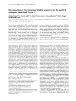
15d-PGJ2 inhibits monocyte adhesion to a brain-derived
endothelial cell line
Activated bEND.3 endothelial cells under pro-inflamma-
tory environment allows increased adherence of leuko-
cytes to its surface to facilitate their migration [6]. In our
in vitro system, bEND.3 cells were activated with TNFα
that caused a significant increase in the adhesion of
monocytes (~9 fold) compared to untreated cells. How-
ever, treatment with15d-PGJ2 (1–10 μM) 30 min prior to
the addition of TNFα significantly inhibited the adhesion
of monocytes (Fig. 1a, b). Prostaglandin production
begins with the liberation of arachidonic acid which
under cyclooxygenase enzymes 1 and 2 gets converted to
PGH2. Specific prostaglandin synthase convert PGH2 into
a series of prostaglandins including PGI2, PGF2α, PGD2
and PGE2 [32]. We also treated the bEND.3 cells with dif-
ferent prostaglandins (PGA1, PGB2, PGD2, PGE1, PGE2,
PGF1α, 15d-PGJ2, PGJ2), arachidonic acid, leukotriene
(LTB4) and thromboxane (TXB4) and observed that PGA1
and PGD2 treatment showed a significant decrease in
TNFα induced adhesion of monocytes, as these are pre-
cursors of 15d-PGJ2 (Fig. 1c). These results suggest the
specificity of 15d-PGJ2 in mediating the inhibition of the
adhesion process of monocytes on activated bEND.3 cells.
15d-PGJ2 did not cause any cell death (assessed by MTT
and LDH release assays) at the concentrations used (data
not shown).
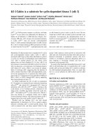
15d-PGJ2 inhibits expression of endothelial CAMs
Extravasation of mononuclear cells the recruitment cas-
cade are orchestrated by cell adhesion molecules on both
endothelial and immune cells [1]. Accordingly, we exam-
ined the effect of 15d-PGJ2 on TNFα induced expression
of CAMs (VCAM-1, ICAM-1 and E-selectin). For this,
bEND.3 cells were pretreated with15d-PGJ2 (5–10 μM)
followed by TNFα (50 ng/ml) treatment. After 2 h of incu-
bation, bEND.3 cells were processed for RNA isolation
and quantitative analysis of CAMs using real time PCR
(qPCR). Treatment with TNFα significantly increased the
mRNA expression of VCAM-1, ICAM-1 and E-selectin as
compared to control cells. 15d-PGJ2 markedly downregu-
lated their expression with a most pronounced effect
observed on expression of VCAM-1 as compared to E-
selectin or ICAM-1 (Fig. 2a, b and 2c). These observations
are in agreement with flow cytometry analysis which also
showed that 15d-PGJ2 treatment significantly reduced the
expression of endothelial CAMs with maximum affect on
VCAM-1 expression (Fig. 2d).
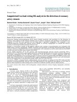
15d-PGJ2 inhibits VCAM-1 expression in a PPAR
γ
independent manner
To determine whether 15d-PGJ
2
mediates its inhibitory
effect through PPARγ, we employed GW9662, an irrevers-
ible PPARγ antagonist. GW9662 (10 μM) did not reverse
15d-PGJ2 mediated inhibition of TNFα induced expres-
sion of VCAM-1 in the endothelial cell line (Fig. 3A).
Another activator of PPARγ, troglitazone, was used to
examine if PPARγ plays any role in expression of VCAM-1
5d-PGJ2 inhibits monocyte adhesion to endothelial cellsFigure 1
5d-PGJ2 inhibits monocyte adhesion to endothelial
cells. bEND.3 cells were incubated with different concentra-
tions of 15d-PGJ2 (1–20 μM) (A) or mentioned prostagland-
ins (5 μM), arachidonic acid (5 μM), Leukotriene 4 (LTB 4, 5
μM) and Thromboxanes 4 (TXB 4, 5 μM) (C) for 30 min fol-
lowed by TNFα (50 ng/ml) stimulation for 6 h. Fluorescently
labeled monocytes were allowed to interact with activated
bEND.3 cells. Adhered monocytes were counted as men-
tioned in 'Material and Methods'. Data calculated as mean ±
SD of 21 fields from 3 different experiments. *** p < 0.001
compared to untreated control cells and !!! p < 0.001 com-
pared to TNFα treated cells. (B) is the pictorial representa-
tion of adhesion under TNFα (50 ng/ml) and 15d-PGJ2 (10
μM) treatment.
Journal of Inflammation 2008, 5:14 />Page 5 of 10
(page number not for citation purposes)
in bEND.3 cells. Troglitazone treatment, similar to 15d-
PGJ2 treatment, inhibited the expression of VCAM-1,
which could not be reversed by GW9662 (Fig. 3a). To
examine the ability of GW9662 on 15d-PGJ2 and troglita-
zone mediated induction of PPARγ transcription, we used
a chimeric receptor system in which the putative ligand-
binding domain of the PPARγ is fused to the DNA binding
domain of the yeast transcription factor galactose-respon-
sive gene 4 (GAL4). The 15d-PGJ
2
and troglitazone
potently activated the PPARγ-dependent chloramphenicol
acetyltransferase (CAT) reporter activity, which was com-
pletely blocked by GW9662 treatment (Fig. 3b). To con-
firm this observation, bEND.3 cells were transfected with
PPARγ wild type (Wt) and dominant-negative (DN)
expression vectors and determined the effects on VCAM-
1mRNA expression. 15d-PGJ2 was able to inhibit the
TNFα induced expression of VCAM-1 in both control and
PPARγ Wt transfected cells. However, transfection with
PPARγ DN was not able to attenuate the 15-dPGJ2 medi-
ated inhibition of VCAM-1 mRNA expression indicating
that the inhibitory effect of 15d-PGJ2 is independent of
PPARγ (Fig. 3c). Treatment with 15d-PGJ2 induced the
PPRE-luciferase activity in transiently transfected PPARγ
Wt expression vector, whereas, it had no effect in PPARγ
15d-PGJ2 inhibits mRNA and protein expression of endothe-lial CAMsFigure 2
15d-PGJ2 inhibits mRNA and protein expression of
endothelial CAMs. bEND.3 cells were pretreated with
15d-PGJ2 (5–20 μM) for 30 min followed by stimulation with
TNFα (50 ng/ml) for 2 h. Cells were harvested in Trizol rea-
gent for RNA isolation and cDNA synthesis. RT-PCR analysis
was done for ICAM-1 (A), VCAM-1 (B) and E-selectin (C).
Results were calculated as mean ± SD for 3 independent
experiments. Samples were examined in triplicates. &&& p <
0.001 compared with control (untreated and unstimulated
cells) and !!! p < 0.001 as compared to TNFα treatment. For
the quantitation of expression of surface CAMs, bEND.3
cells were treated with TNFα (50 ng/ml) in the presence or
absence of 15d-PGJ2 (5–20 μM) for 6 h followed by flow
cytometry analysis (D) (n = 2).
15d-PGJ2 inhibits VCAM-1 in PPARγ independent mannerFigure 3
15d-PGJ2 inhibits VCAM-1 in PPARγ independent
manner. bEND.3 cells were treated with GW9662 (10 μM)
30 min prior to treatment with 15d-PGJ2 (10 μM) or trogli-
tazone (10 μM) followed by TNFα treatment (50 ng/ml).
bEND.3 cells were lysed and processed for immunoblot anal-
ysis for VCAM-1 and β actin expression (A). Endothelial cell
line was cotransfected with PPARγ-GAL4 chimeras and the
reporter plasmid (upstream activating sequences)
5
-TK-CAT.
After 48 h, cells were treated with 15d-PGJ
2
or trogliatzone
in the presence or absence of GW9662 (10 μM) for 24 h.
Cell extracts were subsequently assayed for CAT activity by
ELISA (Roche) (B). pCMV-GAL4-binding domain (without
insert) and (upstream activating sequences)
5
-TK-CAT were
transfected as a control to detect the basal levels of CAT
activity (first lane). Data are mean of three values ± SD. *** p
< 0.001 as compared with untreated cells; !!! p < 0.001 as
compared with 15d-PGJ2 treated cells. (C) Cells were trans-
fected with PPARγ wild type (Wt) and dominant negative
(DN) constructs followed by treatment with 15-dPGJ2 (5
and 10 μM; 30 min) and TNFα (50 ng/ml, 2 h) and processed
for qPCR for detection of VCAM1 mRNA expression as
described in 'Material and Methods' (C). Results were calcu-
lated as mean ± SD for 3 independent experiments. Samples
were run in triplicates. &&& p < 0.001 compared with con-
trol (untreated and unstimulated cells) and !!! p < 0.001 as
compared to TNFα treatment. Cells were co-transfected
with PPARγ wild type (Wt) and dominant negative (DN) (0.5
μg/well) constructs along with PPRE-luc reporter (0.5 μg/
well) and pRL-TK (0.5 μg/well) followed by treatment with
15-dPGJ2 (10 μM) after 24 h. After 24 h incubation, luci-
ferase activity was performed, as described before
pcDNA3.1 was added to normalize the total content of DNA
for transfection. Data are mean ± SD of three different val-
ues. ***, p < 0.001 as compared with untreated cells; !!!, p <
0.001 as compared with 15d-PGJ
2
-treated PPAR wt trans-
fected cells.
Journal of Inflammation 2008, 5:14 />Page 6 of 10
(page number not for citation purposes)
DN transfected cells (Fig. 3d) suggesting that 15d-PGJ2
has the ability to activate PPARγ but its effect on VCAM-1
expression in the bEND.3 endothelial cell line is inde-
pendent of PPARγ.
15d-PGJ2 inhibits NF-
κ
B function in brain-derived
endothelial cell line
To further understand the mechanism of inhibitory action
of 15d-PGJ2 on endothelial CAMs and the process of
adhesion we examined the effect of 15d-PGJ2 on NF-κB
pathway, which is a pleiotropic regulator of many genes
involved in inflammation including CAMs [11]. Using
EMSA, we observed that 15d-PGJ2 inhibited the TNFα
induced binding of the NF-κB complex, in a time and
dose-dependent manner (Fig. 4a). To further define the
inhibitory effect of 15d-PGJ2 on TNFα mediated activa-
tion of the NF-κB pathway, the bEND.3 cells were trans-
fected with the p65/p50 complex along with the NF-κB
luciferase reporter construct. Cells transfected with p65/
p50 exhibited increased reporter activity, which was mark-
edly reduced in a dose-dependent manner with 15d-PGJ2
treatment (Fig. 4b). These observations obtained from
EMSA and transfection studies were further confirmed by
immunostaining for p65 nuclear translocation. Under
TNFα stimulation, p65 translocated to the nucleus and
was markedly attenuated by 15d-PGJ2 treatment (Fig. 4c).
Correspondingly, we also observed that 15d-PGJ2 inhib-
ited the TNFα induced nuclear translocation of p65 and
degradation of IκBα protein in a time and dose-depended
manner (Fig. 4d).
To support the 15d-PGJ2 mediated inhibition on NF-κB
pathway, we examined the effect of 15d-PGJ2 on p65-
DNA binding domain-gal4 transcriptional activity. The
p65-DNA binding domain-gal4 (p65-DNA-gal4) is a chi-
meric-transactivator, which consists of transcriptional
activation domain of NF-κB p65 protein fused to the
DNA-binding domain of GAL4 protein from yeast. As evi-
dent from figure 5, treatment with TNFα induced the tran-
scriptional activity of p65-DBD-gal4 which was
completely blocked by 15d-PGJ2 treatment.
Inhibition of IKK activity by 15d-PGJ2
Based on preceeding results, we examined the effect of
15d-PGJ2 on the activity of IKK, the upstream kinase of
the NF-κB pathway. Cells were treated with 15d-PGJ2 fol-
lowed by TNFα for 15 min and phosphorylation of IKKα
was detected using a specific antibody. As shown in figure
6a, TNFα treatment induced phosphorylation of IKKα in
bEND.3 cells which was completely blocked by 15d-PGJ2
treatment. bEND.3 cells were further cotransiently trans-
fected with IKKα and NF-κB luciferase reporter constructs
and after 24 h, cells were treated with TNFα with or with-
out 15d-PGJ2. TNFα induced the IKKα mediated NF-κB-
reporter activity, which was a significantly downregulated
by 15d-PGJ2 treatment (Fig. 6b). This observation was
further supported when bEND.3 cells were transiently
cotransfected with p65-DBD-gal4 and IKKα expression
vectors. Transient transfection with IKKα significantly
induced p65 transcriptional activity which was com-
pletely blocked by 15d-PGJ2 treatment (Fig. 6c) suggest-
ing that 15d-PGJ2 inhibits NF-κB function by inhibiting
IKKα activity in bEND.3 cells.
Post treatment of 15d-PGJ2 inhibits adhesion of
monocytes on activated brain-derived endothelial cell line
Our results suggested that 15d-PGJ2 inhibits the adhesion
of mononuclear cells on activated endothelial cells by
inhibiting the CAMs expression via downregulation of
NF-κB pathway when pretreated before stimulation with
15d-PGJ2 inhibits TNFα induced NF-κB function in endothe-lial cellsFigure 4
15d-PGJ2 inhibits TNFα induced NF-κB function in
endothelial cells. bEND.3 cells were treated with 15d-
PGJ2 (1–10 μM) and TNFα (50 ng/ml) for various time peri-
ods (5–40 min) and processed for EMSA as described in
'Material and Methods' (A). bEND.3 cells were transiently
transfected with p65, p50 expression vectors along with NF-
κB luciferase reporter construct (0.5 μg/well) and pCMV-β-
galactosidase (0.5 μg/well) followed by treatment with 15d-
PGJ2 (5–20 μM) for 4 h and processed for luciferase and β-
galactosidase activities. Luciferase activity was normalized
with respect to β-gal activity (B). Results were calculated as
mean ± SD for 3 independent experiments. Samples were
run in triplicates. &&& p < 0.001 compared with control and
!!! p < 0.001 compared with TNFα treatment (50 ng/ml).
Cells were treated with 15d-PGJ2(10 μM) for 30 min fol-
lowed by TNFα for 20 min and stained with anti-p65 anti-
body as described in 'Material and Methods' (C). Images
taken at 200× magnification are representative of 6 fields
from each treatment and 3 independent experiments.
Treated and untreated cells were processed for immunoblot
analysis for p65 and IκBα levels (D). Representative blot
from two independent experiments are shown.
Journal of Inflammation 2008, 5:14 />Page 7 of 10
(page number not for citation purposes)
TNFα. We wanted to examine if post treatment with 15d-
PGJ2 could inhibit the adhesion of mononuclear cells on
TNFα-stimulated cells. For this, bEND.3 cells were stimu-
lated with TNFα for 6 h followed by addition of various
concentrations (5–20 μM) of15d-PGJ2. After 30 min of
treatment with 15d-PGJ2, cells were washed and labeled
monocytes were added for adhesion assay. Interestingly,
post treatment with 15d-PGJ2 inhibited adhesion of
mononuclear cells on activated bEND.3 cells (Fig. 7) sug-
gesting that 15d-PGJ2 probably inhibits multiple path-
ways including NF-κB-CAMs expression and other
signaling pathway required for monocyte-endothelial cell
adhesion,.
Discussion
PGs are small lipid molecules that regulate numerous
processes in the body and their biological effects is an area
of concentrated research [33]. The J series of PGs have
been demonstrated to regulate processes like adipogene-
sis, inflammation and tumorigenesis [32]. 15d-PGJ2 is a
metabolite of PGD2 and is produced by mast cells, T cells,
platelets and alveolar macrophages [34]. 15d-PGJ2 is
emerging as a key anti-inflammatory mediator. Consist-
ent with this we have previously shown that 15d-PGJ2 has
an anti-inflammatory role in primary astrocytes [29]. This
study reports for the first time that 15d-PGJ2 inhibits
adhesion of monocytes to TNFα activated bEND.3
endothelial cells by downregulating endothelial CAMs via
inhibition of IKKα-NF-κB pathway but in a PPARγ inde-
pendent manner.
Infiltration of leukocytes is a crucial response in inflam-
matory reactions in numerous disorders where these leu-
kocytes are intended to induce inflammation in CNS
15d-PGJ2 inhibits p65 transcriptional activity in endothelial cellsFigure 5
15d-PGJ2 inhibits p65 transcriptional activity in
endothelial cells. bEND.3 cells were transfected with Gal-
p65 or Gal-DBD along with PTL-luciferase and PRL-TK
reporter constructs as described in Material and Method.
bEND.3 cells were pretreated with 15d-PGJ2 (10 μM) for 30
min followed by TNFα treatment (50 ng/ml). After 6 h of
TNFα treatment, cells were processed for luciferase assay
and results were normalized with PRL-TK luciferase activity
in each sample. Results were calculated as mean ± SD for 3
independent experiments. *** and !!! p < 0.001 compared
with control, @@@ p < 0.001 compared with TNFα treat-
ment.
15d-PGJ2 inhibits TNFα induced IKKα mediated NF-κB reporter activityFigure 6
15d-PGJ2 inhibits TNFα induced IKKα mediated NF-
κB reporter activity. bEND.3 cells were treated with
TNFα (50 ng/ml) in the presence or absence of 15d-PGJ2 (10
μM) followed by detection of pIKKα using its specific anti-
body (Cell Signaling) (A). β actin was used as a control for
equal content of protein loaded. bEND.3 cells were trans-
fected with IKKα, NF-κB luciferase and pCMV-β-galactosi-
dase constructs and treated with 15d-PGJ2 (5–20 μM) and
TNFα (50 ng/ml). After 4 h of TNFα treatment, cells were
processed for luciferase assay as described in 'Material and
Methods' (B). Results were calculated as mean ± SD for 3
independent experiments. &&& p < 0.001 compared with
control, !!! p < 0.001 compared with TNFα treatment and
### p > 0.001 compared with IKKα. bEND.3 cells were
transfected with Gal-p65 or Gal-DBD in the presence or
absence of flag-IKKα along with PTL-luciferase and PRL-TK
reporter constructs as described in Material and Method.
bEND.3 cells were pretreated with 15d-PGJ2 (10 μM) for 30
min followed by TNFα treatment. After 6 h of TNFα treat-
ment (50 ng/ml), cells were processed for luciferase assay
and results were normalized with PRL-TK luciferase activity
in each sample (C). Total DNA content was normalized with
pcDNA3. Results were calculated as mean ± SD for 3 inde-
pendent experiments.
Journal of Inflammation 2008, 5:14 />Page 8 of 10
(page number not for citation purposes)
when BBB is compromised. However, when misdirected,
they destroy healthy cells and matrix components causing
tissue damage [1]. Therefore, in recent years efforts have
been directed to limit the infiltration of mononuclear
cells so as to minimize the tissue injury during the disease
process. In earlier studies in different disease models, it
was reported that 15d-PGJ2 inhibits infiltration of leuck-
ocytes to site of inflammation [29,35]. Since adhesion of
infiltrating cells to endothelium, is a prerequisite for infil-
tration, we investigated the effect of PPAR activator 15d-
PGJ2 on the adhesion process. 15d-PGJ2 was observed to
inhibit the adhesion of monocytes to activated bEND.3
endothelial cells in a dose-dependent manner. These
Results were consistent with previous studies where 15d-
PGJ2 inhibited the adhesion of mononuclear cells to
PMA, IFNγ or IL-1β activated endothelial cells [36,37].
The inhibition of the adhesion process by15d-PGJ2 was
mediated by down regulation of TNFα induced endothe-
lial CAMs expression, namely, VCAM-1, E-selectin and
ICAM-1. Further, this effect was found to be PPARγ inde-
pendent. Our Results were consistent with other reports in
which 15d-PGJ2 and other PPAR activators negatively
modulate endothelial CAMs in vitro [37-39]. Treatment of
bEND.3 cells with 15d-PGJ2 showed effects by attenuat-
ing signaling taking place during adhesion process as well
as downregulating endothelial CAMs expression, thereby
giving a significant additive effect on inhibition on adhe-
sion of monocytes. To further understand the mechanism
of inhibition mediated by15d-PGJ2, we determined its
effect on the NF-κB transcription factor which is known to
be activated by TNFα [9]. 15d-PGJ2 was observed to
inhibit DNA binding of the NF-κB complex in a gel shift
assay. Interestingly, this inhibition was through modula-
tion of upstream targets of the NF-κB pathway. There was
inhibition of TNFα induced degradation of IkBα protein
thereby preventing p65 nuclear translocation. Our study
is supported by other reports of inhibition of NF-κB by
15d-PGJ2, though in different cell types [29,35,40,41].
Thus, our data showed that 15d-PGJ2 inhibits TNFα
induced NF-κB activity and consequently the expression
of endothelial CAMs under our experimental model.
Moreover, we have previously suggested IKK as a target of
15d-PGJ2 in modulating NF-κB pathway in brain glial
cells [29,35], which is consistent in endothelial cells too.
We can conclude from our in vitro data that 15d-PGJ2
inhibits endothelial-monocyte interactions via IKK-NF-
κB-CAMs pathway in endothelial cells. PI3 kinase and Akt
are also known to play an important role in the adhesion
process [42]. The activation of IKK is also regulated via
phosphorylation by Akt [43]. 15d-PGJ2 has been demon-
strated to inhibit the PI3 kinase/Akt pathway in brain glial
cells [29]. PI3 kinase and Akt pathway play important role
in adhesion as we have documented before that inhibi-
tion of PI3Kinase and Akt is able to inhibit the adhesion
of monocytes [27].
Thus, 15d-PGJ2 might be modulating PI3 kinase-Akt-IKK-
NF-κB-CAMs pathway. Interestingly, post treatment with
15d-PGJ2 was also able to inhibit monocyte adhesion on
activated bEND.3 cells, suggesting the possibility that15d-
PGJ2 may also inhibit other signaling pathway/s impor-
tant for firm and sustained adhesion of monocyte on
endothelial cells.
15d-PGJ2 is a natural ligand of PPARγ and has numerous
effects which are PPARγ dependent. Moreover, it has been
shown to has therapeutic potential in various human
autoimmune diseases as well as animal models of autoim-
munity, including arthritis [44-46], ischemia-reperfusion
injury [47,48], Alzheimer's disease [49-51], lupus nephri-
tis [52,53] and EAE [26,54,55]. More recent evidences
have shown that there are effects of 15d-PGJ2 that are
independent of PPARγ activation [32], while, the exact
mechanism of action of 15d-PGJ2 in different systems is
unknown. There are various propositions such as presence
of another cytoplasmic PG receptor [56], recruitment of
p300 by NF-κB [29], inhibition of NF-κB DNA binding by
alkylation of cysteine residue of p65 [57], or ROR depend-
ent mechanism [39].
Post treatment of 15d-PGJ2 inhibits monocyte adhesion to activated endothelial cellsFigure 7
Post treatment of 15d-PGJ2 inhibits monocyte adhe-
sion to activated endothelial cells. bEND.3 cells were
treated with TNFα (50 ng/ml) for 6 h, followed by addition of
different concentrations of 15d-PGJ2 (5–20 μM). After 30
min of incubation with 15-PGJ2, fluorescently labeled mono-
cytes were allowed to interact with activated bEND.3 cells.
Adhered monocytes were counted as mentioned in 'Material
and Methods'. Data calculated as mean ± SD of 21 fields from
3 different experiments. *** p < 0.001 compared to
untreated control cells and @ p < 0.001 compared to TNFα
treated cells.
Journal of Inflammation 2008, 5:14 />Page 9 of 10
(page number not for citation purposes)
Conclusion
All together, the present data shows that 15d-PGJ2 regu-
lates inflammatory responses by inhibiting the infiltration
of leukocytes across the endothelial barrier, which it does
so by inhibiting monocyte adhesion to activated endothe-
lial cells via downregulation of IKK-NF-κB-CAMs pathway
in endothelial cells, independent of PPARγ.
Abbreviations
15d-PGJ 2: 15-deoxy-Delta (12, 14)-prostaglandin J;
CAM: cell adhesion molecule; ICAM: Intercellular cell
adhesion molecule-1; VCAM-1: Vascular cell adhesion
molecule-1; NF-κB: Nuclear factor kappa B; IκB: Inhibi-
tory kappa B; IKK: Inhibitory kappa B kinase.
Competing interests
The authors declare that they have no competing interests.
Authors' contributions
This study is based on an original idea of SG and IS. RP
and SG wrote the manuscript. SG directed and RP per-
formed the in vitro experiments. AKS helped in finalizing
manuscript. All authors read and approved the final man-
uscript.
Acknowledgements
RP and SG are equal contributors for this work. We would like to thank
Drs. Anne G. Gilg and Ramandeep Rattan for editing manuscript and Ms
Joyce Bryan for procurement of chemicals used in this study. These studies
were supported by grants (NS-40144, NS-22576, NS-34741, NS-37766,
and NS-40810) from the NIH and (SCIRF 0406 and SCIRF 0506) from State
of South Carolina Spinal Cord Injury Research Fund Board. This work was
supported by the NIH (NS-22576, NS-34741, NS-37766 and NS-40810)
and from the Extramural Research Facilities Program of the National
Center for Research Resources (Grants C06 RR018823 and No C06
RR015455).
References
1. Ulbrich H, Eriksson EE, Lindbom L: Leukocyte and endothelial
cell adhesion molecules as targets for therapeutic interven-
tions in inflammatory disease. Trends Pharmacol Sci 2003,
24:640-647.
2. Carlos TM, Harlan JM: Leukocyte-endothelial adhesion mole-
cules. Blood 1994, 84:2068-2101.
3. Li H, Cybulsky MI, Gimbrone MA Jr, Libby P: Inducible expression
of vascular cell adhesion molecule-1 by vascular smooth
muscle cells in vitro and within rabbit atheroma. Am J Pathol
1993, 143:1551-1559.
4. Abe Y, Sugisaki K, Dannenberg AM Jr: Rabbit vascular endothelial
adhesion molecules: ELAM-1 is most elevated in acute
inflammation, whereas VCAM-1 and ICAM-1 predominate
in chronic inflammation. J Leukoc Biol 1996, 60:692-703.
5. Moor AC, de Vries HE, de Boer AG, Breimer DD: The blood-brain
barrier and multiple sclerosis. Biochem Pharmacol 1994,
47:1717-1724.
6. Lee SJ, Benveniste EN: Adhesion molecule expression and reg-
ulation on cells of the central nervous system. J Neuroimmunol
1999, 98:77-88.
7. Muller WA: Leukocyte-endothelial cell interactions in the
inflammatory response. Lab Invest 2002, 82:521-533.
8. Pober JS: Immunobiology of human vascular endothelium.
Immunol Res 1999, 19:225-232.
9. Madge LA, Pober JS: TNF signaling in vascular endothelial cells.
Exp Mol Pathol 2001, 70:317-325.
10. Chen CC, Manning AM: Transcriptional regulation of endothe-
lial cell adhesion molecules: a dominant role for NF-kappa B.
Agents Actions Suppl 1995, 47:135-141.
11. Collins T, Read MA, Neish AS, Whitley MZ, Thanos D, Maniatis T:
Transcriptional regulation of endothelial cell adhesion mole-
cules: NF-kappa B and cytokine-inducible enhancers. Faseb J
1995, 9:
899-909.
12. Hayden MS, Ghosh S: Signaling to NF-kappaB. Genes Dev 2004,
18:2195-2224.
13. Ledebur HC, Parks TP: Transcriptional regulation of the inter-
cellular adhesion molecule-1 gene by inflammatory
cytokines in human endothelial cells. Essential roles of a var-
iant NF-kappa B site and p65 homodimers. J Biol Chem 1995,
270:933-943.
14. Mangelsdorf DJ, Thummel C, Beato M, Herrlich P, Schutz G, Umes-
ono K, Blumberg B, Kastner P, Mark M, Chambon P, Evans RM: The
nuclear receptor superfamily: the second decade. Cell 1995,
83:835-839.
15. Tontonoz P, Hu E, Spiegelman BM: Stimulation of adipogenesis in
fibroblasts by PPAR gamma 2, a lipid-activated transcription
factor. Cell 1994, 79:1147-1156.
16. Delerive P, Fruchart JC, Staels B: Peroxisome proliferator-acti-
vated receptors in inflammation control. J Endocrinol 2001,
169:453-459.
17. Daynes RA, Jones DC: Emerging roles of PPARs in inflamma-
tion and immunity. Nat Rev Immunol 2002, 2:748-759.
18. Kliewer SA, Lenhard JM, Willson TM, Patel I, Morris DC, Lehmann JM:
A prostaglandin J2 metabolite binds peroxisome prolifera-
tor-activated receptor gamma and promotes adipocyte dif-
ferentiation. Cell 1995, 83:813-819.
19. Ricote M, Li AC, Willson TM, Kelly CJ, Glass CK: The peroxisome
proliferator-activated receptor-gamma is a negative regula-
tor of macrophage activation. Nature 1998, 391:79-82.
20. Petrova TV, Akama KT, Van Eldik LJ: Cyclopentenone prostaglan-
dins suppress activation of microglia: down-regulation of
inducible nitric-oxide synthase by 15-deoxy-Delta12,14-pros-
taglandin J2. Proc Natl Acad Sci USA 1999, 96:4668-4673.
21. Vaidya S, Somers EP, Wright SD, Detmers PA, Bansal VS: 15-Deoxy-
Delta12,1412,14-prostaglandin J2 inhibits the beta2 integrin-
dependent oxidative burst: involvement of a mechanism dis-
tinct from peroxisome proliferator-activated receptor
gamma ligation.
J Immunol 1999, 163:6187-6192.
22. Kaplan JM, Cook JA, Hake PW, O'Connor M, Burroughs TJ, Zingarelli
B: 15-Deoxy-Delta(12,14)-prostaglandin J(2) (15D-PGJ(2)), a
peroxisome proliferator activated receptor gamma ligand,
reduces tissue leukosequestration and mortality in endo-
toxic shock. Shock 2005, 24:59-65.
23. Genovese T, Cuzzocrea S, Di Paola R, Mazzon E, Mastruzzo C, Cata-
lano P, Sortino M, Crimi N, Caputi AP, Thiemermann C, Vancheri C:
Effect of rosiglitazone and 15-deoxy-Delta12,14-prostaglan-
din J2 on bleomycin-induced lung injury. Eur Respir J 2005,
25:225-234.
24. Cuzzocrea S, Pisano B, Dugo L, Ianaro A, Patel NS, Di Paola R, Gen-
ovese T, Chatterjee PK, Di Rosa M, Caputi AP, Thiemermann C: Ros-
iglitazone and 15-deoxy-Delta12,14-prostaglandin J2, ligands
of the peroxisome proliferator-activated receptor-gamma
(PPAR-gamma), reduce ischaemia/reperfusion injury of the
gut. Br J Pharmacol 2003, 140:366-376.
25. Natarajan C, Bright JJ: Peroxisome proliferator-activated
receptor-gamma agonists inhibit experimental allergic
encephalomyelitis by blocking IL-12 production, IL-12 signal-
ing and Th1 differentiation. Genes Immun 2002, 3:59-70.
26. Diab A, Deng C, Smith JD, Hussain RZ, Phanavanh B, Lovett-Racke
AE, Drew PD, Racke MK: Peroxisome proliferator-activated
receptor-gamma agonist 15-deoxy-Delta(12,14)-prostaglan-
din J(2) ameliorates experimental autoimmune encephalo-
myelitis. J Immunol 2002, 168:2508-2515.
27. Prasad R, Giri S, Nath N, Singh I, Singh AK: Inhibition of phosphoi-
nositide 3 kinase-Akt (protein kinase B)-nuclear factor-
kappa B pathway by lovastatin limits endothelial-monocyte
cell interaction. J Neurochem 2005, 94:204-214.
28. Dignam JD, Lebovitz RM, Roeder RG: Accurate transcription ini-
tiation by RNA polymerase II in a soluble extract from iso-
lated mammalian nuclei. Nucleic Acids Res 1983, 11:1475-1489.
29. Giri S, Rattan R, Singh AK, Singh I: The 15-deoxy-delta12,14-pros-
taglandin J2 inhibits the inflammatory response in primary
Publish with Bio Med Central and every
scientist can read your work free of charge
"BioMed Central will be the most significant development for
disseminating the results of biomedical research in our lifetime."
Sir Paul Nurse, Cancer Research UK
Your research papers will be:
available free of charge to the entire biomedical community
peer reviewed and published immediately upon acceptance
cited in PubMed and archived on PubMed Central
yours — you keep the copyright
Submit your manuscript here:
/>BioMedcentral
Journal of Inflammation 2008, 5:14 />Page 10 of 10
(page number not for citation purposes)
rat astrocytes via down-regulating multiple steps in phos-
phatidylinositol 3-kinase-Akt-NF-kappaB-p300 pathway
independent of peroxisome proliferator-activated receptor
gamma. J Immunol 2004, 173:5196-5208.
30. Giri S, Nath N, Smith B, Viollet B, Singh AK, Singh I: 5-aminoimida-
zole-4-carboxamide-1-beta-4-ribofuranoside inhibits proin-
flammatory response in glial cells: a possible role of AMP-
activated protein kinase. J Neurosci 2004, 24:479-487.
31. Nath N, Giri S, Prasad R, Singh AK, Singh I: Potential targets of 3-
hydroxy-3-methylglutaryl coenzyme A reductase inhibitor
for multiple sclerosis therapy. J Immunol 2004, 172:1273-1286.
32. Harris SG, Padilla J, Koumas L, Ray D, Phipps RP: Prostaglandins as
modulators of immunity. Trends Immunol 2002, 23:144-150.
33. Scher JU, Pillinger MH: 15d-PGJ2: the anti-inflammatory pros-
taglandin? Clin Immunol 2005, 114:100-109.
34. Goetzl EJ, An S, Smith WL: Specificity of expression and effects
of eicosanoid mediators in normal physiology and human
diseases. Faseb J 1995, 9:1051-1058.
35. Rossi A, Kapahi P, Natoli G, Takahashi T, Chen Y, Karin M, Santoro
MG: Anti-inflammatory cyclopentenone prostaglandins are
direct inhibitors of IkappaB kinase. Nature 2000, 403:103-108.
36. Imaizumi T, Matsumiya T, Tamo W, Shibata T, Fujimoto K, Kumagai
M, Yoshida H, Cui XF, Tanji K, Hatakeyama M, et al.: 15-Deoxy-
D12,14-prostaglandin J2 inhibits CX3CL1/fractalkine expres-
sion in human endothelial cells. Immunol Cell Biol 2002,
80:531-536.
37. Jackson SM, Parhami F, Xi XP, Berliner JA, Hsueh WA, Law RE,
Demer LL: Peroxisome proliferator-activated receptor acti-
vators target human endothelial cells to inhibit leukocyte-
endothelial cell interaction. Arterioscler Thromb Vasc Biol 1999,
19:2094-2104.
38. Pasceri V, Wu HD, Willerson JT, Yeh ET: Modulation of vascular
inflammation in vitro and in vivo by peroxisome proliferator-
activated receptor-gamma activators. Circulation 2000,
101:235-238.
39. Migita H, Morser J: 15-deoxy-Delta12,14-prostaglandin J2 (15d-
PGJ2) signals through retinoic acid receptor-related orphan
receptor-alpha but not peroxisome proliferator-activated
receptor-gamma in human vascular endothelial cells: the
effect of 15d-PGJ2 on tumor necrosis factor-alpha-induced
gene expression. Arterioscler Thromb Vasc Biol 2005, 25:710-716.
40. Albrecht EW, Stegeman CA, Heeringa P, Henning RH, van Goor H:
Protective role of endothelial nitric oxide synthase. J Pathol
2003, 199:8-17.
41. Eligini S, Banfi C, Brambilla M, Camera M, Barbieri SS, Poma F, Tremoli
E, Colli S: 15-deoxy-delta12,14-Prostaglandin J2 inhibits tissue
factor expression in human macrophages and endothelial
cells: evidence for ERK1/2 signaling pathway blockade.
Thromb Haemost 2002, 88:524-532.
42. Gerszten RE, Friedrich EB, Matsui T, Hung RR, Li L, Force T, Rosen-
zweig A: Role of phosphoinositide 3-kinase in monocyte
recruitment under flow conditions. J Biol Chem 2001,
276:26846-26851.
43. Ozes ON, Mayo LD, Gustin JA, Pfeffer SR, Pfeffer LM, Donner DB:
NF-kappaB activation by tumour necrosis factor requires
the Akt serine-threonine kinase. Nature 1999, 401:82-85.
44. Fahmi H, Di Battista JA, Pelletier JP, Mineau F, Ranger P, Martel-Pelle-
tier J: Peroxisome proliferator – activated receptor gamma
activators inhibit interleukin-1beta-induced nitric oxide and
matrix metalloproteinase 13 production in human chondro-
cytes. Arthritis Rheum 2001, 44:595-607.
45. Kawahito Y, Kondo M, Tsubouchi Y, Hashiramoto A, Bishop-Bailey D,
Inoue K, Kohno M, Yamada R, Hla T, Sano H: 15-deoxy-
delta(12,14)-PGJ(2) induces synoviocyte apoptosis and sup-
presses adjuvant-induced arthritis in rats. J Clin Invest 2000,
106:189-197.
46. Shan ZZ, Masuko-Hongo K, Dai SM, Nakamura H, Kato T, Nishioka
K: A potential role of 15-deoxy-delta(12,14)-prostaglandin J2
for induction of human articular chondrocyte apoptosis in
arthritis. J Biol Chem 2004,
279:37939-37950.
47. Nakajima A, Wada K, Miki H, Kubota N, Nakajima N, Terauchi Y,
Ohnishi S, Saubermann LJ, Kadowaki T, Blumberg RS, et al.: Endog-
enous PPAR gamma mediates anti-inflammatory activity in
murine ischemia-reperfusion injury. Gastroenterology 2001,
120:460-469.
48. Zingarelli B, Hake PW, Mangeshkar P, O'Connor M, Burroughs TJ,
Piraino G, Denenberg A, Wong HR: Diverse cardioprotective sig-
naling mechanisms of peroxisome proliferator-activated
receptor-gamma ligands, 15-deoxy-Delta12,14-prostaglan-
din J2 and ciglitazone, in reperfusion injury: role of nuclear
factor-kappaB, heat shock factor 1, and Akt. Shock 2007,
28:554-563.
49. Combs CK, Johnson DE, Karlo JC, Cannady SB, Landreth GE: Inflam-
matory mechanisms in Alzheimer's disease: inhibition of
beta-amyloid-stimulated proinflammatory responses and
neurotoxicity by PPARgamma agonists. J Neurosci 2000,
20:558-567.
50. Heneka MT, Klockgether T, Feinstein DL: Peroxisome prolifera-
tor-activated receptor-gamma ligands reduce neuronal
inducible nitric oxide synthase expression and cell death in
vivo. J Neurosci 2000, 20:6862-6867.
51. Munoz U, de Las Cuevas N, Bartolome F, Hermida OG, Bermejo F,
Martin-Requero A: The cyclopentenone 15-deoxy-
delta(12,14)-prostaglandin J2 inhibits G1/S transition and
retinoblastoma protein phosphorylation in immortalized
lymphocytes from Alzheimer's disease patients. Exp Neurol
2005, 195:508-517.
52. Reilly CM, Oates JC, Cook JA, Morrow JD, Halushka PV, Gilkeson GS:
Inhibition of mesangial cell nitric oxide in MRL/lpr mice by
prostaglandin J2 and proliferator activation receptor-
gamma agonists. J Immunol 2000, 164:1498-1504.
53. Reilly CM, Oates JC, Sudian J, Crosby MB, Halushka PV, Gilkeson GS:
Prostaglandin J(2) inhibition of mesangial cell iNOS expres-
sion. Clin Immunol 2001, 98:337-345.
54. Raikwar HP, Muthian G, Rajasingh J, Johnson CN, Bright JJ: PPAR-
gamma antagonists reverse the inhibition of neural antigen-
specific Th1 response and experimental allergic encephalo-
myelitis by Ciglitazone and 15-deoxy-Delta12,14-prostaglan-
din J2. J Neuroimmunol 2006, 178:76-86.
55. Storer PD, Xu J, Chavis JA, Drew PD: Cyclopentenone prostag-
landins PGA2 and 15-deoxy-delta12,14 PGJ2 suppress activa-
tion of murine microglia and astrocytes: implications for
multiple sclerosis. J Neurosci Res 2005, 80:66-74.
56. Hirai H, Tanaka K, Yoshie O, Ogawa K, Kenmotsu K, Takamori Y,
Ichimasa M, Sugamura K, Nakamura M, Takano S, Nagata K: Prostag-
landin D2 selectively induces chemotaxis in T helper type 2
cells, eosinophils, and basophils via seven-transmembrane
receptor CRTH2. J Exp Med 2001, 193:255-261.
57. Straus DS, Pascual G, Li M, Welch JS, Ricote M, Hsiang CH, Sen-
gchanthalangsy LL, Ghosh G, Glass CK: 15-deoxy-delta 12,14-
prostaglandin J2 inhibits multiple steps in the NF-kappa B
signaling pathway. Proc Natl Acad Sci USA 2000, 97:4844-4849.