Báo cáo y học: "Minocycline-induced hypersensitivity syndrome presenting with meningitis and brain edema: a case report'''' doc
Bạn đang xem bản rút gọn của tài liệu. Xem và tải ngay bản đầy đủ của tài liệu tại đây (249.27 KB, 4 trang )
BioMed Central
Page 1 of 4
(page number not for citation purposes)
Journal of Medical Case Reports
Open Access
Case report
Minocycline-induced hypersensitivity syndrome presenting with
meningitis and brain edema: a case report
Nicolas Lefebvre*
1
, Emmanuel Forestier
1
, David Farhi
2
,
Mohseni Zadeh Mahsa
1
, Véronique Remy
1
, Olivier Lesens
1
,
Daniel Christmann
1
and Yves Hansmann
1
Address:
1
Department of Infectious Diseases and Tropical medicine, Teaching Hospital, Strasbourg, France and
2
Department of Dermatology,
Tarnier Hospital, Paris, France
Email: Nicolas Lefebvre* - ; Emmanuel Forestier - ;
David Farhi - ; Mohseni Zadeh Mahsa - ; Véronique Remy - veronique.remy@ch-
cahors.fr; Olivier Lesens - ; Daniel Christmann - ;
Yves Hansmann -
* Corresponding author
Background: Hypersentivity Syndrome (HS) may be a life-threatening condition. It frequently
presents with fever, rash, eosinophilia and systemic manifestations. Mortality can be as high as 10%
and is primarily due to hepatic failure. We describe what we believe to be the first case of
minocycline-induced HS with accompanying lymphocytic meningitis and cerebral edema reported
in the literature.
Case presentation: A 31-year-old HIV-positive female of African origin presented with acute
fever, lymphocytic meningitis, brain edema, rash, eosinophilia, and cytolytic hepatitis. She had been
started on minocycline for inflammatory acne 21 days prior to the onset of symptoms. HS was
diagnosed clinically and after exclusion of infectious causes. Minocycline was withdrawn and
steroids were administered from the second day after presentation because of the severity of the
symptoms. All signs resolved by the seventh day and steroids were tailed off over a period of 8
months.
Conclusion: Clinicians should maintain a high index of suspicion for serious adverse reactions to
minocycline including lymphocytic meningitis and cerebral edema among HIV-positive patients,
especially if they are of African origin. Safer alternatives should be considered for treatment of acne
vulgaris. Early recognition of the symptoms and prompt withdrawal of the drug are important to
improve the outcome.
Background
Hypersentivity Syndrome (HS) is a rare and life-threaten-
ing form of drug reaction [1]. Usual presentation includes
fever, rash, eosinophilia and systemic manifestations.
Mortality may be as high as 10% and is primarily due to
hepatic failure [2]. HS is frequently associated with the use
of sulfonamides, allopurinol, terbinafine, minocycline,
and antiretroviral therapy [3].
Published: 18 May 2007
Journal of Medical Case Reports 2007, 1:22 doi:10.1186/1752-1947-1-22
Received: 12 December 2006
Accepted: 18 May 2007
This article is available from: />© 2007 Lefebvre et al; licensee BioMed Central Ltd.
This is an Open Access article distributed under the terms of the Creative Commons Attribution License ( />),
which permits unrestricted use, distribution, and reproduction in any medium, provided the original work is properly cited.
Journal of Medical Case Reports 2007, 1:22 />Page 2 of 4
(page number not for citation purposes)
Minocycline is widely prescribed for the treatment of
inflammatory forms of acne vulgaris [4]. Although it is
considered to be a safe drug [2], it has been reported to
cause serious adverse events such as hepatitis, auto-
immune syndrome and HS [1,5]. To our knowledge, lym-
phocytic meningitis and brain edema associated with
minocycline-induced HS have not been reported in the lit-
erature. This presentation, which is probably under-recog-
nized, may lead to a diagnostic delay. We report herein
one case.
Case presentation
A 31-year-old female native of Africa was hospitalized
with fever, weakness, nausea, headache, facial edema, and
rash, for 4 days. She had been diagnosed as HIV positive
2 years previously. CD4 cell count was between 300 and
400 cells/mm
3
, viral load was near 150,000 copies/mm
3
,
both steady for several weeks. She had no other relevant
medical history, and was on no treatment for HIV. Three
weeks before the onset of the symptoms, she had been
started on oral minocycline to control an inflammatory
form of acne vulgaris.
At admittance she was unwell and vital signs were: blood
pressure 100/60, temperature 40°C, heart rate 120 beats/
min., respiratory rate 22/min., and oxygen saturation 97%
(room air). On physical examination, she had multiple
erythematous, oedematous and coalescing plaques on the
upper trunk, and on proximal segments of the limbs. Her
palms and soles were erythematous. She also had injec-
tion of the sclera, and edema of the eyelids. Buccal and
genital mucous membranes were unaffected. Palpation
revealed enlarged tender lymph nodes at all sites, but no
accompanying hepatomegaly or splenomegaly. Cardio-
vascular and pulmonary systems were normal, with no
sign of septic shock. Neurological examination was nor-
mal.
Laboratory tests revealed the following values: leucocyto-
sis (24×10
9
/L), eosinophilia (2.8×10
9
/L), neutrophilia
(8×10
9
/L) without lymphocytosis (2.8×10
9
/L), elevated
C-Reactive Protein (17 mg/L) and cytolytic hepatitis (ALT
160 U/L, AST 106 U/L, lactate deshydrogenase 1400 U/L
(LDH)). Multiple blood culture and bacteriological anal-
ysis of urine were sterile. Serological investigation for viral
and bacterial agents, repeated over two weeks showed no
sign of recent infection (Borrelia burgdorferi, Mycoplasma
sp., Chlamydiae sp., Legionella sp., Epstein-Barr virus,
cytomegalovirus, hepatitis A, B and C viruses, measles,
rubella, parvovirus B19, coxsackievirus, echovirus, VZV,
HTLV1, HSV, toxoplasmosis). The antinuclear antibodies
were negative. Chest radiography and echocardiography
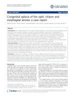
showed no abnormalities, however cerebral CT-scan
showed a diffuse cerebral edema (figure). Electroencepha-
logram was normal. The lumbar puncture revealed lym-
phocytic meningitis with 60 cells/microliter
(lymphocytes: 90%, protein level: 0.80 g/L, glucose level:
0.56 g/L). No microbial agents were found in the cerebro-
spinal fluid (cryptococcal antigen and culture, culture for
bacteriological agents, and polymerase chain reaction
(PCR) for echovirus, coxackies virus, CMV, HSV, VZV).
PCR for HIV in cerebrospinal fluid was not performed.
Histopathology of a skin biopsy sample showed a lym-
phocytic infiltrate into the dermo-epidermis junction
with edema, resulting in a blister detachment. This aspect
was of a lichenoid toxiderma. Lymph node biopsy was not
performed.
Minocycline was discontinued on the fourth day after the
onset of the symptoms. Two days after minocycline with-
drawal, treatment with steroids was introduced (methyl-
prednisolone 60 mg daily) because of the severity of the
symptoms. The patient improved quickly after steroid
administration. Temperature dropped to 37°C within
three days. Within one week the eruption cleared and lym-
phadenopathies disappeared. Biological abnormalities
(eosinophilia, liver enzyme elevation) resolved within
three days. A second brain CT-scan, undertaken 14 days
after the onset of the symptoms, was normal. Lumbar
puncture was not repeated. The steroids were steadily
tailed off (5 mg per week) but a relapse occurred on week
6, when on a dose of 30 mg per day. This relapse was
under the form of a transient and isolated generalized pru-
ritus with no cutaneous nor neurological signs. Skin tests
were not carried out due to the potential risk of severe
reaction. The patient was weaned off steroids over a
period of 8 months and was free of symptoms at dis-
charged form care.
Discussion
Minocycline is widely prescribed for acne vulgaris [4].
Minor adverse effects including nausea, vomiting, dizzi-
ness, photosensitivity and skin eruption have been
described [2]. However, acute and severe reactions such as
HS, autoimmune disorders, serum sickness like reaction,
and pseudotumor cerebri syndrome have also been
reported [1,5-8].
The case described had several features suggesting a diag-
nosis of HS secondary to minocycline treatment. The his-
tory was characteristic (interval of 3 weeks between the
introduction of the drug and the symptoms) and the con-
dition resolved promptly following cessation of minocy-
cline. Moreover, differential diagnoses due to the most
likely infectious diseases were excluded and no evidence
of pseudotumor cerebri syndrome, as it may be observed
with minocycline, was found. While lymphocytic menin-
gitis and cerebral edema have not been described in asso-
ciation with minocycline-induced HS, they have been
reported following use of allopurinol [9].
Journal of Medical Case Reports 2007, 1:22 />Page 3 of 4
(page number not for citation purposes)
HS to minocycline is a nosological entity also called
DRESS syndrome (Drug Reaction with Eosinophilia and
Systemic Symptoms). It occurs within 3 to 4 weeks after
starting therapy, usually in a young patient (21.2 ± 1.8
years-old) [1,5]. As in our patient, HIV-infection and
black African origin have been suggested as risk factors for
minocycline HS [3]. Clinical features include three major
elements: (1) high fever with asthenia; (2) acute general-
ized cutaneous signs often polymorphous (maculo-papu-
lar rash sometimes morbiliform, exanthematous, or
multiform); facial edema is evocative and intense pruritus
is common; and (3) multivisceral involvement [3,5,10].
The most common visceral signs are enlargement of the
lymph nodes, hepatomegaly and splenomegaly [1,5].
Severe reactions may lead to hepatitis, pulmonary infil-
trates with eosinophilia, myocarditis and interstitial
nephritis [1,8]. In 1996, among 13 cases of HS reaction
due to minocycline, none had cerebral involvement [1].
Suggestive blood chemistry includes eosinophilia (often
over 1.5×10
9
/L), cytolytic hepatitis (from mild elevation
of liver enzyme to severe liver failure), LDH elevation and
hemolytic anaemia [1,10]. The skin biopsy may show a
non specific lymphocytic infiltrate or lichenoid interface
dermatitis [1]. The treatment is usually limited to the
withdrawal of minocycline, which is usually followed by
symptomatic relief [5]. Steroids should be restricted to
severe case with threatening renal, liver or lung involve-
ment. They should be used with caution because depend-
ence to steroid is frequent and rebound of the condition
may follow their withdrawal, as observed in this case
report.
Conclusion
As minocycline is a widely used drug, clinicians should be
aware of the risk of HS, even in the presence of neurolog-
ical abnormalities. Early warning signs include an acute
rash with fever, eosinophilia and elevated liver enzymes.
The drug should be immediately and definitively with-
drawn for the patient. If used, minocycline should be
strictly monitored, especially in African or HIV-infected
patients.
Competing interests
The author(s) declare that they have no competing inter-
ests.
Authors' contributions
NL: participated in patient management, diagnosis and
drafted the manuscript.
EF, DF, MMZ: participated in patient management,
reviewing the literature and helped to draft the manu-
script.
OL, VR, YH, DC: helped in patient management and
made final diagnosis.
All authors read and approved the final manuscript.
Acknowledgements
Jacques Margery for his help in the publishing process.
Written consent for publication was obtained from the patient.
References
1. Knowles SR, Shapiro L, Shear NH: Serious adverse reactions
induced by minocycline. Report of 13 patients and review of
the literature. Arch Dermatol 1996, 132(8):934-939.
2. Bernier C, Dreno B: Minocycline. Ann Dermatol Venereol 2001,
128(5):627-637.
3. Roujeau JC, Stern RS: Severe adverse cutaneous reactions to
drugs. N Engl J Med 1994, 331(19):1272-1285.
4. Sapadin AN, Fleischmajer R: Tetracyclines: nonantibiotic prop-
erties and their clinical implications. J Am Acad Dermatol 2006,
54(2):258-265.
5. Shapiro LE, Knowles SR, Shear NH: Comparative safety of tetra-
cycline, minocycline, and doxycycline. Arch Dermatol 1997,
133:1224-1230.
6. Chiu AM, Chuenkongkaew WL, Cornblath WT, Trobe JD, Digre KB,
Dotan SA, Musson KH, Eggenberger ER: Minocycline treatment
and pseudotumor cerebri syndrome. Am J Ophthalmol 1998,
126(1):116-121.
7. Grasset L, Guy C, Ollagnier M: Cyclines and acne: pay attention
to adverse drug reactions! A recent literature review. Rev
Med Interne 2003, 24(5):305-316.
8. Parneix-Spake A, Bastuji-Garin S, Lobut JB, Erner J, Guyet-Rousset P,
Revuz J, Roujeau JC: Minocycline as possible cause of severe and
CT scan showing diffuse cerebral edema in a young woman with hypersensitivity syndrome due to minocyclineFigure 1
CT scan showing diffuse cerebral edema in a young woman
with hypersensitivity syndrome due to minocycline.
Publish with Bio Med Central and every
scientist can read your work free of charge
"BioMed Central will be the most significant development for
disseminating the results of biomedical research in our lifetime."
Sir Paul Nurse, Cancer Research UK
Your research papers will be:
available free of charge to the entire biomedical community
peer reviewed and published immediately upon acceptance
cited in PubMed and archived on PubMed Central
yours — you keep the copyright
Submit your manuscript here:
/>BioMedcentral
Journal of Medical Case Reports 2007, 1:22 />Page 4 of 4
(page number not for citation purposes)
protracted hypersensitivity drug reaction. Arch Dermatol 1995,
131(4):490-491.
9. Mills RM Jr.: Severe hypersensitivity reactions associated with
allopurinol. Jama 1971, 216(5):799-802.
10. MacNeil M, Haase DA, Tremaine R, Marrie TJ: Fever, lymphaden-
opathy, eosinophilia, lymphocytosis, hepatitis, and dermati-
tis: a severe adverse reaction to minocycline. J Am Acad
Dermatol 1997, 36(2 Pt 2):347-350.