Báo cáo y học: "Atypical presentation of renal cell carcinoma: a case report" doc
Bạn đang xem bản rút gọn của tài liệu. Xem và tải ngay bản đầy đủ của tài liệu tại đây (784.74 KB, 3 trang )
BioMed Central
Page 1 of 3
(page number not for citation purposes)
Journal of Medical Case Reports
Open Access
Case report
Atypical presentation of renal cell carcinoma: a case report
Deepak Doshi*
1
, Michael Saab
2
and Nidhi Singh
3
Address:
1
Emergency Medicine, Central Manchester and Manchester Children's University Hospitals, Oxford Road, Manchester M13 9WL, UK,
2
Emergency Medicine, Fairfield General Hospital, Rochdale Old Road, Bury, BL9 7TD, UK and
3
General Medicine, University Hospitals
Birmingham, Raddlebarn Road, Birmingham, B29 6JD, UK
Email: Deepak Doshi* - ; Michael Saab - ; Nidhi Singh -
* Corresponding author
Abstract
A case of Renal Cell Carcinoma (RCC) presenting to the Emergency Department with pyrexia and
rigors is discussed.
Case presentation
A 61 year old female patient presented to the Emergency
Department with feeling unwell, pyrexia, nausea and
headache. She gave a history of fever for the past three
weeks with three episodes of rigors. She also gave a history
of recent loss of appetite and some weight loss. She had
no urinary problems.
On examination she looked pale. A mass was palpable in
the right lower quadrant and lumbar region. Liver and
spleen were not palpable. There were no signs of peritoni-
tis.
Her pulse rate was 125/minute, BP 120/85 and temp 39.6
degree Centigrade. She had blood tests in the Emergency
Department which revealed the following : Hb 12.0 g/dL,
WCC 11.6 × 10(9)/litre and Platelets 237 × 10(9)/litre.
Electrolytes and urea were as follows: Na 133 mmol/litre,
K+ 4.2 mmol/litre, Urea 5.1 mmol/litre and Creatinine 78
micromol/litre. Chest X ray did not reveal any abnormal-
ity.
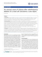
The patient had Ultrasound Scan which showed mass in
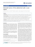
the right Kidney (Figure 1). Her subsequent CT scan as
shown (figure 2) revealed a mass in her right kidney.
She underwent right sided nephrectomy after full staging
procedures and appropriate investigations.
Discussion
Malignant neoplasms involving the kidney may be pri-
mary or secondary tumors. Although metastatic lesions
outnumber primary tumors, secondary renal neoplasms
are usually clinically insignificant and are principally dis-
covered at postmortem examination.
Patients with Renal cell carcinoma (RCC) present with a
range of symptoms, but many are asymptomatic until the
disease is advanced. At presentation, approximately 25
percent of individuals either have distant metastases or
significant local-regional disease. Other patients, even
some with only localized disease, present with a wide
array of symptoms and/or laboratory abnormalities.
Because of this unusual characteristic, RCC has been
labelled the "internist's tumor"[1]. Today, most tumors
are diagnosed incidentally [5,6].
The classic triad of flank pain, hematuria and flank mass
is uncommon (10% cases) and is indicative of advanced
disease. The frequency with which Renal Cell Carcinoma
clinically presents is shown in table 1[2]. The literature
has described several unique clinical presentations of
Published: 6 June 2007
Journal of Medical Case Reports 2007, 1:26 doi:10.1186/1752-1947-1-26
Received: 12 February 2007
Accepted: 6 June 2007
This article is available from: />© 2007 Doshi et al; licensee BioMed Central Ltd.
This is an Open Access article distributed under the terms of the Creative Commons Attribution License ( />),
which permits unrestricted use, distribution, and reproduction in any medium, provided the original work is properly cited.
Journal of Medical Case Reports 2007, 1:26 />Page 2 of 3
(page number not for citation purposes)
Renal Cell Carcinoma in the form of hoarseness or calve-
rial metastasis of which only five cases have been reported
[3,4].
Renal cell carcinoma represents a heterogenous group of
tumors, the most common of which is clear cell adenocar-
cinoma. RCC accounts for 3% of adult tumors. The inci-
dence has increased more than 30% over the past two
decades. It is generally accepted that the increased inci-
dence rates reflect earlier diagnosis at an earlier stage,
largely due to more liberal use of radiological imaging
techniques. However advanced disease has also been diag-
nosed more frequently and mortality has increased as well
[5].
Symptomatic presentation correlates with aggressive his-
tology and advanced disease. Incidental tumours may be
frequently detected in female and elderly patients, as these
groups traditionally seek general medical care more regu-
larly. The mode of presentation can independently predict
an adverse patient outcome. Indicators of symptomatic
presentations include flank pain, flank mass, varicocele,
constitutional symptoms, paraneoplastic syndromes and
bone pain related to metastatic disease [7].
Ultrasound scan was found to be useful screening test, but
CT is the imaging study of choice to identify malignant
features. MRI can be used in equivocal cases [7].
Pre-operative clinical variables may be used instead of the
pathologic stage to determine the risk of recurrence [8].
Conclusion
Renal cell carcinoma presents with various clinical fea-
tures. Atypical presentations of RCC should be considered
for patients presenting with pyrexia of unknown origin.
Competing interests
The author(s) declare that they have no competing inter-
ests.
Authors' contributions
DD wrote the first draft of the manuscript and scanned
photographs for submission. MS proofread the case report
and obtained patient consent. NS performed the literature
search.
Acknowledgements
Written consent was obtained from the patient for publication of the study.
References
1. Gaetani Paolo, Dilene Antonio, Colombo Piergiuseppe, et al.: Calve-
rial metastasis as clinical presentation of Renal Cell Carci-
noma- a report of two cases and review of literature. Clinical
Neurology and Neurosurgery 2005, 107:329-333.
Table 1: Clinical presentation of Renal Cell Carcinoma
Haematuria 40%
Flank pain 40%
Palpable mass 25%
Classic Triad 10%
Weight loss 33%
Fever 20%
Hypertension 20%
Hypercalcemia 5%
Metastasis 30%
This is a picture of renal ultrasoundFigure 1
This is a picture of renal ultrasound. It shows echo-dense
area in the renal area, suggestive of a renal tumour.
RENAL
MASS
Computerised tomography scan image of abdomen showing renal massFigure 2
Computerised tomography scan image of abdomen showing
renal mass.
Renal Mass
Publish with BioMed Central and every
scientist can read your work free of charge
"BioMed Central will be the most significant development for
disseminating the results of biomedical research in our lifetime."
Sir Paul Nurse, Cancer Research UK
Your research papers will be:
available free of charge to the entire biomedical community
peer reviewed and published immediately upon acceptance
cited in PubMed and archived on PubMed Central
yours — you keep the copyright
Submit your manuscript here:
/>BioMedcentral
Journal of Medical Case Reports 2007, 1:26 />Page 3 of 3
(page number not for citation purposes)
2. Greenberg Richard E, Cooper Jeffrey, Knigel Robert L, et al.: Hoarse-
ness, A unique clinical presentation of Renal cell Carcinoma.
Urology 1992, 40:159-161.
3. Kirkali Ziya, Obec Can: Clinical aspects of Renal Cell Carci-
noma. EAU Update Series 2003, 1:89-196.
4. Luciani Lorezo L, Castari Roberto, Tallarigo Carlo: Incidental Renal
Cell Carcinoma- age and stage characterization and clinical
implications; a study of 1092 patients (1982–1997). Urology
2000, 56:58-63.
5. Lee Cheryl T, Katz Jared, Fearn Paul A, Russo Paul: Mode of pres-
entation of Renal Cell Carcinoma- provides prognostic infor-
mation. Urologic Oncology 2000, 7:135-140.
6. Nassir Anmar, Jollimore Jason, Gupta Rekha, Bell David, Norman
Richard: Multilocular cystic Renal Cell Carcinoma; a series of
12 cases and a review of literature. Urology 2002, 60:421-427.
7. Yaycioghi Ozgur, Roberts William W, Chan Theresa, et al.: Prognos-
tic assessment non metastatic Renal Cell Carcinoma – a clin-
ically based model. Urology 2001, 58:141-145.
8. Vincenzo F, Tommaso P, Giovanni N, et al.: Incidental detection
beyond pathological factors as prognostic predictor of Renal
Cell Carcinoma. European Urology 2003, 43:663-669.