Vascular Medicine and Endovascular Interventions - part 3 pot
Bạn đang xem bản rút gọn của tài liệu. Xem và tải ngay bản đầy đủ của tài liệu tại đây (478.17 KB, 34 trang )
59
5
Thrombophilia*
John A. Heit, MD
[Table 5.1]); thrombophilia may also be inherited (Table
5.2). This concept is important because disease susceptibil-
ity does not imply an absolute requirement for primary or
secondary prevention or for treatment. In most persons
with a thrombophilia, thrombosis does not develop. Thus,
thrombophilia(s) must be considered in the context of other
risk factors for incident thrombosis or predictors of recur-
Introduction
Symptomatic thrombosis is caused by dysregulation of the
normal hemostatic response to vessel wall “injury” that
occurs with exposure to a clinical risk factor (e.g., surgery,
trauma, or hospitalization for acute medical illness). Ves-
sel wall injury may be anatomic (e.g., venous endothelial
microtears within vein valve cusps due to stasis or rupture
of a lipid-rich atherosclerotic plaque) or “non-anatomic”
(e.g., cytokine-mediated endothelial expression of adhe-
sion molecules or downregulation of thrombomodulin
expression, related to the “acute infl ammatory response”).
However, the vast majority of persons exposed to a clini-
cal risk factor do not have development of symptomatic
thrombosis. We now recognize that clinical thrombosis is
a multifactorial and complex disease that becomes mani-
fest when a person with an underlying predisposition to
thrombosis (“thrombophilia[s]”) is exposed to additional
risk factors. Emerging evidence suggests that individual
variation in the regulation of the procoagulant, anticoagu-
lant, fi brinolytic, and acute infl ammation or innate immu-
nity pathways most likely accounts for the development
of clinical thrombosis in exposed persons.
Thrombophilia is defi ned as a predisposition to throm-
bosis. Thrombophilia is not a disease per se, but may be as-
sociated with a disease (e.g., cancer), with drug exposure
(e.g., oral contraceptives), or with a specifi c condition (e.g.,
pregnancy or post partum—“acquired thrombophilia”
*Portions of this manuscript have been previously published in Heit
JA. Venous thromboembolism: disease burden, outcomes and risk
factors. J Thromb Haemost. 2005;3:1611-7. Used with permission.
Supported in part by research grants HL66216, HL83141, HL83797,
and RR19457 from the National Institutes of Health and research
grant TS1255 from the Centers for Disease Control and Prevention,
United States Public Health Service; and by Mayo Foundation.
© 2007 Society for Vascular Medicine and Biology
Table 5.1 Acquired or Secondary Thrombophilia
Defi nite causes of thrombophilia
Active malignant neoplasm
Chemotherapy (
L-asparaginase, thalidomide, antiangiogenesis therapy)
Myeloproliferative disorders
Heparin-induced thrombocytopenia and thrombosis
Nephrotic syndrome
Intravascular coagulation and fi brinolysis/disseminated intravascular
coagulation
Thrombotic thrombocytopenic purpura
Sickle cell disease
Oral contraceptives
Estrogen therapy
Pregnancy/postpartum state
Tamoxifen and raloxifene therapy (selective estrogen receptor modulator)
Antiphospholipid antibodies (lupus anticoagulant, anticardiolipin antibody,
anti–β2-glycoprotein-1 antibody)
Paroxysmal nocturnal hemoglobinuria
Wegener granulomatosis
Probable causes of thrombophilia
Infl ammatory bowel disease
Thromboangiitis obliterans (Buerger disease)
Behçet syndrome
Varicose veins
Systemic lupus erythematosus
Venous vascular anomalies (Klippel-Trénaunay syndrome)
Progesterone therapy
Infertility treatments
Hyperhomocysteinemia
Human immunodefi ciency virus infection
Dehydration
Vascular Medicine and Endovascular Interventions
60
most clinical studies have failed to show a consistent asso-
ciation between thrombophilia and myocardial infarction
or stroke.
Unfortunately, no single laboratory assay or simple set
of assays can identify all thrombophilias. Consequently,
a battery of complex and potentially expensive assays is
usually required. Many of the analytes measured in the
laboratory are affected by other conditions (e.g., warfa-
rin decreases protein C and S levels), such that the cor-
rect interpretation of the results can be complicated and
always requires clinical correlation. Detailed descriptions
of the known thrombophilias are included in this chapter,
as well as information on special coagulation laboratory
interpretation.
• The predominant clinical manifestation of throm-
bophilia is VTE
Indications for Thrombophilia Testing:
Why Should I Test for Thrombophilia?
There are no absolute indications for clinical diagnostic
thrombophilia testing. Potential relative indications could
include the following:
– Selected screening of populations that are potentially
“enriched” for thrombophilia (e.g., asymptomatic or
symptomatic family members of patients with a known
familial thrombophilia, especially fi rst-degree relatives);
– Selected screening of populations at increased risk for
thrombosis (e.g., before pregnancy, use of oral contracep-
tion or estrogen therapy, before high-risk surgery, or dur-
ing chemotherapy with angiogenesis inhibitors);
– Testing of symptomatic patients with an incident throm-
botic event (e.g., incident VTE, stillbirth or another com-
rent thrombosis, when estimating the need for primary or
secondary prophylaxis, respectively. With rare exceptions,
the therapy for acute thrombosis is no different for persons
with and without a recognized thrombophilia.
• Thrombophilia is a predisposition or susceptibility to
thrombosis
• Thrombophilia is not a disease, but may be associated
with a disease, drug exposure, or a condition, or may be
inherited
• When estimating the need for primary or secondary
prophylaxis, thrombophilia(s) must be considered in
the context of other risk factors for incident thrombosis
or predictors of recurrent thrombosis, respectively
• With rare exceptions, therapy for acute thrombosis is the
same for those with and without a recognized throm-
bophilia
Thrombophilia may present clinically as one or more of
several thrombotic manifestations or “phenotypes” (Table
5.3). The predominant clinical manifestation of throm-
bophilia is venous thromboembolism (VTE). Although it
is biologically plausible to hypothesize that patients with
atherosclerotic arterial occlusive disease and an underly-
ing thrombophilia who have an atherosclerotic plaque
rupture are more likely to have a symptomatic thrombosis,
Table 5.2 Hereditary (Familial or Primary) Thrombophilia
Defi nite causes of thrombophilia
Antithrombin III defi ciency
Protein C defi ciency
Protein S defi ciency
Activated protein C resistance
Factor V Leiden mutation
Prothrombin G20210A mutation
Homocystinuria
Probable causes of thrombophilia
Increased plasma factors I (fi brinogen), II (prothrombin), VIII, IX, and XI
Hyperhomocysteinemia
Dysfi brinogenemia
Hypoplasminogenemia and dysplasminogenemia
Hypofi brinolysis
Reduced protein Z and Z-dependent protease inhibitor
Reduced tissue factor pathway inhibitor
Possible causes of thrombophilia
Tissue plasminogen activator defi ciency
Increased plasminogen activator inhibitor levels
Methylene tetrahydrofolate reductase polymorphisms
Factor XIII polymorphisms
Increased thrombin-activatable fi brinolysis inhibitor
Table 5.3 Clinical Manifestations of Thrombophilia
Defi nite manifestations
Purpura fulminans (neonatalis or adult)
Superfi cial or deep vein thrombosis, pulmonary embolism
Thrombosis of “unusual” venous circulations (cerebral, hepatic, mesenteric,
and renal veins; possibly arm, portal, and ovarian veins; not retinal vein
or artery)
Warfarin-induced skin necrosis
Possible manifestations
Arterial thrombosis (stroke, acute myocardial infarction)
(Recurrent) fetal loss
Intrauterine growth restriction
Stillbirth
Severe gestational hypertension (preeclampsia)
Abruptio placentae
CHAPTER 5 Thrombophilia
61
plication of pregnancy, incident arterial thrombosis in a
young person without other arterial disease);
– Testing of symptomatic patients with recurrent throm-
bosis, “idiopathic” thrombosis, thrombosis at a young age
(e.g., ≤40 years for venous thrombosis, ≤50 years for arteri-
al thrombosis) or thrombosis in unusual vascular territory
(e.g., cerebral vein, portal vein, hepatic vein, mesenteric
vein, or artery).
With the exception of general population screening,
which is not recommended, all of these potential indica-
tions are controversial and must be considered in the con-
text of the clinical presentation.
Counseling and Screening Asymptomatic Family
Members
Thrombophilia testing, especially genetic testing, of
asymptomatic family members should be done with cau-
tion. Family members (and patients) should receive genetic
counseling before genetic testing, and such testing should
only be performed after obtaining consent. Counseling
should include the reasons for testing, such as the potential
for avoiding clinical thrombosis by risk factor modifi cation
or prophylaxis (both for the family member as well as his
or her children), and the reasons for not testing, such as
stigmatization and mental anguish, the potential effect on
obtaining personal health insurance or employment, and
the possibility of non-paternity.
Thrombophilia testing should only be done if the re-
sults are likely to change medical management. The risk
of idiopathic (“unprovoked”) thrombosis associated
with a thrombophilia, although increased, is still insuffi -
cient to warrant chronic primary prophylaxis (e.g., war-
farin anticoagulation therapy), even for thrombophilias
with high penetrance (e.g., antithrombin defi ciency or
homozygous factor V Leiden carriers) with the possible
exception of paroxysmal nocturnal hemoglobinuria.
Thus, primary “prophylaxis” typically involves either
avoidance or modifi cation of risk exposure or specifi c
prophylactic measures if such exposures are unavoid-
able.
When counseling a family member (or patient) regard-
ing the risk of thrombosis associated with a thrombophilia,
it is most useful to provide the “absolute” risk or incidence
of thrombosis among persons with that particular throm-
bophilia. For example, the relative risk of VTE among
women who are factor V Leiden carriers and using oral
contraceptives is increased about 30-fold; however, the
incidence is only about 300 per 100,000 woman-years, or
about 0.3% per woman-year. Thus, the absolute risk pro-
vides information on both the baseline risk of VTE (about
10-46 per 100,000 woman-years for women of reproduc-
tive age) as well as the relative risk (about 30 for female
factor V Leiden carriers using oral contraceptives).
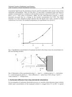
Estimation of the absolute risk of thrombosis should
especially account for the effect of age on the baseline inci-
dence of VTE. For example, among women of perimeno-
pausal age (50-54 years), the incidence of VTE is 123 per
100,000 woman-years, which increases exponentially with
increasing age (Fig. 5.1). Among female factor V Leiden
carriers of perimenopausal age, the relative risk of VTE
associated with hormone therapy may be increased 7- to
15-fold. However, whereas the relative risk for VTE is less
with the use of hormone therapy than with use of oral con-
traceptives, the absolute risk is substantially higher (≈900-
1,800 per 100,000 woman-years, ≈1%-2% per woman-year)
because of the increased incidence with age. Given recent
studies questioning the benefi t of postmenopausal hor-
mone therapy, most women likely would choose to avoid
such therapy if they were known to be factor V Leiden car-
Fig. 5.1 Annual incidence of venous
thromboembolism by age and sex. (From
Silverstein MD, Heit JA, Mohr DN, et al. Trends
in the incidence of deep vein thrombosis and
pulmonary embolism: a 25-year population-
based study. Arch Intern Med. 1998;158:585-
93. Used with permission.)
Vascular Medicine and Endovascular Interventions
62
riers. Thus, it may be relatively cost-effective to perform
thrombophilia screening of an asymptomatic perimeno-
pausal or postmenopausal woman with a known family
history of thrombophilia who is considering hormone
therapy.
Primary Prevention of Incident VTE
Primary prevention of VTE, either by risk factor modifi ca-
tion or by appropriate prophylaxis for patients at risk, is
essential to improve survival and prevent complications.
However, despite improved prophylaxis regimens and
more widespread use of prophylaxis, the overall incidence
of VTE has been relatively constant at about 1 per 1,000
since 1979.
To avoid or modify risk or appropriately target prophy-
laxis, patients at risk for VTE must fi rst be identifi ed. In
the absence of a central venous catheter or active cancer,
the incidence of VTE among children and adolescents
is very low (<1 per 100,000 for age ≤15 years). The inci-
dence increases exponentially after age 50 years, eventu-
ally reaching about 1,000 per 100,000 for persons aged 85
years or older. The incidence of VTE increases signifi cantly
with age for both idiopathic and secondary VTE, which
suggests that the risk associated with advancing age may
be due to the biology of aging rather than simply to an
increased exposure to VTE risk factors with advancing
age. The incidence is slightly higher for women during
childbearing years and for men after age 50 years. The in-
cidence of VTE also varies by race. Compared with white
Americans, black Americans have a 30% higher incidence
and Asian and Native Americans have up to a 70% lower
incidence; Hispanics have an incidence intermediate be-
tween whites and Asian Americans.
Additional independent risk factors for VTE are shown
in Table 5.4. Compared with persons in the community,
hospitalized patients have a greater than 150-fold in-
creased incidence of acute VTE. The population-attribut-
able risk provides an estimate of the burden of disease
in the community that is attributable to a particular risk
factor. For example, the risk factors of hospitalization and
nursing home residence together account for almost 60%
of all incident VTE events occurring in the community.
Thus, hospital confi nement provides an important oppor-
tunity to substantially decrease VTE incidence. Of note,
hospitalization for medical illness and surgery account for
almost equal proportions of VTE (22% and 24%, respec-
tively), which emphasizes the need to provide prophylaxis
to both of these risk groups.
• Primary “prophylaxis” involves either avoidance or
modifi cation of risk exposure or specifi c prophylactic
measures if such exposures are unavoidable
• Compared with persons in the community, hospitalized
patients have a greater than 150-fold increased incidence
of acute VTE
• Hospitalization for medical illness and surgery account
for almost equal proportions of VTE (22% and 24%,
respectively), which emphasizes the need to provide
prophylaxis to both of these risk groups
The risk among surgical patients can be further stratifi ed
on the basis of patient age, type of surgery, and presence
of active cancer. The incidence of postoperative VTE is
increased for surgical patients aged 65 years and older.
High-risk surgical procedures include neurosurgery,
major orthopedic surgery of the leg, renal transplantation,
cardiovascular surgery, and thoracic, abdominal, or pelvic
surgery for malignancy. After controlling for age, type of
surgery, and cancer, additional independent risk factors
for incident VTE after major surgery include increasing
body mass index, intensive care unit confi nement for more
than 6 days, immobility, infection, and varicose veins. The
risk from surgery may be less with neuraxial (spinal or
epidural) anesthesia than with general anesthesia.
Independent risk factors for incident VTE among pa-
tients hospitalized for acute medical illness include in-
creasing age and body mass index, active cancer, neuro-
logic disease with extremity paresis, immobility, fracture,
and prior superfi cial vein thrombosis. Active cancer ac-
counts for almost 20% of incident VTE events occurring
in the community. VTE risk among patients with active
cancer can be further stratifi ed by tumor site, presence of
distant metastases, and active chemotherapy. Although all
of these patients are at risk, the risk appears to be higher
for pancreatic cancer, lymphoma, malignant brain tumors,
Table 5.4 Independent Risk Factors for Deep Vein Thrombosis or
Pulmonary Embolism
Baseline characteristic
Odds
ratio
95% Confi dence
interval
Hospitalization
For acute medical illness 7.98 4.49-14.18
For major surgery 21.72 9.44-49.93
Trauma 12.69 4.06-39.66
Malignancy
Without chemotherapy 4.05 1.93-8.52
With chemotherapy 6.53 2.11-20.23
Prior central venous catheter or transvenous
pacemaker
5.55 1.57-19.58
Prior superfi cial vein thrombosis 4.32 1.76-10.61
Neurologic disease with extremity paresis 3.04 1.25-7.38
Serious liver disease 0.10 0.01-0.71
From Heit JA, Silverstein MD, Mohr DN, et al. Risk factors for deep vein
thrombosis and pulmonary embolism: a population-based case-control
study. Arch Intern Med. 2000;160:809-15. Used with permission.
CHAPTER 5 Thrombophilia
63
cancer of the liver, leukemia, and colorectal and other di-
gestive cancers, and for patients with distant metastases.
Those receiving immunosuppressive or cytotoxic chemo-
therapy, such as
L-asparaginase, thalidomide, angiogen-
esis inhibitors, tamoxifen, and erythropoietin, are at even
higher risk for VTE.
Patients with a central venous catheter or transvenous
pacemaker now account for about 9% of those with inci-
dent VTE in the community. However, warfarin and low-
molecular-weight heparin (LMWH) prophylaxis are not
effective in preventing catheter-induced venous throm-
bosis and are not recommended. Prior superfi cial vein
thrombosis is an independent risk factor for subsequent
deep vein thrombosis (DVT) or pulmonary embolism (PE)
remote from the episode of superfi cial thrombophlebitis.
The risk of DVT imparted by varicose veins is uncertain
and appears to be higher among persons younger than 40
years. Long-haul air travel (>6 hours) is associated with a
slightly increased risk for VTE, which is preventable with
elastic stockings. Studies regarding the protective effect of
coenzyme A reductase inhibitor (statin) therapy against
VTE have provided confl icting results. In addition, the
risk associated with atherosclerosis, or other risk factors
for atherosclerosis such as diabetes mellitus, remains un-
certain. Body mass index, current or past tobacco smok-
ing, chronic obstructive pulmonary disease, and renal
failure are not independent risk factors for VTE. The risk
associated with congestive heart failure, independent of
hospitalization, is low.
Among women, additional risk factors for VTE include
oral contraceptive use and hormone therapy, pregnancy,
and the postpartum period. The greatest risk may occur
during early use of oral contraceptives and hormone
therapy. This risk may be lower for second-generation
oral contraceptives or progesterone alone compared with
fi rst- or third-generation oral contraceptives. For women
with disabling perimenopausal symptoms that cannot be
controlled with non-estrogen therapy, esterifi ed oral estro-
gen or transdermal estrogen therapy may confer less risk
than oral conjugated equine estrogen therapy. Although
VTE can occur anytime during pregnancy, the highest in-
cidence is during the fi rst 2 postpartum weeks, especially
for older mothers. Independent risk factors for pregnancy-
associated VTE include tobacco smoking and prior super-
fi cial vein thrombosis. Women receiving therapy with the
selective estrogen receptor modulators tamoxifen and
raloxifene also are at increased risk for VTE.
Recent family-based studies indicate that VTE is highly
heritable and follows a complex mode of inheritance in-
volving environmental interaction. Inherited decreases in
natural plasma anticoagulants (antithrombin III, protein
C, or protein S) have long been recognized as uncommon
but potent risk factors for VTE. More recent fi ndings of
other decreased natural anticoagulants or anticoagulant
cofactors, impaired downregulation of the procoagulant
system (e.g., activated protein C resistance, factor V Lei-
den), increased plasma concentrations of procoagulant
factors (e.g., factors I [fi brinogen], II [prothrombin], VIII,
IX, and XI), increased basal procoagulant activity, im-
paired fi brinolysis, and increased basal innate immunity
activity and reactivity have added to the list of inherited
or acquired disorders predisposing persons to thrombosis.
These plasma hemostasis-related factors or markers of co-
agulation activation correlate with increased thrombotic
risk and are highly heritable. Inherited thrombophilias
interact with such clinical risk factors as oral contracep-
tives, pregnancy, hormone therapy, surgery, and cancer
to compound the risk of incident VTE. Similarly, genetic
interaction increases the risk of incident VTE. Thus, it may
be reasonable to consider thrombophilia testing of asymp-
tomatic family members with a known history of familial
thrombophilia.
Secondary Prevention of Recurrent VTE
VTE recurs frequently; about 30% of patients have recur-
rence within ten years (Table 5.5). A recent modeling study
suggested that more than 900,000 incident or recurrent VTE
events occurred in the United States in 2002. The hazard
of recurrence varies with the time since the incident event
and is highest within the fi rst 6 to 12 months. Additional
independent predictors of recurrence include male sex,
increasing patient age and body mass index, neurologic
disease with extremity paresis, and active malignancy
(Table 5.6). Other predictors of recurrence include: “idio-
pathic” VTE; a persistent lupus anticoagulant or high-titer
antiphospholipid antibody; antithrombin, protein C, or
protein S defi ciency; compound heterozygous carriers for
Table 5.5 Cumulative Incidence and Hazard of Venous Thromboembolism
Recurrence
Venous thromboembolism recurrence
Time to
recurrence
Cumulative recurrence,
%
Hazard of recurrence,
per 1,000 person-days (SD)
0 days 0.0 0
7 days 1.6 170 (30)
30 days 5.2 130 (20)
90 days 8.3 30 (5)
180 days 10.1 20 (4)
1 year 12.9 20 (2)
2 years 16.6 10 (1)
5 years 22.8 6 (1)
10 years 30.4 5 (1)
Modifi ed from Heit JA, Mohr DN, Silverstein MD, et al. Predictors of
recurrence after deep vein thrombosis and pulmonary embolism: a
population-based cohort study. Arch Intern Med. 2000;160:761-8.
Used with permission.
Vascular Medicine and Endovascular Interventions
64
more than one familial thrombophilia (e.g., heterozygous
for the factor V Leiden and prothrombin G20210A muta-
tions) or homozygous carriers; decreased tissue-factor
pathway inhibitor levels; persistent residual DVT; and
possibly increased procoagulant factor VIII and factor IX
levels.
• VTE recurs frequently; about 30% of patients have re-
currence within 10 years
Data regarding the risk of recurrent VTE among isolated
heterozygous carriers of either the factor V Leiden or the
prothrombin G20210A mutation are confl icting. In a recent
meta-analysis that pooled results from ten studies involv-
ing 3,104 patients with incident VTE, the factor V Leiden
mutation was present in 21.4% of patients and was asso-
ciated with an increased risk of recurrent VTE. Similarly,
pooled results from nine studies involving 2,903 patients
showed that the prothrombin G20210A mutation was
present in 9.7% and was associated with an increased risk
of recurrence. The estimated population-attributable risk
of recurrence was 9.0% and 6.7% for the factor V Leiden
and the prothrombin G20210A mutations, respectively.
An increased D-dimer level measured at least 1 month
after discontinuing warfarin therapy may be a predictor of
DVT recurrence independent of residual venous obstruc-
tion. Secondary prophylaxis with anticoagulation therapy
should be considered for patients with these characteris-
tics. Although the incident event type (DVT alone vs PE) is
not a predictor of recurrence, any recurrence is signifi cant-
ly more likely to be the same as the incident event type.
Because the 7-day case fatality rate is signifi cantly higher
for recurrent PE (34%) than for recurrent DVT alone (4%),
secondary prophylaxis should be considered for incident
PE, especially for patients with chronically reduced cardio-
pulmonary functional reserve.
Diagnostic Thrombophilia Testing: Who
Should Be Tested?
Current Recommendations
Currently recommended indications for thrombophilia
testing include idiopathic or recurrent VTE; a fi rst episode
of VTE at a “young” age (≤40 years); a family history of
VTE (in particular, a fi rst-degree relative with throm-
bosis at a young age); venous thrombosis in an unusual
vascular territory (e.g., cerebral, hepatic, mesenteric, or
renal vein thrombosis); and neonatal purpura fulminans
or warfarin-induced skin necrosis. If two or more of these
thrombosis characteristics are present, the prevalence of
antithrombin, protein C, or protein S defi ciency, and the
factor V Leiden and prothrombin G20210A mutations are
increased. Consequently, a “complete” laboratory inves-
tigation (described below) is recommended for patients
who meet these criteria, whereas more selective testing
(e.g., for activated protein C resistance and factor V Leiden
and prothrombin G20210A mutations) is recommended
for other patients.
The prevalence of hereditary thrombophilia is substantial
among patients with a fi rst VTE (Table 5.7). Because knowl-
edge of a hereditary thrombophilia may be important for
estimating the risk of VTE recurrence, testing for a heredi-
tary thrombophilia should be considered for patients with
a fi rst thrombosis, not limited to patients with recurrent
VTE. Moreover, although patients with idiopathic or recur-
rent VTE may have a higher prevalence of a recognized
thrombophilia, these patients should be considered for
secondary prophylaxis regardless of the results of throm-
bophilia testing. In addition, the prevalence of hereditary
thrombophilia, such as activated protein C resistance, is
substantial among patients with a fi rst episode of VTE at
an older age (Table 5.8). Therefore, testing for a hereditary
thrombophilia should not be limited to patients with a fi rst
episode of VTE before age 40 to 50 years. Although persons
with defi ciency of antithrombin III, protein C, or protein S
are more likely to have thrombosis at a younger age, genetic
interaction (e.g., factor V Leiden or prothrombin G20210A
mutation combined with either antithrombin, protein C,
or protein S defi ciency) compounds the risk of thrombosis
such that testing among older patients should not be lim-
ited to activated protein C resistance (factor V Leiden) or
the prothrombin G20210A mutation.
• Currently recommended indications for thrombophilia
testing include idiopathic or recurrent VTE; a fi rst epi-
sode of VTE at a “young” age (≤40 years); a family his-
tory of VTE (in particular, a fi rst-degree relative with
thrombosis at a young age); venous thrombosis in an
unusual vascular territory (e.g., cerebral, hepatic, me-
Table 5.6 Independent Predictors of Venous Thromboembolism
Recurrence
Characteristic Hazard ratio
95% Confi dence
interval
Age
*
1.17 1.11-1.24
Body mass index
†
1.24 1.04-1.47
Neurologic disease with extremity
paresis
1.87 1.28-2.73
Active malignancy
With chemotherapy 4.24 2.58-6.95
Without chemotherapy 2.21 1.60-3.06
*
Per decade increase in age.
†
Per 10 kg/m
2
increase in body mass index.
From Heit JA, Mohr DN, Silverstein MD, et al. Predictors of recurrence after
deep vein thrombosis and pulmonary embolism: a population-based cohort
study. Arch Intern Med. 2000;160:761-8. Used with permission.
CHAPTER 5 Thrombophilia
65
senteric, or renal vein thrombosis); and neonatal pur-
pura fulminans or warfarin-induced skin necrosis
• The prevalence of hereditary thrombophilia is substan-
tial among patients with a fi rst VTE
Recent evidence suggests that a family history of VTE does
not increase the likelihood of a recognized familial throm-
bophilia. The cumulative lifetime incidence (penetrance)
of thrombosis among carriers of the most common familial
thrombophilia (factor V Leiden) is only about 10%. There-
fore, most patients with an inherited thrombophilia do not
have a family history of thrombosis. Consequently, throm-
bophilia testing should not be limited to symptomatic pa-
tients with a family history of VTE.
The most common presentations of familial throm-
bophilia are DVT of the leg veins and PE. Except for cath-
eter-induced thrombosis, all VTE is most likely associated
with an underlying thrombophilia. Therefore, limiting
testing of patients with thrombosis in unusual vascular
territories will miss most patients with an identifi able fa-
milial (or acquired) thrombophilia.
Several additional issues should be considered regard-
ing testing for a possible thrombophilia. For example, the
prevalence of hereditary thrombophilia among patients
with VTE differs substantially by ethnic ancestry, but
variable testing based on ethnic ancestry has not been
directly addressed. The factor V Leiden and prothrombin
20210 mutation carrier frequencies among asymptomatic
African, Asian, and Native Americans, as well as African
Americans with VTE, are extremely low, such that test se-
lection for hereditary thrombophilia most likely should be
tailored to patient ethnic ancestry. The risk of VTE during
pregnancy or the postpartum period, and the risk of recur-
rent fetal loss, is increased among patients with acquired
or hereditary thrombophilia. Women with VTE during
pregnancy or post partum or recurrent fetal loss should be
tested. Recent evidence suggests that patients with acute
VTE in the presence of active malignancy are also more
likely to have an underlying thrombophilia (e.g., factor V
Leiden). Nevertheless, thrombophilia testing for patients
with thrombosis associated with active cancer or another
Table 5.7 Familial or Acquired Thrombophilia: Estimated Prevalence, and Incidence and Relative Risk of Incident or Recurrent VTE by Type of Thrombophilia
Prevalence, %* Incident VTE Recurrent VTE
Thrombophilia Normal
Incident
VTE
Recurrent
VTE
Incidence
†
(95% CI)
Relative risk
(95% CI)
Incidence
†
(95% CI)
Relative risk
(95% CI)
Antithrombin III defi ciency 0.02-0.04 1-2 2-5 500 (320-730) 17.5 (9.1-33.8) 10,500 (3,800-23,000) 2.5
Protein C defi ciency 0.02-0.05 2-5 5-10 310 (530-930) 11.3 (5.7-22.3) 5,100 (2,500-9,400) 2.5
Protein S defi ciency 0.01-1 1-3 5-10 710 (530-930) 32.4 (16.7-62.9) 6,500 (2,800-11,800) 2.5
Factor V Leiden
‡
3-7 12-20 30-50 150 (80-260) 4.3
§
(1.9-9.7) 3,500 (1,900-6,100) 1.3 (1.0-3.3)
Prothrombin G20210A
‡
1-3 3-8 15-20 350 1.9 (0.9-4.1) … 1.4 (0.9-2.0)
Combined thrombophilias … … … 840 (560-1,220) 32.4 (16.7-62.9) 5,000 (2,000-10,300) …
Hyperhomocysteinemia … … … … … … 2.5
Antiphospholipid Ab … … … … … … 2.5
Factor VIII (>200 IU/dL) … … … … … … 1.8 (1.0-3.3)
Ab, antibody; CI, confi dence interval; VTE, venous thromboembolism.
*In whites.
†
Per 100,000 person-years.
‡
Heterozygous carriers.
§
Homozygous carriers, relative risk=80.
Table 5.8 Age-Specifi c Annual Incidence and Relative Risk of First Lifetime
VTE Among Factor V Leiden Carriers
VTE
Study Age, y
Incidence*
(95% CI)
Relative risk
†
(95% CI)
Ridker et al 1997 40-49
50-59
60-69
≥70
0
197 (72-428)
258 (95-561)
783 (358-1,486)
…
2.7
2.7
4.2
Middeldorp et al 1998 15-30
31-45
46-60
>60
250 (120-490)
470 (230-860)
820 (350-1,610)
1,100 (240-3,330)
≈15
4.3
2.4
2.8
Simioni et al 1999 <15
16-30
31-45
46-60
>60
0
182 (59-424)
264 (86-616)
380 (104-973)
730 (150-2,128)
…
3.6
3.7
1.4
≈700
Heit et al 2005 15-29
30-44
45-59
≥60
0 (0-112)
61 (7-219)
244 (98-502)
764 (428-1,260)
0 (0-1.22)
1.96 (0.24-7.04)
1.73 (0.70-3.56)
3.61 (2.02-5.95)
CI, confi dence interval; VTE, venous thromboembolism.
*VTE incidence per 100,000 person-years.
†
Compared with factor V Leiden non-carriers.
Vascular Medicine and Endovascular Interventions
66
risk exposure (e.g., surgery, hospitalization for acute
medical illness, trauma, neurologic disease with extrem-
ity paresis, or upper extremity thrombosis in the presence
of a central venous catheter or transvenous pacemaker) is
controversial.
Suggested Revised Recommendations
All patients with VTE—regardless of age, sex, race, loca-
tion of venous thrombosis, initial or recurrent event, or
family history of VTE—should be tested for an acquired
or hereditary thrombophilia. Women with recurrent fetal
loss or complications of pregnancy and patients with un-
explained arterial thrombosis also should be tested. These
recommendations are controversial and not universally
accepted.
• Most patients with an inherited thrombophilia do not
have a family history of thrombosis
• Women with VTE during pregnancy or post partum or
recurrent fetal loss should be tested
Diagnostic Thrombophilia Testing: For
What Should I Test?
General Testing
A complete history and physical examination is manda-
tory in evaluating persons with a recent or remote history
of thrombosis, with special attention given to age of onset,
location of prior thromboses, and results of objective di-
agnostic studies documenting thrombotic episodes. An
inquiry regarding interval imaging to establish a new
baseline image is particularly important when diagnosing
recurrent thrombosis in the same vascular territory as a
previous thrombosis. For patients with an uncorroborated
history of DVT, non-invasive venous vascular laboratory
or venous duplex ultrasonographic evidence of venous
outfl ow obstruction (e.g., residual vein thrombosis) or
possibly venous valvular incompetence may be helpful in
corroborating the clinical history.
Patients should be questioned carefully about diseases,
exposures, conditions, or drugs that are associated with
thrombosis (Tables 5.1 and 5.4). A family history of throm-
bosis may provide insight for a potential familial throm-
bophilia, especially in fi rst-degree relatives. Thrombosis
can be the initial manifestation of a malignancy, so a com-
plete review of systems directed at symptoms of occult
malignancy is important, including whether indicated
screening tests for normal health maintenance (e.g., mam-
mography, colon imaging) are current. Ethnic background
should be considered given the extremely low prevalence
of the factor V Leiden and prothrombin G20210A muta-
tions in those of African, Asian, or Native American an-
cestry.
The physical examination should include a careful pe-
ripheral pulse examination as well as examination of the
extremities for signs of superfi cial or deep vein thrombosis
and vascular anomalies. The skin should be examined for
venous stasis syndrome (e.g., leg swelling, stasis pigmen-
tation or dermatitis, or stasis ulcer), varicose veins, and
livedo reticularis, skin infarction, or other evidence of
microcirculatory occlusive disease. Given the strong asso-
ciation of thrombosis with active cancer, a careful exami-
nation for lymphadenopathy, hepatosplenomegaly, and
abdominal or rectal mass should be performed, as well as
breast and pelvic examinations for women, and testicular
and prostate examinations for men.
The laboratory evaluation for patients with thrombosis
should be selective and based on the history and physical
examination (Table 5.9). Specifi c tests may include a com-
plete blood count with peripheral smear, serum protein
electrophoresis, serum chemistries for electrolytes and
liver and renal function, prostate specifi c antigen, carci-
noembryonic antigen, α-fetoprotein, β-human chorionic
gonadotropin, cancer antigen 125, antinuclear antibodies
(double-stranded DNA, rheumatoid factor, extractable
nuclear antigen), and urinalysis. Elevations in hematocrit
or platelet count may indicate the presence of a myelo-
proliferative disorder, which can be associated with either
venous or arterial thrombosis. Secondary polycythemia
can also provide evidence of an underlying occult malig-
nancy. Leukopenia and thrombocytopenia can be found in
paroxysmal nocturnal hemoglobinuria, which is charac-
terized by intravascular hemolysis along with thrombotic
sequelae. The development of thrombosis and thrombo-
cytopenia concurrent with heparin administration should
always prompt consideration of heparin-induced throm-
bocytopenia. The peripheral smear should be reviewed
for evidence of red cell fragmentation that would indicate
microangiopathic hemolytic anemia such as occurs with
intravascular coagulation and fi brinolysis. In patients
with malignancy, chronic intravascular coagulation and
fi brinolysis can result in either venous or arterial thrombo-
sis. A leukoerythroblastic picture with nucleated red cells
or immature white cells suggests the possibility of marrow
infi ltration by tumor.
Chest radiography should be performed along with ap-
propriate imaging studies according to standard health
maintenance guidelines (e.g., Papanicolaou test, mam-
mography, colon imaging). More detailed imaging, such
as angiography or chest, abdominal, or pelvic computed
tomography or magnetic resonance imaging should be
performed only if other independent reasons exist to sus-
pect an occult malignancy or other arterial disease (in the
case of arterial thrombosis). Routine screening for occult
cancer in patients presenting with idiopathic VTE has not
CHAPTER 5 Thrombophilia
67
been shown to improve cancer-related survival and is not
warranted in the absence of clinical features and abnormal
basic laboratory fi ndings suggestive of underlying malig-
nancy. Sputum cytology, an otolaryngologic examination,
and upper gastrointestinal tract endoscopy should be con-
sidered for tobacco smokers or others at risk for esopha-
geal or gastric cancer. In addition to a Papanicolaou test,
endometrial sampling should be considered for women at
risk of endometrial cancer.
Recommended assays for initial and refl ex special co-
agulation testing for a familial or acquired thrombophilia
are provided in Table 5.9. Detailed discussions regarding
the interpretation and nuances of specifi c assays are pro-
vided with the description of the biochemistry, molecular
biology, and epidemiology of each thrombophilia at the
end of this chapter.
• Routine screening for occult cancer in patients present-
ing with idiopathic VTE has not been shown to improve
cancer-related survival and is not warranted in the ab-
sence of clinical features and abnormal basic laboratory
fi ndings
Arterial Thrombosis
Familial or acquired thrombophilia appears to be an un-
usual cause of stroke, myocardial infarction, or other organ
or skin infarction, except in the presence of antiphospholi-
pid antibodies (e.g., lupus anticoagulant, anticardiolipin
antibody, anti–β2-glycoprotein-1 antibodies), heparin-in-
duced thrombocytopenia, myeloproliferative disorders,
homocystinuria, and possibly hyperhomocysteinemia. A
young patient with organ or skin infarction in the absence
of one of the above disorders or risk factors for athero-
sclerosis (e.g., diabetes mellitus, hypertension, hyperlipi-
demia, tobacco exposure) or cardioembolism (e.g., cardiac
arrhythmia), should be carefully evaluated for occult arte-
rial disease (Table 5.10).
Organ infarction should not be deemed to be caused by
a “hypercoagulable disorder” simply because the patient
is young or lacks common risk factors for atherosclerosis
or arterial thromboembolism. A detailed inquiry into con-
stitutional or specifi c symptoms of vasculitis (primary or
secondary), infection (systemic [e.g., endocarditis] or local
[e.g., infected aneurysm with artery-to-artery embolism]),
atheroembolism, trauma (accidental, thermal, or occu-
pational), dissection, vasospasm, or vascular anomaly is
required. Pulse should also be carefully examined, includ-
ing an examination for aneurysmal disease. Evidence of
microcirculatory occlusive disease of the hand, such as
livedo, skin or nailbed infarction, or ulcer, should prompt
Table 5.9 Laboratory Evaluation for Suspected Familial or Acquired
Thrombophilia*
General
Blood: CBC, peripheral smear, ESR, chemistries, PSA, β-HCG, CA 125, ANA
(dsDNA, rheumatoid factor, ENA)
PA/lateral chest radiography, urinalysis, mammography
Colon imaging, especially if no prior screening (proctosigmoidoscopy,
colonoscopy)
Chest imaging for smokers (CT, MRI)
Otolaryngology consultation, especially for smokers
UGI/upper endoscopy
Abdominal imaging (CT, MRI, ultrasonography)
Angiography
Special coagulation laboratory testing
Platelets
HITTS testing: plasma anti-PF4/glycosaminoglycan (heparin) antibodies
ELISA; platelet
14
C-serotonin release assay; heparin-dependent platelet
aggregation
Plasma coagulation
Prothrombin time, aPTT (with phospholipid “mixing” procedure if inhibited)
Thrombin time/reptilase time
Dilute Russell viper venom time (with confi rm procedures)
Mixing studies (inhibitors)
Specifi c factor assays (as indicated)
Fibrinolytic system
Fibrinogen
Plasma fi brin D-dimer
Soluble fi brin monomer complex
Natural anticoagulation system
Antithrombin (activity, antigen)
Protein C (activity, antigen)
Protein S (activity, total and free antigen)
APC-resistance ratio (second generation; factor V–defi cient plasma
mixing study)
Direct genomic DNA mutation testing
Factor V Leiden gene (depending on the result of the APC-resistance ratio)
Prothrombin G20210A
Additional general testing
Anticardiolipin (antiphospholipid) antibodies (IgG and IgM isotypes);
anti–β2-glycoprotein-1 antibodies
Plasma homocysteine (basal, postmethionine load)
Additional selective testing
Flow cytometry for PNH
Plasma ADAMTS-13 activity
Plasminogen (activity)
ANA, antinuclear antibody; APC, activated protein C; aPTT, activated
partial thromboplastin time; β-HCG, β-human chorionic gonadotropin;
CA 125, cancer antigen 125; CBC, complete blood count; CT, computed
tomography; dsDNA, double-stranded DNA; ELISA, enzyme-linked
immunosorbent assay; ENA, extractable nuclear antigen; ESR, erythrocyte
sedimentation rate; HITTS, heparin-induced thrombotic thrombocytopenia
syndrome; MRI, magnetic resonance imaging; PA, posteroanterior; PF4,
platelet factor 4; PNH, paroxysmal nocturnal hemoglobinuria; PSA,
prostate-specifi c antigen; UGI, upper gastrointestinal tract series.
*
Suggested tests that should be performed selectively based on clinical
judgment.
Vascular Medicine and Endovascular Interventions
68
evaluations for endocarditis (infectious and non-infec-
tious), thoracic outlet syndrome or other causes of repeti-
tive arterial trauma (e.g., hypothenar hammer syndrome),
atheroembolism, and thermal injury. Such physical fi nd-
ings in the foot should include a similar search plus an
evaluation for abdominal aortic or popliteal artery aneu-
rysmal disease with athero- or thromboembolism.
• Familial or acquired thrombophilia appears to be an
unusual cause of stroke, myocardial infarction, or other
organ or skin infarction, except in the presence of an-
tiphospholipid antibodies, heparin-induced thrombo-
cytopenia, myeloproliferative disorders, homocystinu-
ria, and possibly hyperhomocysteinemia
Fibromuscular disease typically affects the carotid and
renal arteries and may present as stroke or renal infarcts
due to carotid and renal artery dissection or embolism,
respectively. Because the vascular supply to organs can-
not be directly palpated or observed, arteriography is
required to evaluate organ infarction. In general, duplex
ultrasonography and computed tomography or magnetic
resonance imaging angiography do not provide suffi cient
resolution to exclude these arteriopathies, with the ex-
ception of carotid artery disease. Contrast arteriography
should be performed by a vascular physician (vascular
radiologist, vascular surgeon, or vascular medicine/car-
diologist) who is experienced in diagnosing occult vascu-
lar disease, including careful and detailed selective arteri-
ography of the involved and upstream vascular territory
with selective vasodilator injection and magnifi ed views,
if appropriate.
Timing of Diagnostic Thrombophilia
Testing: When Should I Test?
Many of the natural anticoagulant and procoagulant
plasma proteins are acute-phase reactants. Acute throm-
bosis can transiently decrease the levels of antithrombin III
and occasionally proteins C and S. Consequently, testing
should not be performed during the acute phase of throm-
bosis or during pregnancy. A delay of at least 6 weeks after
the acute thrombosis, or after delivery, usually allows suf-
fi cient time for acute-phase reactant proteins to return to
baseline.
Heparin therapy can lower antithrombin III activity
and antigen levels and can impair interpretation of clot-
based assays for a lupus anticoagulant. A delay of at least
5 days after heparin is withdrawn before testing is usually
feasible. Warfarin therapy decreases the activity and anti-
gen levels of the vitamin K–dependent factors, including
proteins C and S. Rarely, warfarin has also been shown
to elevate antithrombin III levels into the normal range
in those with a hereditary defi ciency. Many authorities
recommend delaying testing until the effects of warfarin
therapy also have resolved. For those in whom tempo-
rary discontinuation of anticoagulation is not practical,
heparin can be substituted for warfarin. However, the ef-
fect of warfarin on protein S levels may not resolve for up
to 6 weeks.
The clinical decision regarding secondary prophylaxis
may depend on the results of special coagulation testing.
Testing for protein C or S defi ciency may be done during
stable warfarin anticoagulation therapy, with adjustment
of the protein C and S levels for the warfarin effect by
comparison with the levels of other vitamin K–depend-
ent proteins with similar plasma half-lives (e.g., factors VII
and II [prothrombin], respectively). If the levels of protein
C or S are within the normal range, the diagnosis of defi -
ciency can be reliably excluded. However, any abnormal
result should be confi rmed after the patient is off warfarin
for a suffi cient amount of time to allow the warfarin effect
to resolve (if possible), or by testing a fi rst-degree family
member. Direct leukocyte genomic DNA testing for the
factor V Leiden and prothrombin G20210A mutations is
unaffected by anticoagulation therapy; such testing can be
performed at any time.
Thrombophilia Diagnostic Testing
• In general, testing should be delayed at least 6-12 weeks
after an acute thrombosis
Table 5.10 Occult Causes of Arterial Thrombosis
Cardioembolism (atrial fi brillation, left ventricular or atrial septal aneurysm,
endocarditis [infectious or non-infectious], ASD or PFO with “paradoxic”
embolism, cardiac tumors)
Artery-to-artery embolism (thromboembolism, cholesterol, tumor, infection)
Arterial dissection (large and small vessel)
Fibromuscular dysplasia (cervical and renal arteries)
Cystic adventitial disease
Arterial aneurysmal disease with thrombosis in situ
Trauma
Arterial entrapment (thoracic outlet syndrome, popliteal entrapment,
common femoral entrapment at the inguinal ligament)
Vasculitis (primary or secondary)
Thromboangiitis obliterans
Arterial wall infection
Vasospasm
Vascular tumors
Vascular anomalies
Thermal injury (erythromelalgia, chilblain, frostbite)
Occupational trauma (hypothenar hammer syndrome, etc)
Hyperviscosity syndromes
Cold agglutinins
Cryoglobulinemia
ASD, atrial septal defect; PFO, patent foramen ovale.
CHAPTER 5 Thrombophilia
69
• Testing for antithrombin defi ciency and a lupus antico-
agulant should be delayed until the patient is off heparin
therapy for at least 48-72 hours
• Testing for protein C and protein S defi ciency should be
delayed until the patient is off warfarin therapy for at
least 1 and 4 weeks, respectively
• A diagnosis of a familial thrombophilia should be con-
fi rmed by repeated diagnostic testing after ensuring that
all acquired causes of defi ciency have been excluded or
corrected, and by testing symptomatic family members
Diagnostic Thrombophilia Testing:
How Should I Manage Patients With
Thrombophilia?
Primary Prophylaxis
All patients should receive appropriate antithrombotic
prophylaxis when exposed to thrombotic risk factors such
as surgery, trauma, or hospitalization for acute medical ill-
ness (Table 5.4). Despite the accumulating evidence that
an underlying thrombophilia increases the risk of clinical
thrombosis among persons exposed to a clinical risk fac-
tor, thrombophilia screening for such persons in the ab-
sence of a known family history of familial thrombophilia
is not recommended at this time. Although the American
College of Chest Physicians guidelines place patients with
“molecular hypercoagulable disorders” in the “high risk”
category for postoperative VTE, current recommendations
regarding VTE prophylaxis for surgery or hospitalization
for medical illness are based solely on clinical character-
istics. In general, prophylaxis regimens are not altered on
the basis of a known inherited or acquired thrombophilia.
However, given the emerging evidence that a throm-
bophilia does increase the risk of symptomatic VTE after
high-risk surgery, in the absence of contraindications,
such patients should be considered for a longer duration
of (out-of-hospital) prophylaxis.
At present, general screening of asymptomatic women
for a thrombophilia before commencing oral contraceptive
therapy or before conception is not recommended. How-
ever, it may be appropriate to screen asymptomatic female
members of a proband with a known familial throm-
bophilia. Anticoagulant prophylaxis is recommended for
asymptomatic women with antithrombin defi ciency dur-
ing pregnancy and the puerperium.
Acute Therapy
In general, patients with a familial or acquired throm-
bophilia and a fi rst VTE should be managed in standard
fashion—with intravenous unfractionated heparin (UFH)
at doses suffi cient to prolong the activated partial throm-
boplastin time (aPTT) into the laboratory-specifi c thera-
peutic range as referenced to plasma heparin levels (0.2-0.4
U/mL by protamine sulfate titration or 0.3-0.7 IU/mL by
anti–factor Xa activity), or with LMWH or fondaparinux.
Among patients with impaired renal function (creatinine
clearance ≤30 mL/min), peak LMWH levels (obtained 3
hours after subcutaneous injection) should be monitored
and the dose adjusted to maintain a heparin level (anti–
factor Xa activity) of 0.5-1.0 IU/mL. Fondaparinux is not
approved for use among patients with renal insuffi ciency.
Patients with a prolonged baseline aPTT due to a lupus
anticoagulant should be treated with LMWH rather than
UFH because of diffi culty in using the aPTT to monitor
and adjust the UFH dose.
Patients with acute DVT may be managed as outpatients.
However, a brief hospitalization may be appropriate for
edema reduction and fi tting of a 30- to 40-mm Hg calf-high
graduated compression stocking for patients with severe
edema. Compared with patients with DVT alone, patients
with PE have signifi cantly worse survival. Such patients
may need to be hospitalized at least briefl y to ensure that
they are hemodynamically stable. Hemodynamically sta-
ble patients with PE and normal cardiopulmonary func-
tional reserve may be managed solely as outpatients with
LMWH therapy. Subsequent oral anticoagulation therapy
should be adjusted to prolong the international normalized
ratio (INR) to a target of 2.5, with a therapeutic range of 2.0
to 3.0. Heparin or oral anticoagulation therapy should be
overlapped by at least 5 days regardless of the INR and
until the INR has been within the therapeutic range for
at least 2 consecutive days. Warfarin therapy for patients
with lupus anticoagulant should not be monitored with
INR point-of-care devices.
Special attention may be required for patients with de-
fi ciencies of antithrombin III or protein C. Some patients
with antithrombin III defi ciency are heparin resistant and
may require large doses of UFH to obtain an adequate an-
ticoagulant effect as measured by the aPTT. Antithrombin
III concentrate can be used in special circumstances such
as recurrent thrombosis despite adequate anticoagula-
tion, unusually severe thrombosis, or diffi culty achieving
adequate anticoagulation. It is also reasonable to treat
antithrombin III–defi cient patients with concentrate be-
fore major surgeries or in obstetric situations when the
risks of bleeding from anticoagulation are unacceptable.
Antithrombin III concentrate appears to have a low risk
of transmitting bloodborne infections and is supplied
as 500 IU/10 mL or 1,000 IU/20 mL. An initial loading
dose should be calculated to increase the antithrombin
III level to 120%, assuming an expected rise of 1.4% per
IU/kg transfused over the baseline antithrombin III level.
For example, for a patient with a baseline antithrombin
III level of 57%, the calculated dose is (120%–57%)/1.4%
=45 IU/kg. The dose should be administered over 10 to 20
Vascular Medicine and Endovascular Interventions
70
minutes, and a 20-minute postinfusion antithrombin level
should be measured. In general, plasma antithrombin lev-
els of 80% to 120% can be maintained by administration of
60% of the initial loading dose every 24 hours.
Hereditary protein C defi ciency can be associated with
warfarin-induced skin necrosis due to a transient hyperco-
agulable state. The initiation of warfarin at standard doses
leads to a decrease in protein C anticoagulant activity to
approximately 50% of baseline within 1 day. Consequent-
ly, treatment with warfarin should be started only after
the patient is fully heparinized, and the dose of the drug
should be increased gradually, after starting from a rela-
tively low dose (2 mg). Those with a history of warfarin-
induced skin necrosis can be anticoagulated with warfarin
after receiving heparin therapy or a source of exogenous
protein C either via fresh frozen plasma or an investiga-
tional protein C concentrate. This offers a bridge until a
stable level of anticoagulation can be achieved.
The total duration of anticoagulation for acute therapy
should be individualized based on the circumstances of
the thrombotic event. In general, a duration of 6 weeks to
3 months of anticoagulation appears to be adequate for
thrombosis related to transient risk factors, whereas pa-
tients with persistent risk factors require 3 to 6 months.
• Hereditary protein C defi ciency can be associated with
warfarin-induced skin necrosis due to a transient hyper-
coagulable state
Secondary Prophylaxis
It is important to make a distinction between acute ther-
apy and secondary prophylaxis. Acute therapy aims to
prevent extension or embolism of an acute thrombosis
and must continue for a suffi cient duration of time and in-
tensity to ensure that the acute thrombus has either lysed
or become organized and the “activated” acute infl amma-
tory/innate immunity system has returned to baseline. As
discussed above, the most appropriate duration of acute
therapy varies among patients but probably is between 3
and 6 months.
Beyond about 6 months, the aim of continued anticoagu-
lation is not to prevent acute thrombus extension or embo-
lism but to prevent recurrent thrombosis (i.e., secondary
prophylaxis). VTE is now viewed as a chronic disease (most
likely because all such patients have an underlying, if not
recognized, thrombophilia) with episodic recurrence. All
randomized clinical trials that have tested different dura-
tions of anticoagulation showed that as soon as anticoagula-
tion is stopped, VTE begins to recur. Thus, anticoagulation
therapy does not “cure” VTE. Considering the full spectrum
of venous thromboembolic disease, the rate of recurrence
after withdrawing acute therapy differs depending on the
duration of anticoagulation, but this is because the rate of
recurrence decreases with increasing time since the incident
event, not the duration of acute therapy.
The decision regarding secondary prophylaxis is com-
plex and depends on estimates of the risk of unprovoked
VTE recurrence while not receiving secondary prophy-
laxis, the risk of anticoagulant-related bleeding, and the
consequences of both, as well as the patient’s preference
(Tables 5.6 and 5.7). Secondary prophylaxis after a fi rst epi-
sode of VTE is controversial and should be recommended
only after careful consideration of the risks and benefi ts. In
general, secondary prophylaxis is not recommended after
a fi rst episode, especially if the event was associated with
a transient clinical risk factor such as surgery, hospitaliza-
tion for acute medical illness, trauma, oral contraceptive
use, pregnancy, or the puerperium.
Secondary prophylaxis may be recommended for:
– Idiopathic, recurrent, or life-threatening VTE (PE, es-
pecially in association with persistently decreased cardi-
opulmonary functional reserve due to chronic cardiop-
ulmonary disease; phlegmasia with threatened venous
gangrene; purpura fulminans);
– Persistent clinical risk factors (active cancer, chronic
neurologic disease with extremity paresis, or other persist-
ent secondary causes of thrombophilia [Table 5.1]);
– A persistent lupus anticoagulant and/or high-titer anti-
cardiolipin or anti–β2-glycoprotein-1 antibody;
– Antithrombin, protein C, or protein S defi ciency;
– Increased basal factor VIII activity or substantial hyper-
homocysteinemia;
– Homozygous carriers or compound heterozygous car-
riers for more than one familial thrombophilia (factor V
Leiden and prothrombin G20210A mutations);
– Possibly a persistently increased plasma fi brin D-dimer
or residual venous obstruction.
The risk of recurrence among isolated heterozygous carri-
ers for either the factor V Leiden or prothrombin G20201A
mutation is relatively low and most likely insuffi cient to
warrant secondary prophylaxis after a fi rst thrombotic
event in the absence of other independent predictors of
recurrence. A family history of VTE is not a predictor of an
increased risk of recurrence and should not infl uence the
decision regarding secondary prophylaxis. However, the
quality of anticoagulation during acute therapy is a predic-
tor of the long-term risk of recurrence. Because of the high
risk of recurrent VTE due to warfarin failure among pa-
tients with active cancer, the most recent American College
of Chest Physicians Consensus Conference on Antithrom-
botic and Thrombolytic Therapy recommended LMWH
as secondary prophylaxis as long as the cancer remains
active.
The risks of recurrent VTE must be weighed against
the risks of bleeding from anticoagulant (warfarin)-based
CHAPTER 5 Thrombophilia
71
secondary prophylaxis. The relative risk of major bleed-
ing is increased about 1.5-fold for every 10-year increase
in age and about 2-fold for patients with active cancer.
Additional risk factors for bleeding include a history of
gastrointestinal tract bleeding or stroke, or one or more
comorbid conditions, including recent myocardial infarc-
tion, anemia (hematocrit <30%), impaired renal function
(serum creatinine >1.5 mg/dL), impaired liver function,
and thrombocytopenia. Moreover, the ability to perform
activities of daily living should be considered because of
the increased risk of bleeding associated with falls. The pa-
tient’s prior anticoagulation experience during acute ther-
apy should also be considered; patients with unexplained
wide variation in the INR or noncompliant patients likely
should not receive secondary prophylaxis. Finally, the
mechanisms by which the anticoagulation effect of warfa-
rin will be monitored and the dose adjusted should be con-
sidered; the effi cacy and safety of such care when rendered
through an “anticoagulation clinic” or when “self-man-
aged” at home are superior to usual medical care. With
appropriate patient selection and management, the risk of
major bleeding can be reduced to ≤1% per year.
Because the risk of VTE recurrence decreases with time
since the incident event, and because the risk of anticoagu-
lant-related bleeding also may vary over time, the need for
secondary prophylaxis must be continually reevaluated. It
is inappropriate to simply recommend “lifelong” or “in-
defi nite” anticoagulation therapy.
Questions
1. A 48-year-old woman presents with a 3-hour history of
progressive headache, nausea, vomiting, aphasia, and
confusion. She is an otherwise healthy non-smoker
with no hypertension but is using a transdermal estro-
gen patch for premature menopause. One brother is on
chronic anticoagulation therapy for a history of a “blood
clot”; two children are well. Magnetic resonance angi-
ography shows left transverse sinus thrombosis with
secondary venous infarction of the left temporal lobe.
She is treated with intravenous standard heparin, man-
nitol, and high-dose corticosteroids, but 3 days later,
brain herniation develops requiring craniotomy and
hematoma evacuation. The heparin is withdrawn. Four
days later, the patient reports left calf pain and swell-
ing, and duplex ultrasonography confi rms an isolated
left calf DVT. Serial duplex ultrasonography performed
the next day shows proximal extension to the femoral
vein, and an inferior vena cava fi lter is placed. She is
dismissed home 15 days after admission, but returns 3
days later with new dyspnea and pleuritic chest pain,
and a ventilation perfusion scan is interpreted as high
probability for PE. She is treated with heparin and war-
farin therapy.
Which of the following statements is(are) true?
a. Special coagulation testing is not indicated because
the transdermal estrogen patch is the cause of her
venous thromboses; moreover, she cannot have a
thrombophilia because she had two uncomplicated
pregnancies and her fi rst thrombosis occurred after
age 40 years.
b. Special coagulation testing should be requested be-
cause the positive family history and thrombosis in
an “unusual” venous circulation (e.g., cerebral sinus)
indicate an underlying hereditary thrombophilia.
c. The cerebral sinus thrombosis and recurrent VTE are
an absolute indication for lifelong anticoagulation
therapy.
d. All of the above.
e. None of the above.
2. The patient described above is referred to you for a sec-
ond opinion regarding a diagnosis of protein S defi cien-
cy and management. The following special coagulation
studies are available for your review:
Assay
First
presentation
Two months
later
Reference
range
Platelet count, ×10
9
/L 480 296 150-450
PT, s (INR) 11.4 (1.1) 23 (2.3) 10-12
aPTT, s 66 36 24-37
dRVVT, s 24 55 24-35
Thrombin time, s >150 19 18-25
Reptilase time, s 22 21 17-23
Fibrinogen, mg/dL 701 455 20-440
Factor II activity, % 109 23 72-140
Factor VII activity, % 100 17 65-160
Factor VIII activity, % 299 121 55-145
Plasma fi brin D-dimer,
ng/mL 4,000-8,000 <250 <250
Soluble fi brin monomer
complex Negative … …
Plasminogen activity, % 137 … 80-123
Antithrombin activity, % 64 90 83-115
Protein C activity, % 112 … 70-130
Protein S
Total antigen, % 120 97 50-120
Free antigen, % 27 5 50-120
Activity, % … 9 60-120
dRVVT, diluted Russell viper venom time; PT, prothrombin time.
(Note: The prolonged aPTT and thrombin time and normal reptilase time
indicate a heparin effect.)
Which statement is correct?
a. On the basis of the admission and follow-up special
coagulation testing, the patient defi nitely has con-
genital protein S defi ciency and requires lifelong anti-
coagulation therapy.
Vascular Medicine and Endovascular Interventions
72
b. A diagnosis of protein S defi ciency cannot be made be-
cause the fi rst testing was performed while on heparin
therapy, and follow-up testing was performed while
on warfarin therapy, both of which decrease protein S
levels.
c. A diagnosis of protein S defi ciency cannot be made
because the fi rst testing was performed while the pa-
tient was acutely ill and receiving hormone therapy,
and follow-up testing was performed while on warfa-
rin therapy, and both circumstances decrease protein
S levels.
d. Admission testing indicates decompensated intravas-
cular coagulation and fi brinolysis that has resolved
with therapy, which strongly suggests an occult ma-
lignancy.
e. The aggressive course of the venous thrombotic
disease in this patient is due to “combined” throm-
bophilias, including thrombocytosis, increased
factor VIII activity, and antithrombin and protein
S defi ciency, which is compounded by hormone
therapy.
3. A 17-year-old girl presents to your institution with acute
right groin pain and right leg swelling. Three weeks
earlier, she underwent an emergent appendectomy else-
where; the surgery was complicated by intraoperative
bleeding and the platelet count was discovered to be
80×10
9
/L. Nine years earlier, she had easy bruising and
thrombocytopenia diagnosed as idiopathic thrombocy-
topenic purpura; she reportedly had a “good response”
to prednisone therapy. Physical examination reveals
right groin tenderness, right leg edema, and livedo re-
ticularis of the legs.
Laboratory investigations:
Complete blood count showed: hemoglobin, 10.3 g/dL;
hematocrit, 29.7%; leukocytes, 10.2×10
9
/L; and platelet
count, 90×10
9
/L.
Antinuclear antibody, positive, 1:160, speckled pattern;
anti-dsDNA antibody, negative; extractable nuclear an-
tigen, negative; anti–smooth muscle antibody, negative;
total complement, normal.
Computed tomography of the abdomen: acute throm-
bosis of right common femoral, external, and iliac
veins and inferior vena cava extending to renal vein
level.
Test Value (reference range)
PT, s 16.2 (10-12)
1:2 mixing normal plasma 14.0
aPTT, s 75 (24-37)
1:2 mixing normal plasma 58
Platelet neutralization procedure 38
Buffer control 65
Test Value (reference range)
dRVVT, s 85 (25-34)
1:2 mixing normal plasma 65
dRVVT confi rm procedure 36
Thrombin time, s 19 (control, 20)
Fibrinogen, mg/dL 640
Factor II activity, % 114
Factor V activity, % 80
Factor VIII activity, % ≥160
Ristocetin cofactor activity, % 230
von Willebrand factor level, % 260
Antithrombin, protein C, and protein S Normal
Anticardiolipin antibody (ELISA)
IgG Positive, 1:1,024
IgM Negative
ELISA, enzyme-linked immunosorbent assay.
Interpretation: Lupus anticoagulant and strongly positive IgG isotype
anticardiolipin antibody.
The most appropriate acute therapy for her DVT is:
a. LMWH because no aPTT monitoring is required.
b. UFH with monitoring of the aPTT and dose adjust-
ment to maintain the aPTT at 1.5 to 2 times normal.
c. UFH with monitoring of heparin levels and dose ad-
justment to maintain the factor Xa activity between
0.5 and 1.0 IU/mL.
d. UFH with monitoring of heparin levels and dose ad-
justment to maintain the anti–factor Xa activity be-
tween 0.5 and 1.0 IU/mL.
e. UFH with monitoring of heparin levels and dose ad-
justment to maintain the factor Xa activity between
0.3 and 0.7 IU/mL.
4. You are asked to see the patient described above for
recommendations regarding warfarin anticoagulation
therapy. You recommend the following:
a. Indefi nite warfarin anticoagulation therapy with
monitoring and dose adjustment to maintain the chro-
mogenic factor Xa activity between 20% and 40%.
b. Warfarin anticoagulation therapy for 6 months with
monitoring and dose adjustment to maintain the INR
at 3.0 to 4.0.
c. Warfarin anticoagulation therapy for 6 months with
monitoring and dose adjustment to maintain the INR
at 2.0 to 3.0.
d. Warfarin anticoagulation therapy with monitoring
and dose adjustment to maintain the chromogenic
factor Xa activity between 20% and 40% for 6 months
and indefi nitely if repeat testing confi rms a persist-
ently positive lupus anticoagulant or high-titer anti-
cardiolipin antibody.
e. Indefi nite warfarin anticoagulation therapy with
monitoring and dose adjustment to maintain the INR
at 2.0 to 3.0.
CHAPTER 5 Thrombophilia
73
5. A 45-year-old obese, non-diabetic woman presents
with right foot pain 2 days after hospital discharge for
bariatric surgery performed 8 days earlier. On examina-
tion, her foot is cold and pale; dorsalis pedis and poste-
rior tibial pulses are absent. Current medication is only
acetaminophen with codeine for pain.
Laboratory studies:
Hemoglobin, 14.0 g/dL; platelet count, 15×10
9
/L; aPTT,
30 s; PT, 12 s.
Which of the following actions should you take next?
a. Begin treatment with aspirin and clopidogrel
b. Begin treatment with standard heparin
c. Begin treatment with LMWH
d. Begin treatment with recombinant hirudin
e. Transfuse platelets
6. A 58-year-old woman who works as a secretary has, in
the past, had two episodes of DVT, one immediately
after the birth of her son. Now she is being evaluated
for atrial fi brillation that has been refractory to medical
treatment. Chronic oral anticoagulation therapy with
warfarin (10 mg daily) has been started. Three days
after beginning warfarin treatment, a necrotic area with
an erythematous border develops on the patient’s left
fl ank.
Which of the following is the most likely cause of these
fi ndings?
a. Lupus anticoagulant
b. Protein C defi ciency
c. Protein S defi ciency
d. Antithrombin defi ciency
e. Heparin cofactor II defi ciency
Suggested Readings
Bauer KA. Management of thrombophilia. J Thromb Haemost.
2003;1:1429-34.
Beyth RJ, Quinn LM, Landefeld CS. Prospective evaluation of an
index for predicting the risk of major bleeding in outpatients
treated with warfarin. Am J Med. 1998;105:91-9.
Bloemenkamp KW, Rosendaal FR, Helmerhorst FM, et al. Higher
risk of venous thrombosis during early use of oral contracep-
tives in women with inherited clotting defects. Arch Intern
Med. 2000;160:49-52.
Blom JW, Doggen CJ, Osanto S, et al. Malignancies, prothrom-
botic mutations, and the risk of venous thrombosis. JAMA.
2005;293:715-22.
Brenner BR, Nowak-Gottl U, Kosch A, et al. Diagnostic stud-
ies for thrombophilia in women on hormonal therapy and
during pregnancy, and in children. Arch Pathol Lab Med.
2002;126:1296-303.
Bucur SZ, Levy JH, Despotis GJ, et al. Uses of antithrombin III
concentrate in congenital and acquired defi ciency states.
Transfusion. 1998;38:481-98.
Buller HR, Sohne M, Middeldorp S. Treatment of venous throm-
boembolism. J Thromb Haemost. 2005;3:1554-60.
Chandler WL, Rodgers GM, Sprouse JT, et al. Elevated hemostat-
ic factor levels as potential risk factors for thrombosis. Arch
Pathol Lab Med. 2002;126:1405-14.
Chee YL, Watson HG. Air travel and thrombosis. Br J Haematol.
2005;130:671-80.
Christiansen SC, Cannegieter SC, Koster T, et al. Thrombophilia,
clinical factors, and recurrent venous thrombotic events.
JAMA. 2005;293:2352-61.
Curb JD, Prentice RL, Bray PF, et al. Venous thrombosis and
conjugated equine estrogen in women without a uterus. Arch
Intern Med. 2006;166:772-80.
Dizon-Townson D, Miller C, Sibai B, et al, National Institute of
Child Health and Human Development Maternal-Fetal Medi-
cine Units Network. The relationship of the factor V Leiden
mutation and pregnancy outcomes for mother and fetus. Ob-
stet Gynecol. 2005;106:517-24.
Folsom AR, Aleksic N, Wang L, et al. Protein C, antithrombin, and
venous thromboembolism incidence: a prospective popula-
tion-based study. Arterioscler Thromb Vasc Biol. 2002;22:1018-
22.
Folsom AR, Cushman M, Heckbert SR, et al. Prospective study
of fi brinolytic markers and venous thromboembolism. J Clin
Epidemiol. 2003;56:598-603.
Folsom AR, Cushman M, Tsai MY, et al. A prospective study of
venous thromboembolism in relation to factor V Leiden and
related factors. Blood. 2002;99:2720-5.
Folsom AR, Cushman M, Tsai MY, et al. Prospective study of the
G20210A polymorphism in the prothrombin gene, plasma pro-
thrombin concentration, and incidence of venous thromboem-
bolism. Am J Hematol. 2002;71:285-90.
Forastiero R, Martinuzzo M, Pombo G, et al. A prospective study
of antibodies to beta2-glycoprotein I and prothrombin, and
risk of thrombosis. J Thromb Haemost. 2005;3:1231-8.
Goodwin AJ, Rosendaal FR, Kottke-Marchant K, et al. A review of
the technical, diagnostic, and epidemiologic considerations for
protein S assays. Arch Pathol Lab Med. 2002;126:1349-66.
Heit JA, Mohr DN, Silverstein MD, et al. Predictors of recurrence
after deep vein thrombosis and pulmonary embolism: a popu-
lation-based cohort study. Arch Intern Med. 2000;160:761-8.
Heit JA, Silverstein MD, Mohr DN, et al. Predictors of survival
after deep vein thrombosis and pulmonary embolism: a popu-
lation-based, cohort study. Arch Intern Med. 1999;159:445-53.
Heit JA, Silverstein MD, Mohr DN, et al. Risk factors for deep
vein thrombosis and pulmonary embolism: a population-
based case-control study. Arch Intern Med. 2000;160:809-15.
Heit JA, Sobell JL, Li H, et al. The incidence of venous throm-
boembolism among Factor V Leiden carriers: a community-
based cohort study. J Thromb Haemost. 2005;3:305-11.
Ho WK, Hankey GJ, Quinlan DJ, et al. Risk of recurrent venous
thromboembolism in patients with common thrombophilia: a
systematic review. Arch Intern Med. 2006;166:729-36.
Key NS, McGlennen RC. Hyperhomocyst(e)inemia and throm-
bophilia. Arch Pathol Lab Med. 2002;126:1367-75.
Kottke-Marchant K, Comp P. Laboratory issues in diagnosing ab-
normalities of protein C, thrombomodulin, and endothelial cell
protein C receptor. Arch Pathol Lab Med. 2002;126:1337-48.
Vascular Medicine and Endovascular Interventions
74
Kottke-Marchant K, Duncan A. Antithrombin defi ciency: issues
in laboratory diagnosis. Arch Pathol Lab Med. 2002;126:1326-
36.
McGlennen RC, Key NS. Clinical and laboratory management
of the prothrombin G20210A mutation. Arch Pathol Lab Med.
2002;126:1319-25.
Middeldorp S, Henkens CM, Koopman MM, et al. The incidence
of venous thromboembolism in family members of patients
with factor V Leiden mutation and venous thrombosis. Ann
Intern Med. 1998;128:15-20.
Press RD, Bauer KA, Kujovich JL, et al. Clinical utility of fac-
tor V Leiden (R506Q) testing for the diagnosis and manage-
ment of thromboembolic disorders. Arch Pathol Lab Med.
2002;126:1304-18.
Ridker PM, Glynn RJ, Miletich JP, et al. Age-specifi c incidence
rates of venous thromboembolism among heterozygous carri-
ers of factor V Leiden mutation. Ann Intern Med. 1997;126:528-
31.
Silverstein MD, Heit JA, Mohr DN, et al. Trends in the incidence
of deep vein thrombosis and pulmonary embolism: a 25-year
population-based study. Arch Intern Med. 1998;158:585-93.
Simioni P, Sanson BJ, Prandoni P, et al. Incidence of venous throm-
boembolism in families with inherited thrombophilia. Thromb
Haemost. 1999;81:198-202.
Smith NL, Heckbert SR, Lemaitre RN, et al. Esterifi ed estrogens
and conjugated equine estrogens and the risk of venous throm-
bosis. JAMA. 2004;292:1581-7.
Van Cott EM, Laposata M, Prins MH. Laboratory evaluation of
hypercoagulability with venous or arterial thrombosis. Arch
Pathol Lab Med. 2002;126:1281-95.
van Dongen CJ, Vink R, Hutten BA, et al. The incidence of recur-
rent venous thromboembolism after treatment with vitamin K
antagonists in relation to time since fi rst event: a meta-analysis.
Arch Intern Med. 2003;163:1285-93.
Vossen CY, Conard J, Fontcuberta J, et al. Familial thrombophilia
and lifetime risk of venous thrombosis. J Thromb Haemost.
2004;2:1526-32.
Vossen CY, Preston FE, Conard J, et al. Hereditary thrombophilia
and fetal loss: a prospective follow-up study. J Thromb Hae-
most. 2004;2:592-6.
Vossen CY, Walker ID, Svensson P, et al. Recurrence rate after
a fi rst venous thrombosis in patients with familial throm-
bophilia. Arterioscler Thromb Vasc Biol. 2005 Sept;25:1992-7.
Epub 2005 Jun 23.
Wu O, Robertson L, Langhorne P, et al. Oral contraceptives,
hormone replacement therapy, thrombophilias and risk of ve-
nous thromboembolism: a systematic review: the Thrombosis:
Risk and Economic Assessment of Thrombophilia Screening
(TREATS) Study. Thromb Haemost. 2005;94:17-25.
Wu O, Robertson L, Twaddle S, et al, The Thrombosis: Risk and
Economic Assessment of Thrombophilia Screening (TREATS)
Study. Screening for thrombophilia in high-risk situations: a
meta-analysis and cost-effectiveness analysis. Br J Haematol.
2005;131:80-90.
75
6
Venous Thromboembolism
Robert D. McBane, MD
cramp, bursting, or “charlie horse” and is confi ned to the
calf, at least initially. The pain can be suffi ciently severe
as to limit weight bearing or ambulation. Often, pain pre-
cedes swelling by several days, but in some patients the
swelling is painless. VTE is frequently preceded by major
surgery, major trauma, or prolonged immobility, such as
a long car ride or plane fl ight. This is not always the case,
however, and a signifi cant minority of patients has no
such identifi able acquired risk factors.
Common symptoms of PE include abrupt onset of dys-
pnea, cough, or syncope. Chest pain is also quite common
and may be pleuritic in nature, although not always so.
Because most PE arises from the deep veins of the legs, a
history of antecedent leg pain or swelling ought to be uni-
versal. This is not the case, however, and in many patients
no such history can be elicited.
Epidemiology
The incidence of VTE exceeds 1 per 1,000 persons, with
approximately 400,000 new cases diagnosed annually in
the United States alone. The 30-day mortality of patients
with a thrombotic event is 30%, and 20% die suddenly
of PE. VTE is therefore the fourth leading cause of death
in Western populations and the third leading cause of
cardiovascular death behind myocardial infarction and
stroke. Of those surviving the thrombotic event, 30%
have development of recurrent VTE within 10 years
and 20% to 30% will have development of the postphle-
bitic syndrome over this time period. VTE is typically a
disease of the elderly, with the incidence of thrombotic
events increasing after age 60 years (Fig. 6.1). The effect
of age is important for several reasons: fi rst, more VTE
cases should be expected as the population ages; and sec-
ond, a young person presenting with VTE is relatively
uncommon, and in these patients an underlying etiology
must be carefully sought.
Introduction
Venous thrombosis can involve any vein. However, the
most common location is the deep veins of the lower
extremities. It is believed that thrombi in these locations
arise within the valve cusps of the deep veins of the calf
and then propagate cephalad. Virchow, in the late 19th
century, postulated that three variables were required for
the pathogenesis of deep vein thrombosis (DVT): vascular
injury, stasis, and a hypercoagulable state. The most dev-
astating complication of DVT occurs when these thrombi
embolize; the vast majority of the emboli are deposited into
the pulmonary arteries resulting in pulmonary embolism
(PE). In patients with a patent foramen ovale, these emboli
infrequently make their way into the arterial circulation
and cause stroke or other arterial occlusion, referred to as
a paradoxic embolism. This section discusses the clinical
presentation, epidemiology, evaluation, and treatment
of venous thromboembolism (VTE), a term that includes
both DVT and PE.
Three variables required for the pathogenesis of
DVT:
• Vascular injury
• Stasis
• Hypercoagulable state
Clinical Presentation
The typical presentation of DVT is the sudden onset of
pain, swelling, or painful swelling of one limb. Bilateral
DVT is unusual and when present typically signifi es an
underlying malignancy. The pain is usually described as a
© 2007 Society for Vascular Medicine and Biology
Vascular Medicine and Endovascular Interventions
76
after convalescence, the risk of recurrent VTE returns to
that of the general population.
Most hospitalized patients have more than one risk fac-
tor for venous thrombosis and these risk factors are most
likely additive. Although surgery and trauma represent
the most important acquired risk factors for VTE, most
thrombotic events occur in non-surgical patients. For
the non-surgical hospitalized patient, major risk factors
include New York Heart Association class III to IV heart
failure, chronic obstructive pulmonary disease exacerba-
tion, sepsis, advanced age, prior VTE, cancer, stroke with
limb paresis, and bed rest.
Risk Factors in Hospitalized Patients
• Surgery and trauma carry the greatest risk for VTE, but
most thrombotic events occur in non-surgical patients
• Major risk factors for non-surgical hospitalized patients
include:
• New York Heart Association class III-IV heart failure
• Chronic obstructive pulmonary disease exacerbation
• Sepsis
• Advanced age
• History of VTE
• Cancer
• Stroke with limb paresis
• Bed rest
Surgery presents a major risk for VTE, which varies by the
type, duration, and indication for surgery, type of anesthe-
sia, associated risk factors, and patient-specifi c variables
including age. The highest risk is associated with ortho-
pedic procedures, especially hip or knee replacement, hip
fracture surgery, and trauma surgery including for spi-
nal cord injury. Patient-specifi c variables include cancer,
congenital thrombophilia, history of VTE, obesity, and
increasing age (>60 years). In general, spinal or epidural
• VTE is the fourth leading cause of death in Western
populations and the third leading cause of cardiovascu-
lar death after myocardial infarction and stroke
• Of persons surviving the venous thrombotic event:
• 30% have recurrent VTE within 10 years
• 20%-30% have postphlebitic syndrome within 10
years
• The incidence of thrombotic events increases after age
60 years
Acquired Risk Factors
For most patients being evaluated for VTE, the thrombotic
event has already occurred. Therefore, the assessment of
risk relates not to the prevention of thrombosis but rather
to the risk of a recurrent event. This risk assessment helps
the clinician stratify patients into high, medium, or low
risk and thereby to prescribe the appropriate duration of
anticoagulant therapy, balancing the risk of recurrence
with the risk of bleeding while receiving the therapy. For
an in-depth review of congenital risk factors and laborato-
ry testing for both congenital and acquired thrombophilia,
see Chapter 5. Independent acquired risk factors for VTE
(in decreasing order of magnitude) include surgery, trau-
ma, hospital or nursing home confi nement, malignancy,
central venous catheter or pacemaker, superfi cial venous
thrombosis, and neurologic disease with extremity paresis
(Table 6.1). Interestingly, liver disease has been found to
be protective, possibly because of inadequate production
of procoagulant factors, most of which are hepatically
synthesized. Many of these risk factors are transient and
therefore do not confer a lifelong risk of recurrent VTE.
Indeed, for variables such as surgery or major trauma, the
risk is clearly increased at the time of the event. However,
Fig. 6.1 Trends in the incidence of deep vein thrombosis adjusted for age
and sex. (Data from Silverstein MD, Heit JA, Mohr DN, et al. Trends in the
incidence of deep vein thrombosis and pulmonary embolism: a 25-year
population-based study. Arch Intern Med. 1998;158:585-93.)
Table 6.1 Acquired Risk Factors for VTE
Risk OR 95% CI
Surgery 21.7 9.4-79.0
Trauma 12.7 4.1-39.7
Hospital/NH 8 4.5-14.2
Malignancy 6.5 2.1-20.2
CVP line/PPM 5.6 1.6-19.6
Superfi cial phlebitis 4.3 1.8-10.6
Paralysis 3 1.3-7.4
Liver disease 0.1 0.0-0.7
CI, confi dence interval; CVP, central venous pressure; NH, nursing
home; OR, odds ratio; PPM, permanent pacemaker; VTE, venous
thromboembolism. Data from Heit JA, Silverstein MD, Mohr DN, et al. Risk
factors for deep vein thrombosis and pulmonary embolism: a population-
based case-control study. Arch Intern Med. 2000;160:809-15.
CHAPTER 6 Venous Thromboembolism
77
anesthesia carries a lower risk than general anesthesia.
Outpatient surgery has a lower associated risk than inpa-
tient surgery. Patients undergoing vascular surgery may
have less risk than other surgeries, possibly because of
the intraoperative use of heparin therapy. Aortic surgeries
carry a higher risk than distal bypass surgeries.
Surgical Risk Factors
• The highest surgical risk is associated with orthopedic
procedures:
• Hip or knee replacement
• Hip fracture surgery
• Trauma surgery (especially for spinal cord injury)
• Patient-specifi c variables include:
• Cancer
• Congenital thrombophilia
• Prior VTE
• Obesity
• Increasing age (>60 years)
It is estimated that more than 100 million women world-
wide use hormonal contraception. VTE is one of the most
disconcerting complications of oral contraception. For
women in their teens, twenties, and thirties, the anticipat-
ed incidence of VTE ranges from 1 in 100,000 to 1 in 10,000
with increasing age. The risk of VTE for oral contraceptive
users is between 3- and 5-fold greater than for non-users
(Table 6.2). This risk is exponentially higher in carriers of
factor V Leiden or prothrombin G20210A gene mutations.
The greatest risk occurs in the fi rst 6 to 12 months of ther-
apy, particularly in fi rst-time users. The risk remains until
the third month after discontinuation of therapy. This risk
is directly proportional to estrogen dose; several studies
have shown that third-generation oral contraceptives
have greater VTE risk than second-generation agents.
Progesterone-only oral contraceptives are associated with
a lower risk than combination preparations.
Data from two large randomized controlled trials enroll-
ing nearly 19,000 women have shown that oral hormone
replacement therapy is associated with a 2- to 3-fold in-
creased rate of VTE compared with non-users (Table 6.2).
The risk appears to be highest in the fi rst 6 to 12 months
of therapy. Limited data suggest that transdermal prepa-
rations carry less risk than oral preparations. Inherited
thrombophilic states increase the risk of VTE exponen-
tially in hormone replacement therapy users compared
with non-users.
Pregnancy increases the incidence of VTE by 3- to 6-fold,
and this incidence is evenly distributed across the three
trimesters (Table 6.2). The risk is higher after cesarean than
vaginal delivery. The puerperium, which encompasses
the 6-week period after delivery, is a higher risk period
than pregnancy. This increased risk of VTE might relate
to high estrogen levels, venous stasis, pelvic trauma with
delivery, and acquired hypercoagulability. This acquired
thrombophilia has been attributed to elevated procoagu-
lant variables (fi brinogen, von Willebrand factor, and fac-
tor VIII) and to decreased natural anticoagulants such as
protein S. Risk factors associated with thrombosis during
pregnancy include increasing age, immobility, obesity,
and prior VTE. DVT occurs three times more frequently in
the left leg than in the right, previously explained by left
iliac compression by the right iliac artery.
Infl ammatory bowel disease is a generally accepted risk
factor for VTE. However, the mechanism underlying this
association remains unclear. The reported incidence of VTE
in this disease is diffi cult to ascertain because most studies
are limited by referral bias. In one population-based study
in Manitoba, the incidence of VTE was 0.5%, which was
signifi cantly greater than expected in the general popu-
lation for both DVT (incidence rate ratio, 3.5; 95% confi -
dence interval [CI], 2.9-4.3) and PE (incidence rate ratio,
3.3; 95% CI, 2.5-4.3). In another study, infl ammatory bowel
disease resulted in VTE at an earlier age compared with
the general population. In this study, however, the overall
incidence was comparable.
Thrombosis is a major source of morbidity in patients
with nephrotic syndrome. The renal vein is the most com-
mon site of venous thrombosis, occurring in approximate-
ly 35% of patients with nephrotic syndrome. Thrombosis
in other venous segments is seen in 20% of cases. Urinary
excretion of antithrombin III, platelet hyperreactivity, and
elevated plasma viscosity are pathophysiologic mecha-
nisms of thrombosis in these patients.
The incidence of VTE in patients with an active ma-
lignancy could be as high as 11%. Patients with cancer
involving the pancreas, gastrointestinal tract, ovary, pros-
tate, and lung are particularly prone to development of
VTE; however, all malignancies carry some association.
Patients with active malignancy who undergo surgery
are at increased risk, with an incidence of thrombosis ap-
proaching 40%. Thrombosis may be the fi rst manifestation
of malignancy in some persons. Trousseau syndrome, or
migratory thrombophlebitis, is a prime example. In a now
classic study, the prevalence of malignancy among 153 pa-
tients with idiopathic VTE was 3.3% at clinical presenta-
Table 6.2 Hormones and VTE
HRT increases VTE risk 2-3 fold
OC increases VTE risk 3-5 fold
VTE risk is greatest in the fi rst 6-12 months of HRT or OC
Risk persists for 3 months after stopping OC
Pregnancy increases VTE risk 3-6 fold
Puerperium is highest pregnancy risk period
HRT, hormone replacement therapy; OC, oral contraception; VTE, venous
thromboembolism.
Vascular Medicine and Endovascular Interventions
78
tion of the thrombus. During the 2-year follow-up period,
a new cancer diagnosis was confi rmed in 7.6%, compared
with an incidence of 1.9% in patients with secondary
thrombosis. If VTE recurred during this time period, the
incidence of new malignancy was 17.1%.
Additional Risk Factors
• Nephrotic syndrome:
• Thrombosis is a major source of morbidity
• Malignancy:
• The incidence of VTE in patients with an active malig-
nancy may be as high as 11%
Recently, an association between idiopathic VTE and
atherosclerosis has been proposed. In a carotid ultra-
sonography study, the odds ratios for carotid plaques in
153 patients with spontaneous thrombosis were 2.3 (95%
CI, 1.4-3.7) versus 146 patients with secondary VTE and
1.8 (95% CI, 1.1-2.9) versus 150 controls. These data have
not yet been replicated.
The association between prolonged travel and VTE is
controversial. Documented associations have been shown
primarily by retrospective studies that assess the history
of recent travel in patients with a new VTE. The associa-
tion appears to be related to duration of travel, with one
study showing increased risk only when travel exceeded
10 hours. In another study, only 56 of 135 million travelers
fl ying to Paris had a confi rmed PE, for a corresponding
rate of 1 per 100 million passengers who traveled less than
6 hours and 1 per 700,000 passengers who traveled more
than 6 hours. Most persons with VTE associated with pro-
longed travel have additional risk factors for thrombosis.
Evaluation
Clinical History and Physical Examination
The history and physical examination historically have
not been believed to be helpful in the assessment of VTE.
However, as is true in all medicine, a careful history and
physical examination are always helpful. Several years
ago, the Wells criteria for the clinical diagnosis of DVT
were published, followed by similar criteria for PE. These
criteria, based on clinical parameters, allow the clinician
to quantify at the bedside the pretest likelihood of ve-
nous thrombosis. The variables in the criteria, if present,
increase the likelihood that the patient has had one or
both of these events (Table 6.3). These variables will ap-
pear self-evident and need not be memorized. Summed
variables from these tables are used to predict the pretest
likelihood that the patient has the disease.
Compression Ultrasonography
In many studies, ultrasonography has shown a sensitivity
of 85% to 90% and a specifi city of 86% to 100% for confi rm-
ing the diagnosis of proximal DVT involving popliteal,
femoral, and iliac veins. Inability to completely compress
the venous segment being interrogated confi rms the di-
agnosis of DVT by this method (Fig. 6.2). The detection of
DVT PE
Points Clinical variable Points Clinical variable
1 Active cancer 3 Clinical symptoms of DVT
1 Paralysis or recent limb casting 3 Alternate explanation less likely than PE
1 Recent immobility >3 days 1.5 Heart rate >100 beats/min
1 Local vein tenderness 1.5 Immobilization or surgery within 4 wk
1 Limb swelling 1.5 Prior VTE
1 Unilateral calf swelling >3 days 1 Hemoptysis
1 Collateral superfi cial vein 1 Malignancy
−2 Alternative diagnosis likely
Sum Sum
>2 High risk >6 High risk
1-2 Moderate risk 2-6 Moderate risk
<1 Low risk <2 Low risk
DVT, deep vein thrombosis; PE, pulmonary embolism; VTE, venous thromboembolism.
Left modifi ed from Wells PS, Anderson DR, Bormanis J, et al. Value of assessment of pretest probability
of deep-vein thrombosis in clinical management. Lancet. 1997;350:1795-8. Used with permission. Right
from Wells PS, Anderson DR, Rodger M, et al. Derivation of a simple clinical model to categorize patients
probability of pulmonary embolism: increasing the models utility with the simpliRED D-dimer. Thromb
Haemost. 2000;83:416-20. Used with permission.
Table 6.3 Clinical Pretest Probability of DVT
and PE
CHAPTER 6 Venous Thromboembolism
79
calf vein thrombi by this technique is reliable but techni-
cally more challenging. Although venography has been
considered the gold standard, clinical experience with
this test is dwindling as fewer studies are currently being
performed.
Computed Tomography
Helical computed tomography (CT) for the diagnosis of
embolism has a sensitivity of between 57% and 100% and
a specifi city of 78% to 100% (Fig. 6.3). These percentages
vary by embolism location, ranging from more than 90%
for central emboli to much lower for segmental or subseg-
mental emboli.
Ventilation Perfusion Scanning
Ventilation perfusion scanning is a valuable tool for the
diagnosis of PE if the results are defi nitive. If the results
are normal or show a high probability of PE (Fig. 6.4),
the clinician can give a defi nitive diagnosis and preclude
further testing. However, many scans yield results inter-
preted as a “possible,” “probable,” “indeterminate,” or
“intermediate” probability of PE, and therefore further
imaging is required. In the classic PIOPED study (Pro-
spective Investigation of Pulmonary Embolism Diagno-
sis), which addressed the utility of ventilation perfusion
scanning in the diagnosis of PE, 72% of patients had “in-
termediate” interpretations and therefore required further
testing. When such non-diagnostic imaging results are ob-
tained, the clinician must use CT imaging or pulmonary
angiography. The requirement for such additional tests is
not ideal because of the time and expense involved.
Pulmonary Angiography
Pulmonary angiography remains the gold standard for
the diagnosis of PE, but it requires expertise in perform-
ance and interpretation that is waning in current practice,
similar to expertise in venography.
Fibrin D-Dimer Testing
Fibrin D-dimer, the soluble degradation product of fi brin
can be measured from a peripheral blood sample using
various assays. D-dimer testing has proved to be very sen-
sitive for the detection of ongoing fi brinolysis but is not
adequately specifi c for thrombosis. A negative D-dimer
result excludes the presence of thrombosis with a negative
predictive value that exceeds 95% and is therefore helpful
in excluding the diagnosis of DVT or PE. Unfortunately,
many clinical scenarios may give a positive D-dimer re-
sult (Table 6.4). A positive D-dimer result carries a poor
positive predictive value of no more than 50% and is not
helpful clinically.
Fig. 6.2 Lower extremity ultrasonography
with (right) and without (left) compression.
Arrows represent the non-compressible
venous segments with thrombus present. FV,
femoral vein; PFV, profunda femoris vein.
Fig. 6.3 Computed tomography angiography of the chest. Acute
pulmonary emboli are noted in both main pulmonary arteries (arrows).
Vascular Medicine and Endovascular Interventions
80
Prevention
In many randomized trials over the past several decades,
the use of primary DVT prophylaxis has been proven to
reduce the rate of VTE and fatal PE. Furthermore, VTE
prophylaxis is widely viewed as cost effective because it
decreases adverse patient outcomes. The risk of bleeding if
DVT prophylaxis is given is a primary concern, but this risk
has not been substantiated by multiple trials of low-dose
unfractionated heparin, low-molecular-weight heparin
(LMWH), or warfarin. Graduated compression stockings
and intermittent pneumatic compression devices have been
shown to decrease the incidence of DVT. These mechanical
methods, however, have not been shown to reduce the risk
of death or PE and in general are less effective than phar-
macologic VTE prophylaxis regimens. Aspirin or other an-
tiplatelet prophylaxis has not been satisfactorily shown to
adequately prevent VTE and therefore is not recommended
for this purpose. Waiting for symptoms of VTE and routine
surveillance for asymptomatic DVT are not reliable strate-
gies for preventing clinically relevant incident VTE and are
not recommended; a priori prevention is preferable.
In patients undergoing surgical procedures, the decision
to provide DVT prophylaxis must be made case by case,
factoring in patient-specifi c risk factors and the general
risks of the anticipated procedure. The patient-specifi c risk
factors include age, history of VTE, concurrent malignan-
cy, and congenital thrombophilias. Surgery-specifi c risk
includes the magnitude of the procedure, orthopedic joint
replacement or hip fracture surgery, surgery for major trau-
ma or associated spinal cord injury, or malignancy- related
surgery. Based on these combined variables, patients are
stratifi ed into categories ranging from low to high risk,
each with associated prophylaxis recommendations. Al-
ternatively, prophylaxis decisions can be group specifi c,
related to the particular surgery planned. An excellent
in-depth review of this topic, including appropriate DVT
prophylaxis for many specifi c surgical procedures, has
been published recently.
• The use of primary DVT prophylaxis has been proven
to reduce the rate of VTE and fatal PE
• Aspirin or other antiplatelet prophylaxis has not been
shown to prevent VTE and is not recommended for this
purpose
Treatment
Typical treatment of VTE includes the simultaneous ini-
tiation of heparin and warfarin therapy. Optimal therapy
includes adequate inhibition of clot propagation, emboli-
zation, and death from embolization, while minimizing
Table 6.4 Conditions That Can Give False-Positive D-Dimer Results
Bleeding
Trauma
Surgery
Pregnancy
Abdominal aortic aneurysm
Malignancy
Fig. 6.4 “High probability” ventilation
perfusion lung scan. This scan shows evidence
of multiple bilateral perfusion mismatches
(lower panel, arrows) consistent with a
high probability of pulmonary embolism.
The ventilation phase scan (upper panel)
is normally distributed. LAO, left anterior
oblique; LPO, left posterior oblique; RAO,
right anterior oblique; RPO, right posterior
oblique.
CHAPTER 6 Venous Thromboembolism
81
the risk of hemorrhagic complications. Only US Food and
Drug Administration–approved and currently available
therapies for clinical use will be highlighted in this section.
Although many promising agents await approval, knowl-
edge of these unapproved agents will not be assessed on
board examination questions.
Unfractionated Heparin
Heparin is a mainstay of therapy for any thrombotic dis-
order. Its anticoagulant properties derive from its inter-
action with antithrombin III (AT) (Fig. 6.5). Heparin in-
duces a conformational change in AT, forming a tertiary
structure that enhances the affi nity of AT for thrombin by
1,000-fold. Once formed, the thrombin-AT complex is es-
sentially irreversible, which effectively inhibits the action
of thrombin.
Heparin is a highly negatively charged proteoglycan
extracted from porcine or bovine intestinal mucosa. Un-
fractionated heparin preparations are heterogeneous, con-
taining heparin chains of variable lengths and molecular
weights ranging from 5,000 to 80,000 kDa. A specifi c penta-
saccharide sequence on the heparin molecule is required
for heparin binding to AT at the heparin binding site, but
only about 25% of the heparin in a preparation contains
this pentasaccharide sequence. Non-specifi c binding to
various cells and plasma proteins neutralizes the antico-
agulant activity of heparin. Because of these factors and
unpredictable distribution volumes and elimination kinet-
ics, therapy with unfractionated heparin must be strictly
monitored by serial activated partial thromboplastin time
(aPTT) measurement for both safety and effi cacy.
The use of unfractionated heparin in the initial treatment
of VTE has been shown to decrease morbidity, mortality,
and recurrent thrombosis, particularly if therapeutic lev-
els are achieved within the fi rst 24 hours of initiation. This
corresponds to prolongation of the baseline aPTT by a fac-
tor of 1.5 to 2.5. Several weight-based nomograms have
been shown to achieve these levels more rapidly. With use
of these nomograms, more than 80% of patients reach the
therapeutic range within 24 hours, with a 5% risk of major
hemorrhage. At an aPTT greater than 2.5-times baseline,
the risk of hemorrhage increases signifi cantly.
When simultaneously using heparin and warfarin, both
drugs are continued either until the international normal-
ized ratio (INR) has reached therapeutic levels (2.0-3.0) or
for 5 days, whichever is longer. The reason for prolonged
heparin therapy is to ensure that procoagulant vitamin
K–dependent proteins (factors II, VII, IX, and X) are suf-
fi ciently depleted while protecting against thrombosis
during this period of hypercoagulability, when vitamin
K–dependent anticoagulant (protein C and S) stores are
likewise being depleted.
Heparin resistance is relatively common, affecting
25% of persons with VTE. This resistance is defi ned as a
heparin requirement exceeding 35,000 units per day. Sev-
eral causes of heparin resistance primarily relate to non-
specifi c binding of heparin to plasma proteins and cells.
Common culprits include elevated circulating factor VIII
and fi brinogen. Other possibilities for heparin resistance
include AT defi ciency, increased heparin clearance, or
medications such as aprotinin or nitroglycerin. Therapeu-
tic options include simply increasing the heparin dose or
changing to LMWH or a direct thrombin inhibitor. It is
Fig. 6.5 Mechanism of antithrombin activation by heparin. TAT, thrombin–antithrombin III complex.
Vascular Medicine and Endovascular Interventions
82
not appropriate to test for AT defi ciency while the patient
is receiving heparin, because heparin therapy alone can
result in moderate decreases of AT.
Heparin-induced thrombocytopenia (HIT) can com-
plicate heparin therapy in 5% to 7% of patients receiving
heparin and typically occurs 5 to 10 days after initiation.
Suggested monitoring for this complication includes
measuring platelet count every other day. The diagnosis
of HIT is suspected if the platelet count decreases by 50%
or more, or if thrombocytopenia is present with counts
less than 100×10
9
/L. Thrombotic events can occur in up
to 50% of cases and the risk can last for 30 days or more
after heparin withdrawal. For this reason, an alternative
antithrombotic agent must be initiated if the diagnosis of
HIT is made and heparin is withdrawn.
Treatment of VTE
• Initiation of heparin and warfarin, both of which are
continued until either the INR has reached therapeutic
levels (2.0-3.0) or for 5 days, whichever is longer
• Heparin resistance is relatively common, affecting
25% of those with VTE
• HIT may complicate heparin therapy in 5%-7% of
patients receiving heparin and occurs 5-10 days after
initiation
• LMWH given once daily, or in two divided daily doses
Low-Molecular-Weight Heparin
LMWH is produced by depolymerizing unfractionated
heparin using chemical or enzymatic methods (Fig. 6.6).
This yields a preparation of small heparin fragments of
a more uniform molecular weight (4,000-6,000 kDa).
LMWH requires AT for activity, but unlike the unfraction-
ated heparin-AT complex, the LMWH-AT complex has a
higher specifi city for factor Xa than for thrombin. Because
LMWH is a smaller molecule, the tertiary structure re-
quired for the thrombin-AT interaction cannot be devel-
oped and its affi nity is therefore increased for factor Xa,
which does not require this tertiary intermediate. Because
of its uniformity of size and more neutral charge, LMWH
has much less non-specifi c binding, improved bioavail-
ability, and more predictable pharmacology. Dosing is
safe and effective when based on patient body weight and
does not affect the aPTT.
LMWH given once daily or in two divided daily doses
provides effective initial anticoagulant therapy for pa-
tients with VTE. Recurrent thrombotic events, major hem-
orrhage, and mortality rates are equal to or less than those
with unfractionated heparin. Weight-based dosing with-
out monitoring makes this drug particularly attractive for
the outpatient treatment of DVT. Treatment of PE with
LMWH is also very effective; however, initial therapy
should begin in the hospital to ensure hemodynamic sta-
bility. HIT is a rare complication of LMWH, occurring in
less than 1% of patients.
LMWH therapy does not require monitoring under
normal circumstances. However, monitoring should be
considered for pregnant or morbidly obese patients and
for those on prolonged therapy or with renal insuffi ciency.
Under such conditions, it is reasonable to obtain an anti–
factor Xa activity level. This should be measured 4 hours
after the last LMWH dose; the therapeutic level should be
Fig. 6.6 Mechanism of antithrombin
activation by low-molecular-weight (LMW)
heparin.
CHAPTER 6 Venous Thromboembolism
83
0.5 to 1.0 IU/mL if receiving twice-daily dosing or 1.0 to
2.0 IU/mL with once-daily dosing. In most patients, serial
monitoring is not necessary.
Fondaparinux
Fondaparinux can be thought of as an ultra-low-molecu-
lar-weight heparin. This drug is a synthetic pentasaccha-
ride analog of the sequence necessary for AT activation at
the heparin binding site (Fig. 6.7). It is given subcutane-
ously and has an elimination half-life of 17 to 21 hours.
Fondaparinux is eliminated primarily by the kidneys and
is most likely contraindicated in patients with severe renal
impairment (creatinine clearance <30 mL/min). Because
of its very low molecular weight and essentially neutral
net charge, this drug does not bind signifi cantly to other
plasma proteins and, specifi cally, does not bind platelet
factor 4. Therefore, the risk of HIT developing is negligi-
ble. Its net neutral charge also means that fondaparinux is
not inhibited by protamine as are unfractionated heparin
and LMWH. However, no antidote for bleeding is avail-
able for patients receiving this drug.
Fondaparinux, like LMWH, does not require monitor-
ing. Routine coagulation tests such as prothrombin time
and aPTT are relatively insensitive measures and unsuit-
able for monitoring. Like LMWH, the anti–factor Xa activ-
ity of fondaparinux can be measured if necessary.
Fondaparinux has been shown to provide superior
DVT prophylaxis in the setting of hip fracture surgery.
For hip and knee replacement surgeries, fondaparinux
was as effective as (in one trial) or more effective than (in
two trials) LMWH (enoxaparin) in preventing VTE. Major
bleeding occurred in 2% to 3% of patients receiving fon-
daparinux in these trials compared with 0.2% to 2% for
enoxaparin. The MATISSE-DVT trial randomly assigned
2,205 patients to receive either fondaparinux (at one of
three doses) or enoxaparin for 5 days while initiating war-
farin. At 3 months, the rates of recurrent VTE and major
bleeding were similar between the two treatment arms. In
the MATISSE-PE trial, patients were randomly assigned
to receive either fondaparinux (one of three doses) or
unfractionated heparin given intravenously. As in MA-
TISSE-DVT, the rates of recurrent VTE and major bleeding
were similar between the two treatment arms at 3 months.
Fondaparinux appears to be as effective as LMWH or un-
fractionated heparin in the initial treatment of VTE.
Warfarin
Warfarin blocks the hepatic carboxylation of vitamin
K–dependent coagulation factors (prothrombin and fac-
tors VII, IX, and X), thus inhibiting the activation of pro-
thrombin to thrombin. Carboxylation is required for cal-
cium binding and the shape reconfi guration necessary for
incorporation of the proteins into activation complexes on
the platelet phospholipid bilayer. With either the inhibi-
tion of carboxylation or calcium sequestration (e.g., with
citrate or ethylenediaminetetraacetic acid), coagulation
factor activation is brought to a standstill.
Inhibition of vitamin K reductase activity by warfa-
rin can be overridden by exogenous vitamin K, which is
predominantly in the reduced form (Fig. 6.8). Effective
anticoagulation requires that these factors be inhibited
to 20% to 25% activity. For factor VII, this inhibition can
be accomplished within the fi rst day of warfarin initia-
tion. The prothrombin time is very sensitive to factor VII
levels and may begin to rise as factor VII is depleted.
Factor X and prothrombin, by comparison, have much
longer half-lives and may remain relatively normal for
several days. Warfarin also blocks the carboxylation of
the endogenous anticoagulants protein C and protein S.
Protein C has a half-life similar to that of factor VII and is
Fig. 6.7 Mechanism of antithrombin
activation by fondaparinux.