Recurrent Hernia Prevention and Treatment - part 5 docx
Bạn đang xem bản rút gọn của tài liệu. Xem và tải ngay bản đầy đủ của tài liệu tại đây (968.25 KB, 41 trang )
165
VI
How to Create a Recurrence After Incisional Hernia Repair
Simons: I think there is a place for randomizing the small
hernias, suture vs. mesh. We are going to do a trial with
Rotterdam on umbilical hernia looking for what do
you do in 2-cm hernias or 1-cm hernias. I don’t know
whether you have to use a mesh in that case. There is
only little evidence, and we should randomize these
patients.
Halm: In our study we advised abandoning suture repair.
Now you say that when you have to do a suture repair
you have to do it in the following way. Maybe you should
go one step further and say never do suture repair, and
follow the patients until they have serious problems. Is
there any indication for doing suture repair in the first
place? It gives so many problems; one should never do
it anyway.
Simons: Are you talking about the non-operative treat-
ment?
Halm: Yes, perhaps the non-operative treatment is a far
better choice than the suture repair.
Simons:
I think in asymptomatic patients there is a lot
of room for non-operative treatment. Don’t operate on
people that don’t complain, and in very large hernias I
send them home also, because the risks don’t outweigh
the benefit.
Simons: Covering the mesh or trying to close the abdomi-
nal wall over the mesh vs. leaving the defect as it was or
only approximating it. When you leave a defect, do you
suture the borders of the fascia to the mesh or do you just
stick to stitches that you have at the bilateral sides?
Flament: In my opinion, closure of the tissue in front of
the mesh is only to prevent contact between the skin and
the mesh. Sometimes, if we want to close the muscles, we
use some relaxing incisions, but not very often. We use
anything we can, e.g. a small amount of the peritoneal
sac, but we never stitch the limits of the abdominal wall
to the prosthesis.
Simons: In what percentage would you estimate that you
leave a defect after the Rives-Stoppa-Flament repair?
Flament: If we give enough tension on the prosthesis, we
usually close the fascia in all cases.
Kingsnorth:
The Rives technique in the hand of experts
produces extremely good results. There are no national
surveys; we don’t really know what proportion of general
surgeons uses this technique. But it is my impression that
most general surgeons will choose the onlay technique be-
cause it is simpler. Do you think we should have a random-
ized trial concerning sublay vs. onlay. We have never had
one; the two techniques have been around for 30 years, but
a randomized trial has never been done? Why?
Flament: I don’t know. Maybe everybody believes that his
technique is the best and has good results. If you promote
a prospective trial on the two techniques I will never see
the results.
Kingsnorth:
All we can say is that it produces good results
in the hand of experts and we can say nothing more than
that. We don’t know whether it produces good results in
the hand of ordinary general surgeons.
Flament: The only objection we have with the Chevrel
procedure is the need for big skin flaps, sometimes with
necrosis. Chevrel saw a lot of seromas before he glued
the prosthesis.
Kingsnorth:
Do you think a recommendation of this
meeting would be to encourage the industry to support
a trial of sublay vs. onlay?
Flament: Maybe.
Fitzgibbons:
I just would like to make a point: you
showed that the Reverdins needle goes through the skin.
Do you routinely do this or do you ever bring it out in
the subcutaneous tissue?
Flament: As someone said, usually we have fatty patients.
The needle with the stitches is not long enough when
you have 10 cm of fat below the skin, so to go through
the skin you have to use a long needle. As I have shown
in other communications, the laparoscopist use the Gor
needle which looks exactly like the Reverdin needle to pass
transfixing stitches in laparoscopic procedures.
Introduction
Nowadays, prosthetic repair is the standard technique to
repair incisional hernias. Basically there are three meth-
ods for implantation of prosthetic meshes when used for
reconstruction of abdominal wall defects: inlay, onlay or
sublay. The choice of each method is predominantly based
on the surgeon’s preference. For a proper reconstruction
the prosthetic mesh must have a sufficient overlap with
the fascia. The onlay and sublay techniques both provide a
proper overlap between the mesh and the fascia, whereas
the inlay technique does not provide enough contact be-
20.2 Open Onlay Mesh Reconstruction for Incisional Hernia
T.S. V R, O.R. B, R.P. B
Schumpelick.indd 165Schumpelick.indd 165 05.04.2007 8:51:15 Uhr05.04.2007 8:51:15 Uhr
166 Incisional Hernia
20
tween the myoaponeurotic fascia and the mesh to guar-
antee proper anchorage. Therefore the latter technique
must be abandoned [1].
The onlay technique is simple, no extensive adhesioly-
sis is needed, and fixation of the mesh is easy and can
be an attractive alternative to the more difficult sublay
technique.
Operative Technique
The skin and subcutaneous fat are dissected free from
the hernia sac and the anterior fascia, far laterally. The
hernia is reduced and the fascia is closed primarily, if
possible. When primary closure is not possible, the peri-
toneum covering the bowels or the greater omentum
is used as an interface between the intra-abdominal
viscera and the mesh. Subsequently, a prosthetic mesh
is positioned on the ventral fascia, with an overlap of
at least 5 cm between the fascia and the mesh. The
prosthetic mesh is fixed to the fascia with non-resorb-
able sutures or staples. The prosthetic mesh must be
firmly fixed to the fascial edges to prevent herniation
between the ventral fascia and the mesh [1]. Scarpa´s
fascia and skin are closed over the prosthetic mesh.
(
⊡
Figure 20.1a,b
) If no full thickness skin is available
the greater omentum or a composite myocutaneous flap
should is used to cover the prosthetic mesh [2].
Patients and Methods
From 1996 to 2000, 17 patients (9 women and 8 men)
with a ventral hernia were operated using the onlay
technique using polypropylene mesh. All patients re-
ceived standard thrombo-embolic and antibiotic pro-
phylaxis.
The records of the patients were reviewed. The fol-
lowing data were extracted from the medical record:
size and cause of the hernia, pre- and postoperative
mortality and morbidity, with special attention to
wound complications. All patients were invited to come
to the outpatient clinic for physical examination of the
abdominal wall, at least 1 year after operation.
Results
Reconstruction was performed under clean condi-
tions in all patients. The cause of the hernia was open
treatment of generalized peritonitis in four patients
and a recurrent hernia in two patients. In four patients
the abdominal wall was closed primarily, covered with
an onlay polypropylene mesh. In 9 patients the fascial
gap was bridged with an onlay polypropylene mesh.
In all patients, the mesh was fixed to the fascia with
iron staples.
The postoperative course was uneventful in four
patients. Wound complications occurred in 13 patients:
one patient had a wound infection, two patients had
skin necrosis and 12 patients had a seroma. In one of
these 12 patients the seroma became infected after
puncture, another patient developed skin necrosis
secondary to seroma.
Two patients died within 1 year after the operation,
not related to the hernia operation. Fifteen patients were
seen in the outpatient clinic after a median follow-up
of 18.5 months (range 12–28 months). Three patients
had a recurrent hernia (20%), five patients complained
about a rigid abdominal wall.
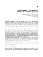
⊡
Fig. 20.1a,b. Reconstruction of an incisional hernia using the onlay reconstruction. a The rectus abdominis muscle is approximated
in the midline. The polypropylene mesh should be fixed to the fascia with an overlap of at least 5 cm in all directions and with a
double row of non-resorbable sutures. b The fascia cannot be approximated under the mesh. Omentum is placed between mesh
and bowels. The inner row of sutures should be positioned from the fascial edges. If this inner row of sutures is placed away of the
fascial edge, the intra-abdominal pressure might push the mesh away from the fascia and a recurrence can easily to occur
ab
Schumpelick.indd 166Schumpelick.indd 166 05.04.2007 8:51:15 Uhr05.04.2007 8:51:15 Uhr
167
VI
How to Create a Recurrence After Incisional Hernia Repair
Discussion
Abdominal wall hernia reconstruction using an onlay
polypropylene mesh seems the most straightforward
method, but is associated with serious postoperative
complications.
The prosthetic mesh can be used in two ways. First,
as a support when the fascia can be closed primarily.
Then the mesh can be positioned either as an onlay
or a sublay, because the biomechanical circumstances
are similar. Still, the sublay technique is preferred since
wound complications such as seroma formation and in-
fection are rather frequent. Using the sublay technique,
the retromuscular position will prevent the exposure
of the prosthesis if wound complications occur. Sec-
ond, prosthesis can be used to bridge fascial defects if
the fascia cannot be closed primarily [1, 3–5]. Under
these circumstances, the sublay technique, where the
intra-abdominal pressure (0.2–2.0 kPa) presses the
prosthesis against the ventral abdominal wall, is pre-
ferred as well. If properly fixed, the forces on the mesh
are counteracted by the abdominal wall, thus prevent-
ing reherniation [6]. The sutures in concert with the
fibro-collagenous tissue that surrounds the prosthetic
mesh will counteract the small sheering forces on the
prosthesis (
⊡
Fig. 20.2).
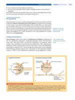
When using the onlay technique, the intra-abdomi-
nal pressure is not counteracted and the much larger
forces will put a continuous stress on the fixating su-
tures and the fibro-collagenous tissue, with the risk of
tearing the prosthesis from the fascia (
⊡
Fig. 20.3
). Al-
though the sublay mesh reconstruction is superior, the
onlay mesh reconstruction might be helpful in selected
patients, for example, to prevent contact between the
prosthesis and the bowel and when the sublay technique
is not possible for technical reasons.
In the literature, ten series report the results of onlay
mesh reconstruction [7–16] (
⊡
Table 20.3
). All but one
of the series are retrospective case series. The number
of patients included varies from 9–70. The series have
a wide range of follow-up and the method of follow-up
was mentioned in none of the studies. The reherniation
rate varied between 0 and 13%. The reherniation rate
in our series was 20%, but it is the only series where
all patients were seen in the outpatients’ clinic after an
adequate follow-up period. The results are similar to
other series with adequate follow-up [4].
Several prosthetic materials can be used to repair
incisional hernias. Expanded-polytetrafluoroethylene
(ePTFE) patch and polypropylene mesh (PPM)-based
prosthesis are the most frequently used prosthetic ma-
terials. PPM is the preferred prosthetics material when
the onlay technique is used. First, because the anchorage
of the prosthesis to the adjacent fascia is superior to
the ePTFE patch. Fixation of the ePTFE patch depends
solely on the fixating sutures, because the micropores
(20 µm) in ePTFE patch are too small to allow ingrowth
of fibro-collagenous tissue [17, 18]. PPM is completely
⊡
Fig. 20.2. Due to the intra-abdominal pressure, a rehernia-
tion occurred
⊡
Fig. 20.3.
In an intact abdominal wall
the intra-abdominal pressure (I.A.P.) is
compensated by the muscle strain (MR).
In the midline of the abdominal wall there
always a muscle strain to the lateral border
caused by the oblique abdominal muscles
and compensated by the opposite site,
there is a balance. The intra-abdominal pres-
sure (I.A.P.) on the inner row of sutures of an
onlay reconstruction is not compensated
by muscle strain (MR), but the muscle still
gives a constant strain to the lateral border
(M). This result is a constant force on the
sutures (in black)
Schumpelick.indd 167Schumpelick.indd 167 05.04.2007 8:51:16 Uhr05.04.2007 8:51:16 Uhr
168 Incisional Hernia
20
incorporated into fibro-collagenous tissue and firmly
anchors to the adjacent fascia. Second, because PPM
is rather resistant against infection, whereas infected
ePTFE patches have to be removed. Since wound in-
fections occur in 17–50% of patients, the use of ePTFE
patch to repair incisional hernias by the onlay technique
is too risky [19–21]. Korenkov et al. performed a ran-
domized clinical trial comparing onlay polypropylene
mesh repair with suture repair and onlay dermal graft
repair [16]. This trial is the only randomized clinical
trial comparing onlay reconstruction with two differ-
ent biomaterials. Wound complications occurred in
20%. Although none of the meshes had to be removed
because of infection, the trial was stopped because of
the high complication rate.
In our series, 76% of patients suffered from seroma
after the operation, compared to 0–31% in other series
(
⊡
Table 20.3). Seromas are a consequence of the large
subcutaneous wound surface that is created to fix the
prosthetic mesh with an adequate overlap to the fascia.
Seromas are a frequent complication after reconstruc-
tion of large abdominal wall hernias occurring in up
to 30% [19, 22]. Moreover, wound infections are fre-
quent. In our series, 24% of patients suffered a wound
infection, which is similar to the frequency found in
other series [14, 16]. Wound infection may also occur
secondary to skin necrosis. Separation of the epigas-
tric perforating arteries endangers the vascular supply
of the skin, which may interfere with wound healing
and may result in skin necrosis and subsequent infec-
tion.
In conclusion, onlay prosthetic repair of abdominal
wall hernias is easy but, because of the increased chance
of reherniation and loss of the prosthesis in the case of
wound complications, the use of onlay prosthetic repair
must be discouraged and be performed only when the
superior sublay repair is not possible.
Acknowledgements. The authors wish to thank Mr. F.
Bosch (Tilburg, The Netherlands), medical illustrator,
for making the illustrations.
References
1. de Vries Reilingh TS, van Geldere D, Langenhorst B, de Jong
D, van der Wilt GJ, van Goor H et al. Repair of large midline
incisional hernias with polypropylene mesh: comparison of
three operative techniques. Hernia 2004; 8(1): 56–59
⊡
Table 20.3. Onlay technique
Author Year Patients Complications
n (%)
Reherniation
n (%)
Follow-up
mean (range)
months
Larson 1978 19 10 0 ? (12–60)
Deitel 1979 36 14 (11%) 2 (6%) 42 (?)
Lewis 1984 50 15 (10%) 3 (6%) 30 (?)
Wagman 1985 19 10 0 14 (?)
Molloy 1991 50 10 (20%) 4 (8%) 45 (6–120)
Liakakos 1994 49 14 (8%) 4 (8%) ? (0–16)
Birolini 2000 20 15 (25%) 0 ? (12–84)
Korenkov 2002 70 14 (20%) 6 (9%) 14 (11–24)
De Vries
Reilingh
2004 17 13 (76%) 3 (20%) 18.5 (12–28)
Machairas 2004 43 19 (21%) 4 (9%) 54.4 (4–106)
Kingsnorth 2004 16 15 (31%) 2 (13%) ? (6–60)
Schumpelick.indd 168Schumpelick.indd 168 05.04.2007 8:51:17 Uhr05.04.2007 8:51:17 Uhr
169
VI
How to Create a Recurrence After Incisional Hernia Repair
2. Bleichrodt RP, Malyar AW, de Vries Reilingh TS, Buyne OR,
Bonenkamp JJ, van Goor H. The omentum-polypropylene
sandwich technique: an attractive method to repair large
abdominal wall defects in the presence of contamination
or infection. Hernia 2007; 11(1): 71–74
3. Burger JW, Luijendijk RW, Hop WC, Halm JA, Verdaasdonk EG,
Jeekel J. Long-term Follow-up of a Randomized Controlled
Trial of Suture Versus Mesh Repair of Incisional Hernia. Ann
Surg 2004; 240(4): 578–585
4. Luijendijk RW, Hop WC, van den Tol MP, de Lange DC,
Braaksma MM, IJzermans JN et al. A comparison of suture
repair with mesh repair for incisional hernia. N Engl J Med
2000; 343(6): 392–398
5. Stoppa RE. The treatment of complicated groin and incisional
hernias. World J Surg 1989; 13(5): 545–554
6. Klinge U, Klosterhalfen B, Conze J, Limberg W, Obolenski
B, Ottinger AP et al. Modified mesh for hernia repair that is
adapted to the physiology of the abdominal wall. Eur J Surg
1998; 164(12): 951–960
7. Birolini C, Utiyama EM, Rodrigues AJJ, Birolini D. Elective
colonic operation and prosthetic repair of incisional hernia:
does contamination contraindicate abdominal wall prosthe-
sis use? J Am Coll Surg 2000 191(4): 366–372
8. Deitel M, Vasic V. A secure method of repair of large ventral
hernias with Marlex mesh to eliminate tension. Am J Surg
1979; 137(2): 276–277
9. Larson GM, Harrower HW. Plastic mesh repair of incisional
hernias. Am J Surg 1978; 135(4): 559–563
10. Liakakos T, Karanikas I, Panagiotidis H, Dendrinos S. Use of
Marlex mesh in the repair of recurrent incisional hernia. Br
J Surg 1994; 81(2): 248–249
11. Molloy RG, Moran KT, Waldron RP, Brady MP, Kirwan WO.
Massive incisional hernia: abdominal wall replacement with
Marlex mesh. Br J Surg 1991; 78(2): 242–244
12. Wagman LD, Barnhart GR, Sugerman HJ. Recurrent midline
hernial repair. Surg Gynecol Obstet 1985; 161(2): 181–182
13. Kingsnorth AN, Sivarajasingham N, Wong S, Butler M. Open
mesh repair of incisional hernias with significant loss of do-
main. Ann R Coll Surg Engl 2004; 86(5): 363–366
14. Machairas A, Misiakos EP, Liakakos T, Karatzas G. Incisional
hernioplasty with extraperitoneal onlay polyester mesh. Am
Surg 2004; 70(8): 726–729
15. Lewis RT. Knitted polypropylene (Marlex) mesh in the repair
of incisional hernias. Can J Surg 1984; 27(2): 155–157
16. Korenkov M, Sauerland S, Arndt M, Bograd L, Neugebauer
EA, Troidl H. Randomized clinical trial of suture repair, poly-
propylene mesh or autodermal hernioplasty for incisional
hernia. Br J Surg 2002; 89(1): 50–56
17. de Vries Reilingh TS, Malyar AW, Walboomers XF et al. Im-
pregnation of e-PTFE abdominal wall patches with silver
salts and chlorhexidine diminishes biocompability and is
associated with an increased reherniation rate (submitted)
18. van der Lei B, Bleichrodt RP, Simmermacher RK, van Schilf-
gaarde R. Expanded polytetrafluoroethylene patch for the
repair of large abdominal wall defects. Br J Surg 1989; 76(8):
803–805
19.
de Vries Reilingh TS, van Goor H, Charbon J et al. Repair of
large midline abdominal wall hernias: Components Separa-
tion Technique versus Prosthetic Repair. Interim analysis of a
randomised controlled trial. World J Surg 2007; 31: 756–763
20. Lowe JB, Garza JR, Bowman JL, Rohrich RJ, Strodel WE. En-
doscopically assisted “components separation” for closure
of abdominal wall defects. Plast Reconstr Surg 2000; 105(2):
720–729; quiz 730
21. de Vries Reilingh TS, van Goor H, Rosman C, Bemelmans
MH, de Jong D, van Nieuwenhoven EJ et al. “Components
separation technique” for the repair of large abdominal wall
hernias. J Am Coll Surg 2003; 196(1): 32–37
22. Conze J, Kingsnorth AN, Flament JB, Simmermacher R, Arlt
G, Langer C et al. Randomized clinical trial comparing light-
weight composite mesh with polyester or polypropylene
mesh for incisional hernia repair. Br J Surg 2005; 92(12):
1488–1493
Discussion
Flament: I am surprised that no one has mentioned re-
laxing incisions today, because with them a suture repair
may be achieved in cases where non-absorbable meshes
are not suitable, e.g. in infected cases. Main part of the on-
lay repair by Chevrel was a relaxing incision of the ante-
rior sheath of the rectus muscle and a prosthesis covering,
reinforcing and recreating the anterior rectus sheath. That
is a little different from what you have shown compared
to the 400 cases of Chevrel published in Hernia.
deVries Reilingh: There is a randomized clinical trial in-
cluding patients for Ramirez technique with and without
mesh reinforcement, and the mesh is placed in the sublay
position, not onlay. We choose this technique because of
the large wound complication described by onlay mesh
plasty and also with the Ramirez technique, and it seems
not suitable to put a mesh in areas where they might
cause problems.
Kurzer:
I was interested, but not surprised, to see your high
rate of wound complication and abdominal wall stiffness.
I am interested that Prof. Flament and his colleagues have
a vast experience with sublay mesh and have shown over
many years that it works very well. Prof. Kingsnorth, with
respect, is advocating a randomized trial of a bad opera-
tion against a good operation done badly, and I can’t see
the point in doing that. Do a good operation well. We
should be teaching the people to do the good operation, not
doing more randomized clinical trials of two very different
operations, one of which doesn’t work well at all. I am
pleased that you are moving over to sublay mesh.
Chan:
In my study and review we have taken a lot of
onlay mesh, that’s all I can tell you, especially for big ones.
It just doesn’t work, because most of the time the defect is
just so big, its too tight to put it in, so it just won’t work,
I would recommend not to use it at all.
Kingsnorth:
I would like to speak up in favour of the
onlay technique. Firstly, we must not ignore the results
of Prof. Chevrel, that are every bit as good as the sub-
Schumpelick.indd 169Schumpelick.indd 169 05.04.2007 8:51:17 Uhr05.04.2007 8:51:17 Uhr
170 Incisional Hernia
20
lay technique; we cannot call the onlay a bad operation.
Secondly, I think it is very versatile; the best place for the
sublay technique is only in the upper abdomen because
you can then put it in front of the posterior rectus sheath;
once you get below the linea arcuata, you then only have
peritoneum, that often tears and then you have mesh in
direct contact with bowel, so I think in the lower abdomen
the onlay technique maybe advantageous. We must give
the onlay technique a chance, it is more versatile, it is
easier, and general surgeons are capable of using it under
more circumstances than the sublay technique.
Schumpelick: I would also like to say something in fa-
vour of the onlay technique, even as a sublay man. In
the recurrent cases, where the retromuscular space is al-
ready obliterated by a mesh, it is sometimes very difficult
to place another mesh in the same space. With the new
meshes you can do an onlay repair. The main problem
with the old meshes in the onlay position was infection,
something we don’t see with the new large pore meshes
that are better integrated. And even in the case of infec-
tion there is no need for explantation. We have done some
in this technique with good results.
Introduction
Since 1993, experience in minimally invasive incisional
hernia repair has accumulated such that we now have
some basic understanding of how to optimize the tech-
nical outcome of this procedure. In this review we will
summarize technical maneuvers which we believe will
minimize the risk of recurrence after minimally invasive
incisional herniorrhaphy. The conclusions and recom-
mendations of this review are based on our own clinical
experience [1] and a review of the surgical literature. As is
the case in most areas of surgery, the recommendations
given in this review are based on uncontrolled clinical se-
ries and expert opinion; there are little to no data available
from randomized controlled trials in the field of minimally
invasive incisional hernia surgery.
Methods
An internet search of the literature was performed
(PubMed/National Library of Medicine, www.ncbi.
nlm.nih.gov/entrez/) using various combinations of the
following keywords: minimally invasive, laparoscopic,
ventral, incisional, hernia. The inclusion criteria were
papers that contained adequate data on > 10 patients
undergoing minimally invasive incisional or ventral
herniorrhaphy. To be included, a paper needed to de-
scribe patient demographics, surgical technique, peri-
operative events, and some follow-up/recurrence data.
In addition to internet search, the references of selected
papers were searched manually to identify any possible
manuscripts that were missed (none were found with
this secondary search). In some instances, a group of
authors had multiple publications on the same series
of patients; in these cases only the most recent update
of a given patient series was included in the present
review.
Results for Hernia Recurrence
A total of 53 manuscripts met the inclusion criteria
(
⊡
Table 20.4); these papers described 5227 minimally
invasive incisional or ventral herniorrhaphies (a com-
prehensive analysis will be submitted for later publi-
cation.) Certain aspects of herniorrhaphy technique
were virtually identical among all 53 manuscripts:
intraperitoneal sublay of prosthetic mesh which ex-
tended beyond the margins of hernia in all directions,
with no excision of the hernia sac. The papers differed
in the type of mesh used, the amount of mesh overlap
of the defect, and in the technique of mesh fixation
(see discussion below). The rate of hernia recurrence in
these 5227 published procedures was 3.98%. Of course,
this result is mostly the product of specialty centers in
which minimally invasive surgery is prominent, so the
recurrence rate for all operators is likely to be higher.
The results from the 53 manuscripts of this review also
is subject to publication bias (i.e., better results have a
greater likelihood of being submitted than mediocre
results). The reported recurrence rate from open in-
20.3 Technical Factors Predisposing to Recurrence After Minimally Invasive Incisional
Herniorrhaphy
C.T. F, L.E. L, M.A. C
Schumpelick.indd 170Schumpelick.indd 170 05.04.2007 8:51:18 Uhr05.04.2007 8:51:18 Uhr
171
VI
How to Create a Recurrence After Incisional Hernia Repair
⊡
Table 20.4. Papers included in review of minimally invasive incisional/ventral hernia surgery
Ref. no. Year Authors Institution Procedures
[7] 1997 Holzman et al. Duke 121
[8] 1998 Toy et al. Multicenter 144
[9] 1998 Tsimoyiannis et al. Hatzikosta General Hospital, Ioannina 111
[10] 1999 Koehler et al. Martha‘s Vineyard Hospital 132
[11] 1999 Kyzer et al. Tel Aviv Univ 153
[12] 1999 Sanders et al. Tulane Univ, Henry Ford Hospital 112
[13] 2000 Chari et al. Meridia Huron Hospital, Cleveland 114
[14] 2000 Chowbey et al. Sir Ganga Ram Hospital, New Delhi 202
[15] 2000 DeMaria et al. MCV, Richmond 121
[16] 2000 Farrakha Abu Dhabi, UAE 118
[17] 2000 Reitter et al. UI Peoria, IL 149
[18] 2000 Szymanski et al. Scarborough Hospital, Canada 144
[19] 2001 Birgisson, Park et al. UKY 164
[20] 2002 Andreoni et al. UNC Chapel Hill 113
[21] 2002 Aura et al. Aulnay-Sous-Bois, France 186
[22] 2002 Bageacu et al. Saint-Etienne, France 159
[23] 2002 Ben-Haim et al. Tel Aviv Univ 100
[24] 2002 Berger et al. Baden-Baden 150
[25] 2002 Gillian et al. Southern Maryland Hospital 100
[26] 2002 Kirshtein et al. Ben Gurion Univ, Beer Sheva, Israel 103
[27] 2002 Kua et al. Royal Brisbane Hospital, Queensland, Austral 130
[28] 2002 Lau et al. Univ Hong Kong Med Ctr 111
[29] 2002 Parker et al. Univ South Carolina 150
[30] 2002 Raftopoulos et al. UI Chicago 150
[31] 2002 Salameh et al. Baylor, Houston TX 129
[32] 2002 van‘t Riet et al. Erasmus U Med Ctr, Rotterdam 125
Schumpelick.indd 171Schumpelick.indd 171 05.04.2007 8:51:18 Uhr05.04.2007 8:51:18 Uhr
172 Incisional Hernia
20
⊡
Table 20.4. Continued
Ref. no. Year Authors Institution Procedures
[33] 2002 Wright et al. Hennepin County Med Ctr, Minneapolis 190
[34] 2003 Carbajo et al. Valladolid, Spain 270
[35] 2003 Chelala et al. Univ Hosp Tivoli, Belgium 120
[36] 2003 Chowbey et al. Sir Ganga Ram Hospital, New Delhi 134
[37] 2003 Eid et al. UPitt, VAMC Pitt, UMN 179
[38] 2003 Heniford et al. Carolinas Medical Center, UKY, Emory, UTN 850
[39] 2003 LeBlanc et al. Min Invas Surg Inst, Baton Rouge 200
[40] 2003 McGreevy et al. Dartmouth-Hitchcock Med Ctr, VAMC VT 165
[41] 2003 Mizrah et al. Ben Gurion Univ, Beer Sheva, Israel 231
[42] 2003 Rosen et al. Cleveland Clinic 114
[43] 2004 Bamehriz and Birch McMaster Univ, Hamilton, Can 128
[44] 2004 Bencini and Sanchez Florence, Italy 164
[45] 2004 Bower et al. East Carolina Univ, Greenville 100
[46] 2004 Franklin et al. Texas Endosurgery Institute, MGH, Monterrey 384
[1] 2004 Frantzides et al. NWU, UNMC, UTN 208
[47] 2004 Gal et al. Bugat Pal Hosp, Hungary 115
[48] 2004 Kannan et al. Changi General Hosp, Singapore 120
[49] 2004 McKinlay and Park Univ Maryland 170
[50] 2004 Moreno-Egea et al. Murcia, Spain 190
[51] 2004 Muysoms et al. Ghent, Belgium 152
[52] 2004 Sanchez et al. Florence 190
[53] 2004 Ujiki et al. NWU, UHawaii, Hines VA 100
[54] 2004 Verbo et al. Catholic Univ, Rome Italy 145
[55] 2005 Angele et al. Ludwig-Maximilians Univ, Munich 128
[56] 2005 Johna Loma Linda Univ, CA 118
[57] 2005 Olmi et al. Monza, Italy 150
[58] 2005 Perrone et al. Washington Univ 121
Schumpelick.indd 172Schumpelick.indd 172 05.04.2007 8:51:19 Uhr05.04.2007 8:51:19 Uhr
173
VI
How to Create a Recurrence After Incisional Hernia Repair
cisional herniorrhaphy (not reviewed here) is widely
variable, from several percent to 20% or more. Need-
less to say, a prospective randomized comparison of
open vs. minimally invasive incisional hernia repair has
not been done. Considering the inherent advantages of
minimally invasive surgery, however, it would be rea-
sonable to predict that the overall results (including
recurrence, infection, pain, patient satisfaction, etc.) of
the minimally invasive approach would be as least as
good, if not better, than the open approach.
Technical Factors: Entry and Exposure
For any laparoscopic procedure, the surgeon can
minimize the risk of port-site hematoma by transil-
luminating the abdominal wall prior to trocar inser-
tion. This maneuver minimizes the risk of abdominal
wall vessel laceration. It is not clear, however, whether
a port site hematoma predisposes a patient to recur-
rent hernia. In order to prevent port-site hernia, the
surgeon should close all port sites for trocars > 5 mm,
and for 5mm if the site has become stretched or en-
larged [2].
Probably the first major technical issue that the sur-
geon encounters during a minimally invasive incisional
hernia is intra-abdominal exposure. Retrospective anal-
ysis has determined, not surprisingly, that inadequate
dissection of the hernial defects will increase the risk
of hernia recurrence [3]. Nearly all authors of the 53
manuscripts of the present review stress complete ex-
posure of the ventral abdominal wall with takedown of
all adhesions to the viscera. The entire incision needs to
be visualized. Such a maneuver will prevent the surgeon
from missing a small, asymptomatic defect which later
could enlarge into a symptomatic one. This is especially
important with long midline incisions closed with run-
ning nonabsorbable suture, in which the so-called Swiss
cheese abdomen (i.e., multiple small hernias deriving
from the cutting action of the suture) can develop. Small
hernias can be hidden in a mass of dense adhesions, so
complete adhesiolysis is essential.
Technical Factors: Mesh Type
The next choice of potential consequence during min-
imally invasive incisional hernia repair is the mesh
type. Expanded Polytetrafluoroethylene (ePTFE) was
the prosthetic material used in the majority of proce-
dures in 41 (77%) of the 53 manuscripts; of these 41
papers, 33 (62%) specified their ePTFE as the dual-
surface construct available from W. L. Gore and As-
sociates, Inc. (i.e., DualMesh). This mesh has a closed
structure surface on the side facing the viscera; this
is intended to reduce tissue attachment. The other
side (facing the abdominal wall) has a macroporous
structure (corduroy), which is intended to enhance
tissue attachment. Interestingly, an improvised dual-
surface mesh for minimally invasive incisional her-
niorrhaphy already was in use by the early 1990s [4].
This was a bilaminar prosthesis consisting of a sheet
of ePTFE and a sheet of polypropylene sewn together;
the polypropylene side was applied to the abdominal
wall while the ePTFE side contacted the viscera. This
dual-surface arrangement encouraged tissue ingrowth
on the abdominal wall side, thereby increasing the ro-
bustness of the repair, yet minimized intestinal reaction
to the mesh. So far, published clinical experience with
the dual-surface mesh configuration has shown it to be
safe. To our knowledge, there have been no published
cases of primary erosion of ePTFE into the viscera after
incisional herniorrhaphy with ePTFE. In laparoscopic
incisional hernia repair the prosthesis is typically placed
in direct contact with the viscera which, in the case of
heavy-weight polypropylene mesh, introduces the risk
of visceral erosion. The dual-surface mesh configura-
tion appears not to have this risk.
The use of ePTFE has undergone a resurgence with
the advent of minimally invasive incisional hernia
repair. This material was less popular in open hernia
repair because it was more prone to infection and in-
corporated less well than other materials (e.g., poly-
propylene). Since mesh infection appears to be less of
a problem with the minimally invasive approach, and
with the introduction of the dual-surface product which
incorporates strongly into the abdominal wall yet is
benign to the viscera, dual-surface ePTFE has become
the material of choice for the majority of the authors
in this review. It should be noted, however, that there
are a number of light-weight/composite polypropylene
hernia meshes now available which may be suitable (or
even better) alternatives to ePTFE. Long-term compara-
tive data in patients are not available.
Technical Factors: Mesh Overlap
As indicated above, the universal approach to minimally
invasive repair of hernia of the ventral abdominal wall
in manuscripts of this review is sublay positioning of
prosthetic mesh, a technique originally described in
open surgery by Rives and Flament [5] and also by
Stoppa in the groin [6]. For repairs of this type, one
Schumpelick.indd 173Schumpelick.indd 173 05.04.2007 8:51:19 Uhr05.04.2007 8:51:19 Uhr
174 Incisional Hernia
20
requirement for the mesh is that it should have adequate
overlap (a more accurate term would be underlap) of
the hernial defect [3]. That is, the margin of the mesh
should extend beyond the margin of the defect by an
appropriate amount throughout the defect’s entire cir-
cumference. The range of mesh overlap in the 53 manu-
scripts of this review is shown in
⊡
Fig. 20.4. Most (60%)
of the authors favoured a minimum of 3cm of overlap;
24% indicated 4cm or more. One might hypothesize
that the recurrence rate would decrease as the overlap
increased, but this is not supported by plotting these
two variables, as shown in
⊡
Fig. 20.4 (it should be ad-
mitted that this is a relatively unscientific manipulation
of uncontrolled data). The final answer to an appropri-
ate amount of mesh overlap during minimally invasive
incisional herniorrhaphy is not known, although 3cm
most commonly is chosen. The optimal distance most
likely is dependent on multiple variables, and may not
be simply defined by “more is better.”
Technical Factors: Mesh Fixation
One of the more controversial issues in minimally
invasive incisional herniorrhaphy is the technique of
mesh fixation. At a minimum, the laparoscopically
performed sublay technique requires some fixation to
keep the mesh anterior while pneumoperitoneum is
present. Further fixation beyond this would be intended
to prevent mesh migration/ slippage with subsequent
reherniation. The basic choices for fixation are (1)
tacking/ stapling, (2) transabdominal fixation sutures,
or (3) a combination of both. Of the 53 manuscripts in
this review, 44 contained sufficient details regarding
mesh fixation; 69% of the papers utilized a combina-
tion of tacking/stapling and fixation sutures, while 29%
utilized tacking/stapling alone (one paper used sutures
alone). A plot of fixation technique vs. recurrence rate
is shown in
⊡
Fig. 20.5
; there was no statistical differ-
ence in recurrence with respect to fixation. Neverthe-
less, given that a common cause of recurrent herniation
is mesh slippage, it would seem reasonable to use the
maximum amount of mesh fixation (i.e., lots of tacks/
staples + lots of fixation sutures). Unfortunately, fixa-
tion sutures are associated with long-term abdominal
pain, and they also require additional stab incisions
in the skin and more operating time. We have spoken
with surgeons who anecdotically claim that their recur-
rence rate is less with the combined use of tacks/staples
and sutures, but controlled data are lacking. Further-
more, there are details of fixation technique (e.g., spi-
ral tacks vs. straight staples, single vs. multiple rows
of tacks, spacing between tacks and/or sutures, etc.),
which further complicate the fixation issue. One of us
(C.T.F.) utilizes a single row of straight staples at 1cm
intervals (having obtained a 1.4% recurrence rate [1],
while the other (M.A.C.) has changed his technique to
a single row of spiral tacks at 1cm intervals with 2–0
polypropylene transabdominal fixation sutures placed
every 5–7cm. The first author (C.T.F.) places each staple
radially so that one end is buried into the PTFE while
the other end takes tissue. In addition, he is careful that
each staple enters the abdominal wall perpendicularly
(using the two-handed stapling technique) to ensure
maximum tissue penetration. It is this type of technical
detail that could make the difference between a 1% vs.
a 5% recurrence rate. In any event, it is difficult to rec-
ommend one fixation technique over another without
ANOVA: p=0.545
mesh overlap [cm]
0
5
15
recurrence rate [%]
2.0
10
20
2.5 3.0 3.5 4.0 5.04.5
⊡
Fig. 20.4. Plot of hernia recurrence rate
vs. minimum mesh overlap of the hernial
defect for minimally invasive incisional/
ventral herniorrhaphy. Complete data
were available from 45 of the 53 manu-
scripts shown in
⊡
Table 20.4
Schumpelick.indd 174Schumpelick.indd 174 05.04.2007 8:51:19 Uhr05.04.2007 8:51:19 Uhr
175
VI
How to Create a Recurrence After Incisional Hernia Repair
controlled data. This is another area of surgery which
will continue to be dictated by training environment,
local experience, and so forth.
Technical Factors: Infection
Wound infection has been shown to be an independent
risk factor for recurrence after open incisional hernia
repair in numerous clinical series (data not reviewed
here). Port-site infection after laparoscopic incisional
hernia repair usually can be handled with antibiotics
and local care without endangering the mesh; infec-
tion of ePTFE mesh itself, however, invariably means
mesh removal with subsequent hernia recurrence.
Although seemingly less common with the minimally
invasive approach, mesh infection still had an incidence
of 0.89% in the 5227 procedures of this review. There
are a number of recommendations (expert opinion,
not necessarily standard of care) to minimize the risk
of major wound/mesh infection in minimally invasive
incisional herniorrhaphy:
▬ pre-operative bowel preparation (mechanical and
oral antibiotics);
▬ appropriate use of antibiotic prophylaxis;
▬ use of an antimicrobial-impregnated adhesive
drape;
▬ avoidance of ePTFE contact with skin;
▬ changing surgical gloves prior to handling the
mesh;
▬ careful surgical dissection with minimal blood
loss;
▬
deferral of operation in the presence of incisional
inflammation or stitch abscess.
Smoking should be minimized/eliminated pre-op-
eratively, as this has been shown to be a risk factor for
failure in open incisional herniorrhaphy. If the patient
develops a large seroma postoperatively, then the sur-
geon should avoid the temptation of aspiration/drain-
age. The vast majority of these seromas will resolve
without intervention; unnecessary violation of the space
may introduce bacteria.
An issue related to infection is the management of
intra-operative small bowel perforation. This compli-
cation occurred in 81 (1.6%) of the 5227 cases of this
review. Details on the management of these cases were
not available for all of them. In general, however, a
surgeon has at least three options when a small bowel
perforation is recognized intra-operatively: (1) convert
to an open procedure, repair the enterotomy, and close
the hernial defect primarily without a mesh; (2) if there
is no enteric spillage, then repair the enterotomy lapa-
roscopically and complete the mesh herniorrhaphy as
planned; (3) repair the enterotomy laparoscopically,
place the patient on IV antibiotics for several days, and
then perform the minimally invasive incisional hernior-
rhaphy with mesh (usually the authors choice). There
are variations to these options, but the essential choice
is conversion vs. laparoscopic bowel repair and herni-
orrhaphy vs. laparoscopic bowel repair with delayed
herniorrhaphy. The idea of placing a piece of PTFE in
the face of potential enteric contamination (option 2
above) may not seem safe, but there are numerous suc-
cessful examples of this management in the 53 articles
of this review. Since the incidence of this complication
is relatively low, it will be difficult to ascertain the op-
timal management, especially with respect to patient
comorbidities. Consequently, treatment for each case
⊡
Fig. 20.5. Plot of hernia recurrence rate
vs. technique of mesh fixation for mini-
mally invasive incisional/ventral hernior-
rhaphy. Complete data were available
from 44 of the 53 manuscripts shown in
⊡
Table 20.4
t-test: p=0.894
tacks or staples
and sutures
0
2
8
recurrence rate [%]
10
12
tacks or
staples only
6
4
Schumpelick.indd 175Schumpelick.indd 175 05.04.2007 8:51:21 Uhr05.04.2007 8:51:21 Uhr
176 Incisional Hernia
20
of intra-operative small bowel perforation will depend
on the characteristics of the injury, surgeon’s bias and
experience, patient comorbidities, and so on. Intra-op-
erative colon injuries are more rare; since the bacterial
concentration in the colon is at least a millionfold of that
in the small bowel, however, one should be wary of simul-
taneous repair of a colon injury and mesh placement.
Summary
At this relatively early stage in the history of minimally
invasive repair of ventral/incisional hernia, a few rec-
ommendations for optimizing technique and reducing
recurrence may be given:
1. Completely, yet carefully, expose the entire incision
and anterior abdominal wall.
2. For intraperitoneal mesh placement, a dual-surface
mesh which incorporates into the abdominal on one
side while remaining relatively nonreactive to the
viscera on the other appears optimal.
3. The ideal amount of mesh overlap of the defect is
not known; a 3cm overlap seems reasonable.
4. The optimal form of mesh fixation needs to be stud-
ied by a carefully designed and controlled trial. At
this point tacks/staples ± fixation sutures are the
most popular techniques.
5. Minimize the risk of mesh infection; have a plan
ready in the event of an intra-operative small bowel
enterotomy.
6. Close all port sites for trocars >5mm.
Acknowledgements. Supported in part by a grant to
MAC from the United States National Institutes of
Health (K08 GM00703).
References
1. Frantzides CT, Carlson MA, Zografakis JG, Madan AK, Moore
RE. Minimally invasive incisional herniorrhaphy: a review of
208 cases. Surg Endosc 2004; 18(10): 1488–1491
2. Tonouchi H, Ohmori Y, Kobayashi M, Kusunoki M. Trocar site
hernia. Arch Surg 2004; 139(11): 1248–1256
3. Lowham AS, Filipi CJ, Fitzgibbons RJ Jr., Stoppa R, Wantz GE,
Felix EL, Crafton WB. Mechanisms of hernia recurrence after
preperitoneal mesh repair. Traditional and laparoscopic. Ann
Surg 1997; 225(4): 422–431
4.
Frantzides CT, Carlson MA. Minimally invasive ventral hernior-
rhaphy. J Laparoendosc Adv Surg Tech A 1997; 7(2): 117–120
5. Flament JB, Avisse C, Palot JP, Delattre JF. Biomaterials: Prin-
ciples of Implantation. In: Schumpelick V, Kingsnorth AN
(eds) Incisional hernia. Springer, Berlin Heidelberg New
York, 1999
6. Stoppa R, Ralmiaramanana F, Henry X, Verhaeghe P. Pros-
thetic repair of recurrent groin hernias. In: Schumpelick V,
Kingsnorth AN (eds) Incisional hernia. Springer, Berlin Hei-
delberg New York, 1999
7. Holzman MD, Purut CM, Reintgen K, Eubanks S, Pappas TN.
Laparoscopic ventral and incisional hernioplasty. Surg En-
dosc 1997; 11(1): 32–35
8. Toy FK, Bailey RW, Carey S et al. Prospective, multicenter
study of laparoscopic ventral hernioplasty. Preliminary re-
sults. Surg Endosc 1998; 12(7): 955–959
9. Tsimoyiannis EC, Tassis A, Glantzounis G, Jabarin M, Siakas
P, Tzourou H. Laparoscopic intraperitoneal onlay mesh re-
pair of incisional hernia. Surg Laparosc Endosc 1998; 8(5):
360–362
10. Koehler RH, Voeller G. Recurrences in laparoscopic incisional
hernia repairs: a personal series and review of the literature.
Jsls 1999; 3(4): 293–304
11. Kyzer S, Alis M, Aloni Y, Charuzi I. Laparoscopic repair of post-
operation ventral hernia. Early postoperation results. Surg
Endosc 1999; 13(9): 928–931
12. Sanders LM, Flint LM, Ferrara JJ. Initial experience with lapa-
roscopic repair of incisional hernias. Am J Surg 1999; 177(3):
227–231
13. Chari R, Chari V, Eisenstat M, Chung R. A case controlled study
of laparoscopic incisional hernia repair. Surg Endosc 2000;
14(2): 117–119
14. Chowbey PK, Sharma A, Khullar R, Mann V, Baijal M, Vashistha
A. Laparoscopic ventral hernia repair. J Laparoendosc Adv
Surg Tech A 2000; 10(2): 79–84
15. DeMaria EJ, Moss JM, Sugerman HJ. Laparoscopic intra-
peritoneal polytetrafluoroethylene (PTFE) prosthetic patch
repair of ventral hernia. Prospective comparison to open
prefascial polypropylene mesh repair. Surg Endosc 2000;
14(4): 326–329
16. Farrakha M. Laparoscopic treatment of ventral hernia. A
bilayer repair. Surg Endosc 2000; 14(12): 1156–1158
17. Reitter DR, Paulsen JK, Debord JR, Estes NC. Five-year
experience with the „four-before“ laparoscopic ventral
hernia repair. Am Surg 2000; 66(5): 465 468; discussion
468–469
18. Szymanski J, Voitk A, Joffe J, Alvarez C, Rosenthal G. Tech-
nique and early results of outpatient laparoscopic mesh
onlay repair of ventral hernias. Surg Endosc 2000; 14(6):
582–584
19. Birgisson G, Park AE, Mastrangelo MJ Jr., Witzke DB, Chu UB.
Obesity and laparoscopic repair of ventral hernias. Surg En-
dosc 2001; 15(12): 1419–1422
20. Andreoni KA, Lightfoot H, Jr., Gerber DA, Johnson MW, Fair
JH. Laparoscopic incisional hernia repair in liver transplant
and other immunosuppressed patients. Am J Transplant
2002; 2(4): 349–354
21. Aura T, Habib E, Mekkaoui M, Brassier D, Elhadad A. Lapa-
roscopic tension-free repair of anterior abdominal wall
incisional and ventral hernias with an intraperitoneal Gore-
Tex mesh: prospective study and review of the literature. J
Laparoendosc Adv Surg Tech A 2002; 12(4): 263–267
22. Bageacu S, Blanc P, Breton C, Gonzales M, Porcheron J, Cha-
bert M, Balique JG. Laparoscopic repair of incisional hernia:
a retrospective study of 159 patients. Surg Endosc 2002;
16(2): 345–348
Schumpelick.indd 176Schumpelick.indd 176 05.04.2007 8:51:21 Uhr05.04.2007 8:51:21 Uhr
177
VI
How to Create a Recurrence After Incisional Hernia Repair
23. Ben-Haim M, Kuriansky J, Tal R, Zmora O, Mintz Y, Rosin D,
Ayalon A, Shabtai M. Pitfalls and complications with lapa-
roscopic intraperitoneal expanded polytetrafluoroethylene
patch repair of postoperative ventral hernia. Surg Endosc
2002; 16(5): 785–788
24. Berger D, Bientzle M, Muller A. Postoperative complications
after laparoscopic incisional hernia repair. Incidence and
treatment. Surg Endosc 2002; 16(12): 1720–1723
25. Gillian GK, Geis WP, Grover G. Laparoscopic incisional and
ventral hernia repair (LIVH): an evolving outpatient tech-
nique. Jsls 2002; 6(4): 315–322
26. Kirshtein B, Lantsberg L, Avinoach E, Bayme M, Mizrahi S.
Laparoscopic repair of large incisional hernias. Surg Endosc
2002; 16(12):1717–1719
27. Kua KB, Coleman M, Martin I, O’Rourke N. Laparoscopic
repair of ventral incisional hernia. ANZ J Surg 2002; 72(4):
296–299
28. Lau H, Patil NG, Yuen WK, Lee F. Laparoscopic incisional her-
nioplasty utilising on-lay expanded polytetrafluoroethylene
DualMesh: prospective study. Hong Kong Med J 2002; 8(6):
413–417
29. Parker HH, 3rd, Nottingham JM, Bynoe RP, Yost MJ. Laparo-
scopic repair of large incisional hernias. Am Surg 2002; 68(6):
530–533; discussion 533–534
30. Raftopoulos I, Vanuno D, Khorsand J, Ninos J, Kouraklis G,
Lasky P. Outcome of laparoscopic ventral hernia repair in
correlation with obesity, type of hernia, and hernia size. J
Laparoendosc Adv Surg Tech A 2002; 12(6): 425–429
31. Salameh JR, Sweeney JF, Graviss EA, Essien FA, Williams MD,
Awad S, Itani KM, Fisher WE. Laparoscopic ventral hernia re-
pair during the learning curve. Hernia 2002; 6(4): 182–187
32. van’t Riet M, Vrijland WW, Lange JF, Hop WC, Jeekel J, Bonjer
HJ. Mesh repair of incisional hernia: comparison of laparo-
scopic and open repair. Eur J Surg 2002; 168(12): 684 689
33. Wright BE, Niskanen BD, Peterson DJ, Ney AL, Odland MD,
VanCamp J, Zera RT, Rodriguez JL. Laparoscopic ventral her-
nia repair: are there comparative advantages over traditional
methods of repair? Am Surg 2002; 68(3): 291 295; discussion
295–296.
34. Carbajo MA, Martp del Olmo JC, Blanco JI, Toledano M, de
la Cuesta C, Ferreras C, Vaquero C. Laparoscopic approach
to incisional hernia. Surg Endosc 2003; 17(1): 118–122
35.
Chelala E, Gaede F, Douillez V, Dessily M, Alle JL. The suturing
concept for laparoscopic mesh fixation in ventral and incisional
hernias: preliminary results. Hernia 2003; 7(4): 191–196
36. Chowbey PK, Sharma A, Khullar R, Soni V, Baijal M. Lapa-
roscopic ventral hernia repair with extraperitoneal mesh:
surgical technique and early results. Surg Laparosc Endosc
Percutan Tech 2003; 13(2): 101–105
37. Eid GM, Prince JM, Mattar SG, Hamad G, Ikrammudin S,
Schauer PR. Medium-term follow-up confirms the safety
and durability of laparoscopic ventral hernia repair with
PTFE. Surgery 2003; 134(4): 599 603; discussion 603–604
38. Heniford BT, Park A, Ramshaw BJ, Voeller G. Laparoscopic
repair of ventral hernias: nine years’ experience with 850
consecutive hernias. Ann Surg 2003; 238(3): 391–399; dis-
cussion 399–400
39. LeBlanc KA, Whitaker JM, Bellanger DE, Rhynes VK. Laparo-
scopic incisional and ventral hernioplasty: lessons learned
from 200 patients. Hernia 2003; 7(3): 118–124
40. McGreevy JM, Goodney PP, Birkmeyer CM, Finlayson SR,
Laycock WS, Birkmeyer JD. A prospective study comparing
the complication rates between laparoscopic and open
ventral hernia repairs. Surg Endosc 2003; 17(11): 1778–
1780
41. Mizrahi S, Lantsberg L, Kirshtein B, Bayme M, Avinoah E.
The experience with a modified technique for laparoscopic
ventral hernia repair. J Laparoendosc Adv Surg Tech A 2003;
13(5): 305–307
42. Rosen M, Brody F, Ponsky J, Walsh RM, Rosenblatt S, Dup-
erier F, Fanning A, Siperstein A. Recurrence after laparoscopic
ventral hernia repair. Surg Endosc 2003; 17(1): 123–128
43. Bamehriz F, Birch DW. The feasibility of adopting laparoscopic
incisional hernia repair in general surgery practice: early
outcomes in an unselected series of patients. Surg Laparosc
Endosc Percutan Tech 2004; 14(4): 207–209
44. Bencini L, Sanchez LJ. Learning curve for laparoscopic ventral
hernia repair. Am J Surg 2004; 187(3): 378–382
45. Bower CE, Reade CC, Kirby LW, Roth JS. Complications
of laparoscopic incisional-ventral hernia repair: the ex-
perience of a single institution. Surg Endosc 2004; 18(4):
672–675
46. Franklin ME Jr., Gonzalez JJ Jr., Glass JL, Manjarrez A. Laparo-
scopic ventral and incisional hernia repair: an 11-year experi-
ence. Hernia 2004; 8(1): 23–27
47. Gal I, Balint A, Szabo L. Results of laparoscopic repair of
abdominal wall hernias using an ePTFE-polypropylene
composite mesh (in German). Zentralbl Chir 2004; 129(2):
92–95
48. Kannan K, Ng C, Ravintharan T. Laparoscopic ventral her-
nia repair: local experience. Singapore Med J 2004; 45(6):
271–275
49. McKinlay RD, Park A. Laparoscopic ventral incisional hernia
repair: a more effective alternative to conventional repair
of recurrent incisional hernia. J Gastrointest Surg 2004; 8(6):
670–674
50. Moreno-Egea A, Torralba JA, Girela E, Corral M, Bento M,
Cartagena J, Vicente JP, Aguayo JL, Canteras M. Immediate,
early, and late morbidity with laparoscopic ventral hernia
repair and tolerance to composite mesh. Surg Laparosc En-
dosc Percutan Tech 2004; 14(3): 130–135
51. Muysoms F, Daeter E, Vander Mijnsbrugge G, Claeys D. Lap-
aroscopic intraperitoneal repair of incisional and ventral
hernias. Acta Chir Belg 2004; 104(6): 705–708
52. Sanchez LJ, Bencini L, Moretti R. Recurrences after laparo-
scopic ventral hernia repair: results and critical review. Hernia
2004; 8(2): 138–143
53. Ujiki MB, Weinberger J, Varghese TK, Murayama KM, Joehl
RJ. One hundred consecutive laparoscopic ventral hernia
repairs. Am J Surg 2004; 188(5): 593–597
54. Verbo A, Petito L, Pedretti G, Lurati M, D‘Alba P, Coco C. Use
of a new type of PTFE mesh in laparoscopic incisional hernia
repair: the continuing evolution of technique and surgical
expertise. Int Surg 2004; 89(1): 27–31
55. Angele MK, Lohe F, Dietz J, Hernandez-Richter T, Jauch KW,
Heiss MM. Laparoscopic incisional hernia repair – an alterna-
tive to the conventional procedure? [German]. Zentralbl Chir
2005; 130(3): 255–259
56. Johna S. Laparoscopic incisional hernia repair in obese pa-
tients. Jsls 2005; 9(1): 47–50
Schumpelick.indd 177Schumpelick.indd 177 05.04.2007 8:51:21 Uhr05.04.2007 8:51:21 Uhr
178 Incisional Hernia
20
57. Olmi S, Magnone S, Erba L, Bertolini A, Croce E. Results of
laparoscopic versus open abdominal and incisional hernia
repair. Jsls 2005; 9(2): 189–195
58. Perrone JM, Soper NJ, Eagon JC, Klingensmith ME, Aft RL,
Frisella MM, Brunt LM. Perioperative outcomes and compli-
cations of laparoscopic ventral hernia repair. Surgery 2005;
138(4): 708 715; discussion 715–716
Discussion
Itani: One of the issues that nobody addresses with lapa-
roscopic surgery is the issue of cosmesis. As you know, in
open surgery in all these deformed abdominal walls it is
very easy to remove the scar, doing an abdominal plasty
if needed, remove excess skin, but you cannot do that with
the laparoscopic procedure.
Frantzides:
You can do that with a laparoscopic proce-
dure at the latest stage, which means a second operation
later on.
LeBlanc: One thing that you didn’t mention when you
look at the fixation, and I know that you are not a pro-
ponent of suture as I am, there is no good consensus, but
a lack of adequate follow-up in the majority of series
that allow anyone to make a firm determination. There
are only two or three series that have followed up be-
yond 2 or 3 years, so there are just not enough data; we
need more prospective randomized trials to answer that
question.
Schumpelick.indd 178Schumpelick.indd 178 05.04.2007 8:51:22 Uhr05.04.2007 8:51:22 Uhr
VI
⊡
Fig. 21.1.
The four compartments of the
abdominal wall, separated by the Linea alba
(dark blue arrow), the lateral margins of the
rectus sheath (light blue arrows) and the os-
seous frame (grey arrows)
21 Anatomical Limitations –
Where Are the Layers?
J. C, A. P
The surgical armamentarium to solve the persisting
problem of incisional hernia has grown over the decades
and recently expanded essentially by the laparoscopic
techniques. However, the multitude of techniques is a
typical sign that no procedure meets all requirements
to answer every fascial defect of the abdominal wall.
This might be explained by the different and difficult
anatomy of the abdominal wall in the median com-
partment and both lateral compartments and their
complicated transition zone of muscles, fascias and
aponeuroses (
⊡
Fig. 21.1).
The muscles of the abdominal wall, antagonistic to
the muscles of the back, are important for every move-
ment of the trunk. They are essential for erect position,
regulate the intra-abdominal pressure, support defeca-
tion and furthermore they are permanently involved in
supporting breathing.
From topographic-anatomical aspects the abdo-
minal wall closes the skeletal gap between the lower
thoracic aperture and the pelvis, the so-called lacuna
sceleti sternopubica, according to August Rauber. The
abdominal wall consists of different muscles, fascial
structures, aponeuroses, peritoneum and intercalated
nerves and vessels fixed within the osseous frame
[1, 2].
On both sides of the midline the rectus abdominis
muscle runs in vertical direction from the fifth to the
seventh rib to the pubic bone (
⊡
Fig. 21.2a
). The muscle
is separated by three to four horizontal tendineous inter-
sections that fix the muscle to the anterior rectus sheath.
At the lower insertion it is overlayed by the rudimentary
pyramidalis muscle. The medial compartment is mainly
a single muscle layer structure that is surrounded by
the rectus sheath. This collageneous structure orgin-
Schumpelick.indd 179Schumpelick.indd 179 05.04.2007 8:51:22 Uhr05.04.2007 8:51:22 Uhr
180 Incisional Hernia
21
ates from the aponeuroses of the oblique muscles of the
lateral compartment of the abdominal wall.
The lateral compartment of the abdominal wall is
formed by three oblique muscles that run in different
directions [3]. The external oblique muscle runs in a
cranial to caudal direction from the fifth to the twelfth
rib to the iliac crest, pubic tubercle and linea alba
(
⊡
Fig. 21.2b). Beneath this structure lies the internal
oblique muscle, presenting different parts with different
fibre directions (
⊡
Fig. 21.2c). This muscle originates
from the iliac crest, the lumbodorsal fascia and from
the lateral part of the inguinal ligament; it terminates at
the ribs and the linea alba. Between these two muscles
an avascular layer of loose connective tissue can be
found. The transverse muscle runs more horizontally
from the seventh to the twelfth rib, the deep sheet of
the lumbodorsal fascia, the iliac crest and the lateral
part of the inguinal ligament of Poupart to the xiphoid
process, the linea alba and the medial parts of the pubic
bone (
⊡
Fig. 21.2a). Between these muscles the neuro-
vascular bundles are intercalated.
The rectus sheath presents a different architecture
above and below the arcuate line (
⊡
Fig. 21.3). Above
this variable line the anterior rectus sheath is formed by
the aponeurosis of the external oblique muscle and the
ventral part of the aponeurosis of the internal oblique
muscle. The posterior rectus sheath, on the other hand,
is formed by the posterior part of the aponeurosis of
the internal oblique muscle and the aponeurosis of the
transverse muscle. Approximately 3–5cm below the
umbilicus the structures forming the posterior rectus
sheath above also join the anterior rectus sheath. The
zone where this change takes place is the arcuate line of
Douglas (
⊡
Fig. 21.2a). According to these conditions,
the posterior lamina of the rectus sheath beneath the
arcuate line is formed only by the transversal fascia.
Incisional hernia repair with mesh is principally an
augmentation of the abdominal wall. To achieve suf-
ficient and stable mesh integration, a tissue overlap of
5cm has been shown to be the minimum to prevent
hernial recurrence at the mesh border. The amount of
overlap seems to be independent of the mesh position
within the abdominal wall, with exception of the inlay
technique where the prosthesis is placed to bridge the
fascial defect; but even in the laparoscopic bridging
technique a sufficient overlap is postulated.
In the onlay technique, where the meshes are
placed epifascially, there are no anatomical limitations.
The mesh implantation with a sufficient overlap can be
easily performed. Limitation must be expected only if
the fascial defects are neighbouring osseous structures
such as the xiphoid process, the ribs or pubic bone.
The same applies for the open or laparoscopic IPOM
techniques, where the mesh is placed onto the parietal
peritoneum within the abdominal cavity. The extension
to osseous structures is achievable in the pubic region
by dissection of the urinary bladder and opening the
preperitoneal space as in the inguinal TAPP procedure.
To cover defects which are bordered by osseous struc-
tures in the upper abdomen, the mesh is placed onto
⊡
Fig. 21.2a–c. Schematic drawings of the muscular and fascial components of the abdominal wall. a M.rectus abdominis and
M.transversus abdominis (star: arcuate line of Douglas in the posterior lamina of the rectus sheath; arrow: semilunar line of Spighel).
b M.obliquus abdominis externus abdominis. c M.obliquus internus abdominis; note the different fibre directions in the different
parts of the muscle
Schumpelick.indd 180Schumpelick.indd 180 05.04.2007 8:51:23 Uhr05.04.2007 8:51:23 Uhr
181
VI
Anatomical Limitations – Where Are the Layers?
the diaphragm with limited options for mesh fixation.
It should be kept in mind that mesh-related compli-
cations threaten if meshes are in direct contact with
intra-abdominal structures.
The standard procedure for incisional hernias of the
midline is the sublay technique. The mesh is covered by
tissue of the abdominal wall on both sides, the rectus
muscle externally and the posterior rectus sheath in-
ternally, thus preventing a direct contact with the intes-
tines. A sufficient mesh subduction cranial and caudal
of the defect can be achieved by incision of the posterior
rectus sheath on both sides of the linea alba, opening
the preperitoneal space that appears like a fatty triangle
[4]. In the case of neighbouring osseous structures, the
preparation can be extended into the retroxiphoidal or
retropubic area (
⊡
Fig. 21.4) [5].
This is different when the defect neighbours or
crosses the lateral margin of the rectus sheath, as oc-
curs in transverse or pararectal incisional hernias.
Due to the different muscular and fascial composition
of the lateral and medial compartment, the prepara-
tion of a mesh layer is more challenging. In the lateral
compartment the ideal anatomical layer is between the
external and internal oblique muscle. This avascular
connective tissue plane is known from the abdominal
wall separation technique of Ramirez [6]. In the case
below L. arcuata
above L. arcuata
⊡
Fig. 21.3. Anatomical-topographical
view of the components of the rectus
sheath above and below the arcuate line
⊡
Fig. 21.4. Mesh position and neigh-
bouring osseous structures in different
techniques
Schumpelick.indd 181Schumpelick.indd 181 05.04.2007 8:51:24 Uhr05.04.2007 8:51:24 Uhr
182 Incisional Hernia
21
of incisional hernia defects crossing the compartments,
a mesh extension from the medial-retromuscular to
the lateral-intermuscular layer (between external and
internal oblique muscle) is a possibility to fulfil the pos-
tulates of mesh repair.
References
1. Prescher A, Lierse W (2000) Anatomie der ventralen
Leibeswand. In: Schumpelick V (Hrsg) Hernien. Thieme,
Stuttgart, pp 1–27
2. Prescher A (1999) Surgical anatomy of the abdominal wall.
In: Incisional Hernia. Springer, Berlin Heidelberg New York,
pp 45–60
3. Klinge U, Prescher A, Klosterhalfen B, Schumpelick V (1997)
Development and pathophysiology of abdominal wall de-
fects. Chirurg 68: 293–303
4. Conze J, Prescher A, Klinge U, Saklak M, Schumpelick V (2004)
Pitfalls in retromuscular mesh repair for incisional hernia: the
importance of the “fatty triangle”. Hernia 8: 255–259
5. Conze J, Prescher A, Kisielinski K, Klinge U, Schumpelick V
(2004) Technical consideration for subxiphoidal incisional
hernia repair. Hernia 9(1):84-7
6. Ramirez OM, Ruas E, Dellon AL (1990) “Components separa-
tion” method for closure of abdominal-wall defects: an ana-
tomic and clinical study. Plast Reconstr Surg 86:519–526
Discussion
Frantzides: I don’t advocate an overlap of 2 cm, but what
I use personally is at least 3cm overlap. The data show,
however, based on the 53 papers that I have reviewed,
that it doesn’t matter, there is no statistical significant
difference if the overlap is 2 or 5cm.
Conze:
If we talk about evidence and prospective stud-
ies, there are only two studies, that is the study from
Luijendyk/The Netherlands and the Vypro I study. The
Luijendyk study had an overlap of 2cm and didn’t close
the fascia in front of the mesh in all cases. This study,
with the follow-up by Burger, has a high recurrence rate
and is always mentioned to show the limitations of this
technique; but we should also look at the limitations of
this study protocol, where augmentation and bridging
techniques are mixed together. In the Vypro I study there
was an overlap of 5cm, with a result of 12% recurrences
after 24 months compared to 23% in the Luijendyk study.
So I believe there is considerable importance concerning
the overlaps and again, the mesh polymer and structure
has also a great impact.
Deysine:
You have presented us with a challenge that
will demand another conference. Basically, if you ap-
proach a flank hernia, e.g. postnephrectomy, it is easy
to anchor the mesh in the front, but then at the top you
have to anchor it to the rib and in the lower abdomen
you have nothing to anchor to. There is no answer to
this. Most of the talks on abdominal ventral hernia
repair don’t face this problem. It will require a lot of
imagination, so I congratulate you on opening this prob-
lem.
Conze: It’s not only in the talks that you don’t find this
topic, its also missing in all the hernia books.
Bendavid: I have seen at least six cases of iliac crest her-
nias that were quite generous, and I have never had any
problem, because all I have done was drill holes, up to
nine of them, and anchor a Marlex or polypropylene mesh
of any kind.
Conze:
How is the mobilization of the patient afterwards?
I am afraid that might cause some limitations, most cer-
tainly if you take heavy-weight meshes.
Bendavid: None whatsoever.
Flament: The only point where I disagree totally with you
is when you write “no mesh fixation to body structures”.
At the end of the 19th century, anatomists showed that
with three stitches through the Cooper ligament you can
lift the cadaver. Why not use these thick structures, e.g.
the iliac crest, to put stitches in?
Conze:
I personally believe that the abdominal wall is
something dynamic and I want to keep it like this. Mesh
fixation to osseous structures will have an influence on
the mobility and dynamic.
Schumpelick:
We have learned from Rene Stoppa that
a large overlap is better than fixation, and there is no
fixation in the Stoppa procedure!
Schumpelick.indd 182Schumpelick.indd 182 05.04.2007 8:51:27 Uhr05.04.2007 8:51:27 Uhr
VI
22 Biomechanical Data – “ Hernia Mechanics”:
Hernia Size, Overlap and Mesh Fixation
R. S, U. K, O. S, M. B, K. J, V. S
Introduction
Mesh repair has not been able to eliminate hernia recur-
rence. Therefore several possible biomechanical causes
have been accused: the size of the prosthesis, the extent of
surgical dissection, the overlap of the mesh and whether
it is properly secured, all have been shown to affect the
risk of recurrence after hernia repair.
Secure mesh fixation is intended to prevent the risk of
recurrence due to implant dislocation caused by abdomi-
nal shear forces. The fixation of biomaterials is required
until sufficient ingrowth has made collagen impregnation
sufficiently strong to ensure repair of the fascial defect.
Whereas there is controversy about the need for fixation
in preperitoneal inguinal hernia repair ( sublay position),
there is consent that an additional mesh fixation in an-
terior inguinal ( onlay position) and all types of incisional
hernia repairs seems to be essential.
In preperitoneal repairs, the fixation of the prosthe-
sis is postulated to be strong enough based only on the
physiological intra-abdominal pressure and no additional
suturing or fixing is mandatory in the case of a sufficient
overlap. On reviewing the literature, a lack of biomechani-
cal data regarding this problem becomes apparent. There-
fore we developed a standardized hernia simulation model
to investigate possible correlations between hernia size,
overlap and mesh fixation.
Design of the Hernia Test Stand
and Methods
In co-operation with the Fraunhofer Institute for Pro-
duction Technologies, Aachen, a standardized test stand
was realized to simulate abdominal wall hernias and
their reconstruction in a sublay and onlay setup. Ac-
cording to our previous investigations, the physiologi-
cal landmarks to simulate different abdominal peak
pressures of up to 200 mmHg and an abdominal wall
elasticity of 20 to 30% at a pressure level of 150 mmHg
were set.
The so called hernia test stand” (
⊡
Fig. 22.1) is char-
acterized by four main components:
▬
The pressure chamber to simulate the abdominal
cavity. This includes a highly elastic and ultrathin
silicone sac to display the peritoneum, which can be
insufflated by air pressure.
▬ The standardized abdominal wall is patterned by a
silicone sheet of 20 to 30% of elasticity combined
with fresh porcine muscular tissue as mesh layer.
▬ The digital imaging unit to monitor the face of
contact and mesh deformation during abdominal
pressure enhancement.
▬ The measurement device to determine the protru-
sion of the mesh and abdominal wall during ab-
dominal pressure enhancement
Schumpelick.indd 183Schumpelick.indd 183 05.04.2007 8:51:28 Uhr05.04.2007 8:51:28 Uhr
184 Incisional Hernia
22
By replacing the genuine abdominal wall by a standard-
ized silicone membrane with comparable biomechanical
properties, it is possible to eliminate a main source of
errors due to varying anatomical specimen. The porcine
muscular tissue as mesh layer performs no mechani-
cal work but serves as gliding and fixation sheet for
the mesh.
Therefore it is possible to investigate the impact of
a varying overlap, defect size, mesh or fixation tech-
nique in a model of otherwise static biomechanical
parameters.
Overlap and Mesh Fixation: Sublay Setup
Using this standardized in vitro model of the abdomi-
nal wall, the compressive, tensile and shear forces were
simulated at abdominal pressures of 0–200 mmHg.
Mesh deformation and dislocation at the abdominal
wall and mesh protrusion into the bridged defect were
determined during abdominal pressure enhancement
in a sublay setup (
⊡
Figs.22.1 and 22.2
). The biome-
chanical properties of ten most frequently used meshes
(Marlex®, Atrium®, Premilene LP®, Mersilene®, Dual
⊡
Fig. 22.1. Standardized model for abdominal wall hernia simulation, the hernia test stand. Monitoring of the mesh dislocation
(left) and protrusion of the mesh and abdominal wall (right) during pressure enhancement
⊡
Fig. 22.2. Circular defect in a simulated
sublay repair
peritoneum
mesh
muscular tissue
fascia
adominal pressure
Sublay
Schumpelick.indd 184Schumpelick.indd 184 05.04.2007 8:51:28 Uhr05.04.2007 8:51:28 Uhr
185
VI
Biomechanical Data – „Hernia Mechanics“: Hernia Size, Overlap and Mesh Fixation
mesh®, Ti-mesh light®, Ti-mesh extra light®, Vypro®,
Vypro II® and Ultrapro®) were examined.
During consecutive test series with a varying defect
size of 1 to 3 cm and a decreasing overlap starting by
3 cm (defect to overlap = 1:1) the critical overlap of
non-fixation, associated with mesh dislocation through
the defect, was detected. The material properties of all
ten meshes brought into the trial are found to be suf-
ficiently stable. There was no rupture or destruction of
any single mesh found following simulated abdominal
pressures of up to 200 mmHg.
Without fixation, in all
cases a relative movement between the mesh and the
muscular layer of the abdominal wall was observed
(
⊡
Fig. 22.3). Starting by a 3-cm overlap and a defect
size of 3 cm (defect tooverlap = 1:1), only a minimal
relative movement was monitored. In the case of a 2 cm
overlap (defect to overlap = 3:2) the gliding is significant,
but it was not yet possible to push a mesh through the
defect at a peak pressure of 200 mmHg (
⊡
Fig. 22.3).
The critical overlap was determined at a defect-overlap
ratio of 2:1. In a simulated defect of 3 cm and a 1.5 cm
overlap nine in ten meshes dislocated through the defect
at a mean pressure of 160 mmHg with a range of 125
to 200 mmHg. In the same setup, at the critical over-
lap of 1.5 cm bridging a 3-cm defect, the meshes were
fixed at certain points using either eight single sutures
(Prolene® 2/0) or glued to the whole muscular surface
by 1ml fibrin sealant (Tissucol®). The quality of fixation
was assessed by analyzing the mesh deformation, mesh
dislocation and the protrusion of the abdominal wall
during pressure enhancement.
Fixing meshes by suturing or gluing reliably pre-
vents the mesh dislocation even at a peak pressure of
200 mmHg (
⊡
Fig. 22.4). None of the ten meshes was
observed to be pushed through the defect. Analyzing
⊡
Fig. 22.3. Simulation of a
sublay repair without mesh
fixation (initial overlap 2 cm)
and movement of the mesh
on the muscle during pressure
enhancement
⊡
Fig. 22.4. Post trial: sublay simula-
tion (1.5 cm overlap) following non-fixa-
tion (dislocation through the defect at
150 mmHg) and fibrin sealant (no move-
ment or dislocation after pressures up to
200 mmHg)
⊡
Fig. 22.5. Face of contact between mesh
(Ultrapro®) and abdominal wall following
different fixation techniques (non-fixation
vs. eight single sutures vs. fibrin glue. Mesh
in a sublay position bridging a 3-cm defect
by a 1.5-cm overlap
0 mmHg 100 mmHg 150 mmHg post trial
brin
sutures
non-
xation
Schumpelick.indd 185Schumpelick.indd 185 05.04.2007 8:51:30 Uhr05.04.2007 8:51:30 Uhr
186 Incisional Hernia
22
the protrusion of the mesh and abdominal wall leads
to a lower protrusion and higher stability of the mesh
and abdominal wall compound for the glued group.
The difference was significant comparing fibrin with
non-fixation at pressures between 0 and 150 mmHg, but
did not even reach a significant level comparing sutures
and fibrin at pressures of up to 200 mmHg.
The surface of contact between mesh and abdominal
wall was assessed by digital imaging (
⊡
Fig. 22.5
). It
could be demonstrated that any kind of fixation pre-
vents the mesh from dislocation through the defect, but
a fixation of the whole surface of the mesh by gluing
guarantees a secure position of the implant on the layer
without folding after pressure release.
Fixation of Meshes: Onlay Setup
An onlay setup was chosen to evaluate the biomechani-
cal properties and stability of mesh fixation following
various suture techniques and fibrin sealant in a simu-
lated Lichtenstein repair. The onlay mesh was fixed by
four and six single sutures (Prolene® 2/0) or by the use of
fibrin glue (Tissucol®). In two further test series an ad-
ditional closure of the 3 cm hernia orifice was performed
by a running suture (Prolene® 2/0). The simulated ab-
dominal pressure was continuously elevated until a mesh
dislocation was observed. Furthermore, the pressure
level was determined when the hernia orifice re-opened,
depending on the technique of repair (
⊡
Fig. 22.6
).
In onlay repair sealing the meshes with fibrin is
superior to four single sutures and at least equivalent
to all mere suture techniques. The best biomechani-
cal results can be achieved by combining a continuous
suture closure of the defect and mesh fixation using
fibrin glue. Based on the data gathered, the clinical use
of mesh fixation with fibrin sealant is considered to
be sufficiently safe and stable as far as inguinal hernia
surgery is concerned.
Conclusions
In sublay repair, a permanent mesh fixation is not man-
datory if the overlap exceeds a defect-overlap ratio of
2:1. Therefore an overlap of more than 2 to 3 cm in all
directions should be sufficient for most inguinal defects.
These data gathered in vitro are in accordance with the
clinical research results of individual series. In the case
of large medial hernia defects, it could be indicative
for additional mesh fixation. Fibrin gluing represents a
reliable method of mesh fixation to prevent early mesh
dislocation even at peak pressures of 200mmHg.
Larger longitudinal defects such as found in inci-
sional hernia have to be further investigated using a
recently designed modified test device. Preliminary
results will lead us to a requested overlap of at least 4 to
6 cm. Whether a postulated ratio of defect-overlap has to
exceed 2:1 has to be verified by further investigations.
Discussion
Miserez: Firstly, there was a paper published 2 or 3 years
ago with a similar high-pressure chamber where Vypro
did not perform very well; with a collapse of the mesh
in the different experimental setups. Secondly, I am still
struggling with the tensile strength of the abdominal wall,
hernia orice open
mesh dislocation
4 SS
0
100
200
50
150
250
6 SS 4 SS and RS Fibrin Fibrin and RS
⊡
Fig. 22.6. Onlay simulation and fixation
of the mesh by single sutures (SS) or fibrin
sealant (fibrin) and additional closure of
the defect by running sutures (RS)
Schumpelick.indd 186Schumpelick.indd 186 05.04.2007 8:51:31 Uhr05.04.2007 8:51:31 Uhr
187
VI
Biomechanical Data – „Hernia Mechanics“: Hernia Size, Overlap and Mesh Fixation
16 or 32 N, is this in both the transversal and longitudinal
direction the same or is it different?
Schwab:
They explanted, we have standardized. These
are the calculated forces, we don’t know the forces.
Kukletta: It is very impressive how fibrin glue, which is
not a glue, improved your results. Did you think of using
a glue that really is a glue, because fibrin doesn’t have any
adhesive strength?
Schwab:
Although fibrin is not glue it has adhesive
strength; you can take some fibrin and lift a Swiss
chocolate of 200 g without any problem. It is not a
question of taking a glue at each edge point. The fu-
ture could be to coat the mesh with a glue so that the
mesh doesn’t move at all. The peaks at square millime-
tres are smaller than if you have three or four fixation
points.
Carlson:
My summary of how to perform a suture repair:
in general don’t do it except in very limited indications
like small defects (fingertip size) or infected wounds.
Flament: In open incisional hernia repair we favour the
open sublay technique, for most but not all types of in-
cisional hernia, with a low rate of septic complications,
and good functional and cosmetic results. When this is
not possible, we need a very dedicated mesh for intra-
abdominal placement. We must not forget that these
hernias are a general disease, and we must have a very
long follow-up, since 8–10% of the recurrences occur after
10 years. So retromuscular seems better, though I still do
not understand why not fix the mesh also to the bone as
you stitch the mesh all around the abdominal wall? For
a small number of patients we need a good prosthesis for
intra-abdominal placement.
Concluding Remarks
Schumpelick.indd 187Schumpelick.indd 187 05.04.2007 8:51:31 Uhr05.04.2007 8:51:31 Uhr
VII
How to Treat the Recurrent
Incisional Hernia
23 Open Repair 191
24 Laparoscopical Repair 223
Schumpelick.indd 189Schumpelick.indd 189 05.04.2007 8:51:32 Uhr05.04.2007 8:51:32 Uhr
VII
23 Open Repair
Introduction
Even with the routine use of mesh, repair of an incisional
hernia is a challenge. Increasing evidence of impaired
wound healing in these highly selected patients supports
routine use of an open prefascial, retromuscular mesh repair
(
⊡
Fig. 23.1). Basic pathophysiological principles dictate
that a wide overlap underneath healthy tissue is required
for a successful long-term outcome and prevention of re-
currence. The extent of overlap should be 5 cm in all direc-
tions: surrounding the fascial wound closure, subxiphoidal,
underneath the ribs, below the arcuate line and retropubic.
Generally, it is not the preparation of the hernia sac or the
dissection of the mesh area that generates difficulty during
the management of recurrent incisional hernias, but it is the
patients and their comorbid conditions which may cause
some troubles and delay the reconvalescence.
Pathophysiological Concept
For decades incisional hernia was hypothesized to be
caused mainly by technical problems with suture tech-
nique. Consequently, correction of this problem was
undertaken by a repeat, but more meticulous, suture
repair with a variety of configurations to prevent re-
recurrence. Additional doubling of the fascia to rein-
force the abdominal wall was performed in some cases.
Whereas the intra-operative aspects were ostensibly
satisfying, the long-term results were disappointing.
Recurrence rates of 50% after suture repair of an in-
cisional hernia were reproduced in several studies [2,
19]. It was the introduction of mesh by Usher et al. in
1958 that opened a new era [20]. Reinforcement of the
abdominal wall with strong polyester or polypropylene
nets produced a resilient scar–mesh compound that
23.1 How to Treat the Recurrent Incisional Hernia: Open Repair in the Midline
V. S, U. K, R. R, J. C, K. J
⊡
Fig. 23.1. Mesh position in retromuscular,
prefascial (sublay) position
Schumpelick.indd 191Schumpelick.indd 191 05.04.2007 8:51:32 Uhr05.04.2007 8:51:32 Uhr