Báo cáo y học: " Delayed treatment of basilar thrombosis in a patient with a basilar aneurysm: a case report" docx
Bạn đang xem bản rút gọn của tài liệu. Xem và tải ngay bản đầy đủ của tài liệu tại đây (402.28 KB, 4 trang )
BioMed Central
Page 1 of 4
(page number not for citation purposes)
Journal of Medical Case Reports
Open Access
Case report
Delayed treatment of basilar thrombosis in a patient with a basilar
aneurysm: a case report
T Fakhouri
1
and LD McCullough*
2
Address:
1
The University of Connecticut Medical School, 263 Farmington Ave, Farmington, CT 06001, USA and
2
Department of Neurology, MC-
1840, the University of Connecticut Health Center, 263 Farmington Ave, Farmington, CT 06001, USA
Email: T Fakhouri - ; LD McCullough* -
* Corresponding author
Abstract
Introduction: Acute occlusion of the basilar artery is a neurological emergency that has a high
risk of severe disability and mortality. Delayed thrombolysis or endovascular therapy has been
performed with some success in patients who present after 3 hours of symptom onset. Here we
present the first case of delayed intra-arterial thrombolysis of a basilar artery thrombosis
associated with a large saccular aneurysm.
Case presentation: A 73-year-old Caucasian man with a history of smoking and alcohol abuse
presented to the Emergency Department complaining of diplopia and mild slurred speech and who
progressed over 12 hours to coma and quadriparesis. He was found to have a large basilar tip
aneurysm putting him at high risk for hemorrhage with lytic treatment.
Conclusion: The treatment options for basilar thrombosis are discussed. Aggressive treatment
options should be considered despite long durations of clinical symptoms in basilar thrombosis,
even in extremely high risk patients.
Introduction
Stroke is the leading cause of long-term disability in the
US. As life expectancies increase, the burden of this dis-
ease will continue to grow. The only FDA approved ther-
apy for stroke is intravenous tissue plasminogen activator
(tPA) but this agent must be administered within 3 hours
of symptom onset. There has been an increasing use of
interventional therapies, that is to say, clot retrieval
devices and intra-arterial (IA) thrombolytics administra-
tion in patients with severe strokes presenting outside the
time window for intravenous tPA. Although there are
numerous reports in the literature which demonstrate that
delayed IA thrombolysis may improve outcome, espe-
cially in posterior circulation strokes with stuttering symp-
toms [1], there has not been a report of the use of this
therapy in a patient with an associated basilar tip aneu-
rysm. Here we present a patient who progressed over >12
hours to complete basilar occlusion and quadriparesis
who was treated with lytics despite the long duration of
symptoms and a large basilar tip aneurysm.
Case presentation
A 73-year-old Caucasian man with a history of smoking
and alcohol abuse presented to the Emergency Depart-
ment (ED) complaining of the abrupt onset of diplopia
and mild slurred speech upon awakening at 8 a.m. He ini-
tially thought the symptoms were due to fatigue. Approx-
imately 4 hours after his initial symptoms, he noted
minimal right-sided weakness and came to the ED. On
admission, the patient was awake and alert but was
Published: 18 November 2008
Journal of Medical Case Reports 2008, 2:353 doi:10.1186/1752-1947-2-353
Received: 2 May 2008
Accepted: 18 November 2008
This article is available from: />© 2008 Fakhouri and McCullough; licensee BioMed Central Ltd.
This is an Open Access article distributed under the terms of the Creative Commons Attribution License ( />),
which permits unrestricted use, distribution, and reproduction in any medium, provided the original work is properly cited.
Journal of Medical Case Reports 2008, 2:353 />Page 2 of 4
(page number not for citation purposes)
slightly confused and not oriented to place. Physical
examination revealed mild dysarthria, a partial left 3rd
cranial nerve palsy with mild abduction of the left eye and
a 4 mm pupil that was minimally reactive, with a briskly
reactive 2.5 mm pupil on the right. He had a mild right
central 7th nerve palsy as well as mild right upper extrem-
ity weakness with a pronator drift. Sensory exam was
intact to pin prick and touch. Computed tomography
(CT) scanning of the head was obtained urgently and
showed no hemorrhage or acute ischemic changes. An
urgent computed tomography angiogram (CTA) was per-
formed that demonstrated a tortuous tip of the basilar
artery with "possible aneurysm vs. clot". A second inci-
dental 5 mm aneurysm was seen in the Middle Cerebral
Artery. As the patient was clinically stable, and it was
unclear if there was a clot in an underlying basilar aneu-
rysm, he was admitted to the neurological intensive care
unit (ICU) for observation. He was placed on aspirin (325
mg) at that time.
Over the next 8 hours, the patient became progressively
obtunded and developed a 2/5 quadriparesis, a complete
left 3rd cranial nerve palsy, and complete right ophthal-
moplegia. At approximately 14 hours after the onset of
symptoms, he was urgently intubated for airway control,
decreasing mental status and loss of gag reflex. He was
brought to the endovascular suite for angiography and
possible IA tPA for a progressive basilar thrombosis
despite the known aneurysm. A near occlusion was found
at the basilar tip after the origins of the superior cerebellar
arteries, which was associated with a large, complex calci-
fied basilar tip aneurysm, and which was likely a nidus for
clot formation. From a position just proximal to the
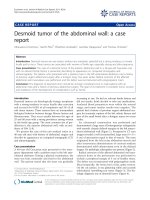
occlusion, 10 mg of tPA was infused in a pulsatile fashion
over 30 minutes. Angiography showed improved flow
through the distal basilar artery, with no change in the
appearance of the calcified dilation of the basilar tip or
extravasation of dye (Figure 1). The patient was sent to the
ICU. Diffusion magnetic resonance imaging (MRI) the
following morning showed multiple tiny diffusion bright
lesions in the cerebellum, thalamus and midbrain. This
was consistent with an aborted basilar artery occlusion
with evidence of ischemia throughout the entire basilar
artery vascular territory (Figure 2). On examination the
following afternoon, the patient had near complete reso-
lution of symptoms, and was discharged to rehabilitation
9 days later with a mild right 6th cranial nerve palsy. He
was discharged on Warfarin with an International Nor-
malized Ratio (INR) of 2.0 as the ischemic changes seen
on MRI were minimal, suggesting a low risk of hemor-
rhagic conversion. No dye extravasation was seen from
the aneurysm. It was felt that he was at high risk for recur-
rent thrombosis. The plan was for non-emergency
endovascular coiling of the basilar tip aneurysm 2 months
after discharge; however, the patient was lost to follow-up
and did not return to the vascular clinic.
Discussion
Due to a lack of data from randomized clinical trials,
administration of IA thrombolysis for patients with basi-
lar artery occlusion remains controversial [2]. Given the
poor natural history of the disease and the low (<20%)
estimated spontaneous recanalization rate, some neurolo-
gists feel that the survival benefit of IA thrombolysis pre-
dicted from published case series is sufficient evidence for
its use. Among the 10 studies of IA thrombolysis for acute
basilar occlusion published in the English literature, there
was an aggregate recanalization rate of 64%, and an over-
all 48% absolute risk reduction for death among patients
who recanalized versus those who did not [3]. Without
recanalization, the likelihood of good outcome is less
than 5% [4]. Reports have varied regarding the effect of
delayed administration of IA thrombolysis for basilar
Cerebral angiogram demonstrating calcified abnormality in the distal basilar arteryFigure 1
Cerebral angiogram demonstrating calcified abnor-
mality in the distal basilar artery. Flow is seen in the dis-
tal basilar artery after 10 mg of tissue plasminogen activator
was infused in a pulsatile fashion over 30 minutes.
Journal of Medical Case Reports 2008, 2:353 />Page 3 of 4
(page number not for citation purposes)
occlusion. One case series reported that treatment beyond
the 6-hour window resulted in no increased risk of hem-
orrhage among 20 patients treated for basilar occlusion
[1]. A similar study of 26 patients showed no association
between survival and the treatment interval [5], while
another reported a statistically significant decrease in reca-
nalization rate in patients treated beyond 6 hours of
symptom onset [6]. A recent report suggested that even
symptomatic chronic basilar occlusions (>80 days) may
be improved by vascular intervention [7], representing the
ability of this area to survive despite low cerebral blood
flow.
Diffusion-weighted magnetic resonance image showing punctate diffusion bright lesions in the cerebellum (panels a and b), tha-lamus (c), and midbrain (d)Figure 2
Diffusion-weighted magnetic resonance image showing punctate diffusion bright lesions in the cerebellum
(panels a and b), thalamus (c), and midbrain (d).
Publish with BioMed Central and every
scientist can read your work free of charge
"BioMed Central will be the most significant development for
disseminating the results of biomedical research in our lifetime."
Sir Paul Nurse, Cancer Research UK
Your research papers will be:
available free of charge to the entire biomedical community
peer reviewed and published immediately upon acceptance
cited in PubMed and archived on PubMed Central
yours — you keep the copyright
Submit your manuscript here:
/>BioMedcentral
Journal of Medical Case Reports 2008, 2:353 />Page 4 of 4
(page number not for citation purposes)
For the patient described in this report, delayed IA throm-
bolysis was pursued as a final measure to reverse a rapid
deterioration in his condition despite concerns regarding
the structural abnormalities in the distal basilar artery.
Post-lysis MRI demonstrated small areas of injury
throughout the vascular territory of the basilar artery that
in conjunction with his deteriorating clinical exam, sug-
gested that the entire basilar territory (that is to say, thala-
mus, midbrain and medulla) was at risk for infarct (Figure
2). His declining mental status was likely due to thalamic
ischemia. Despite the delay in treatment, we were able to
salvage this "at risk" territory and the patient was left with
minimal deficits.
Conclusion
This case illustrates a complex management issue in a
patient with basilar thrombosis in the setting of a large
basilar tip aneurysm. The possibility that flow changes
could occur with aggressive endovascular treatment and
reperfusion that could lead to aneurysm rupture needed
to be considered in the risk/benefit assessment of treat-
ment. In addition, although it has been well described in
the literature that late (>3 hours) treatment of basilar
occlusion can lead to good outcomes, and that the natural
history without treatment is bleak, there are no large pro-
spective trials showing the benefit of late intervention.
Many of these procedures are done outside of the classic
"therapeutic window", as was done in this patient due to
his rapid clinical deterioration. The risks of these proce-
dures when done outside of a clinical trial must be dis-
cussed with the patient (if possible) and family, especially
in a high-risk, unusual case such as this. To date, the use
of IA thrombolytics in dissecting arterial aneurysms or for
thrombosis during aneurysm coiling has been described
in the literature [7] but this is the first report of a sponta-
neous thrombosis in a saccular aneurysm treated with
delayed thrombolytics.
Abbreviations
CTA: computed tomography angiogram; ED: emergency
department; IA: intra-arterial; ICU: intensive care unit;
MRI: magnetic resonance imaging; Strength testing is
listed as 2/5 as a standard neurological score where 5/5 is
maximal strength; tPA/Altepase: tissue plasminogen acti-
vator
Consent
Written informed consent was obtained from the patient
for publication of this case report and any accompanying
images. A copy of the written consent is available for
review by the Editor-in-Chief of this journal.
Competing interests
The authors declare that they have no competing interests.
Authors' contributions
TF was a major contributor in writing the manuscript and
performing the literature review. LDM interpreted the
patient data and was a major contributor in writing the
manuscript. Both authors read and approved the final
manuscript.
Acknowledgements
Drs Gary R Spiegel and Stephen K. Ohki interpreted the images, treated
the patient and provided patient data. Dr McCullough is supported by NIH
grants 5R01NS050505 and 5R01NS055215-02.
References
1. Ezaki Y, Tsutsumi K, Onizuka M, Kawakubo J, Yagi N, Shibayama A,
Toba T, Koga H, Miyazaki H: Retrospective analysis of neurolog-
ical outcome after intra-arterial thrombolysis in basilar
artery occlusion. Surg Neurol 2003, 60(5):423-429.
2. Ford GA: Intra-arterial thrombolysis is the treatment of
choice for basilar thrombosis: con. Stroke 2006,
37(9):2438-2439.
3. Smith WS: Intra-arterial thrombolytic therapy for acute basi-
lar occlusion: pro. Stroke 2007, 38(2 Suppl):701-703.
4. Schonewille WJ, Algra A, Serena J, Molina CA, Kappelle LJ: Outcome
in patients with basilar artery occlusion treated convention-
ally. J Neurol Neurosurg Psychiatry 2005, 76:1238-1241.
5. Cross DT 3rd, Moran CJ, Akins PT, Angtuaco EE, Diringer MN: Rela-
tionship between clot location and outcome after basilar
artery thrombolysis. Am J Neuroradiol 1997, 18(7):1221-1228.
6. Arnold M, Nedeltchev K, Schroth G, Baumgartner RW, Remonda L,
Loher TJ, Stepper F, Sturzenegger M, Schuknecht B, Mattle H: Clini-
cal and radiological predictors of recanalisation and out-
come of 40 patients with acute basilar artery occlusion
treated with intra-arterial thrombolysis. J Neurol Neurosurg Psy-
chiatry 2004, 75(6):857-862.
7. Yu W, Kostanian V, Fisher M: Endovascular recanalization of
basilar artery occlusion 80 days after symptom onset. Stroke
2007, 38(4):1387-1389.
8. Bendok BR, Padalino DJ, Levy EI, Qureshi AI, Guterman LR, Hopkins
LN: Intravenous abciximab for parent vessel thrombus dur-
ing basilar apex aneurysm coil embolization: case report and
literature review. Surg Neurol 2004, 62(4):304-311.