Báo cáo y học: " Difficulties in establishing a timely diagnosis of pulmonary artery sarcoma misdiagnosed as chronic thrombo-embolic pulmonary disease: a case report" ppsx
Bạn đang xem bản rút gọn của tài liệu. Xem và tải ngay bản đầy đủ của tài liệu tại đây (1.52 MB, 5 trang )
BioMed Central
Page 1 of 5
(page number not for citation purposes)
Journal of Medical Case Reports
Open Access
Case report
Difficulties in establishing a timely diagnosis of pulmonary artery
sarcoma misdiagnosed as chronic thrombo-embolic pulmonary
disease: a case report
Ivanka Djordjevic*
1
, Tatjana Pejcic
1
, Milan Rancic
1
, Milan Radovic
1
,
Petar Bosnjakovic
2
, Tatjana Radjenovic-Petkovic
1
, Desa Nastasijevic-
Borovac
1
, Slavica Golubovic
1
and Dragana Dacic
1
Address:
1
The Clinical Center Nis, Clinic for lung diseases, Department for non specific lung diseases, Bul. Dr. Zorana Djindjica 48, 18 000 Nis,
Republic of Serbia and
2
The Clinical Center Nis, Institute for Radiology, Bul. Dr. Zorana Djindjica 48, 18 000 Nis, Republic of Serbia
Email: Ivanka Djordjevic* - ; Tatjana Pejcic - ; Milan Rancic - ;
Milan Radovic - ; Petar Bosnjakovic - ; Tatjana Radjenovic-Petkovic - ;
Desa Nastasijevic-Borovac - ; Slavica Golubovic - ; Dragana Dacic -
* Corresponding author
Abstract
Introduction: Pulmonary artery sarcomas are rare neoplasms that are often confused with
chronic thrombo-embolic disease, as both can have similar clinical and imaging presentation.
Case presentation: In this report, we present a case of a 50-year-old man initially diagnosed with
chronic thrombo-embolic pulmonary disease, but who was later found to have pulmonary artery
sarcoma with poor survival prognosis. We review the clinical and imaging characteristics of the two
diseases and discuss the difficulties in establishing a timely diagnosis.
Conclusion: Similar clinical features and imaging presentation of pulmonary artery sarcoma and
chronic thrombo-embolic pulmonary disease make definitive diagnosis difficult. This case report
also illustrates and emphasizes that in any case with no predisposition factors for embolism, no
evidence of deep venous thrombosis and pulmonary emboli, and inadequate relief of symptoms
with anticoagulation, an alternative diagnosis of pulmonary artery sarcoma should be considered.
If pulmonary artery sarcoma is diagnosed late in the course of the disease, there is usually a poor
survival outcome.
Introduction
Pulmonary artery sarcoma is a rare tumor of the cardiovas-
cular system. Because of its rarity and insidious growth, it
is often mistaken for pulmonary embolism [1,2]. Clinical
symptoms, as well as imaging characteristics often associ-
ated with pulmonary embolism, are also very common in
patients with pulmonary artery sarcoma which, in many
instances, delays the correct diagnosis. [3-6]. We present a
case of a man who experienced a thrombus in the main
pulmonary artery and was later diagnosed with pulmo-
nary artery sarcoma with poor survival outcome.
Case presentation
A 50-year-old man was admitted to our clinic with a 20-
day history of cough, small amounts of haemoptysis, fever
and exhaustion. There was no past medical history of pre-
Published: 16 February 2009
Journal of Medical Case Reports 2009, 3:64 doi:10.1186/1752-1947-3-64
Received: 24 January 2008
Accepted: 16 February 2009
This article is available from: />© 2009 Djordjevic et al; licensee BioMed Central Ltd.
This is an Open Access article distributed under the terms of the Creative Commons Attribution License ( />),
which permits unrestricted use, distribution, and reproduction in any medium, provided the original work is properly cited.
Journal of Medical Case Reports 2009, 3:64 />Page 2 of 5
(page number not for citation purposes)
disposition factors for embolism or episodes of venous
thrombo-embolism.
During the hospitalization, the patient developed a mas-
sive haemoptysis. Laboratory blood tests showed elevated
inflammatory parameters, but platelets and plasma coag-
ulation function parameters were in the normal range:
platelet: 254 × 10
9
/L; fibrinogen: 278 mg/dl; bleeding
time, Duke methods: < 4 minutes; partial thromboplasin
time: 29 seconds; prothrombin time: 12.7 seconds;
thrombin time: 17 seconds; protein C: 121%; protein S:
93%. A computerized tomography (CT) scan of the lungs
showed a few areas of pulmonary consolidation on the
right side and a few enlarged mediastinal lymph nodes
(Figure 1). The presumptive fibre optic bronchoscopy
findings were bronchogenic carcinoma; however, his-
topathological findings did not confirm a malignancy.
The patient was given symptomatic therapy which led to
a complete clinical recovery, and he was discharged with
follow-up recommended.
Two months later the patient developed intensive chest
pain, medium amounts of haemoptysis and fever. Labora-
tory tests showed three-fold elevated inflammatory
parameters and D-dimer value. Chest radiography was
significant since it showed prominent parenchymal den-
sity in the lateral right low-lung field as well as heart
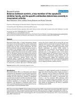
enlargement. On CT scan (Figure 2), we identified differ-
ent locations of pulmonary consolidation and pleural
thickening on the right side, as well as a dilated pulmo-
nary artery trunk. Venous duplex ultrasound of the lower
extremities was negative for deep-vein thrombosis. How-
ever, echosonography showed right ventricular hypertro-
phy; perfusion scintigraphy uncovered a complete
absence of the perfusion of the right lung, and pulmonary
angiography (Figure 3) showed a complete occlusion of
the right pulmonary artery due to external compression or
intra-luminal infiltration. The patient was placed on low-
density heparin for a presumptive diagnosis of pulmonary
embolism. There were no further episodes of haemopty-
sis, and the patient was discharged with a recommended
anticoagulant therapy.
A further 6 months later, despite the anticoagulant ther-
apy treatment, the patient developed massive haemopty-
sis. Transoesophageal echosonography showed a mass in
the pulmonary trunk. On the basis of contrast-enhanced
CT scan (Figure 4) and magnetic resonance imaging
(MRI) (Figure 5) of the heart and blood vessels showing a
large filling defect in the pulmonary artery trunk and in
the right branch, massive pulmonary embolism was diag-
nosed. A right dorsal posterior pleural effusion was noted
and 25 ml of haemorrhage pleural fluid was drained.
The patient underwent surgery with embolectomy of a
suspected thrombotic mass in the pulmonary trunk; how-
ever, histopathology revealed an angiosarcoma of the pul-
monary artery. He was haemodynamically unstable, and
due to respiratory insufficiency, invasive mechanical ven-
tilation had to be carried out. Three weeks later, he died
due to heart and lung failure.
Computerized tomography scan of the lung: a few areas of pulmonary consolidation on the right side and a few enlarged mediastinal lymph nodes can be seenFigure 1
Computerized tomography scan of the lung: a few
areas of pulmonary consolidation on the right side
and a few enlarged mediastinal lymph nodes can be
seen.
Computerized tomography scan of the lung showing different locations of pulmonary consolidation and pleural thickening on the right side and dilated pulmonary artery trunkFigure 2
Computerized tomography scan of the lung showing
different locations of pulmonary consolidation and
pleural thickening on the right side and dilated pul-
monary artery trunk.
Journal of Medical Case Reports 2009, 3:64 />Page 3 of 5
(page number not for citation purposes)
Discussion
Pulmonary artery sarcoma is a rare tumor of the cardiovas-
cular system. The reported age for its appearance ranges
from 13 to 86 years of age, with the majority of cases
occurring in middle age. The aetiology of these tumors is
obscure. It has been suggested that they arise from the
mesenchymal cells of the muscle angle of the bulbus
cordis. The most frequent histopathological type is leio-
myosarcoma or "undifferentiated spindle-cell sarcoma".
Angiosarcoma accounts for 7% of cases. The histopatho-
logical classification does not seem to be useful clinically
or prognostically [1,2].
Because of its rarity and insidious growth characteristics,
pulmonary artery sarcoma is often mistaken for pulmo-
nary embolism, leading to inappropriate therapy such as
prolonged anticoagulation or thrombolysis [1]. The
symptoms often associated with pulmonary embolism
can be present in patients with pulmonary artery sarcoma
[1,3,7], including sudden onset of chest pain, dyspnoea
and haemoptysis, cough or right-heart failure. However,
patients with pulmonary artery sarcoma generally experi-
ence a slowly progressing decline over several weeks or
Pulmonary angiography showing a large filling defect causing complete obstruction of the right pulmonary arteryFigure 3
Pulmonary angiography showing a large filling defect
causing complete obstruction of the right pulmonary
artery.
Contrast-enhanced computerized tomography scan of large blood vessels showing a large filling defect in the pulmonary artery trunk and in one-third of the left main pulmonary artery, as well as a complete occlusion of the right branch due to emboli or some infiltrative massFigure 4
Contrast-enhanced computerized tomography scan
of large blood vessels showing a large filling defect in
the pulmonary artery trunk and in one-third of the
left main pulmonary artery, as well as a complete
occlusion of the right branch due to emboli or some
infiltrative mass.
Contrast-enhanced magnetic resonance image of the large blood vessels showing a large filling defect in the pulmonary artery trunk and complete occlusion of the right branch with a discreet increase in signal intensity after contrast injection (arrows)Figure 5
Contrast-enhanced magnetic resonance image of the
large blood vessels showing a large filling defect in
the pulmonary artery trunk and complete occlusion
of the right branch with a discreet increase in signal
intensity after contrast injection (arrows).
Journal of Medical Case Reports 2009, 3:64 />Page 4 of 5
(page number not for citation purposes)
months, characterized by symptoms of weight loss, fever
and severe fatigue, as commonly seen in malignancy [8].
The symptoms observed in our patient were related to the
infiltration or invasion of the adjacent bronchus, which
lead to the initial presumed diagnosis of bronchogenic
carcinoma.
Laboratory studies are of little value in establishing the
diagnosis. It is not uncommon to observe anaemia or an
elevated erythrocyte sedimentation rate [3]; we also found
elevated inflammatory parameters, which are non-specific
findings. These findings are unusual in pulmonary embo-
lism.
Pulmonary artery sarcoma develops within the pulmo-
nary trunk or pulmonary valve region and is frequently
associated with a multicentric origin in the outflow track
of the right ventricle. Also, perfusion defects remain static
or progressive over time rather than changing with fibri-
nolysis or recurring, as one might expect in thrombo-
embolic disease [3]. Approximately 40% of patients
develop a direct invasion or metastasis to the lung, while
systemic spread to kidneys, brain or adrenal glands occurs
in about 20% of cases [3,9].
Chest radiographic findings are varied. The most common
finding is an abnormal hilar shadow that has the appear-
ance of an enlarged pulmonary artery or mass projecting
into the lung parenchyma. Other common findings are
pulmonary consolidation, atelectasis or pulmonary nod-
ules or masses, presumably the result of the embolic phe-
nomenon with or without infarction or metastases in the
lungs [8,10]. Pleural effusion, usually haemorrhagic with
normal cytological findings, is a common finding [8] and
was also observed in our patient.
However, in pulmonary thrombo-embolic disease, imag-
ing changes are most likely bilateral or recurrent, depend-
ing on the aetiology and therefore on the direction of
blood flow and distribution of emboli [5,6,11].
In our case, despite anticoagulation, the imaging features
remained persistent. Furthermore, a unilateral central pul-
monary embolus is relatively uncommon and suggests a
possibility of malignancy [4-7].
Pulmonary artery sarcoma and chronic thrombo-embolic
pulmonary disease can be easily confused on CT or MRI
scan, because both are characterized by intra-luminal fill-
ing defects and pulmonary arterial dilatation [4,7]. How-
ever, there are radiographic criteria that may help
differentiate the two entities. CT findings consistent with
malignancy include filling defects occupying the entire
luminal diameter of the pulmonary arteries and extra-
luminal extension of the tumor. In addition, pulmonary
artery sarcomas may be indicated by areas of inhomoge-
neous, high or low attenuation, representing haemor-
rhage or necrosis, soft-tissue density in pulmonary
arteries, or enhancement after administration of gadopen-
tetate dimeglumine on the MRI [4,7]. Gadolinium-
enhanced MRI is another potentially useful diagnostic
tool in differentiating between intra-luminal tumors and
large thrombi [4,7,12].
There are no specific findings on echosonography, per-
fusion lung scan or pulmonary angiography perfusion
lung scan that would reliably differentiate embolic
obstruction from obstruction caused by a tumour, with
the exception of commonly static perfusion defects and a
positive gallium scan in the cases of sarcomatous obstruc-
tion [3,4,12].
In cases of pulmonary artery sarcoma, early diagnosis and
radical surgical resection offer the only opportunity for
prolonged survival [1,13], with only three cases reported
to survive for longer than 3 years. Surgical resection
increases the chances of survival by approximately 12
months, and there is limited evidence that this may be fur-
ther extended by neo-adjuvant and/or adjuvant irradia-
tion and chemotherapy [14,15]. The role of radiation
therapy and postoperative anticoagulation therapy is still
not clearly defined [1,9]. For patients with extensive medi-
astinal involvement or metastatic disease, limited tumour
resection or bypass procedure may offer significant pallia-
tion benefit and enhance survival. The prognosis is
mainly dependent on local recurrence. The average time
of survival without an intervention is 6 weeks [7,14].
In our case, a diagnosis was established too late, the dis-
ease was in an advanced stage and there was no appropri-
ate therapy modality.
Conclusion
Our case illustrates and emphasizes that pulmonary artery
sarcoma should be included in the differential diagnosis
of pulmonary thrombo-embolic disease in cases where: a)
symptoms do not respond to anticoagulation, b) no
source of thrombi and emboli can be detected, and c) pul-
monary nodules and/or metastases develop on follow-up.
If pulmonary artery sarcoma is diagnosed after the occur-
rence of distal metastases or involvement of adjacent
mediastinal structure, there is a poor survival outcome.
Abbreviations
CT: computerized tomography; MRI: magnetic resonance
imaging
Consent
Written informed consent was obtained from the patient's
next-of-kin (sister) for publication of this case report and
Publish with BioMed Central and every
scientist can read your work free of charge
"BioMed Central will be the most significant development for
disseminating the results of biomedical research in our lifetime."
Sir Paul Nurse, Cancer Research UK
Your research papers will be:
available free of charge to the entire biomedical community
peer reviewed and published immediately upon acceptance
cited in PubMed and archived on PubMed Central
yours — you keep the copyright
Submit your manuscript here:
/>BioMedcentral
Journal of Medical Case Reports 2009, 3:64 />Page 5 of 5
(page number not for citation purposes)
any accompanying images. A copy of the written consent
is available for review by the Editor-in-Chief of this jour-
nal.
Competing interests
The authors declare that they have no competing interests.
Authors' contributions
ID was a chief author of the manuscript, researched the
case, contributed to the concept, design and definition of
intellectual content along with the literature search, data
acquisition & analysis and manuscript preparation. TP
and TRP assisted with the analysis of the data, helped sub-
stantially with the discussion and contributed to the man-
uscript. MR and MR helped in data analysis and
manuscript preparation, editing and review. DNB, SG and
DD assisted with the details of the case report and have
been involved in drafting the manuscript. PB analyzed
and interpreted computerized tomography and magnetic
resonance imaging and helped with the discussion. All
authors have read and approved the final manuscript.
Acknowledgements
The authors wish to thank all physicians from the clinic who participated in
the medical care of the patient described in this report. Also, we are grate-
ful to Nikola Ilic and Marta Djordjevic for technical assistance in the com-
puter processing of images.
References
1. Mattoo A, Fedullo PF, Kapelanski D, Ilowite JS: Pulmonary artery
sarcoma. Chest 2002, 122(2):745-7.
2. Pagni S, Passik CS, Riordan C, D'Agostino RS: Sarcoma of the main
pulmonary artery: an unusual etiology of recurrent pulmo-
nary emboli. J Cardiovasc Surg 1999, 40:457-461.
3. Parish JM, Rosenow EC, Swensen SJ, Crotty TB: Pulmonary artery
sarcoma. Clinical features. Chest 1996, 110:1480-1488.
4. Cox JE, Chiles C, Aquino SL, Savage P, Oaks T: Pulmonary artery
sarcomas: a review of clinical and radiological features. J
Comput Assist Tomogr 1997, 21:750-755.
5. Au VWK, Veitch E, Gustafson S, Kermeen F, Sage MR: Radiological
inestigation of pulmonary embolism: an audit in teaching
hospital. J HK Coll Radiol 2005, 8:141-145 [ />publ/Journal/counter.php?ref=vol8no3/original_article_1.pdf].
6. Garg K: CT of pulmonary thromboembolic disease. Radiol Clin
North Am 2002, 40:111-122.
7. Kaplinsky EJ, Favaloro RR, Pombo G, Perrone SV, Vigliano CA, Sch-
nidt JL, Bougen RP: Primary pulmonary artery sarcoma resem-
bling chronic thromboembolic pulmonary disease. Eur Respir
J 2000, 16:1202-1204.
8. Burke AP, Virami R: Sarcomas of the large vessels. A clinico-
pathological study. Cancer 1993, 71:1761-1773.
9. Long HQ, Qin Q, Xie CH: Response of pulmonary artery inti-
mal sarcoma to surgery, radiotherapy and chemotherapy: a
case report. J Med Case Reports 2008, 2:217.
10. Sheikh Hina, Gregorio Remigio, Yousem Samuel: Final Diagnosis –
Rhabdomyosarcoma of pulmonary artery trunk. University of
Pittsburgh, School of Medicine, Department of Pathology Online Case Stud-
ies. Case 303 – Progressive Shortness of Breath, Pulmonary Pathology
[ />].
11. Odev K, Acikgozoglu S, Gormus N, Aribas O, Kiresi D, Solak H: Pul-
monary embolism due to cardiac hydatid disease: imaging
findings of unusual complication of hydatid cyst. Eur Radiol
2002, 12:627-633.
12. Yoshiaki O, Toru S, Teiji T, Takeyoshi K, Satoru F, Naoki M, Yasuo B:
Pulmonary artery sarcoma diagnosed by using intravascular
ultrasound images. Thorax 1999, 54:748-749.
13. Nakahira A, Ogino H, Sasaki H, Katakami N: Long-term survival of
pulmonary artery sarcoma produced by aggressive surgical
resection and adjuvant chemotherapy. Eur J Cardiothorac Surg
2007, 32:388-390.
14. Kathiravel Y, Westwood D, Macemon J, Singh H: An international
surgical collaboration for the management of pulmonary
artery sarcoma: a New Zealand experience. J NZ Med Assoc
2007, 120:1257.
15. Hsing JM, Thakkar SG, Borden EC, Budd GT: Intimal pulmonary
artery sarcoma presenting as dyspnea: a case report. Int
Semin Surg Oncol 2007, 4:14.