Báo cáo y học: "Sequence similarity between the erythrocyte binding domain 1 of the Plasmodium vivax Duffy binding protein and the V3 loop of HIV-1 strain MN reveals binding residues for the Duffy Antigen Receptor for Chemokines" ppsx
Bạn đang xem bản rút gọn của tài liệu. Xem và tải ngay bản đầy đủ của tài liệu tại đây (746.53 KB, 10 trang )
RESEARC H Open Access
Sequence similarity between the erythrocyte
binding domain 1 of the Plasmodium vivax Duffy
binding protein and the V3 loop of HIV-1 strain
MN reveals binding residues for the Duffy
Antigen Receptor for Chemokines
Michael J Bolton
1
, Robert F Garry
2*
Abstract
Background: The surface glycoprotein (SU, gp120) of the human immunodeficiency virus (HIV) must bind to a
chemokine receptor, CCR5 or CXCR4, to invade CD4+ cells. Plasmodium vivax uses the Duffy Binding Protein (DBP)
to bind the Duffy Antigen Receptor for Chemokines (DARC) and invade reticulocytes.
Results: Variable loop 3 (V3) of HIV-1 SU and domain 1 of the Plasmodium vivax DBP share a sequence similarity.
The site of amino acid sequence similarity was necessary, but not sufficient, for DARC binding and contained a
consensus heparin binding site essential for DARC binding. Both HIV-1 and P. vivax can be blocked from binding to
their chemokine receptors by the chemokine, RANTES and its analog AOP-RANTES. Site directed mutagenesis of
the heparin binding motif in members of the DBP family, the P. knowlesi alpha, beta and gamma proteins
abrogated their binding to erythrocytes. Positively charged residues within domain 1 are required for binding of
P. vivax and P. knowlesi erythrocyte binding proteins.
Conclusion: A heparin binding site motif in members of the DBP family may form part of a conserved erythrocyte
receptor binding pocket.
Introduction
Human immunodeficiency virus type 1 (HIV-1) and the
human malaria parasite Plasmodium vivax both use che-
mokine receptors in obligate steps of cell invasion. HIV-
1 uses CCR5 and CXCR4 as the major coreceptors for
infecting CD4+ cells (macrophages, T-lymphocytes, and
other cell types) in vivo,whileP. vivax uses the Duffy
antigen receptor for chemokines (DARC) for invading
human reticulocytes [1,2]. Alleles of CCR5 and DARC
associated with decreased functional protein e xpress ion
confer resistance to HIV and P. vivax, respectively, and
chemokines can inhibit in vitro infection by either
pathogen [1,3-5]. The HIV surface glycoprotein (SU,
gp120) undergoes a conformational change upon
binding to CD4 and then presents a chemokine receptor
bindi ng surface predicted to include a hydrophobic core
surrounded by positive residues contributed by con-
served and variable regions including the base of the
V3 loop. The V3 loop putatively extends toward the cell
surface and contacts the chemokine receptor at a second
site in the second extracellular loop. Individual amino
acid mutations i n the V3 loop can change chemokine
receptor specificity.
P. vivax and the simian malaria, P. knowlesi, use Duffy
binding proteins (PvDBP and PkDBP) to invade human
erythrocytes. These proteins belong to a family of ery-
throcyte binding proteins with conserved regions. The
erythrocyte binding domains of PvDBP and PkDBP (or
P. knowlesi a protein) have been shown to map to the
330 amino-acid cysteine-rich region II known as the
Duffy-binding-like (DBL) domains [6 ]. Other me mbers
of the family include the P. knowlesi b and g proteins
* Correspondence:
2
Department of Microbiology and Immunology Tulane University
1430 Tulane Avenue New Orleans, Louisiana 70112 USA
Full list of author information is available at the end of the article
Bolton and Garry Virology Journal 2011, 8:45
/>© 2011 Bolton and Garry; licensee BioMed Centr al Ltd. This is an Open Access article distributed under the terms of the Creative
Commons Attribution License ( whic h permits unrestricted use, distribution, and
reproduction in any medium, provided the original work is properly cited.
and the P. falciparum erythrocyte-binding antigen (EBA-
175), which use DBLs to bind to other receptors.
Here we repo rt the identification o f an amino acid
sequence similarity between the V3 loop of HIV-1 strain
MN and a site in Plasmodium erythrocy te binding pro-
teins that contains a consensus heparin binding site. Both
HIV-1 and P. vivax can be blocked from bindi ng to their
chemokine receptors by the chemokine RANTES. Muta-
genesis studies suggest that the heparin b inding site
motif in members of the DBP family may form part of a
conserved erythrocyte receptor binding pocket.
Materials and methods
Sequence comparisons
William Pearson’s LALIGN program, which implements
a linear-space local similarity algorithm, was u sed to
perform regional alignments. Sequence and structural
comparisons were performed for the V3 loop of SU of
HIV-1 strain MN, accession: AAT67509; P. vivax DBP,
ACD76813; P. knowlesi DBP, XP_002261904; P. falci-
parum erythrocyte binding protein EBA-175 (F1), acces-
sion AAA29600. Plasmodium proteins are members of
pfam05424 (a member of the superfamily cl05146).
Erythrocytes
Blood was collected in 10% citrate phosphate dextrose
(CPD) and stored at 4°C unwashed for up to 4 weeks,
or washed in RPMI with malaria supplements and
stored in malaria culture medium at 50% hematocrit for
up to 2 weeks. The DARC+ human erythrocyte s used in
the erythrocyte binding assay and the P. knowlesi ery-
throcyte invasion assay had the phenotype Fy(a
-
b
+
)as
determined by standard blood banking methods using
anti-Fya and anti-Fyb antisera (Gamma Biologicals,
Houston, TX). Erythrocytes were washed three times in
DMEM (Gibco BRL) and resuspended to a hematocrit
of 10% in complete DMEM for the erythrocyte binding
assay. Erythrocytes used in the P. knowlesi erythrocyte
invasion assay were washed three times and resuspended
to a hematocrit of 10% using malaria complete RPMI.
Cell Culture and Transfection of COS-7 Cells
COS-7 cells (ATCC CRL 1651; Rockville, MD) were cul-
tured in DMEM with 10% heat inactivated FBS (Gibco
BRL) in a humidified 5% CO
2
incubator at 37°C. Cells
were seeded in po lystyrene dishes with 3.5-cm diameter
wells and grown for 24 h to 30-50% confluence before
transfection with 1 mg of pHVDR22 plasmid DNA and
10 ml of Lipofectamine (Gibco BRL).
P. knowlesi in vitro culture
Whole blood from rhesus macaques was collected in
10% CPD and allowed to separate overnight at 4°C.
The erythrocyte phase was washed in RPMI with
L-glutamine and supplemented with 25 mM HEPES,
300 mM hypoxanthine, 10 mM thymidine, 1.0 mM
sodium pyruvate, and 11 mM glucose. This RPMI with
malaria supplements was then used to prepare malaria
culture medium by adding to a final concentration of
0.24% sodium bicarbonate and 0.2% Albumax-I (Life
Tech, Gibco BRL). Cultures were maintained at a hema-
tocrit of 10% in malaria culture medium under an atmo-
sphere of 5% O
2
,5%CO
2
, balanced N
2
(Air Liquide,
Houston, TX) at 38°C.
Percoll Purification of Schizont-infected Erythrocytes
Cultures of P. knowlesi at 5-10% infected erythrocytes
were washed three times in RPMI with malaria supple-
ments and 10% FBS and brought up to a hematocrit of
10%. A 50% Percoll solution was made by adding
0.45 volumes 1X PBS, 0.05 volumes 10X PBS and
0.5 volumes Percoll (Sigma). Two ml of the washed cul-
ture was overlaid on 2 ml of the 50% Percoll solution in
a 4 ml polystyrene tube and centrifuged for 20 min at
2100 RPM in a Sorvall centrifuge. The ring of cells at
the interface was removed, pooled and washed three
time in 1X PBS. The pellet was brought up in malaria
culture medium to 2 × 10
7
cells/ml.
PvRII Erythrocyte Binding Assay
COS-7 cells were transfected by Lipofectamine with
1-2 mg of pHVDR22 DNA, a plasmid kindly provided
by L. Miller which expresses region I I of the DBP of P.
vivax on the cell surface as a chimera with the HSV gD
protein [7] Duffy Fy (a-b+) erythrocytes were washed
three times in RPMI 1640, resu spended to a hematocrit
of 1% in 1 ml of complete DMEM with the chemokines
RANTES, MIP-1a, SDF-1 or AOP-RANTES at concen-
trations of 0, 0.1, 1, 10, and 100 nM for 1 h at 37°C
(Peprotech, Gryphon Pharmaceuticals, San Francisco,
CA). This suspension was swirled over aspirated COS-
7 cells 40-60 h after transfection and allowed t o settle
over 2 h at 37°C. The COS-7 cells were then washed
three times with 2 ml of PBS to remove nonadherent
erythrocytes. The number of adherent erythrocyte
rosettes was scored in 20 randomly chosen fields at a
magnification of 40 using an inverted microscope. Per-
cent inhibition was determined by dividing the number
of rosettes in the presence of chemokines by the num-
ber at a concentration of 0 nM. The 50% inhibitory con-
centration (IC
50
) was determined by the mean of t hree
separate experiments to use in a semi-log cubic spline
curve fit with the DeltaSoft 3 software (Biometallics,
Inc., Princeton, NJ).
P. knowlesi Erythrocyte Invasion Assay
Human Duffy Fy(a
-
b
+
) erythrocytes were washed in
complete malaria medium and 2 × 10
7
washed cells
Bolton and Garry Virology Journal 2011, 8:45
/>Page 2 of 10
were added to increasing concentrations of chemokines
in malaria culture medium at final volume of 900 ml for
1h at roo m temperature. To each tube of chemokine-
treated erythrocytes, 100 ml or 2 × 10
6
schizont-infected
erythrocytes was added and placed in a well of a poly-
styrene 24-well plate (Becton-Dickinson). The cultures
were maintained under a blood-gas atmosphere at 3 8°C
for 8 h to allow the infected erythrocytes to rupture and
release free mero zoites capable of infecting new erythro-
cytes and developing to ring-stage trophozoites. The
culture was centrifuged at 2100 RPM for 3 min and a
thin smear was made from the pellet. The thin smear
was fixed with methanol and stained with Leukostat
Solution B (100 mg Eosin Y+300 ml 37% formaldehyde +
400 mg sodium phosphate dibasic + 500 mg p otassium
phosphate monobasic, q.s. to 100 ml with dH
2
O), rinsed,
and stained with Leukostat Solution C (47 mg Methylene
Blue + 44 mpp Azure A + 400 mg sodium phosphate
dibasic + 500 mg potassium phosphate monobasic, q.s to
100 ml with dH
2
O). The percentage of erythrocytes
infected with ring-stage trophozoites per 2000 erythrocytes
was determined at 1000X. Inhibition of invasion
expressed as % inhibition was determined by dividing the
percentage of ring-stage parasites by the percentage of
ring-stage parasites at 0 nM chemokine, multiplying b y
100 and subtracting this value from 100 [1].
Statistical analysis
The software StatView (Brainpower, Inc., Calabasas, CA),
was used to determine the statistical difference between
the inhibitory concentrations of RANTES, AOP-
RANTES, and MIP-1a, using a two-way ANOVA test.
Plasmids
The plasmids pHVDR22, pHKADR22, pHKBDR22 and
pHKGDR22 encode for the region II (amino acids
198-522) of the P. vivax DBP and region II of the
P. knowlesi a, b and g genes, respectivel y, in the context
of the HSV gD protein. These plasmids have been pre-
viously described and were kindly provided by the
laboratory of Louis H. Miller. These plasmids were
created from the plasmid pRE4, which contains an
SV40 origin of replication, a Rous sarcoma virus LTR as
a promoter, the coding region of the HSV glycoprotein
D (HSV gD) inserted in the HindIII cloning site, and the
SV40 early polyadenylat ion signal. The HSV gD features
a 25 amino acid signal peptide at the amino terminus, a
24 amino acid hydro phobic transmembrane region, a
30 amino acid cytoplasmic tail at the carboxy terminus,
and two epitopes at amino acids 11-19 and 272-279 that
can be targeted specifically with the monoclonal antibo-
dies ID3 and DL6, respectively. The region II sequences
were inserted between the unique Apa I and Pvu II
restriction sites.
Cloning and Site Directed Mutagenesis
Mutants of the region II expressing plasmids were gen-
erated by three strategies: inverse PCR, PCR and restric-
tion digestion, or PCR-based site directed mutagenesis.
Each mutant was sequenced by Research Genetics, Inc.
(Huntsville, Ala.) to confirm proper construction at the
site of mutation.
The following constructs were made from the
pHVDR22 plasmid:
pv22d32 This construct contains a deletion in amino
acids 216-247 of the RII of P. vivax, which corresponds
to the V3-like peptide region with similarity to
the V3 loop and comprises cysteines C1 to C4 of region
II. For lack of proper restriction enzyme sites, an
inverse PCR strategy was use to amplify the entire
pHVDR22 plasmid flanking the site to be deleted. The
primers 5’TGT ATG AAG GAA CTT ACG AAT TTG
G3’ and 5’TTT CAT TAC AGT ATT TTG AAG3’ were
first phosphorylated with T
4
kinase then used with the
long range, high fidelity DeepVent polymerase (New
England Biolabs, Inc., Beverly, MA) to amplify the pro-
duct under the following thermocycling conditions:
5 minutes at 94°C initial denaturing, then 35 cycles at
94°C for 60 seconds, 55°C for 60 seconds, 72°C for
3 minutes. The product was digested with DPN I to
eliminate methylated input plasmid DNA, then blunt-
end ligated with high concentration ligase (Gibco BRL).
pv22MNV3 This construct replaces the 32 amino acid
V3-like peptide of the P. vivax RII with the V3 loop of
HIV-1 strain MN. To amplify the V3 loop of HIV-1
MN
by PCR, PM-1 cells were infected with HIV-1
MN
(donated by Dr. James Robinson, Tulane University
Medical Center) and genomic DNA was isolated from
infected cultures. This DNA includes proviral DNA and
wasusedastemplateforaPCRwiththeprimers
P2 5’GAC GCT GCG CCC ATA GTG CTT CCT G3’
and P5 5’ ACA CAT GGAATT CGGCCAGTA GT3’
which are homologous to conserved regions of the env
gene of HIV and amplify the region between nucleotides
6884 and 7783, which includes the V3 loop.
This PCR product then served as template in a nested
PCR of the HIV-1
MN
V3 loop using the primers
HVMN-F 5’ AAT TGTACAAGACCCAACTAC3’ and
HVMN-r 5’ATGTGCTTGTCT TATAGTTCC3’ .This
nested PCR was carried out using the DeepVent enzyme
to generate blunt ends. The product o f the second,
nested PCR was gel purified. The gel-purified amplicon
wasthenre-amplifiedina300mlPCRusingHVMN-r
and HVMN-F primers, which were first phosphorylated
with T
4
poly N kinase. This reamplification product was
column purified and blunt-end ligated to the inverse
PCR product described in the preparation of pvD32.
The sequenced con struct matched the MN V3 sequence
as previously published.
Bolton and Garry Virology Journal 2011, 8:45
/>Page 3 of 10
pv22suf32 This construct was designe d to determine if
the 32-aa V3 -like peptide of P. vivax RII is sufficient for
DARC binding by deleting all flanking RII amino acids.
The primers used to create this construct were 5’ CAA
AAT CAG CTG AT G AAA AAC TGT AAT TAT3’
and 5’CAA ATT GGG CCC TTC CTT CAT ACA TAA
TTG3’ and contain the restriction sites for Apa I and
Pvu II. The pHVDR22 plasmid was digested with Apa I
and Pvu II, and the digested vector was separated from
the insert by gel electrophoresis and extracted using the
QIAEX II gel extraction kit (Qiagen Inc., Valencia, CA).
The PCR product was also digested with Apa I and Pvu
II and ligated to the digested vector.
pv22d5C1 This construct deletes amino acids 198-216,
or the 5’ flanking region to C1. This was created using
the primers 5’TGT ATG AAG GAA CTT ACG AAT
TTG G3’ and 5’ GGG GCC TTG GGC CCT GTC ACA
AC3’, the product of which was digested with Apa I and
Pvu II and cloned into the digested vector as described
for psuf32
pv22d3C4 This construct deletes amino acids 247 to
522 or the 3’ flanking region to C4. This was created
using the primers 5’ CCGGTCCTGGACCAGCTG
ACG3’ and 5’TTT CAT TAC AGT ATT TTG AAG3’
the product of which was digested with Apa I and Pvu
II and c loned into the digested vector as described in
psuf32
pv22d5C4 This construct deletes amino acids 198 to
247 or the 5’ flanking region to C4 This was created
using the primers 5’ CAATTACAGCTGAAGGAA
CTT ACG AAT TTG3’ and 5’ GGG GCC TTG GGC
CCT GTC ACA AC3’ the product of which was
digested with Apa I and Pvu II and cloned into the
digested vector as described in pv22suf32
pv22KARA The Stratagene QuickChange kit (Promega)
was used to mutate the heparin binding consensus site in
PvRII at amino acids 217-226 from YKRKRRERDW to
YARKAREADW using the primers 5’ GTA ATT ATG
CGA GAA AA G CTC GGG AAG CAG ATT GG3’ and
5’ CCA ATC TGC TTC CCG AGC TTT TCT CGC
ATA ATT AC3’. These primers also introduce an Ava I
site as a silent mutation for screening.
pv22KAKA The Stratagene QuickChange kit was used
to mutate a second potential heparin binding consensus
site at amino acids 364-373, between C5 and C6, from
SVKKRLKGNF to SVKARLAGNF using the primers 5’
GAT GTA CTC AGT TAA AGC AAG ACT TAA
GGG G3’. These primers al so introduce an Afl II site as
a silent mutation for screening
pv22KA The Stratagene Quick Change kit was used to
introduce a s ingle alanine substitution in the heparin
binding consensus site at amino acids 217-226 from
YKRKRRERDW to YKRARRERDW. 5’ CTC TTT CCC
GACGAGCTCTCTTATAATTACAG3’ and
5’ CTG TAA TTA TAA GAG AGC TCG TCG GGA
AAG AG3’.
These primers also introduce a Sac I site as
a silent mutation for screening.
The following mutants were made from the
pHKADR22, pHKBDR22, or pHKGDR22 plasmids using
the Stratagene QuickChange kit:
pkalpha22KARA This mutant was designed to change
the heparin binding consensus site in pHKADR22 at
amino acids 217-226 from DKRKRGERD to DARKA-
GEAD using the primers 5’GTCCCAATCTGCTTC
CCC GCG AGC TCT CGC ACT ACC ACA CTT G and
5’ CAA GCG TAA TGA TGC GAG AGC TCG CGG
GGA AGC AGA TTG GGA C3’ . These primers also
introduce a Sac I site as a silent mutation for screening.
pkbeta22KARA This mutant was designed to change
the heparin binding consensus site in pHKBDR22 at
amino acids 217-226 from NKRKRGTRD to NARKAG-
TAD using the primers 5’ CAG TCC CAA TCT GCT
GTC CCG CGA GCT TCT GCA TTA TTA CAC C3’
and 5’GGT GTA ATA ATG CGA GAG CTC GCG GGA
CAG C AG ATT GGG ACT G3 ’. These primers also
introduce a Sac I site as a silent mutation for screening.
pkgamma22KARA This mutant was designed to change
the heparin binding consensus site in pHKGDR22 at
amino acids 217-226 from DKRKRGERD to DARKA-
GEAD using the primers 5’GTCCCAATCTGCTTC
CCC GCG AGC TCT CGC ACT ACC ACA CTT G and
5’ CAA GCG TAA TGA TGC GAG AGC TCG CGG
GGA AGC AGA TTG GGA C3’ . These primers also
introduce a Sac I site as a silent mutation for screening.
Immunofluorescence Staining
Transfected cells used in the erythrocyte binding assay
were rinsed in PBS and incubated for 1 h at 37°C with
monoclonal antibodies that bind to amino acids
11-19 and 272-279 of the mature HSV gD protein
found in pHVDR22. These primary antibodies, ID3 or
DL6 (provided by Drs. Gary Cohen and Ro selyn Eisen-
berg), were used at a 1:2000 dilution in PBS containing
10% FBS. The cells were rinsed with PBS and incubated
at 37°C with fluorescein conj ugated anti-mouse antibo-
dies at 1:100 in PBS containing 10% FBS. Untransfected
COS-7 cells were also stained as a negative staining con-
trol. The cells were then fixed with 4% paraformalde-
hyde for 15 min, and observed for surface expression of
the products of the transfected plasmids using an
inverted fluorescence microscope.
Results
Homologous sequences in Plasmodium erythrocyte
binding proteins and the V3 loop of HIV-1 SU
The common use of chemokine receptors by HIV-1 SU
and PvDBP suggested the possibility that these proteins
may share struct ural or functio nal motifs. A homology
Bolton and Garry Virology Journal 2011, 8:45
/>Page 4 of 10
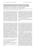
search lead to identification of an amino-acid s equence
similarity between the V3 loop of HIV-1 strain MN and
a 32-aa site within region II of PvDVP, which m aps to
domain 1 (Figure 1). Homologous sequences are present
in the cysteine-rich regions of the P. falciparum erythro-
cyte binding protein EBA-175 (F1) and P. knowlesi DBP.
There is a consens us glycosaminoglycan (GAG) binding
sequence (BBXB, where B is a basic amino acid, K or R)
in the HI V-1 MN, P. vivax and P. knowlesi sequences.
P. falciparum EBA-175 has two GAG binding sites of
the BBBxxB type in tandem.
Blocking of PvRII binding to DARC by RANTES and AOP-
RANTES
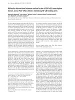
The erythrocyte binding assay of Chitnis and Miller [6]
was used to determine the inhibitory concentrations o f
chemokines for region II of the P. vivax DBP binding to
DARC. Both RANTES and AOP-RANTES elicited a
dose-response inhibition of binding (Figure 2). MIP-1a
is known not to bind to DARC, and was included as a
negative control. SDF-1, the n atural ligand of CXCR-
4 and an inhibitor of X4 viruses, has not been tested for
DARC binding in the published literature, and did not
inhibit binding in the erythrocyte binding assay. The
A
B
HIV-1
V3 loo
p
P. knowlesi
V3-like loo
p
CTRPNYNKRKRIHIGPGRAFY-TTKNI-IGTIR-QAHC
C ND-KRKRGERDWDC PAEKDVCISVRRYQL-C
C RE-KRK-GMK-WDCKKKNDRNYVCIPDRRIQL-C
HIV-1 SU (V3 loop)
P. vivax DBP
P. knowlesi DBP
P. falciparum EBP
C NY-KRKRRERDWDC NTKKDVCIPDRRYQL-C
P. falciparum
V3-like loo
p
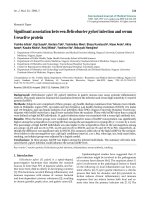
Figure 1 Similarities between peptides in the V3 loop of HIV-1 and conserved Plasmodium erythrocyte binding proteins.PanelA:
Homologous sequences in the cysteine-rich regions of the P. falciparum erythrocyte binding protein EBA-175 (F1), P. knowlesi DBP (a), P. vivax
DBP, and the V3 loop of HIV-1 strain MN. Identical or similar amino acids are boxed in yellow or green in both panels. KRKR (in green box) is a
consensus glycosaminoglycan (GAG) binding sequence (BBXB, where B is a basic amino acid, K or R) in the HIV-1, P. vivax and P. knowlesi
sequences. P. falciparum EBA-175 has two GAG binding sites of the BBBxxB type in tandem (underlined). Panel B: Secondary structure of the V3
loop of HIV-1 SU strain MN as determined by Sharon et al. [14] is shown (PDB: 1NJ0). Addition residues not contained in this structure were
modeled in SWISS-MODEL (dashed oval). Structures are shown for the V3 loop-like regions of P. knowlesi DBP as determined by Singh et al. [10]
(2C6J) and P. falciparum EBA-175 as determined by Tolia et al. [15] (1ZRL).
Bolton and Garry Virology Journal 2011, 8:45
/>Page 5 of 10
IC
50
for RANTES and AOP-RANTES were 2.09 nM and
1.51 nM, respectively. The difference in response as
determined by a two-way ANOVA test was significant
between RANTES and MIP-1a and between AOP-
RANTES and MIP-1a, but not between RANTES and
AOP-RANTES (p < 0.05).
Blocking of P. knowlesi invasion of DARC+ human
erythrocytes by RANTES and AOP-RANTES
A standard erythrocyte invasion assay was used to deter-
mine the chemokine inhibitory concentrat ions o f
DARC-dependent invasion of human DARC+ erythro-
cytes by P. knowlesi. Both RANTES and AOP-RANTES
elicited a dose-response inhibition of invasion (Figure 3).
MIP-1a was again used as a control. The IC
50
of
RANTES and AOP-RANTES in the infection assay was
0.053 nM and 0.062 nM, respectively. The IC
50
for each
inhibitor in the invasion assay was more than a log
lower than the IC
50
in the erythrocyte binding assay.
The d ifference in response as determined by a two-way
ANOVA test was significant between RANTES and
MIP-1a and between AOP-RANTES and MIP-1a,but
not between RANTES and AOP-RANTES (p < 0.05).
The V3-like peptide is necessary, but not sufficient for
DARC binding
The pHVDR22 plasmid expresses region II of the P. vivax
DBP and binds to DARC+ erythrocytes when expressed
on the surface of COS-7 cells. Region II includes
12 conserved cysteine residues, C1-C12, and the 32 amino
acid V3-like peptide spans C1-C4. Deletion mutants lack-
ing the V3-like peptide or adjacent sequences were made
from pHVDR22 and tested for their ability to bind to
DARC+ erythrocytes when expressed on COS-7 cells. The
expression of each construct on the surface of COS-7 cells
was confirmed by immunofluorescence sta ining, and the
number of COS-7 cells stained was 5-10% of the popula-
tion. The same number of COS-7 cells transfected with
the parental pHVDR22 plasmid were stained and visua-
lized by immunofluourescence.
The pv22d32 construct that specifically deleted the
V3-like peptide completely failed to bind to DARC+
erythrocytes in the erythrocyte binding assay (Figure 4A).
This suggests t hat the DBP V3-like peptide is necessary
forDARCbinding.Thedeletionofallflanking
sequences to the V3-like peptide, accomplished in the
pv22suf32 mutant, also abrogated binding, showing that
the DBP V3-like peptide is not sufficient for DARC bind-
ing. This confirms that there are other areas of region II
necessary for binding. Truncation of the amino acids
flanking the DBP V3-like peptide toward the amino ter-
minus, as accomplished in the pv22d5C construct, had
only a small effect on binding. However, truncation of
the region flanking the DBP V3-li ke peptide to the car-
boxy terminus, in pv22d3C4, abrogated binding. This
suggests that es sential binding residues are located in the
C-terminal, but not the N-terminal regions flanking the
DBP V3-like peptide. To confirm the need for the DBP
V3-like peptide in addition to the C-terminal flanking
region , truncation of the amino-terminal end of region II
up to and including the DBP V3-like peptide, in con-
struct pv22d5C4, again abrogated binding.
RANTES
AOP-RANTES
MIP-1α
SDF-1
120
100
80
60
40
20
0
-20
-40
Chemokine concentration
(
nM
)
Binding inhibition
(
%
)
0 0.1 1 10010 1000
Figure 2 Chemokine inhibition of PvRII bindi ng to DARC+
erythrocytes. The erythrocyte rosette assay of Chitnis and Miller [6]
was used to quantify chemokine inhibition of PvRII Binding to
DARC+ Erythrocytes. Binding was determined by subtracting the
number of COS-7 cells expressing pvRII with rosettes of chemokine-
treated DARC+ human erythrocytes (per 20 fields at 200X
magnification) from the number with rosettes of untreated
erythrocytes, and dividing by the number with rosettes of untreated
erythrocytes. The data shown are the mean of three separate
experiments.
120
100
80
60
40
20
0
Chemokine concentration
(
nM
)
Invasion inhibition
(
%
)
RANTES
AOP-RANTES
MIP-1α
0 0.01 0.1 1 10010
Figure 3 Chemokine inhibition of P. knowlesi invasion of DARC
+ erythrocytes. Inhibition of P. knowlesi invasion of DARC+
erythrocytes was determined by subtracting the number of
chemokine-treated DARC+ human erythrocytes invaded by P.
knowlesi merozoites (per 2000 erythrocytes) from the number of
untreated DARC+ human erythrocytes invaded by P. knowlesi
merozoites, and dividing by the number of untreated, invaded
erythrocytes.
Bolton and Garry Virology Journal 2011, 8:45
/>Page 6 of 10
A polycation sequence within the DBP V3-like Peptide is
necessary for DARC Binding
To determine if the polycation sequence in the DBP is
necessary for DARC binding, site directed mutagenesis
was used to introduce alanine substitutions for three
positively charged amino acids at K221, R224, and R227.
The pv22KARA mutant contains these substitutions and
does not bind DARC+ erythrocytes when expressed on
COS-7 cells (Figure 4B). The six positively charged
amino acids at this site of PvRII create several possible
consensus heparin binding sequences of the patterns
BBXB or BBBXXB, where B is a basic amino acid and ×
is any amino acid, including a basic one. To determine
how sensitive binding is to loss of charge at this the site,
a single alanine substitution was made at K223 in the
pv22KA construct. This mutant was capable of binding
to DARC+ erythrocytes as well as the wild type
pHVDR22 protein.
One other site in PvRII, at amino acids 364-373,
between C5 and C6, contains a polycationic site which
conforms to a consensus heparin binding sequence. The
pv22KAKA construct introduces two alanine substitu-
tions for lysine residues in this second consensus
heparin binding site at K367 and K370. The DBP region
II expressed from this construct is able to bind to
DARC+ erythrocytes on the surface of COS-7 cells as
well as wild type pHVDR22.
The polycationic site has a conserved Role in the DBP
protein family for binding to Diverse Receptors
Studies by Ranjan and Chitnis h ave identified a site in
PvRII in the C-terminal flanking regio n to the DBP V3-
like peptide, between C4-C7, that contain residues
necessary for DARC binding [8]. This study also showed
that the C1-C4 region of the P. knowlesi b protein, a
member of the DBP family that does not bind to DARC,
was capable of substituting for the P. vivax C1-C4.
Upon closer inspection, the polycationic site is well con-
served in the DBP family, with great similarity between
proteins that bind different receptors (Figure 4C). The
P. knowlesi a and g proteins have an identical consensus
heparin binding site, but only a binds to DARC. To see
if the polycationic site may play a similar role in
the binding proteins of other members of the DBP
family, the same three alanine substitutions found in
pv22KARA were introduced by site directed mutagen-
esis into the plasmids pHKADR22, pHKBDR22, and
pHKGDR22. This yielded the constructs pkalphaKARA,
pkbetaKARA, and pkgammaKARA, which contain the
K221,R224,andR227alaninesubstitutionintheP.
knowlesi a, b,andg genes, respectively. All three of
these mutants failed to bind rhesus erythrocytes when
expressed in COS-7 cells.
Discussion
HIV-1 binds to chemokine receptors such as CCR5 and
CXCR4 using SU, and can be inhibited from in vivo
infection by mutation of the chemokine receptors or by
incubation with chemokines, such as RANTES. Likewise,
P. vivax uses its DBP to bind to DARC and can be
inhibited by null mutations in the receptor or in vitro
pHVDR22
pv22d32
pv22suf32
pv22d3C4
pv22d5C1
pv22d5C5
pv22KARA
pv22KA
pv22KAKA
pkalphaKARA
pkbetaKARA
pkgammaKARA
BINDING
100
%
0
%
0
%
0
%
80
%
0
%
0
%
100
%
100
%
0
%
0
%
0
%
C1 C4 C5 C6 C12
YKRKRRERDW
YARKAREADW
YKRKRRERDW
YKRARRERDW
SVKKRLKGNF
SVKARLAGNF
A
C
B
DKRKRGERD
DARKAGEAD
NKRKRGTRD
NARARGTAD
DARKA
G
EAD
DKRKRGERD
Figure 4 Mutants of the region II of Erythrocyte Binding
Proteins. Panel A. P. vivax DBP region II is shown in blue with
conserved cysteines C1, C4, C5, C6 and C12 shown. Deletion
mutants in the are shown with the V3-like peptide (amino acids
216-247, between C1-C4) highlighted in red. Primers flanking this
site, facing outward, were used to create pv22d32 (delete 32 amino
acids) by inverse PCR. The other mutants were created with primers
facing inward and containing restriction enzyme sites. Percent
binding is expressed as number of rosettes compared to pHVDR22.
Panel B. Site directed mutagenesis using the Stratagene
QuickChange kit was used to make alanine substitutions within the
consensus heparin binding site of the V3-like peptide (R22KARA,
R22KA), or another consensus site (R22KAKA) at amino acids 364-
373, between conserved cysteines C5-C6. Panel C. Site directed
mutagenesis was used to created the same KARA mutation in the
conserved heparin binding site between C1-C4 of the P. knowlesi a,
b and g proteins.
Bolton and Garry Virology Journal 2011, 8:45
/>Page 7 of 10
by MGSA or IL-8. Here, we show that the chemokine,
RANTES, and its analog AOP-RANTES, kno wn to
block HIV-1 SU binding to CCR5, also blocks P. vivax
DBP binding to DARC. This demonstrates that natural
and designed chemokine inhibitors can be cross-protec-
tive to both pathogens, and may have important
implications for drug and vaccine development in
co-endemic areas.
The overlap in chemokine inhibition of both HIV-
1andPlasmodium infection suppo rts a hypothesis that
SU and PvDBP have convergently evolved to mimic
chemokines in such a way that the two proteins have
structural similarities. The N-terminus extracellular
domain of DARC is involved in binding to bo th
region II of the PvDBP and to chemokines, just as the
N-terminus of CCR5 is critical for SU and chemo-
kine binding. In particular, negatively charged and
sulfotyrosine residues in the CCR5 N-terminus and
CXCR4 extracellular domain have important interac-
tions with the C4/V3 stem of SU, and positively charged
residues are implied to be important components of the
SU chemokine receptor binding surface. Similarly, Pv-
DBL or Pka-DBL have important interactions with
sulphated tyrosine (Tyr 41) residue on DARC [9] The
results of a homology search identifying an amino-acid
sequence similarity between the V3 loop of HIV-1 strain
MN and a 32-aa site within region II of PvDBP contain-
ing a polycati onic site (Figure 1). Other members of the
EBP family share this homology or “V3-like peptide” .
The crystal structure of the P. knowlesi DBL domain
(Pka-DBL), which binds to DARC during infection of
human erythrocytes, shows that this structure is indeed
similar, with disulfide bridges between C1 and C4 and
between C2 and C3 forming a random coil structure
designated domain 1 [10].
To investigate the role of the V3-like peptide in DARC
bindi ng we used an established erythrocyte binding assay
and made mutants of the region II PvDBP expression
vector. Deletion of the 32-aa V3-like peptide in construct
pv2 2d32, or deleti on of the flanking regi ons in construct
pv22suf32, abrogated binding to DARC, suggesting that
the V3-like peptide was necessary but not sufficient for
binding (Figure 4A). In particular, the region between the
conserved cysteines C4-C12 was necessary for binding as
demonstrated by binding of pv22d5C1 and nonbinding of
pv22d3C4, but the C4-C12 region was also not sufficient
as shown by nonbinding of pv22d5C4. It is possible that
the deletions we have made change the folding of the
receptor binding site, with the ex ception of the del etion
of amino acids 198-216. Previous work by Ranjan and
Chitnis [8] using chimeras of region II between the P.
vivax DBP and P. knowlesi b protein, which does not
bind DARC but sialic acid, revealed the entire C4-
C7 region of PvDBP region II is necessary for DARC
binding, which our data corroborate. Of note, their data
show that region C4-C7 is sufficient for binding, but this
does not mean that other regions are not involved bind-
ing of the full-length protein. The authors of the chimeric
data suggest that residues outside of C4-C7 influence the
fine specificity of the DBL binding domain [8]. This
mightbecomparabletothespecificityofchemokine
receptor binding attributed to small changes in the
V3 loop, which mimics the b hairpin structure in chemo-
kines, while conserved portions of the SU molecule cre-
ate the structural backbone of the chemokine receptor
binding surface [11].
The polycationic site within the V3-like peptide is con-
served in the EBP family and contains consensus heparin
binding sequences of the patterns BBXB or BBBXXB,
where B is a basic amino acid and × is any amino acid,
including a basic one. It was previously shown that poly-
anions inhibit DARC-binding by the P. knowlesi a pro-
tein, and we have determined that this also to be true of
the PvDBP region II (Bolton et al., in preparation). We
made site-directed mutations to substitute alanines for
positively charged amino acids at K221, R224, and R227.
This mutant, designated pv22KARA, did not bind. Such
minor changes make it less likely that this mutant does
not bind due to folding error than to contributions these
residues make directly to receptor binding. To deter-
mimne if the polycationic site was sensitive to a
single alanine substitution, we created a mutation at
K223 which did not change binding in pv22KA. This
mutant protein still contained consensus heparin binding
sequences and five positively charged residues at the site.
We also mutated the only other polycationic site in the
PvDBP region II that conforms t o a consensus heparin
binding sequence at amino acids 364-373 by substituting
alanines at K367 and K370. pv22KAKA was still able to
bind. These data show that the polycation site in the V3-
like peptide is discretely involved in DARC binding and
suggestthemultiplepositivechargesplayaredundant
role at the site.
Previous site-directed mutagenesis experiments have
identified residues Tyr 94, Asn 95, Lys 96, Arg 1 03, Leu
168 and Ile 175 on domain 2 as required for recognition
of DARC on human erythrocytes [12-14]. Based on the
crystal structure of the Pka-DBL these residues lie close to
a set of positively charged residues Lys 96, Lys 100, Arg
103 and Lys 177 that have been suggested to interact with
the sulphate group on DARC Tyr 41 [10]. Mapping the
polycationic site we found to be sensitive to alanine substi-
tuions onto the crystal structure shows that it is adjacent
to the putative binding site residues and may provide such
an interaction with the sulphated Tyr 41 (Figure 5).
The P. knowlesi a, b,andg proteins share the V3-like
loop and polycation site homology in region II with
PvDBP, though only the a pro tein binds DARC. We
Bolton and Garry Virology Journal 2011, 8:45
/>Page 8 of 10
introduced similar alanine mutations into three posi-
tively- charged amino acids of each of the three P. know-
lesi EBPs at the polycationic site. In all 3 cases this
eliminated normal binding to rhesus erythrocytes. In the
case of the P. knowlesi a protein this reinforces the con-
clusion that the site c an contribute t o DARC binding.
The P. knowlesi b,andg proteins, however, don’tbind
to DARC. The receptor f or the P. knowlesi b protein is
sialic acid, which is negatively charged for which the
polycation site might contribute to a positively charged
binding pocket. A chimera produced by Ranjan and
Chitnis [8] with the C1-C4 region exchanged between
the P. knowlesi b protein and P. vivax DBP bound to
DARC on rhesus erythrocytes, without the removal of
sialic acid residues required for native P. vivax DBP to
bind to rhesus DARC. It is possible that a closer homol-
ogy between the polycationic site of P. knowlesi a and b
proteins, each of which contain 5 cationic residues ver-
sus the 6 cationic residues of the P. vivax DBP polyca-
tionic site, allows for this change in speci ficity. Another
chimera in the same study with the P. vivax polycationic
site is able to bind to rhesus erythrocytes in the same
manner as the P. knowlesi b protein, but only in the
presence of C4-C5 of the P. knowlesi b protein. This
again suggests that the homology of the polycationic site
within the EBP family may a llow for a redundant func-
tion in receptor binding, but the role of the polycationic
site is in conjunction with other residues in r egion II
which together allow for efficient receptor bindi ng. The
results presented here, in conjunction with previ ous stu-
dies, indicate that the he parin binding site motif in
members of the DBP family may form part of a con-
served erythrocyte receptor binding pocket.
Acknowledgements
This work was supported by National Institutes of Health grant RR018229.
Author details
1
Vaccine and Infectious Disease Institute, Fred Hutchinson Cancer Research
Center Division of Allergy and Infectious Diseases University of Washington
1100 Fairview Avenue Seattle, Washington 98109 USA.
2
Department of
Microbiology and Immunology Tulane University 1430 Tulane Avenue New
Orleans, Louisiana 70112 USA.
Authors’ contributions
MJB performed the investigations described in this study. MJB and RFG
conceived of the study, and RFG participated in its design and coordination
and helped to draft the manuscript. Both authors read and approved the
final manuscript.
C16
K19
R20
K21
K22
R40
R41
C29
C36
C45
Y94
N95
K96
R103
L168
I175
V3-like loop
Domain 1
Domain 2
Domain 3
Figure 5 T hree dimensional location of heparin binding motif in relation to known binding residues on t he crystal structure of
recombinant Pka-DBL. The crystal structure of the recombinant Pka-DBL that binds to human DARC is shown with previously described
binding residues Tyr 94, Asn 95, Lys 96, Arg 103, Leu 168 and Ile 175 [10,12-14] highlighted in magenta in domain 2 of the molecule. The
heparin binding motif on the V3-like peptide is highlighted in yellow cysteines C1-C4 and blue for the basic residues (lysine and arginine) in
domain 1 of the molecule.
Bolton and Garry Virology Journal 2011, 8:45
/>Page 9 of 10
Competing interests
The authors declare that they have no competing interests.
Received: 29 November 2010 Accepted: 31 January 2011
Published: 31 January 2011
References
1. Horuk R, Chitnis CE, Darbonne WC, Colby TJ, Rybicki A, Hadley TJ, Miller LH:
A receptor for the malarial parasite Plasmodium vivax: the erythrocyte
chemokine receptor. Science 1993, 261(5125):1182-1184.
2. Simmons G, Wilkinson D, Reeves JD, Dittmar MT, Beddows S, Weber J,
Carnegie G, Desselberger U, Gray PW, Weiss RA, Clapham PR: Primary,
syncytium-inducing human immunodeficiency virus type 1 isolates are
dual-tropic and most can use either Lestr or CCR5 as coreceptors for
virus entry. J Virol 1996, 70(12):8355-8360.
3. Liu R, Paxton WA, Choe S, Ceradini D, Martin SR, Horuk R, MacDonald ME,
Stuhlmann H, Koup RA, Landau NR: Homozygous defect in HIV-
1 coreceptor accounts for resistance of some multiply-exposed
individuals to HIV-1 infection. Cell 1996, 86(3):367-377.
4. Miller LH, Mason SJ, Clyde DF, McGinniss MH: The resistance factor to
Plasmodium vivax in blacks. The Duffy-blood-group genotype, FyFy. N
Engl J Med 1976, 295(6):302-304.
5. Cocchi F, DeVico AL, Garzino-Demo A, Arya SK, Gallo RC, Lusso P:
Identification of RANTES, MIP-1 alpha, and MIP-1 beta as the major HIV-
suppressive factors produced by CD8+ T cells. Science 1995,
270(5243):1811-1815.
6. Chitnis CE, Miller LH: Identification of the erythrocyte binding domains of
Plasmodium vivax and Plasmodium knowlesi proteins involved in
erythrocyte invasion. J Exp Med 1994, 180(2):497-506.
7. Chitnis CE, Chaudhuri A, Horuk R, Pogo AO, Miller LH: The domain on the
Duffy blood group antigen for binding Plasmodium vivax and P.
knowlesi malarial parasites to erythrocytes. J Exp Med 1996,
184(4):1531-1536.
8. Ranjan A, Chitnis CE: Mapping regions containing binding residues within
functional domains of Plasmodium vivax and Plasmodium knowlesi
erythrocyte-binding proteins. PNAS 1999, 96(24):14067-14072.
9. Choe H, Moore MJ, Owens CM, Wright PL, Vasilieva N, Li W, Singh AP,
Shakri R, Chitnis CE, Farzan M: Sulphated tyrosines mediate association of
chemokines and Plasmodium vivax Duffy binding protein with the Duffy
antigen/receptor for chemokines (DARC). Molecular Microbiology 2005,
55(5):1413-1422.
10. Singh SK, Hora R, Belrhali H, Chitnis CE, Sharma A: Structural basis for
Duffy recognition by the malaria parasite Duffy-binding-like domain.
Nature 2006, 439(7077):741-744.
11. Sharon M, Kessler N, Levy R, Zolla-Pasner S, Görlach M, Anglister J:
Alternative comformations of HIV-1 V3 loops mimic β hairpins in
chemokines, suggesting a mechanism for co-receptor specificity.
Structure 2003, 11(2):225-236.
12. Singh SK, Singh AP, Pandey S, Yazdani SS, Chitnis CE, Sharma A: Definition
of structural elements in Plasmodium vivax and P. knowlesi Duffy-
binding domains necessary for erythrocyte invasion. Biochemical Journal
2003, 374(1):193-198.
13. VanBuskirk KM, Sevova E, Adams JH: Conserved residues in the
Plasmodium vivax Duffy-binding protein ligand domain are critical for
erythrocyte receptor recognition. PNAS 2004, 101(44):15754-15759.
14. Hans D, Pattnaik P, Bhattacharyya A, Shakri AR, Yazdani SS, Sharma M,
Choe H, Farzan M, Chitnis CE: Mapping binding residues in the
Plasmodium vivax domain that binds Duffy antigen during red cell
invasion. Molecular Microbiology 2005, 55(5):1423-1434.
15. Tolia NH, Enemark EJ, Sim BK, Joshua-Tor L: Structural basis for the EBA-
175 erythrocyte invasion pathway of the malaria parasite Plasmodium
falciparum. Cell 2005, 122(2):183-93.
doi:10.1186/1743-422X-8-45
Cite this article as: Bolton and Garry: Seque nce similarity between the
erythrocyte binding domain 1 of the Plasmodium vivax Duffy binding
protein and the V3 loop of HIV-1 strain MN reveals binding residues for
the Duffy Antigen Receptor for Chemokines. Virology Journal 2011 8:45.
Submit your next manuscript to BioMed Central
and take full advantage of:
• Convenient online submission
• Thorough peer review
• No space constraints or color figure charges
• Immediate publication on acceptance
• Inclusion in PubMed, CAS, Scopus and Google Scholar
• Research which is freely available for redistribution
Submit your manuscript at
www.biomedcentral.com/submit
Bolton and Garry Virology Journal 2011, 8:45
/>Page 10 of 10