Báo cáo y học: " New technical approach for the repair of an abdominal wall defect after a transverse rectus abdominis myocutaneous flap: a case report" pot
Bạn đang xem bản rút gọn của tài liệu. Xem và tải ngay bản đầy đủ của tài liệu tại đây (808.66 KB, 6 trang )
BioMed Central
Page 1 of 6
(page number not for citation purposes)
Journal of Medical Case Reports
Open Access
Case report
New technical approach for the repair of an abdominal wall defect
after a transverse rectus abdominis myocutaneous flap: a case
report
Daniel A Kaemmer*, Joachim Conze, Jens Otto and Volker Schumpelick
Address: Department of Surgery, Medical Faculty, Rheinish-Westphalian Technical University, D-52074 Aachen, Germany
Email: Daniel A Kaemmer* - ; Joachim Conze - ; Jens Otto - ;
Volker Schumpelick -
* Corresponding author
Abstract
Introduction: Breast reconstruction with autologous tissue transfer is now a standard operation,
but abnormalities of the abdominal wall contour represent a complication which has led surgeons
to invent techniques to minimize the morbidity of the donor site.
Case presentation: We report the case of a woman who had bilateral transverse rectus
abdominis myocutaneous flap (TRAM-flap) breast reconstruction. The surgery led to the patient
developing an enormous abdominal bulge that caused her disability in terms of abdominal wall and
bowel function, pain and contour. In the absence of rectus muscle, the large defect was repaired
using a combination of the abdominal wall component separation technique of Ramirez et al and
additional mesh augmentation with a lightweight, large-pore polypropylene mesh (Ultrapro
®
).
Conclusion: The procedure of Ramirez et al is helpful in achieving a tension-free closure of large
defects in the anterior abdominal wall. The additional mesh augmentation allows reinforcement of
the thinned lateral abdominal wall.
Introduction
Abnormalities of the abdominal wall contour after breast
reconstruction with autologous tissue transfer have previ-
ously been reported as problematic, with a lower abdom-
inal bulge being the most frequently reported
abnormality [1]. Although the cosmetic results and
patient satisfaction seem to be good in most cases with
regards to shape, symmetry and muscular function, differ-
ences become obvious in the morbidity of the donor site
[2-5]. Modifications and new techniques have been devel-
oped to reduce complications, but none of these modifi-
cations is able to prevent contour abnormalities of the
donor site completely [6], and new techniques, which pre-
serve the anterior rectus sheath are limited in their use by
anatomic variations [2].
In addition to the aesthetic disturbance, these defects can
also lead to adverse interference of the abdominal wall
functions, as a thrust bearing for the intraabdominal pres-
sure and as an antagonist of the back muscles and part of
the respiratory system. To date, these side effects have not
attracted attention in the literature and no therapeutic
approaches have been reported.
Here we present the case of a woman with an extreme
bulge of the lower abdominal wall following bilateral
Published: 16 April 2008
Journal of Medical Case Reports 2008, 2:108 doi:10.1186/1752-1947-2-108
Received: 16 August 2007
Accepted: 16 April 2008
This article is available from: />© 2008 Kaemmer et al; licensee BioMed Central Ltd.
This is an Open Access article distributed under the terms of the Creative Commons Attribution License ( />),
which permits unrestricted use, distribution, and reproduction in any medium, provided the original work is properly cited.
Journal of Medical Case Reports 2008, 2:108 />Page 2 of 6
(page number not for citation purposes)
transverse rectus abdominis myocutaneous flap (TRAM-
flap) breast reconstruction. This was repaired using a com-
bination of the abdominal wall component separation
technique of Ramirez et al [7] and additional mesh aug-
mentation.
Case presentation
We report the case of a 61-year-old woman who was suf-
fering from lower abdominal bulge formation, chronic
constipation, as well as a feeling of permanent abdominal
constriction and pain. These symptoms appeared eight
months after bilateral breast reconstruction, which was
performed following subcutaneous mastectomy that was
necessary owing to ductal carcinoma in situ. The breast
reconstruction was conducted using a non-muscle-spar-
ing pedicled TRAM-flap transposition. The defect created
at the donor site within the abdominal wall after harvest-
ing the rectus muscle was closed using a continuous
suture with resorbable suture material. An additional aug-
mentation was performed by the implantation of a
resorbable polyglactin mesh placed on the fascial suture.
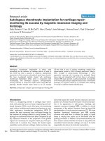
The patient presented at the authors' outpatient clinic
eight months after reconstruction. At that time her body
mass index was 18.9 and she was suffering from a lower
abdominal bulge formation (Figure 1). An ultrasound
examination revealed an abdominal wall defect measur-
ing 18 × 20 cm, with no detectable rectus abdominis mus-
cle remaining, resembling a large rectus diastasis. A
preoperative endoscopy of the colon showed signs of
adhesions in the colon sigmoideum and transversum, but
no other pathologies; the laboratory values were normal.
Apart from an appendectomy performed 20 years ago, the
patient had undergone no other previous abdominal sur-
gery. In addition to the annoying large bulge in this oth-
erwise slim patient, the pain experienced during everyday
Abdominal contour before and after reconstructionFigure 1
Abdominal contour before and after reconstruction. (A) The preoperative abdominal contour (lateral view). (B) The
abdominal contour six weeks after the reconstruction (lateral view). In addition to minimizing the abdominal bulge, Ramirez et
al's technique is able to shape the lateral abdominal wall in an aesthetic manner; lateral bulging was avoided using mesh augmen-
tation.
Journal of Medical Case Reports 2008, 2:108 />Page 3 of 6
(page number not for citation purposes)
movement and impairment of bowel function led to an
explorative laparotomy and an attempt to reconstruct the
abdominal wall.
Following adequate preparations with intestinal irriga-
tion, a re-incision through the midline scar was per-
formed. On entering the peritoneal cavity, several dense
adhesions of small intestine to the abdominal wall and
interenteric to the colon were found. These were carefully
dissolved without causing injury to the intestine. Further
exploration revealed a near-total absence of both abdom-
inal rectus muscles; residual muscle fibres could be
detected only at the lateral side of the rectus sheath. The
initially implanted absorbable mesh was not identified,
and the ultrasonographic finding of a diastasis-like defect
with lateralization of both lineae semilunares was veri-
fied. Following a wide-ranging mobilization of the epifas-
cial subcutaneous tissue, the remaining parts of the
anterior rectus sheaths and minimal lateral parts of the
rectus muscles were exposed. The herniation sac was
partly resected, leaving sufficient material to facilitate a
peritoneal closure of the abdominal cavity. In order to
reach an adaptation of both lateralized anterior rectus
sheaths, a component separation of the abdominal wall
(Ramirez procedure) was performed. In the absence of an
intact rectus abdominis muscle and anterior rectus sheath,
only a vertical incision lateral to the linea semilunaris and
separation in the plane between oblique external and
internal muscle was used. A two-layer closure of the fascia
in the midline was performed using a non-resorbable sin-
gle-stitch suture of the posterior wall, and a continuous
suture with a slowly resorbable suture material for the
remaining anterior rectus sheath. The lateral defects
between the external oblique muscle and linea semiluna-
ris were covered with a halfmoon-shaped lightweight
polypropylene mesh (Ultrapro
®
; Ethicon, Norderstedt,
Germany) on each side (Figure 2). Punctual mesh fixation
was achieved using resorbable 3/0 single-stitch sutures
(Dexon
®
; Braun, Germany). A subcutaneous suction drain
was placed on top of each mesh, after which wound clo-
sure was achieved with a continuous intracutaneous
suture using non-resorbable material.
The patient's recovery was uneventful; during her hospital
stay she wore an elastic abdominal belt and was provided
with analgesics and physical therapy with intense respira-
tory training. The suction drains and suture material were
removed on schedule, the postoperative ultrasonography
was without pathological findings and minimal postoper-
ative seroma resolved. The patient was discharged from
hospital and made subsequent visits to the outpatient
clinic. At 12 months after surgery she remained satisfied
with the outcome.
Discussion
The TRAM-flap technique developed by Hartrampf et al
[8] in 1982 is now well established. Long-term evalua-
tions of any complications and aesthetic outcome have
been conducted which state that, for the TRAM-flap, the
rate of ('true') hernia or abdominal bulge is about 0–5%
[5]. Modifications of the original technique have been
developed, including muscle- and fascia-sparing tech-
niques [9] as well as free flaps [10] and mesh implanta-
tion [11]. These modifications have reduced the incidence
of complications, such as hernia and bulge formation, in
the remaining abdominal wall.
The anterior rectus sheath is one of the major components
maintaining the integrity of the abdominal wall and con-
tour; consequently, flaps which preserve this structure
completely have been evaluated [12]. The deep inferior
epigastric perforator flap (DIEP-flap) is an alternative,
widely used modification, and surgeons have also
described and used the superficial inferior epigastric artery
flap (SIEA-flap) or gluteal artery perforator flap (GAP-
flap) [13]. These flaps preserve the anterior rectus sheath
and therefore minimize the risk of a hernia or bulge for-
mation, although this has been described in the case of
DIEP-flaps and is considered to be a result of denervation.
The myocutaneous flap has no advantages in terms of
autologous tissue volume and the possibility of modelling
symmetric and natural-looking breasts. SIEA-flaps can
only be used if a superficial inferior epigastric artery is
present and is sufficient to perfuse the flap, but in this
select patient group it may be used as the first choice [2].
Today, GAP-flaps are considered as a fall-back technique
and are used only if abdominal cutaneous tissue and fat is
not appropriate for the reconstruction.
In the case described in this report the bilateral non-mus-
cle-sparing TRAM-flap transfer led to an enormous
abdominal bulge that caused disability for the patient in
many different ways. To date, no standard surgical proce-
dure has been developed to treat these defects. Damage to
the TRAM-flap resulted in a broad defect in the area of the
harvested rectus muscle that could not be reversed (Figure
3). The principal idea of any repair should be to recon-
struct the abdominal wall integrity with closure of the fas-
cial defect. In 1990, Ramirez et al [7] described a
component separation technique which allowed a mid-
line advancement of the abdominal wall of up to 10 cm
on each side, without the need for musculofascial flaps.
Moreover, this technique provides an innervated and vas-
cularized compound for dynamic support by dividing the
abdominal wall components along an avascular plane.
Additional mesh augmentation was not used in the origi-
nal component separation method described by Ramirez
et al. The anterior rectus sheath was opened and the rectus
Journal of Medical Case Reports 2008, 2:108 />Page 4 of 6
(page number not for citation purposes)
muscle was separated from the posterior rectus sheath and
moved medially. In the present case, because there was
almost no rectus muscle remaining, it was necessary to
omit this step. A longitudinal incision was made lateral to
the border of the rectus sheath and separation continued
in the more-or-less avascular plane between the external
and internal oblique muscles, leaving the external oblique
lateral to the subsequently closed midline incision. To the
best of the authors' knowledge, the component separation
technique has been performed previously with only one
rectus muscle remaining, but never without any rectus
muscle on either side. In contrast, it was stated that at least
one innervated rectus was required to re-establish the
integrity of the abdominal wall [7].
The idea of mesh augmentation in the midline was aban-
doned owing to the fact that, in the present patient, there
was no typical incisional hernia pathophysiology but
rather an abdominal wall defect that had been created
deliberately, and this made a collagen defect unlikely. The
use of mesh material was reduced to only augmenting the
thinned lateral abdominal wall, to prevent any possible
postoperative bulging of the internal oblique and trans-
verse muscles. For the same contouring reasons, and to
avoid extensive adhesion formation, a mesh prosthesis
placed intraperitoneally using an onlay technique
(IPOM) [14] was not used. Furthermore, this technique
would have required replacement rather than augmenta-
tion of the abdominal wall.
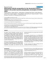
Mesh augmentation using two halfmoon-shaped lightweight polypropylene meshes placed on the defects between the external oblique muscles and lineae semilunaresFigure 2
Mesh augmentation using two halfmoon-shaped lightweight polypropylene meshes placed on the defects
between the external oblique muscles and lineae semilunares. The meshes were fixed using resorbable single-stitch
sutures. After a midline incision and adhesiolysis, the abdominal wall components were separated along the avascular plane
between the internal and external oblique abdominal muscles. A midline closure in two layers was performed using non-
resorbable single-stitch sutures and continuous slowly resorbable suture for the posterior wall and anterior rectus sheath,
respectively.
Journal of Medical Case Reports 2008, 2:108 />Page 5 of 6
(page number not for citation purposes)
An extensive epifascial preparation might put the blood
circulation of the skin at risk. In slim patients, where the
subcutaneous layer is not usually pronounced, the addi-
tional use of excessive foreign material should be consid-
ered carefully. The use of lightweight, large-pore
polypropylene meshes appears to reduce the risk of any
major foreign-body reaction that might lead to shrinkage
of the mesh area or to a reduction in abdominal wall
mobility [15]. The textile features of this new mesh gener-
ation are more adapted to the physiology of the abdomi-
nal wall and are predisposed to its augmentation [16].
Conclusion
It has been shown that a reconstruction of the abdominal
wall midline is possible and maintainable in the absence
of both rectus muscles, using the component separation
technique of Ramirez et al. A modification is suggested
using additional mesh augmentation to cover the thinned
lateral abdominal wall, using a lightweight polypropylene
mesh prosthesis.
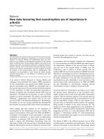
Schema of the abdominal wallFigure 3
Schema of the abdominal wall. (A) The normal abdominal wall. (B) Left: postoperative conditions after bilateral TRAM-
flap. Right: abdominal bulge that developed in the present case. (C) Conditions after abdominal wall component separation,
before double-layer midline closure. (D) Postoperative conditions after mesh augmentation.
Publish with BioMed Central and every
scientist can read your work free of charge
"BioMed Central will be the most significant development for
disseminating the results of biomedical research in our lifetime."
Sir Paul Nurse, Cancer Research UK
Your research papers will be:
available free of charge to the entire biomedical community
peer reviewed and published immediately upon acceptance
cited in PubMed and archived on PubMed Central
yours — you keep the copyright
Submit your manuscript here:
/>BioMedcentral
Journal of Medical Case Reports 2008, 2:108 />Page 6 of 6
(page number not for citation purposes)
Competing interests
The author(s) declare that they have no competing inter-
ests.
Authors' contributions
DAK assisted with the surgery, designed the case report,
collated the information, performed the literature search
and prepared the manuscript. JC assisted with the surgery,
was involved in all investigations and assisted in provid-
ing a critical appraisal and review of the manuscript. JO
prepared the images, advised on the format and design
and assisted in providing a critical appraisal of the manu-
script. VS performed the surgery, was involved in all inves-
tigations and assisted in the literature search, writing and
editing of the manuscript. All authors have reviewed and
approved the final manuscript.
Consent
Written informed consent was obtained from the patient
for publication of this case report and accompanying
images. A copy of the written consent is available for
review by the Editor-in-Chief of this journal.
References
1. Nahabedian MY, Dooley W, Singh N, Manson PN: Contour abnor-
malities of the abdomen after breast reconstruction with
abdominal flaps: the role of muscle preservation. Plast Recon-
str Surg 2002, 109:91-101.
2. Chevray PM: Breast reconstruction with superficial inferior
epigastric artery flaps: a prospective comparison with TRAM
and DIEP flaps. Plast Reconstr Surg 2004, 114:1077-1083.
3. Blondeel N, Vanderstraeten GG, Monstrey SJ, Van LK, Tonnard P,
Lysens R, Boeckx WD, Matton G: The donor site morbidity of
free DIEP flaps and free TRAM flaps for breast reconstruc-
tion. Br J Plast Surg 1997, 50:322-330.
4. Clough KB, O'Donoghue JM, Fitoussi AD, Vlastos G, Falcou MC: Pro-
spective evaluation of late cosmetic results following breast
reconstruction: II. Tram flap reconstruction. Plast Reconstr
Surg 2001, 107:1710-1716.
5. Mizgala CL, Hartrampf CR Jr, Bennett GK: Assessment of the
abdominal wall after pedicled TRAM flap surgery: 5- to 7-
year follow-up of 150 consecutive patients. Plast Reconstr Surg
1994, 93:988-1002.
6. Nahabedian MY, Momen B: Lower abdominal bulge after deep
inferior epigastric perforator flap (DIEP) breast reconstruc-
tion. Ann Plast Surg 2005, 54:124-129.
7. Ramirez OM, Ruas E, Dellon AL: 'Components separation'
method for closure of abdominal-wall defects: an anatomic
and clinical study. Plast Reconstr Surg 1990, 86:519-526.
8. Hartrampf CR, Scheflan M, Black PW: Breast reconstruction with
a transverse abdominal island flap. Plast Reconstr Surg 1982,
69:216-225.
9. Kroll SS, Marchi M: Comparison of strategies for preventing
abdominal-wall weakness after TRAM flap breast recon-
struction. Plast Reconstr Surg 1992, 89:1045-1051.
10. Baldwin BJ, Schusterman MA, Miller MJ, Kroll SS, Wang BG: Bilateral
breast reconstruction: conventional versus free TRAM. Plast
Reconstr Surg 1994,
93:1410-1416.
11. Bucky LP, May JW Jr: Synthetic mesh. Its use in abdominal wall
reconstruction after the TRAM. Clin Plast Surg 1994, 21:273-277.
12. Guerra AB, Metzinger SE, Bidros RS, Rizzuto RP, Gill PS, Nguyen AH,
Dupin CL, Allen RJ: Bilateral breast reconstruction with the
deep inferior epigastric perforator (DIEP) flap: an experi-
ence with 280 flaps. Ann Plast Surg 2004, 52:246-252.
13. Blondeel PN: The sensate free superior gluteal artery perfora-
tor (S-GAP) flap: a valuable alternative in autologous breast
reconstruction. Br J Plast Surg 1999, 52:185-193.
14. Rasim ZM, Alzahrani MA, Sigman HH, Meakins JL, Fried GM: Com-
parison of adhesion formation and tensile strength after
three laparoscopic herniorrhaphy techniques. Surg Laparosc
Endosc 1997, 7:133-136.
15. Conze J, Krones CJ, Schumpelick V, Klinge U: Incisional hernia:
challenge of re-operations after mesh repair. Langenbecks Arch
Surg 2006, 392:453-457.
16. Klinge U, Klosterhalfen B, Ottinger AP, Junge K, Schumpelick V:
PVDF as a new polymer for the construction of surgical
meshes. Biomaterials 2002, 23:3487-3493.