Báo cáo y học: "Multiplex Amplification Refractory Mutation System Polymerase Chain Reaction (ARMS-PCR) for diagnosis of natural infection with canine distemper virus" potx
Bạn đang xem bản rút gọn của tài liệu. Xem và tải ngay bản đầy đủ của tài liệu tại đây (2.31 MB, 9 trang )
Chulakasian et al. Virology Journal 2010, 7:122
/>Open Access
RESEARCH
© 2010 Chulakasian et al; licensee BioMed Central Ltd. This is an Open Access article distributed under the terms of the Creative Com-
mons Attribution License ( which permits unrestricted use, distribution, and reproduc-
tion in any medium, provided the original work is properly cited.
Research
Multiplex Amplification Refractory Mutation
System Polymerase Chain Reaction (ARMS-PCR) for
diagnosis of natural infection with canine
distemper virus
Songkhla Chulakasian
1
, Min-Shiuh Lee
2
, Chi-Young Wang
1
, Shyan-Song Chiou
3
, Kuan-Hsun Lin
1
, Fong-Yuan Lin
1
,
Tien-Huan Hsu
1
, Min-Liang Wong
1
, Tien-Jye Chang*
1
and Wei-Li Hsu*
3
Abstract
Background: Canine distemper virus (CDV) is present worldwide and produces a lethal systemic infection of wild and
domestic Canidae. Pre-existing antibodies acquired from vaccination or previous CDV infection might interfere the
interpretation of a serologic diagnosis method. In addition, due to the high similarity of nucleic acid sequences
between wild-type CDV and the new vaccine strain, current PCR derived methods cannot be applied for the definite
confirmation of CD infection. Hence, it is worthy of developing a simple and rapid nucleotide-based assay for
differentiation of wild-type CDV which is a cause of disease from attenuated CDVs after vaccination. High frequency
variations have been found in the region spanning from the 3'-untranslated region (UTR) of the matrix (M) gene to the
fusion (F) gene (designated M-F UTR) in a few CDV strains. To establish a differential diagnosis assay, an amplification
refractory mutation analysis was established based on the highly variable region on M-F UTR and F regions.
Results: Sequences of frequent polymorphisms were found scattered throughout the M-F UTR region; the identity of
nucleic acid between local strains and vaccine strains ranged from 82.5% to 93.8%. A track of AAA residue located 35
nucleotides downstream from F gene start codon highly conserved in three vaccine strains were replaced with TGC in
the local strains; that severed as target sequences for deign of discrimination primers. The method established in the
present study successfully differentiated seven Taiwanese CDV field isolates, all belonging to the Asia-1 lineage, from
vaccine strains.
Conclusions: The method described herein would be useful for several clinical applications, such as confirmation of
nature CDV infection, evaluation of vaccination status and verification of the circulating viral genotypes.
Background
Canine distemper is a highly contagious disease caused
by canine distemper virus (CDV), which belongs to the
genus Morbillivirus of the family Paramyxoviridae.
Although CDV primarily infects canids, infection of
other terrestrial and aquatic carnivores has been reported
[1-7]. CDV infection causes a systemic disease with
severe immunosuppression involving primary replication
of the virus in macrophages and lymphocytes of the
respiratory tract, as well as in various lymphoid tissues
[8].
The genome of CDV is approximately 15.7 kb in length
and consists of a single-stranded, negative-sense RNA
encoding the following eight viral proteins: two tran-
scriptase-associated proteins (the phosphoprotein P and
the large protein L) and the nucleocapsid protein (N) that
encapsidates the viral RNA, a single envelope-associated
matrix (M) protein and two glycoproteins: haemaggluti-
nin/attachment protein (H) and a fusion protein (F) [9].
The F protein is responsible for viral fusion with host
* Correspondence: ,
1
Department of Veterinary Medicine, College of Veterinary Medicine, National
Chung Hsing University, 250 Kou Kuang Road, Taichung 402, Taiwan
3
Graduate Institute of Microbiology and Public Health, College of Veterinary
Medicine, National Chung Hsing University, 250 Kou Kuang Road, Taichung
402, Taiwan
Full list of author information is available at the end of the article
Chulakasian et al. Virology Journal 2010, 7:122
/>Page 2 of 9
cells. The open reading frame of the F gene encodes 662
amino acids, which comprise a pre-signal peptide (Fsp),
the F1 subunit and the F2 subunit; the latter two subunits
are produced via post-translational proteolysis of the pri-
mary translation precursor product, designated pre-F0
[10,11].
CDVs worldwide could be clustered into six major
genetic lineages; America, European, Asia-1, Asia-2, Arc-
tic, and Vaccine [12-16]. Over the last five decades, CDV
isolates from the latter lineage, such as Onderstepoort,
and Snyder Hill, were applied in vaccine production and
used as conventional distemper vaccines [17,18].
Recently, a new vaccine based on the contemporary vac-
cine strain (Vaccine X, GenBank: EU072198
) has been
used for immunisation. Sequence analysis, however,
revealed that the contemporary strain used for Vaccine X
is genetically distinct from the other CDVs in the vaccine
lineage (used in conventional distemper vaccines).
Canine distemper is an incurable multisystemic viral
disease that causes respiratory signs, gastrointestinal dis-
orders, and progressive neurological signs. Prevention of
CDV infection mainly relies on the use of live attenuated
vaccines. Current routine serological tests detecting
serum antibody titers are difficult to distinguish that ani-
mals have been vaccinated or late in infection as the mod-
ified live vaccines may result in a false positive in the first
few weeks after immunisation. This rise the difficulties
not only in the epidemic surveillance monitoring CDV
outbreaks in domestic and wild animals, but also in the
clinical diagnosis as a reference for treatment strategies,
either continuing therapy or euthanasia. Recently, several
molecular based assays have been established [12,15,19-
21] to definitively clarify CDV infection. These molecular
methods can only differentiate wild type and conven-
tional vaccine strians. However, they are not able to iden-
tify the contemporary vaccine strain from the circulating
wild type CDVs and thus it is possible that dogs vacci-
nated with the contemporary vaccine could be regarded
as wild type CDV infection.
The goal of this study is to establish a simple and rapid
assay for differentiating CDV of natural infection from
that of vaccination which could be broadly adopted in
countries where both conventional and contemporary
distemper vaccines are commonly used in the vaccination
program. Our previous report showed that there was a
remarkable genetic diversity in the Fsp region among dif-
ferent CDV isolates [22], we further examined variation
of Fsp and its upstream non-coding region (M-F untrans-
lated region; M-F UTR) between circulating wild-type
CDV and the vaccine strains in Taiwan. Toward this
objective, CDVs from local isolates and three commonly
used vaccines were sequenced and subjected to phyloge-
netic analysis. In addition, based on the determined
divergent sequences, a multiplex ARMS-PCR system and
enzyme recognition profile for the F gene and its
upstream non-coding region were accordingly developed
and successfully applied to the differentiation of vaccine
strains and field isolates.
Results
Sequence and phylogenetic analysis
To determine the phylogenetic relationships among of
CDV field and vaccine strains, considering the limited
sequence information of F gene from other countries,
phylogenetic analysis of H gene was conducted to deter-
mine the lineage relationship of various CDV strains. Ini-
tially, full-length H gene sequences of the seven local
CDV were identified (GenBank: FJ705230
to FJ705239).
Consistent with our previous report [23], the local strains
originated from CDV-Asia-1 lineage. Furthermore,
unlike vacc-Q and Vacc-N (Onderstepoort strain), Vacc-P
was distinct, placed near strains of America (additional
file 1).
By means of PCR with primer set CDV-F/R (the loca-
tion was illustrated in Fig 1), sequences of F gene plus the
upstream M-F intergenic region (nucleotides 4325-5325)
were identified from seven CDV confirmed cases (namely
TW1 to TW7) that were used to represent local strains
(Asia-1 lineage) and three most commonly used commer-
cial vaccines in Taiwan. These sequences were submitted
to GenBank (GenBank: FJ694842
to FJ694848 for the
field isolates and FJ694849
, FJ694850 and FJ694851 for
vaccines N, P and Q, respectively). Alignment of the
nucleotide sequences using Clustal W demonstrated that
the sequence identities among local isolates ranged from
96.8-100%, while those of the vaccine isolates were lower
at 86.2-96.3% (Table 1). Interestingly, the nucleotide
sequence identity could be as low as 82.5%, when local
and commercial vaccine isolates were compared (range,
82.5-93.8%). Additionally, since Vacc-P was genetically
distinct from other CDV vaccine strains, the lineages ori-
gin of Vacc-P strain is necessary to be clarified. Based on
the sequence alignment of full length F gene, Vacc-P has
its nucleotide identity as high as 99.3% when comparing
with Vaccine X strain (GenBank: EU072198
) (Data not
shown), which clearly manifested that Vacc-P strain
might be derived from the contemporary CDV vaccine
strain as Vaccine X. These findings indicated that the
sequence variation of CDV circulating in Taiwan and the
currently used commercial vaccines is significant. Also,
the contemporary distemper vaccines, such as Vacc-P
and Vaccine X, are commonly used in Taiwan.
Phylogenetic analysis of these nucleotide sequences in
conjunction with CDV strains from other continents
available in the GenBank database was then carried out.
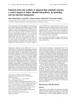
The phylogenetic tree, as shown in Fig. 2, demonstrated
that all local isolates formed a single clade, which was dis-
tant from the vaccine isolates and other field isolates
Chulakasian et al. Virology Journal 2010, 7:122
/>Page 3 of 9
from America. Among the vaccine strains, Vacc-P, the
contemporary vaccine strain, was freestanding and
located between our local isolates and the CDV strains
isolated in America. This contrasted with Vacc-N and
Vacc-Q, which were clustered in the same group as the
Onderstepoort vaccine strain. A bootstrap value of 100
for this clade suggests a robust phylogenetic grouping.
Noticeably, the sequence variation events among the
local isolates and the commercial vaccines observed in
the M-F intergenic region and the pre-signal peptide
region of F gene were well scattered (Fig. 3A).
If these results are examined as a whole, all of the local
isolates were found to be closely related to strains belong-
ing to the Asia-1 lineage, which is distant and phylogenet-
ically distinct from the vaccine strains. Additionally, the
analysis of the three commercial vaccines indicated that
two out of the three seem to have originated from a com-
mon ancestor similar to other vaccine strains (Onderste-
poort and Convac), while only Vacc-P strain has a closer
phylogenetic relationship with our local strains.
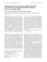
Figure 1 Schematic illustration of the CDV genome and the locations of the primers used in this study. The primer pairs CDF-F and CDF-R were
designed for the first round amplification. Two inner primer sets F-vacc/R-vacc and F-wt/R-wt were simultaneously used for the second round multi-
plex ARMS-PCR. The F-wt and R-vacc were designed to differential amplification of field and vaccine strains, respectively. Arrows indicate the direction
of primers.
M
F
Fsp
4325
5325
CDF-F
CDF-R
F-vacc
R-vacc
F-wt
R-wt
AAA
TGC
300bp
N
P/C/V
M
F
H
L
590bp
Table 1: Comparison of the nucleotide sequences of field isolates from Taiwan with commercial CDV vaccines using the
CDV M-F UTR and part of the F gene (nucleotides 4325-5325)
Percentage identity
TW-1 TW-2 TW-3 TW-4 TW-5 TW-6 TW-7 Vacc N Vacc P Vacc Q
TW-1 98.5 99.3 98.1 97.6 98.0 98.1 83.3 93.6 85.1
TW-2 1.5 98.6 97.8 97.9 97.7 97.8 82.7 93.8 85.0
TW-3 0.7 1.4 98.2 97.7 98.1 98.2 82.9 93.5 84.8
TW-4 1.9 2.2 1.8 96.9 99.9 100.0 83.1 93.5 84.9
TW-5 2.4 2.1 2.3 3.2 96.8 96.9 82.5 93.2 85.0
TW-6 2.0 2.3 1.9 0.1 3.3 99.9 83.0 93.4 84.8
TW-7 1.9 2.2 1.8 0.0 3.2 0.1 83.1 93.5 84.9
Vacc N 19.3 20.2 19.9 19.6 20.4 19.7 19.6 86.2 96.3
Vacc P 6.7 6.5 6.8 6.8 7.2 7.0 6.8 15.6 88.9
Vacc Q 16.9 17.1 17.3 17.2 17.0 17.3 17.2 3.8 12.2
Divergence
Chulakasian et al. Virology Journal 2010, 7:122
/>Page 4 of 9
Differentiation of the vaccine strains and the field CDV
isolates by Multiplex ARMS-PCR
Amplification refractory mutation system (ARMS)-PCR,
also called allele-specific oligonucleotide PCR, was origi-
nally designed for the detection of known sequence poly-
morphisms, such as point mutations [24]. Using just two
pairs of primers in a single PCR tube, this method can
simultaneously amplify both mutant and wild type alleles,
plus it allows for the amplification of an internal DNA
control. This technique has been applied to the genotyp-
ing, analysis of genetic disorders [25-27], and the diagno-
sis of several different virus infections [26,28,29]. The
discrimination of amplification mainly depends on the
mismatch nucleotide at the most 3'-terminus of primer
[24]. The allele-specific (or lineage-specific) priming of
the PCR process will only permit amplification to occur
when the most 3'-terminal nucleotide matches with its
target sequences (Fig 1).
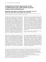
Alignment of the sequences revealed the substitution of
three adenines at positions 530-532 in all three vaccine
strains; while the sequences at the same positions in the
local isolates are T/CGC (marked with square in Fig. 3A).
Interestingly, this T/CGC, located 35 nucleotide down-
stream from Fsp start codon, also can be observed in
other Asia-1 CDVs, including strains from Taiwan (49
strains) and China, published in GenBank database (Fig.
3B). This apparent variation allowed the design of a geno-
type-specific primer that would differentiate local strains
from the vaccine strains. With this in mind, in order to
increase the discrimination power, the last three nucle-
otides at the 3'-end of the forward F-wt and the reverse R-
vacc primers were designed to specifically target this par-
ticular region of the wild type or field isolates, respec-
tively. In addition, two universal outer primers, reverse R-
wt and forward F-vacc were designed to act as primer
pairs for nested ARMS-PCR amplification (Fig. 1).
The region made up of nucleotides 4325-5325, which
corresponds to part of the M gene, the intergenic spacer
between the M and F genes and part of the F gene, was
initially synthesised from the cDNAs of the seven CDV
field isolates and the three commercial vaccines using the
primer set CDF-F and CDF-R. The resulting amplicons
were subsequently amplified using the two type specific
primers sets, F-vacc/R-vacc and F-wt/R-wt (Fig. 1). The
second-round PCR products represent the various
genetic clusters. As illustrated in Fig. 4A, all commercial
vaccine isolates were recognised by the primers F-vacc
and R-vacc and yielded products that were 590 bp in
length, while all seven local isolates yielded 300 bp-prod-
ucts when amplified by primers F-wt and R-wt.
Moreover, in order to further evaluate whether this
multiplex assay can be used to characterise vaccine
strains among the local strains, we performed PCR with
the two sets of primers and different combinations of
templates, such as one of the vaccine strains with or with-
out the presence of a field isolate. The results consistently
produced the correct 590 bp and 300 bp PCR products
according to the templates present in the amplification
(Fig. 4B). No cross-reactivity between the heterotypic
primer pairs and the CDV strains was observed, this indi-
cates that the multiplex ARMS-PCR is able to distinguish
local isolates from vaccine strains even in a mixed popu-
lation.
Base on the sequence homogeneity in agreement with
the vaccine lineage, within the similar position of 590bp-
product in other CDV strains from GenBank, three ade-
nosines (AAA) located 35 nucleotide downstream from
Fsp start codon, were observed in CDV isolates from
Asia-2, America, Europe lineages (Fig. 3B), indicating
that the forward F-vacc and reverse R-vacc primers,
designed to circumstantially target AAA motif (Fig 1), are
potentially able to amplify 590bp-products for recogni-
tion of these three CDV lineages. Thus, in order to spec-
ify these CDVs from vaccine strain, the further
genotyping assay is needed to develop.
Figure 2 Phylogenetic analysis of various CDV strains based on
the nucleotide sequence of part of the F protein and the inter-
genic region between the M gene and the F gene (nucleotides
4325-5325). Only bootstrap values greater than 70 are shown and the
branch lengths are proportional to genetic distance.
Local isolates
Chulakasian et al. Virology Journal 2010, 7:122
/>Page 5 of 9
Figure 3 Sequence alignment of partial F gene. (A)The F gene nucleotide sequences, including the intergenic region between M and F gene (M-
F UTR), of field strains from Taiwan and three commercial vaccine strains were analysed. The numbering starts at the first amino acid of the M-F UTR.
Only amino acids that differ from the majority sequence are shown. Identical residues are represented by dots. The substitution of the AAA present
in the vaccine strains, which was used to design the differentiating primers for ARMS-PCR, is indicated by a square box. (B) The region consisting of
TCG motif, located 35 nucleotide downstream from the start codon (ATG) was comparatively aligned with various CDV lineages: Asia-1 strain; TW-KS2,
TW-TP1, TW-KL1, HeB-07, JL-07, NM, ZD01, BS0610 (GenBank: EU192013
, EU191985, EU191988, EU327874, EU327875, EF596903, EF596904, EU934234),
Asia-2 strain; 007 Lm (GenBank: AB474397
), Europe strain; 5047/91, R252/72, Rockborn, X65509 (GenBank: AF026240, AF026243, AF026244, X65509),
America strain; A75/17, 01-2689, 5804, 5804P, 00-2601 (GenBank: AF164967
, AY649416, AY386315, AY386 316, AY443350), and Vaccine strain; Vaccine
X, Snyder Hill and Onderstepoort (GenBank: EU072198
, GU138403, AF305419). Omitted sequences are represented by dots.
A
B
M Fsp F
Majority
Tw1
Tw2
Tw3
Tw4
Tw5
Tw6
Tw7
Vacc-N
Vacc-P
Vacc-Q
Majority
Tw1
Tw2
Tw3
Tw4
Tw5
Tw6
Tw7
Vacc-N
Vacc-P
Vacc-Q
Majority
Tw1
Tw2
Tw3
Tw4
Tw5
Tw6
Tw7
Vacc-N
Vacc-P
Vacc-Q
Chulakasian et al. Virology Journal 2010, 7:122
/>Page 6 of 9
Genotyping of CDV vaccine strains by restriction fragment
length polymorphism (RFLP)
Within 590 nucleotides, the recognition site of BamH I
was observed in contemporary vaccine, but not in CDV-
Vaccine cluster. The RFLP analysis was performed to dif-
ferentiate contemporary vaccine from other vaccine
strains. As expected, a smaller fragment of 504 bp was
detected from Vacc-P amplicon digested with BamH I,
whereas the other two vaccines remained undigested
(Fig. 4C). Thus, these results indicated that the RFLP
analysis may be applied for further characterized the con-
temporary vaccine strain from other vaccine strains.
Discussion
In this study, differential ARMS-PCR and RFLP genotyp-
ing system were established on the basis of the genetic
divergence spanning from the intergenic region of the M
and F genes to the Fsp region of F gene. The level of
genetic variation of the F gene between the vaccine and
circulating CDV strains in Taiwan was documented in
our previous study [22]. Here we showed that, in addition
to the F gene, low nucleotide similarity was found across
the intergenic region of the M and F genes between the
vaccine and field strains. Our results are consistent with a
previous report, in which the genetic divergence of the
M-F UTR was approximately two-fold higher than that of
the most divergent coding sequence of the H gene [30].
Interference due to the presence of pre-existing anti-
bodies produced by vaccination or a previous infection
will affect the results of any serological diagnosis of CDV.
In order to reinforce the interpretation resulting from
serology based methods, the development of a method
that allows the diagnosis and differentiation of CDV
acquired by natural infection from that used for vaccina-
tion is worthwhile. Martella et al (2007) developed an RT-
PCR genotyping system based on the lineage-specific
nucleotide polymorphisms scattered over the H gene.
Their system was used to characterise the major CDV lin-
eages; European, Asia-1, Asia-2, Arctic, and Vaccine
strains [12]. However, because of limitations in primer
design, this system was not able to amplify CDV belong-
ing to the America cluster and vaccine X. Very recently,
another multiplex PCR assays was reported by Si et al
(2010); in which primers targeting H gene was designed
to distinguish field strains from China and strains from
vaccine cluster, i.e. Onderstepoort [21]. Likewise, Uema
et al (2005) reported that presence of EcoRV and Ssp I
enzyme recognition sites of H gene in Asia strains was
able to differentiate those without these sequences i.e.
vaccine strains [14]. However, within the same DNA frag-
ment, the Ssp I site also found in Vacc-P and American
CDVs (Data not shown), indicating that this method was
not able to differentiate contemporary vaccine strains
from CDVs in Asia-1 and Asia-2 lineages. Therefore, the
PCR genotypic system and RFLP assay targeting on H
gene described previously will be jeopardized when the
vaccine derived from contemporary virus strain were
generally conducted.
In this study, the highly conserved TGC at positions
530-532 in pre-signal peptide (Fsp) of the local strains
(Fig. 3A) allowed us to design genotype specific primer
pairs to distinguish local CDV strains (Asia-1) from three
vaccines, including the contemporary strains (Fig. 1). As
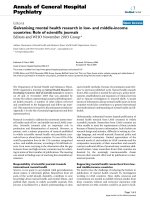
Figure 4 Differential diagnosis of natural canine distemper virus
infection by multiplex ARMS-PCR. (A) Results of a multiplex PCR us-
ing the two primer pairs: F-vacc/R-vacc and F-wt/R-wt. As indicated by
the arrowheads, a 590 bp product corresponding to vaccine tempalte
was specifically amplified from cDNA of Vacc-P, Vacc-Q and Vacc-N
(lane 1 to 3); the 300 bp product was only amplified from the local
strains (lanes 4-10). Note: Bands with a higher molecular weight, indi-
cated with an arrow, were products amplifed by the outer primer set,
F-vacc and R-wt. (B) Characterisation of CDV strains by the two sets of
genotype specific primers in combination with various templates,
namely Vacc-P (lane 1), Vacc-Q (lane 2), a local strain (lane 3), Vacc-P
and a local strain (lane 4), Vacc-Q and a local strain (lane 5) and a neg-
ative control without template (lane 6). As indicated with arrowheads,
the amplicons corresponding to a specific template, the vaccine
strains (590 bp) and the local strains (300 bp), can be differentiated. (C)
RFLP analysis of CDV vaccine strains. A unique BamH I recognition site
was found in Vacc-P and CDV isolates in America lineage, but not in
Vacc-N, Vacc-Q and other CDVs in Vaccine lineage. As shown in the
lower panel, digestion of Vacc-P PCR product with BamH I enzyme re-
sulted in a smaller DNA fragment (~500 bp; lane 1), whereas DNA ob-
tained from Vacc-Q and Vacc-N remained intact (590 bp; lane 2 and 3).
590bp
300bp
(bp)
1500
1000
500
300
590bp
300bp
1000
500
300
(bp)
590bp
300bp
1000
500
300
(bp)
A
B
C
(bp)
3000
1500
1000
500
300
1 2 3
BamH I
87
590 bp
~500 bp
Vacc-P and America lineage
Vacc-N, vacc-Q
Chulakasian et al. Virology Journal 2010, 7:122
/>Page 7 of 9
expected, the size difference between the vaccine specific
and field strain specific products provided a simple and
reliable method of identification and differentiation of
CDV (Fig. 4A), even when mixed templates from field
and vaccine strains were used (Fig. 4B). Although the
identity of the Fsp amino acid sequence, when the Taiwan
strains and vaccine strains are compared, was as low as
64-67% [22], surprisingly, an analysis of Fsp region in var-
ious CDV strains in GenBank database indicates that
TGC motif used to specifically target local isolates is
highly conserved among the Asia-1 lineage. These find-
ings demonstrated that our assay will be able to reliably
differentiate field CDV (Asia-1 lineage, as tested in pres-
ent study) from the two major lineages of conventional
vaccines, namely Vacc-N/Vacc-Q and contemporary vac-
cine, namely Vacc-P.
In addition to differential diagnosis of natural CDV
infection, the highly genetic variation of M-F UTR
throughout 590 nucleotides allowed us to design the
RFLP genotyping system based on the unique restriction
enzyme profile. In this region, the recognition site of
BamH I was observed in contemporary vaccine, America,
and Asia-2 clusters, but not in CDV-Vaccine cluster. Fur-
thermore, the restriction patterns of enzyme Apo I and
Bgl I were different among contemporary vaccine, Amer-
ica and Asia-2 lineages (Table 2). Taking together, the
RFLP assay with these restriction enzymes could be
potentially used in for genotyping those CDV lineages
that will be useful for identification of CDV infection
acquired from other lineage and also for monitoring the
evolution of CDV viruses. Notwithstanding, due to the
limitation of clinical specimens from other geographic
areas, we was able to affirm the differentiation of circulat-
ing CDV-Asia1 strains from vaccines and contemporary
vaccine.
Conclusions
At present, control of CDV relies on immunisation with
vaccines, mostly live attenuated vaccines. A multiplex
ARMS-PCR assay developed in this study can be consid-
ered as a practical and robust tool for the rapid differenti-
ation of current circulating CDV and vaccine strains
based on the sequence polymorphism in the F gene and
its upstream M-F UTR. When used clinically, this assay,
for the first time, is able to effectively identify the origin
of a CDV infection and, most importantly, confirm the
presence of a natural CDV infection.
Methods
Sample collection and preparation
Regardless of vaccination history, seven isolates of CDV
were obtained from dogs' nasal swabs with the clinical
suspicion of canine distemper provided by the Veterinary
Teaching Hospital of National Chung Hsing University
and by the Taichung City Animal Protection and Health
Inspection Center. Nasal swabs were homogenised in 1
ml of phosphate buffered saline (PBS) and then centri-
fuged at 8,000 g for 1 min. Supernatants were collected
and kept at -80°C for further experiments. In addition,
three live-attenuated commercial vaccines, Vacc-P, Vacc-
N and Vacc-Q, all currently used in Taiwan, were also
included in this study.
Table 2: Comparison of the restriction enzyme profile within 590 nucleotide of non-coding region between M and F gene
(nucleotide 4403-4492) in different CDV lineages.
lineages Isolates Restriction enzyme recognition site Expected size of fragments (base pair)
BamH I Bgl I Apo I
Vaccine Onderstepoort
Synder Hill
Vacc-Q
Vacc-N
+ 27, 263
Contemporary
vaccine
Vacc-P +++ 27, 40, 19, 504
America 00-2601
00-2689
98-2645
98-2646
98-2654
+-++ 27, 59, 196, 308
Asia-2 007 Lm +- - 27, 59, 504
Chulakasian et al. Virology Journal 2010, 7:122
/>Page 8 of 9
Purification of the nucleic acid, reverse transcription and
amplification of F gene
Total nucleic acid was extracted from the supernatants of
swabs and vaccines using the RNeasy Mini 50 kit (QIA-
GEN) according to the manufacturer's instructions. Total
RNA (1 μg) and random 8-mer primers (50 μM) were
denatured at 65°C for 5 min and cooled down on ice. To
synthesise the first-strand cDNA, the RNA and primers
were mixed in 5 × reaction buffers, 0.1 M DDT, 0.5 mM
of each deoxynucleotide, 200 U SuperScript III reverse
transcriptase (Invitrogen) and 40 U RNase inhibitor. A
total of 20 μL of the mixture was initially incubated at
25°C, then the reaction was held at 65°C for 60 min and
finally it was terminated by incubation at 70°C for 10 min.
Following this, the first round amplification was con-
ducted by polymerase chain reaction (PCR) with the
outer primers CDF-F: 5'-AGAGTGCAAAATAGTAA-
GAATCCAAGC-3' and CDF-R: 5'-GAAAGAGACTG-
GCTATTCCGATGC-3', which amplified a fragment
containing the M gene (115 downstream nucleotides;
4325-4439), M-F UTR (495 nucleotides; 4440-4934) and
the F gene (first 391 nucleotides; 4935-5325) (Fig. 1).
Thermocycling conditions for amplification started with
an initial denaturation at 95°C for 5 min and then the
reaction mixture was subjected to 35 cycles of heat dena-
turation at 95°C for 1 min, primer annealing at 55°C for 2
min, DNA extension at 72°C for 2 min; this was followed
by a final extension at 72°C for 7 min. The identity of the
resulting PCR products was verified by direct automated
sequencing.
The Multiplex ARMS-PCR assay
The F gene products from the first round PCR were then
further simultaneously amplified by multiplex ARMS-
PCR using two primer sets in order to distinguish the
vaccine and field strains. The specific primer sets, namely
F-wt and R-vacc, were designed according to the different
sequences obtained and specifically targeted either the
field isolates or the vaccine strains (Fig. 1). The primers
used for vaccine strain amplification were F-vacc: 5'-
CATCAGCCATGATCAGGGTCTTTTC-3' and R-vacc:
5'-GGGCGGTCTTGTTGGGTATGTGTTT-3'. The
primers used for field strain amplification were F-wt: 5'-
AATTCCCAAAAAATCCAAACCCTGC-3' and R-wt:
5'-GATTGCCGCCTCTTGAACCAGGAA-3'. The
amplification conditions for the multiplex-nested ARMS-
PCR were 95°C for 5 min followed by 35 cycles of dena-
turation at 95°C for 1 min, annealing at 55°C for 2 min,
DNA extension at 72°C for 2 min and a final extension at
72°C for 7 min. All amplification cycles were performed
in a DNA thermal cycle (GeneAmp PCR system 2700).
The PCR products were resolved by 1.2% agarose gel
electrophoresis with Health safe nucleic acid stain. Prod-
uct sizes were determined with reference to a 100 base
pair (bp) DNA Ladder.
Restriction Fragment Length Polymorphism (RFLP) analysis
For genotyping, the PCR product amplified with primers
F-vacc and R-vacc was isolated by using the Purelink™
PCR purification kit (Invitrogen), and resulting product
was further digested with restriction enzyme BamH I
(New England Biolabs). A 4 ml-aliquot was digested with
1.5 U of BamH I at 37°C for 90 min according to the man-
ufacturer's recommendation. The resulting restriction
fragments were resolved by 1.2% Tris acetate-EDTA-aga-
rose gel electrophoresis.
Phylogenetic analysis
Several CDV strains were selected for phylogenetic analy-
sis. The nucleotide sequence accession numbers in the
GenBank database for the F gene and its upstream region,
M-F UTR, sequences of the reference strains used in this
study are: A75/17-USA (GenBank: AF164967
), Raccoon
00-2601-USA (GenBank: AY443350
), Raccoon 00-2689-
USA (GenBank: AY649446
), Raccoon 98-2646 (GenBank:
AY542312
), Raccoon 98-2654 (GenBank: AY466011),
Raccoon 98-2645 (GenBank: AY445077
) and Onderste-
poort (GenBank: AF305419
).
Nucleotide sequences corresponding to the CDV F and
H genes were aligned using the CLUSTAL W multiple
alignment method with BioEdit software [31] and com-
pared with other previously published sequences
reported in GenBank. The phylogeny of the nucleotide
and amino acid alignments were analysed using distance
matrix methods (DNADIST for nucleotide sequence and
PROTDIST for amino acid sequence, followed by
NEIGHBOR) using the PHYLIP software package [32].
The datasets were subjected to bootstrap analysis based
on 1,000 re-samplings of the original data and the SEQ-
BOOT program was used to produce a majority-rule
consensus tree.
Additional material
Competing interests
The authors declare that they have no competing interests.
Additional file 1 Phylogenetic analysis of CDV strains based on the
deduced 331 amino acid sequence of the H protein. Only bootstrap val-
ues greater than 70 are shown, and branch lengths are proportionate to
genetic distances. The accession numbers of H gene sequences of the ref-
erence strains are: Onderstepoort (AF378705), Convac (Z35493), SnyderHill
(AF259552), Yanaka (D85755), Ueno (D85753), Hamamatsu (D85754), KDK1
(AB025271), Raccoon dog-Japan (AB016776), Dog98-002 (AB025270),
Dog5B (AY297453), DogHM-3 (AB040767), Dog26D (AB040766), Dog5VD
(AY297454), Dog-TW (AY378091), Dog5804-Germany (AY386315), Giant
Panda-China (AF178038), Dog-China (AF172411), PDV-2 Siberian seal
(X84998), Dog-Turkey (AY093674), Dog91A-Denmark (AF478544), Dog91B-
Denmark (AF478546), DogDen (AF478543), Dogiso-Den (AF478547), Dog
Denmark (Z47761), Raccoon-USA (Z47764), Raccoon01-2689-USA
(AY649446), Raccoon01-2676-USA (AY498692), Raccoon01-2690-USA
(AY465925), Raccoon00-2601-USA (AY443350), Jevelina-USA (Z47765) and
A75-17 (AF164967).
Chulakasian et al. Virology Journal 2010, 7:122
/>Page 9 of 9
Authors' contributions
SC conducted most of this work under supervision of W-L H and T-J C. M-S L, C-
Y W, and S-S C participated in clinical sample collection. K-H L, F-Y L, and T-H H
participated in the sequence analysis of H gene under supervision of M-L W. All
authors have read and approved the manuscript.
Acknowledgements
The authors wish to thank Dr. Sarah M. Richart (Department of Biology and
Chemistry, Azusa Pacific University, CA, USA) for editorial assistance and Taic-
hung City Animal Protection and Health Inspection Center, Taichung, Taiwan
for the sample collection. This study was supported by the Bureau of Animal
and Plant Health Inspection and Quarantine, the Council of Agriculture (grant
number: GA97104), and National Scientific Council (grant number: NSC96-
2313-B-005-016-MY3), Taiwan.
Author Details
1
Department of Veterinary Medicine, College of Veterinary Medicine, National
Chung Hsing University, 250 Kou Kuang Road, Taichung 402, Taiwan,
2
Animal
Health Research Institute, Council of Agriculture, 376 Chung-Cheng Road,
Tamsui, Taipei 251, Taiwan and
3
Graduate Institute of Microbiology and Public
Health, College of Veterinary Medicine, National Chung Hsing University, 250
Kou Kuang Road, Taichung 402, Taiwan
References
1. Appel MJ, Summers BA: Pathogenicity of morbilliviruses for terrestrial
carnivores. Vet Microbiol 1995, 44:187-191.
2. Blixenkrone-Moller M, Svansson V, Have P, Orvell C, Appel M, Pedersen IR,
Dietz HH, Henriksen P: Studies on manifestations of canine distemper
virus infection in an urban dog population. Vet Microbiol 1993,
37:163-173.
3. Gemma T, Watari T, Akiyama K, Miyashita N, Shin YS, Iwatsuki K, Kai C,
Mikami T: Epidemiological observations on recent outbreaks of canine
distemper in Tokyo area. J Vet Med Sci 1996, 58:547-550.
4. Roelke-Parker ME, Munson L, Packer C, Kock R, Cleaveland S, Carpenter M,
O'Brien SJ, Pospischil A, Hofmann-Lehmann R, Lutz H, et al.: A canine
distemper virus epidemic in Serengeti lions (Panthera leo). Nature
1996, 379:441-445.
5. Barrett T: Morbillivirus infections, with special emphasis on
morbilliviruses of carnivores. Vet Microbiol 1999, 69:3-13.
6. Martella V, Pratelli A, Cirone F, Zizzo N, Decaro N, Tinelli A, Foti M,
Buonavoglia C: Detection and genetic characterization of canine
distemper virus (CDV) from free-ranging red foxes in Italy. Mol Cell
Probes 2002, 16:77-83.
7. Guiserix M, Bahi-Jaber N, Fouchet D, Sauvage F, Pontier D: The canine
distemper epidemic in Serengeti: are lions victims of a new highly
virulent canine distemper virus strain, or is pathogen circulation
stochasticity to blame? J R Soc Interface 2007, 4:1127-1134.
8. Krakowka S: Mechanisms of in vitro immunosuppression in canine
distemper virus infection. J Clin Lab Immunol 1982, 8:187-196.
9. von Messling V, Oezguen N, Zheng Q, Vongpunsawad S, Braun W,
Cattaneo R: Nearby clusters of hemagglutinin residues sustain SLAM-
dependent canine distemper virus entry in peripheral blood
mononuclear cells. J Virol 2005, 79:5857-5862.
10. Cherpillod P, Zipperle L, Wittek R, Zurbriggen A: An mRNA region of the
canine distemper virus fusion protein gene lacking AUG codons can
promote protein expression. Arch Virol 2004, 149:1971-1983.
11. Merz DC, Scheid A, Choppin PW: Importance of antibodies to the fusion
glycoprotein of paramyxoviruses in the prevention of spread of
infection. J Exp Med 1980, 151:275-288.
12. Martella V, Elia G, Lucente MS, Decaro N, Lorusso E, Banyai K, Blixenkrone-
Moller M, Lan NT, Yamaguchi R, Cirone F, et al.: Genotyping canine
distemper virus (CDV) by a hemi-nested multiplex PCR provides a rapid
approach for investigation of CDV outbreaks. Vet Microbiol 2007,
122:32-42.
13. Bolt G, Jensen TD, Gottschalck E, Arctander P, Appel MJ, Buckland R,
Blixenkrone-Moller M: Genetic diversity of the attachment (H) protein
gene of current field isolates of canine distemper virus. J Gen Virol 1997,
78(Pt 2):367-372.
14. Uema M, Ohashi K, Wakasa C, Kai C: Phylogenetic and restriction
fragment length polymorphism analyses of hemagglutinin (H) protein
of canine distemper virus isolates from domestic dogs in Japan. Virus
Res 2005, 109:59-63.
15. Hashimoto M, Une Y, Mochizuki M: Hemagglutinin genotype profiles of
canine distemper virus from domestic dogs in Japan. Arch Virol 2001,
146:149-155.
16. Demeter Z, Lakatos B, Palade EA, Kozma T, Forgach P, Rusvai M: Genetic
diversity of Hungarian canine distemper virus strains. Vet Microbiol
2007, 122:258-269.
17. Brown AL, Vitamvas JA, Merry DL Jr, Beckenhauer WH: Immune response
of pups to modified live-virus canine distemper-measles vaccine. Am J
Vet Res 1972, 33:1447-1456.
18. Haig DA: Canine distemper: immunization with avianized virus.
Onderstepoort J Vet Res 1956, 17:19-53.
19. Mochizuki M, Hashimoto M, Hagiwara S, Yoshida Y, Ishiguro S: Genotypes
of canine distemper virus determined by analysis of the hemagglutinin
genes of recent isolates from dogs in Japan. J Clin Microbiol 1999,
37:2936-2942.
20. Schatzberg SJ, Li Q, Porter BF, Barber RM, Claiborne MK, Levine JM, Levine
GJ, Israel SK, Young BD, Kiupel M, et al.: Broadly reactive pan-
paramyxovirus reverse transcription polymerase chain reaction and
sequence analysis for the detection of Canine distemper virus in a case
of canine meningoencephalitis of unknown etiology. J Vet Diagn Invest
2009, 21:844-849.
21. Si W, Zhou S, Wang Z, Cui S: A multiplex reverse transcription-nested
polymerase chain reaction for detection and differentiation of wild-
type and vaccine strains of canine distemper virus. Virol J 2010, 7:86.
22. Lee MS, Tsai KJ, Chen LH, Chen CY, Liu YP, Chang CC, Lee SH, Hsu WL: The
identification of frequent variations in the fusion protein of canine
distemper virus. Vet J 2010, 183:184-190.
23. Chan KW, Hsieh HH, Wang HC, Lee YJ, Sung MH, Wong ML, Hsu WL:
Identification, expression and antigenic analysis of recombinant
hemagglutinin proteins of canine distemper virus. J Virol Methods 2009,
155:18-24.
24. Newton CR, Graham A, Heptinstall LE, Powell SJ, Summers C, Kalsheker N,
Smith JC, Markham AF: Analysis of any point mutation in DNA. The
amplification refractory mutation system (ARMS). Nucleic Acids Res
1989, 17:2503-2516.
25. Vannucchi AM, Pancrazzi A, Bogani C, Antonioli E, Guglielmelli P: A
quantitative assay for JAK2(V617F) mutation in myeloproliferative
disorders by ARMS-PCR and capillary electrophoresis. Leukemia 2006,
20:1055-1060.
26. Chen Q, Lu P, Jones AV, Cross NC, Silver RT, Wang YL: Amplification
refractory mutation system, a highly sensitive and simple polymerase
chain reaction assay, for the detection of JAK2 V617F mutation in
chronic myeloproliferative disorders. J Mol Diagn 2007, 9:272-276.
27. Newton CR, Heptinstall LE, Summers C, Super M, Schwarz M, Anwar R,
Graham A, Smith JC, Markham AF: Amplification refractory mutation
system for prenatal diagnosis and carrier assessment in cystic fibrosis.
Lancet 1989, 2:1481-1483.
28. Gramegna M, Lampertico P, Lobbiani A, Colucci G: Detection of the
hepatitis B virus major pre-core mutation by the amplification
refractory mutation system technique. Res Virol 1993, 144:307-309.
29. Liang TJ, Bodenheimer HC Jr, Yankee R, Brown NV, Chang K, Huang J,
Wands JR: Presence of hepatitis B and C viral genomes in US blood
donors as detected by polymerase chain reaction amplification. J Med
Virol 1994, 42:151-157.
30. Liermann H, Harder TC, Lochelt M, von Messling V, Baumgartner W,
Moennig V, Haas L: Genetic analysis of the central untranslated genome
region and the proximal coding part of the F gene of wild-type and
vaccine canine distemper morbilliviruses. Virus Genes 1998, 17:259-270.
31. Hall TA: BioEdit: a user-friendly biological sequence alignment editor
and analysis program for Windows 95/98/NT. Nucleic Acids Symp Ser
1999, 41:95-99.
32. Felsenstein J: PHYLIP - Phylogeny Inference Package (Version 3.2).
Cladistics 1989, 5:164-166.
doi: 10.1186/1743-422X-7-122
Cite this article as: Chulakasian et al., Multiplex Amplification Refractory
Mutation System Polymerase Chain Reaction (ARMS-PCR) for diagnosis of
natural infection with canine distemper virus Virology Journal 2010, 7:122
Received: 11 March 2010 Accepted: 10 June 2010
Published: 10 June 2010
This article is available from: 2010 Chulakasian et al; licensee BioMed Central Ltd. This is an Open Access article distributed under the terms of the Creative Commons Attribution License ( ), which permits unrestricted use, distribution, and reproduction in any medium, provided the original work is properly cited.Virology Journal 2010, 7:122

![Báo cáo Y học: The membrane-bound [NiFe]-hydrogenase (Ech) from Methanosarcina barkeri : unusual properties of the iron-sulphur clusters docx](https://media.store123doc.com/images/document/14/rc/ee/medium_eeh1395026426.jpg)