Pediatric Epilepsy Diagnosis and Therapy - part 7 pps
Bạn đang xem bản rút gọn của tài liệu. Xem và tải ngay bản đầy đủ của tài liệu tại đây (640.73 KB, 92 trang )
38 • DOSAGE FORM CONSIDERATIONS IN THE TREATMENT OF PEDIATRIC EPILEPSY
529
10% to 20%, which makes it difficult to administer an
effective dose (82).
Oxcarbazepine
Clemens et al performed a study in 10 healthy volunteers
to characterize the bioavailability of rectally administered
oxcarbazepine suspension (300 mg/5 mL) diluted 50%
with water. Mean relative bioavailability calculated from
plasma AUCs was 8.3% (SD 5.5%) for monohydroxy
derivative (MHD) and 10.8% (SD 7.3%) for OXC. The
C
max
and AUC differed significantly between routes for
both MHD and OXC (P Ͻ 0.01). The total amount of
MHD excreted in the urine following rectal administra-
tion was 10 Ϯ 5% of the amount excreted following oral
administration. Oral absorption was consistent with pre-
vious studies. The most common side effects were head-
ache and fatigue with no discernable difference between
routes. MHD bioavailability following rectal administra-
tion of OXC suspension is significantly less than after oral
administration, most likely because of OXC’s poor water
solubility. It is unlikely that adequate MHD concentra-
tions can be reached by rectal administration of diluted
OXC suspension (83).
Paraldehyde
Rectally administered paraldehyde has been widely used
to control severe seizures, particularly in children (84,
85). However, information on the efficacy, toxicity, and
pharmacokinetics is limited. Rectal bioavailability is 75%
to 90% versus 90% to 100% for the oral route. Time
to peak concentrations after rectal administration is 2.5
hours versus 0.5 hours for oral administration. Paralde-
hyde should be diluted with an equal volume of olive oil
or vegetable oil to reduce mucosal irritation.
Phenobarbital
There is no commercially available rectal dosage form
for phenobarbital. Graves and coworkers gave seven
volunteers phenobarbital sodium parenteral solution
rectally and intramuscularly (86). After rectal administra-
tion absorption was 90% complete, with a time to peak
concentration of 4.4 hours versus 2.1 hours for the IM
injection. Suppositories containing phenobarbital sodium
are more rapidly absorbed than phenobarbital acid given
either orally or intramuscularly (87, 88).
Phenytoin
Occasionally, there arises a need to administer phenytoin
rectally, although no commercial rectal dosage form is
available. Several studies of investigational suppository
formulations have failed to demonstrate absorption.
Rectal administration of phenytoin sodium parenteral
solution in dogs produced low but measurable serum
concentrations, but absorption was slow (89). Rectal
administration of phenytoin is not recommended.
Valproic Acid
Valproic acid absorption has been studied after rectal
administration of diluted syrup and suppositories. Rectal
absorption of the commercially available syrup is complete,
with peak concentrations occurring approximately 2 hours
after a dose (90–92). High osmolality necessitates 1:1 dilu-
tion of the syrup to minimize catharsis. The syrup has
been used to treat status epilepticus when other therapy is
ineffective. Various suppository formulations are absorbed
well, albeit more slowly than the syrup, with time to peak
concentration occurring in 2 to 4 hours (93, 94).
Topiramate
Topiramate is also readily absorbed following rectal
administration. In a study of twelve healthy subjects who
received either 100 or 200 mg of topiramate orally and
a 200 mg dose of topiramate given rectally, the relative
bioavailability (F
rel
), which was determined by calculat-
ing the dose-normalized areas under the concentration
time curves, was 0.72 Ϯ 0.18 h/L for the rectal dose
and 0.76 Ϯ 0.20 h/L for the oral dose.The relative bio-
availability for topiramate administered rectally was
0.95 Ϯ 0.17 with a range of 0.68 Ϯ 1.2 (95).
Zonisamide
Nagatomi et al investigated two zonisamide supposito-
ries compared with IV and oral dosing in rats (96). The
bioavailability of the hydrophilic base was 96%, and that
from the lipophilic base was 108%. The C
max
following
both rectal suppositories was significantly greater than
an equal oral dose, and T
max
occurred faster after the
hydrophilic-based suppository (2 hrs) than after either
the lipophilic-based or the oral dose (4 hrs).
STUDIES OF OTHER
ADMINISTRATION ROUTES
Buccal/Sublingual
Buccal/sublingal administration of diazepam and loraz-
epam has been recommend by some clinicians as a part
of routine clinical practice. However, there are no studies
documenting their efficacy. Limited data exist for pharma-
cokinetic and efficacy for this route of administration.
Buccal administration of midazolam was studied
in 10 healthy adults in a study in which 2 mL of the
IV • GENERAL PRINCIPLES OF THERAPY
530
intravenous preparation of midazolam 5 mg/mL fla-
vored with peppermint was held in the mouth for 5 min-
utes, then spat out. The researchers found that changes
on electroencephalography were observed within 5 to
10 minutes of administration of the drug, suggesting
rapid absorption and onset of effect (97). In a random-
ized controlled trial conducted in a hospital emergency
department, the safety and efficacy of buccal midazolam
were compared with those of rectal diazepam (98). The
dose used for each drug was determined by the age of the
child, with a target dose of about 0.5 mg/kg (from 2.5
mg for children aged 6 to 12 months; 4 mg for those 1
to 4 years; 7.5 mg for those 5 to 9 years; and 10 mg for
those 10 years or older). A total of 219 episodes of acute
seizures in 177 children were treated.Therapeutic success
was defined as cessation of seizure within 10 minutes of
drug administration without respiratory depression and
without seizure recurrence within 1 hour. A postivie out-
come was achieved in 56% of patients treated with buc-
cal midazolam, compared with 27% of patients treated
with rectal diazepam (P Ͻ0.001; odds ratio [OR] 4.1,
95% CI 2.2–7.6). Median time to seizure termination was
8 minutes (range: 5–20 minutes) for buccal midazolam
and 15 minutes (range: 5–31 minutes) for rectal diazepam
(P ϭ 0.01; hazard ratio [HR] 0.7; 95% CI 0.5–0.9).
Greenblatt et al compared the pharmacokinetics of
sublingual lorazepam with IV, IM, and oral LZP (99). Ten
healthy volunteers randomly received 2 mg of LZP in the
following five formulations: IV injection, IM injection,
oral tablet, sublingual administration of the oral tablet,
and sublingual administration of a specially formulated
tablet. Peak plasma concentrations, time to peak concen-
trations, elimination half life, and relative bioavailability
were not significantly different among the formulations.
Peak concentrations were highest for the IM route, fol-
lowed by oral and sublingual; time to peak concentrations
was most rapid for the IM route, followed by sublingual
and oral. Mean relative bioavailabilities were high for all
routes: IM (95.9%), oral (99.8%), sublingual of oral tab-
let (94.1%) and sublingual of special tablet (98.2%).
It should be noted, however, that the efficacy, safety,
duration of effect, and ease of buccal/sublingal adminis-
tration by nonmedical caregivers have not been evaluated
in settings outside of hospitals.
INTRANASAL
Several benzodiazepines possess the physical, chemical,
and pharmacokinetic properties required of effective
nasal therapies. Among the benzodiazepines considered
for intranasal administration, midazolam has been most
extensively studied. In one randomized, open-label trial
involving 47 children with prolonged (Ͼ10 minutes)
febrile seizures, the safety and efficacy of intranasal
midazolam (0.2 mg/kg) were compared with those of
intravenous diazepam (0.3 mg/kg) administered over
5 minutes (100). Intranasal midazolam was as safe and
effective as intravenous diazepam and resulted in earlier
cessation of seizures as a result of rapid administration.
However, the role of intranasal midazolam in treat-
ing seizure emergencies remains to be established. There
are no adequately controlled trials demonstrating the
safety and efficacy of intranasal midazolam for out-of-
hospital treatment. Moreover, the short elimination half-
life of midazolam—especially in patients taking enzyme-
inducing drugs—raises concern as to whether its duration
of effect is satisfactory in out-of-hospital settings.
Intranasal lorazepam has also been studied (101).
Intranasal LZP was absorbed with a mean percent bioavail-
ability of 77.7 Ϯ 11.1%. A double-peak concentration-
time curve was observed, indicating possible secondary
oral absorption. The time to peak concentration was vari-
able, ranging from 0.25–2 hours. Lorazepam’s relatively
limited lipid solubility as compared with that of mid-
azolam or diazepam results in a slower rate of absorption
and onset of action.
Diazepam has a lipid solubility and potency com-
parable with those of midazolam and a much longer
elimination half-life, properties that make it a good can-
didate for intranasal administration. The bioavailability
of a novel intranasal diazepam formulation has been
compared with that of intranasal midazolam in healthy
volunteers (n ϭ 4) (102). Both midazolam and diazepam
were rapidly absorbed, but diazepam’s absorption was
more extensive and its half-life longer than that of mid-
azolam. Compared with rectally administered diazepam,
the nasal diazepam formulation is absorbed to the same
extent, but appears to be more rapidly absorbed, resulting
in attainment of maximum concentrations as much as
30 minutes earlier (103).
Nasogastric Tubes
A nasogastric (NG) tube offers an alternative route of
drug delivery. However, drug may adhere to the tubing,
clog the tubing, or not be absorbed. Occlusion of the tube
by the drug is also a concern. Tube occlusions may require
replacement of the tube, which is both costly and incon-
venient for the patient. Recently, it has been demonstrated
that sustained-release carbamazepine (Carbatrol
®
) can
be opened, mixed with 0.9% sodium chloride or apple
juice as diluents, and reliably delivered through an NG
tube or feeding tube 12 French or greater in size (104,
105). Topiramate has also been reported to be effective
in patients with status epilepticus when given through
an NG tube (106).
However, absorption from nasogastric tubes is
not always comparable to orally administered formula-
tions. When patients who are receiving tube feedings are
38 • DOSAGE FORM CONSIDERATIONS IN THE TREATMENT OF PEDIATRIC EPILEPSY
531
switched from IV phenytoin (fosphenytoin) to oral phe-
nytoin administered via a nasogastric tube, there appears
to be decreased absorption of the oral formulation. This
seems to occur regardless of whether the suspension
or the oral capsule dosage form is used. Although the
mechanism has not been clearly documented, it has been
postulated that phenytoin may bind to proteins in the
enteral feeding. Also, the enteral feeding may increase
the GI motility, which may decrease the absorption (107).
Sometimes very large oral doses may need to be given to
maintain the desired serum concentrations in patients
receiving phenytoin and enteral feedings via a nasogastric
tube. Some practitioners try to stop the enteral feedings
for two hours before and two hours after the dose of phe-
nytoin. IM fosphenytoin would be an alternative (3).
SUMMARY
The selection of AED dosage forms is very important in
pediatric epilepsy. Patients may be unwilling or unable
to take oral solid dosage forms. Therefore, the avail-
ability of alternative oral dosage forms such as suspen-
sions, solutions, and sprinkles is important. Patients
who experience concentration-dependent side effects or
breakthrough seizures may realize improved control by
switching to an alternative dosage form. For example, a
controlled-release formulation will provide lower peaks
and higher troughs, facilitating better seizure control with
less toxicity.
Although it has been the practice to crush oral solids
and mix the contents with food, this is not always desir-
able. Some products, such as Phenytek
®
, Depakote-ER,
Depakote
®
, and Tegretol-XR
®
, lose the properties they
were designed to provide if the structure of the prepa-
ration is disrupted. In some cases, the rate or extent of
absorption may be altered when the drug is given with
food. It also has been a custom to compound pediatric
dosage forms extemporaneously. This is an important way
to provide drug in a form that young children can take.
However, clinicians should be cautious about extempora-
neous compounding of pediatric formulations unless they
can determine the amount of drug in the formulation,
the stability of the product, and the bioavailability. This
requires an assay for the compounded product and an
assay of the drug in blood. In addition, with compounded
drugs, someone should taste the preparation before it is
given to the patient. For example, gabapentin has a very
bitter taste when it is put into solution. Therefore, when
a drug is compounded for pediatric delivery, the new
formulation should be tested to ensure that it is being
delivered properly. Specialized dosage forms generally
are more expensive.
Caregivers should be thoroughly educated in drug
administration techniques for children. When carefully
instructed, caregivers can properly administer medications
(108). Drug administration techniques are summarized
in Tables 38-4, 38-5, and 38-6. When doses are given
as “teaspoonfuls,” caregivers should have a calibrated
device for measuring the dose rather than using a com-
mon utensil. The volume of “standard” teaspoons varies
up to fourfold. Drugs given rectally, such as diazepam,
require special caregiver education.
Clinical assessment, selection of a drug, and deter-
mination of the dose require special attention in the
TABLE 38-4
Medication Administration Guidelines for Infants
Use a calibrated dropper or oral syringe.
Support the infant’s head while holding the infant in lap.
Give small amounts of medication to prevent choking.
If desired, crush non–enteric-coated tablets to a powder
and sprinkle on small amounts of food.
Provide physical comforting to calm the infant while
administering medications.
TABLE 38-5
Medication Administration Guidelines for
Toddlers
Allow child to choose a position in which to take
medications.
Disguise the taste with a small volume of flavored drink
or food. Rinse mouth with flavored drink to remove
aftertaste.
Use simple commands in the toddler’s jargon to obtain
cooperation. Allow the toddler to choose which medi
cations to take first. Allow toddler to become familiar
with the oral dosing device.
TABLE 38-6
Medication Administration Guidelines for
Preschool Children
Place tablet or capsule near back of tongue and provide
water or a flavored liquid to aid in swallowing.
Do not use chewable tablets if the child’s teeth are
loose. Use a straw to administer medications that may
stain teeth.
Use a rinse with a flavored drink to minimize aftertaste.
Allow child to help make decisions about dosage forms,
place of administration, which medication to take
first, and the type of flavored drink to use.
IV • GENERAL PRINCIPLES OF THERAPY
532
pediatric patient, as does the selection of the appropri-
ate formulation and dosage form. This last step in the
therapeutic plan plays a pivotal role in the ultimate suc-
cess of therapy. The objective is to ensure the regular
and consistent delivery of drug to the brain. When con-
ventional oral tablets and capsules are inappropriate
or impractical, alternate formulations, dosage forms,
and routes of administration should be considered.
The clinician also must assess the ability of the care-
giver to correctly prepare, measure, and administer
medications and instruct caregivers about proper drug
administration.
References
1. Rowland M, Tozer TN. Clinical pharmacokinetics: concepts and applications. 2nd ed.
Philadelphia: Lea & Febiger, 1989.
2. Gibaldi M. Biopharmaceutics and clinical pharmacokinetics. 3rd ed. Philadelphia: Lea &
Febiger, 1984.
3. Winter ME, Tozer TN. Phenytoin. In: Burton ME, Shaw LM, Schentag JL, Evans WE,
eds. Applied Pharmacokinetics and Pharmacodynamics: Principles of Therapeutic Drug
Monitoring. 4th ed. Philadelphia: Lippincott.
4. Ansel HC, Popovich NG. Pharmaceutical dosage forms and drug delivery systems. 5th
ed. Philadelphia: Lea & Febiger, 1990.
5. Stewart BH, Kugler AR, Thompson PR, Bockbrader HN. A saturable transport mechanism
in the intestinal absorption of gabapentin is the underlying cause of lack of proportionality
between increasing dose and drug levels in plasma. Pharm Res 1993; 10:276–281.
6. Tyrer JH, Eadie MJ, Sutherland JM, Hooper WD. Outbreak of anticonvulsant intoxica-
tion in an Australian city. Br. Med J [Clin Res] 1970; 4:271–273.
7. Bochner F,Hooper WD,Tyrer JH,Eadie MJ.Factors involved in an outbreak of phenytoin
intoxication. J Neurol Sci 1972; 16:481–487.
8. Hamilton RA, Garnett WR, Kline BJ, et al. The effect of food on valproic acid absorption.
Am J Hosp Pharm 1981; 38:1490–1493.
9. Levy R, Pitlick W, Troupin A, et al. Pharmacokinetics of carbamazepine in normal man.
Clin Pharmacol Ther 1975; 17:657–668.
10. Carter BL, Garnett WR, Pellock JM, et al. Interaction between phenytoin and three
commonly used antacids. Ther Drug Monit 1981; 3:333–340.
11. Stewart CG, Hampton EM. Effect of maturation on drug disposition in pediatric patients.
Clin Pharm 1987; 6:548–564.
12. Painter MJ, Pippenger C, MacDonald H, et al. Phenobarbital and diphenylhydantoin
levels in neonates with seizures. J Pediatr 1978; 92:315–319.
13. Kearns GL, Reed MD. Clinical pharmacokinetics in infants and children: a reappraisal.
Clin Pharmacokinet 1989; 17(Suppl):29–67.
14. Shargel, L, Wu-Pong W, Yu ABC. Bioavailability and bioequivalence. In: eds. Applied
Biopharmaceutics and Pharmacokinetics, 5th ed. New York: McGraw-Hill, 2005:
453–499.
15. American Medical Association. Featured report: generic drugs (A-02). .
assn.org/ama/pub/category/print/15279.html.
16. American Academy of Neurology. Assessment: generic substitution for antiepileptic
medication. Neurology 1990; 40:1641–1643.
17. Liow K, Barkley GL, Pollard JR, Harden CL, et al. AAN position statement on AED
generics. Neurology 2007; 68:1249–1250.
18. Clemens P, Riss JR, Kriel RL, Cloyd JC. Administration of antiepileptic drugs by alternate
routes: review. in press.
19. deBoer AG, Moolenaar F, deLeed LGJ, et al. Rectal drug administration: clinical phar-
macokinetic considerations. Clin Pharmacokinet 1982; 7:285–311.
20. Carmichael RR, Mahoney DC, Jeffrey LP. Solubility and stability of phenytoin sodium
when mixed with intravenous solutions. Am J Hosp Pharm 1980; 37:95–98.
21. Kostenbauder HD, Rapp RP, McGovern JP, et al. Bioavailability and single-dose phar-
macokinetics of intramuscular phenytoin. Clin Pharmacol Ther 1975; 18:449–456.
22. Serrano EE, Wilder BJ. Intramuscular administration of diphenylhydantoin. Histologic
follow-up. Arch Neurol 1974; 31:276–278.
23. Leppik IE, Boucher R, Wilder BJ, Murthy VS, et al. Phenytoin prodrug: preclinical and
clinical studies. Epilepsia 1989; 30(Suppl):S22–S26.
24. Fisher JH, Cwik MS, Sibley CB, Doyo K. Stability of fosphenytoin sodium with intra-
venous solutions in glass bottles, polyvinyl chloride, and polypropylene syringes. Ann
Pharmacother 1997; 31:553–559.
25. Eldon MA, Loewen GR, Viogtman RE, et al. Pharmacokinetics and tolerance of fosphe-
nytoin and phenytoin administered intravenously to healthy subjects. Can J Neurol Sci
1993; 20(Suppl 4):S180.
26. Jamerson BD, Dukes GE, Grouwer KLR, et al. Venous irritation related to intravenous
administration of phenytoin versus fosphenytoin. Pharmacotherapy 1994; 14:47–52.
27. Garnett WR, Kugler AR, O’Hara KA, Driscoll SM, et al. Pharmacokinetics of fosphe-
nytoin following intramuscular administration of fosphenytoin substituted for oral phe-
nytoin in epileptic patients. Neurology 1995; 45:A248.
28. Ramsay RE, Wider BJ, Uthman BM, et al. Intramuscular fosphenytoin (Cerebyx) in
patients requiring a loading dose of phenytoin.
Epilepsy Res 1997; 181–187.
29. Wilder BJ, Campbell K, Ramsey RE, et al. Safety and tolerance of multiple doses of
intramuscular fosphenytoin substituted for oral phenytoin in epilepsy and neurosurgery.
Arch Neurol 1996; 53:764–768.
30. Fitzsimmons WE, Garnett WR, Comstock TJ, et al. Comparison of the single dose bio-
availability and pharmacokinetics of extended phenytoin sodium capsules and phenytoin
oral suspension. Epilepsia 1986; 27:464–468.
31. Food and Drug Administration. New prescribing directions for phenytoin. FDA Drug
Bull 1978; 8:27–28.
32. Jung D, Powell JR, Walson P, Perrier D. Effect of dose on phenytoin absorption. Clin
Pharmacol Ther 1980; 28:479–485.
33. Goff DA, Spunt KAL, Jung D, Bellur SN, et al. Absorption characteristics of three phe-
nytoin sodium products after administration of oral loading doses. Clin Pharmacol 1984;
3:634–638.
34. Sarkar MA, Karnes HT, Garnett WR. Effects of storage and shaking on the settling
properties of phenytoin suspension. Neurology 1989; 39:202–209.
35. Sherry J. Bioequivalence of Phenytek™ 300 mg capsules. CNS News 2002; (Special
Report, August):12–16.
36. Maas B, Garnett WR, Comstock TJ, et al. A comparison of the relative bioavailability
and pharmacokinetics of carbamazepine tablets and chewable tablet formulations. Ther
Drug Monit 1987; 9:28–33.
37. Graves NG, Kriel RL, Jones-Saete C, et al. Relative bioavailability of rectally administered
carbamazepine suspension in humans. Epilepsia 1985; 26:429–433.
38. Garnett WR, Carson, Pellock JM, et al. Comparison of carbamazepine and 10-11-
diepoxide carbamazepine plasma levels in children following chronic dosing with Tegretol
suspension and Tegretol tablets. Neurology 1987; 37(Suppl):93.
39. Thakker KM, Mangat S, Garnett WR, et al. Comparative bioavailability and steady
state fluctuations of Tegretol commercial and carbamazepine OROS tablets in adult and
pediatric patients. Biopharm Drug Dispos 1992; 13:559–569.
40. Garnett WR, Levy B, McLean AM, et al. A pharmacokinetic evaluation of twice-daily
extended-release carbamazepine and four-times daily immediate-release carbamazepine
in patients with epilepsy. Epilepsia 1998; 39:274–279.
41. Stevens RE, Limsakun T, Evans G, Mason DH Jr. Controlled, multidose, pharmacokinetic
evaluation of two extended-release carbamazepine formulations (Carbatrol and Tegretol-
XR). J Pharm Sci 1998 Dec; 87(12):1531–1534.
42. Fischer JH, Barr AN, Palovcek FP, et al. Effect of food on the serum concentration profile
of enteric-coated valproic acid.
Neurology 1988; 38:1319–1320.
43. Cloyd JC. Pharmacokinetic pitfalls of present antiepileptic medications. Epilepsia 1991;
32(Suppl 5):S53–S65.
44. Cloyd JC, Kriel RL, Janes-Saete CM, et al. Comparison of sprinkle vs syrup formulations
of valproate for bioavailability, tolerance and preference. J Pediatr 1992; 120:634–638.
45. Depakote
®
(divalproex sodium delayed release tablets). In: Physician’s Desk Reference.
57th ed. Montvale, NJ: Thompson PDR, 2003; 430–437.
46. Depakote-ER
®
(divalproex sodium extended-release tablets). In: Physician’s Desk Refer-
ence. 57th ed. Montvale, NJ: Thompson PDR, 2003:437–441.
47. Velasco M, Ford JL, Rowe P, Rajabi-Siahboomi AR. Influence of drug: hydroxypro-
pyl methylcellulose ratio, drug and polymer particle size and compression force on
the release of diclofenac sodium from HPMC tablets. J Controlled Release 1999; 57:
75–85.
48. Ford JL, Rubinstein MH, McCaul F, Hogan JE, et al. Importance of drug type, tablet
shape and added diluents on drug release kinetics from hydroxypropylmethylcellulose
matrix tablets. Int J Pharm 1987; 40:223–234.
49. Dutta S, Zhang Y, Selness DS, et al. Comparison of the bioavailability of unequal doses of
divalproex sodium extended-release formulation relative to the delayed release formula-
tion in healthy volunteers. Epilepsy Res 2002; 49:1–10.
50. Kernitsky L, O’Hara KA, Jiang P, Pellock JM. Extended-release divalproex in child and
adolescent outpatients with epilepsy. Epilepsia 2005; 46(3):440–443.
51. Depacon
®
(valproate sodium injection). In: Physician’s Desk Reference. 57th ed. Mont-
vale, NJ: Thompson PDR, 2003:416–421.
52. Morton LD, O’Hara KA, Coots PB, Ibrahim M, et al. Intravenous valproate experience
in pediatric patients. Epilepsia 2002; 43(Suppl 7):62.
53. Cloyd JC, Dutta S, Cao G, et al.Valproate unbound fraction and distribution volume
following rapid infusions in patients with epilepsy. Epilepsy Res 2003; 53:19–27.
54. Garnett WR. Antiepileptics. In: Schumacher GE, ed. Therapeutic Drug Monitoring.
Norwalk, CN: Appleton and Lange, 1995:345–395.
55. Felbatol
®
(felbamate tablets and suspension). In: Physician’s Desk Reference. 61st ed.
Montvale, NJ: Thompson PDR, 2004:1915–1919.
56. Neurontin
®
(gabapentin capsules, tablets, oral solution). In: Physician’s Desk Reference.
61st ed. Montvale, NJ: Thompson PDR, 2007:2487–2492.
57. Lamictal
®
(lamotrigine tablets and chewable/dispersible tablets). In: Physician’s Desk
Reference. 61st ed. Montvale, NJ: Thompson PDR, 2007:1481–1490.
58. Topamax
®
(topiramate tablets, sprinkle capsules). In: Physician’s Desk Reference, 61st
ed. Montvale, NJ: Thompson PDR, 2007:2404–2413.
59. Gabatril
®
(tiagabine tablets). In: Physician’s Desk Reference. 61st ed. Montvale, NJ:
Thompson PDR, 2007:984–988.
38 • DOSAGE FORM CONSIDERATIONS IN THE TREATMENT OF PEDIATRIC EPILEPSY
533
60. Keppra
®
(levetiracetam tablets). In: Physician’s Desk Reference. 61st ed. Montvale, NJ:
Thompson PDR, 2007:3314–3323.
61. Trileptal
®
(oxcarbazepine tablets and oral suspension). In: Physician’s Desk Reference.
61st ed. Montvale, NJ: Thompson PDR, 2007:2300–2306.
62. Zonegran
®
(zonisamide capsules). In: Physician’s Desk Reference. 61st ed. Montvale,
NJ: Thompson PDR, 2007:1101–1105.
63. Lyrica (pregabalin capusules. In: Physician’s Desk Reference. 61st ed. Montvale, NJ:
Thompson PDR, 2007:2539–2545.
64. Graves NM, Kriel RL. Rectal administration of antiepileptic drugs in children. Pediatr
Neurol 1987; 3:321–326.
65. Jensen PK, Abild K, Poulsen MN. Serum concentration of clonazepam after rectal admin-
istration. Acta Neurol Scand 1983; 68:417–420.
66. Rylance GW, Poulton J, Cherry RC, et al. Plasma concentrations of clonazepam after
single rectal administration. Arch Dis Child 1986; 61:186–188.
67. Johannessen SI, Henriksen O, Munthe-Kaas AW, et al. Serum concentration profile stud-
ies of tablets and suppositories of valproate and carbamazepine in healthy subjects and
patients with epilepsy. In: Levy RH, Pitlick WH, Eichelbaum M, Meijer J, eds. Metabolism
of Antiepileptic Drugs. New York: Raven Press, 1984:61–71.
68. Brouard A, Fonta JE, Masselin S, et al. Rectal administration of carbamazepine gel. Clin
Pharm 1990; 9:13–14.
69. Moolenaar F, Bakker S, Visser J, et al. Biopharmaceutics of rectal administration of
drugs in man. IX. Comparative biopharmaceutics of diazepam after single rectal, oral,
intramuscular and intravenous administration in man. Int J Pharm 1980; 5:127–137.
70. Lombroso CT. Intermittent home treatment of status and clusters of seizures. Epilepsia
1989; 30(Suppl):S11–S14.
71. Dhillon S, Oxley J, Richens A. Bioavailability of diazepam after intravenous, oral and rec-
tal administration in adult epileptic patients. Br J Clin Pharmacol 1982; 13:427–432.
72. Hoppu K, Santavuori P. Diazepam rectal solution for home treatment of acute seizures
in children. Acta Paediatr Scand 1981; 70:369–372.
73. Albano A, Reisdorff J, Wiegenstein JG. Rectal diazepam in pediatric status epilepticus.
Am J Emerg Med 1989; 70:168–172.
74. Dreifuss FE, Rosman NP, Cloyd JC, Pellock JM, et al. A comparison of rectal diazepam
gel and placebo for acute repetitive seizures. N Engl J Med 1998; 338(26):1869–1875.
75. Kriel RL, Cloyd JC, Hadsall RS, et al. Home use of rectal diazepam for cluster and
prolonged seizures: efficacy adverse reactions, quality of life, and cost analysis. Pediatr
Neurol 1991; 7:13–17.
76. Grossmann R, Maytal J, Fernando J. Rectal administration of felbamate in a child with
Lennox-Gastaut syndrome. Neurology 1994; 44(10):1979.
77. Kriel RL, Birnbaum AK, Cloyd JC, et al. Failure of absorption of gabapentin after rectal
administration. Epilepsia 1997; 38:1242–1244.
78. Birnbaum AK, Kriel RL, Im Y, Remmel RP. Relative bioavailability of lamotrigine chew-
able dispersible tablets administered rectally. Pharmacotherapy 2001; 21:158–162.
79. Birnbaum AK, Kriel RL, Burkhardt RT, Remmel RP. Rectal absorption of lamotrigine
compressed tablets. Epilepsia 2000; 41:850–853.
80. Dooley JM, Tibbles JAR, Rumney PG, et al. Rectal lorazepam in the treatment of acute
seizures in childhood. Ann Neurol 1984; 18:312–313.
81. Graves NM, Kriel RL. Bioavailability of rectally administered lorazepam. Clin Neuro-
pharmacol
1987; 10:555–559.
82. Malinovsky J-M, Lejus C, Servin F, et al. Plasma concentrations of midazolam after I.V.,
nasal or rectal administration in children. Br J Anaesthesia 1993; 70:617–620.
83. Clemens PL, Cloyd JC, Kriel RL, Remmel RP. Relative bioavailability, metabolism, and
tolerability of rectally administered oxcarbazepine suspension. Clin Drug Investig 2007;
27:243–250.
84. Anthony RM, Andorn AE, Sunshine I, et al. Paraldehyde pharmacokinetics in ethanol
abusers. Fed Proc 1977; 36:285.
85. Curless RG, Holzman BH, Ramsay RE. Paraldehyde therapy in childhood status epilep-
ticus. Arch Neurol 1983; 40:477–480.
86. Graves NM, Holmes GB, Kriel RL, et al. Relative bioavailability of rectally administered
phenobarbital sodium parenteral solution.
Ann Pharmacother 1989; 23:565–568.
87. Matsukura M, Higashi A, Ikeda T, et al. Bioavailability of phenobarbital by rectal admin-
istration. Pediatr Pharmacol 1981; 1:259–265.
88. Minkov E, Lambov N, Kirchev D, Bantutova I, et al. Biopharmaceutical investigation of
rectal suppositories. Part 2(1): Pharmaceutical and biological availability of phenobarbital
and phenobarbital-sodium. Pharmazie 1985; 40:257–259.
89. Fuerst RH, Graves NM, Kriel RL, et al. Absorption and safety of rectally administered
phenytoin. Eur J Drug Metab Pharmacokinet 1988; 13:257–260.
90. Cloyd JC, Kriel RL. Bioavailability of rectally administered valproic acid syrup. Neurology
1981; 31:1348–1352.
91. Scanabissi E, DalPozzo D, Franzoni E, et al. Rectal administration of sodium valproate
in children. Ital J Neurol Sci 1984; 5:189–193.
92. Snead OC, Miles MV. Treatment of status epilepticus in children with rectal sodium
valproate. J Pediatr 1985; 106:323–325.
93. Moolenaar F, Greving WJ, Huizinga T. Absorption rate and bioavailability of valproic acid
and its sodium salt from rectal dosage forms. Eur J Clin Pharmacol 1980; 17:309–315.
94. Holmes GB, Rosenfeld WE, Graves NM, et al. Absorption of valproic acid suppositories
in human volunteers. Arch Neurol 1989; 48:906–909.
95. Conway JM, Birnbaum AK, Kriel R L, Cloyd JC. Relative bioavailability of topiramate
administered rectally. Epilepsy Res 2003; 54:91–96.
96. Nagatomi A, Mishima M, Tsuzuki O, Ohdo S, et al. Utility of a rectal suppository
containing the antiepileptic drug zonisamide. Biol Pharm Bull 1997; 20(8):892–896.
97. Scott RC, Besag FMC, Boyd SG, et al. Buccal absorption of midazolam: Pharmacokinetics
and EEG pharmacodynamics. Epilepsia 1998; 39:290–294.
98. McIntyre J, Robertson S, Norris E, et al. Safety and efficacy of buccal midazolam versus
rectal diazepam for emergency treatment of seizures in children: a randomized controlled
trial. Lancet 2005; 366:205–210.
99. Greenblatt DJ, Divoll M, Harmatz JS, Shader RI. Pharmacokinetic comparison of sublin-
gual lorazepam with intravenous, intramuscular, and oral lorazepam. J Pharm Sci 1982;
71(2):248–252.
100. Lahat E, Goldman M, Barr J, et al. Comparison of intranasal midazolam with intravenous
diazepam for treating febrile seizures in children: prospective randomized study.
Br Med
J 2000; 321:83–86.
101. Wermeling DP, Miller JL, Archer SM, Manaligod JM, et al. Bioavailability and pharma-
cokinetics of lorazepam after intranasal, intravenous, and intramuscular administration.
J Clin Pharmacol 2001; 41:1225–1231.
102. Riss JR, Cloyd JC, Kriel RL. Bioavailability and tolerability of a Novel Intranasal Diaz-
epam Formulation in Healthy Volunteers. American Academy of Neurology, San Diego,
CA, April 4, 2006.
103. Cloyd JC, Lalonde RL, Beniak TE, et al. A single-blind, crossover comparison of the
pharmacokinetics and cognitive effects of a new diazepam rectal gel with intravenous
diazepam. Epilepsia 1998; 39:520–526.
104. Garnett WR, Huffman J, Welsh S. Administration of Carbatrol
®
(carbamazepine
extended-release capsules) via feeding tubes. Epilepsia 1999; (Suppl):498.
105. Riss JR, Kriel RL, Kammer NM, et al. Administration of Carbatrol
®
to children with
feeding tubes. Pediatr Neurol 2002; 27(3):193–195.
106. Towne AR, Garnett LK, Waterhouse EJ, et al. The use of topiramate in refractory status
epilepticus. Neurology 2003 Jan 28; 60(2):332–334. Review.
107. Au Yeung SC, Ensom MH. Phenytoin and enteral feedings: does evidence support an
interaction? Ann Pharmacother 2000 Jul-Aug; 34(7–8):896–905. Review.
108. McMahon SR, Rimsza ME, Bay RC. Parents can dose medication accurately. Pediatrics
1997; 100:330–333.
535
Principles of Drug
Interactions: Implications
for Treatment with
Antiepileptic Drugs
harmacokinetic interactions, some-
times leading to adverse clinical sit-
uations, have long been recognized
as an occasionally unavoidable
facet of antiepileptic drug (AED) treatment (1, 2). Since
the mid-1990s, a number of newer AEDs have entered
the marketplace, both in the United States and globally.
One general advantage of these newer medications is an
improved pharmacokinetic profile, including a reduced
potential for participating in drug-drug interactions, as
compared to the older medications.
The aim of this chapter is to summarize in-vitro
and in-vivo data regarding drug interactions with both
the newer as well as the older, traditional AEDs in terms
of absorption, distribution, protein binding, and hepatic
induction and inhibition. Clinical implications of these
interactions will also be discussed.
PATIENTS AT RISK
Patients perhaps at the greatest risk for drug interac-
tions are usually those who are the most severely ill.
This includes patients in the intensive care unit, geriatric
patients, premature neonates, and young children. Drug
Barry E. Gidal
interactions may be a significant contributor to both
patient morbidity and mortality (3, 4).
Clinicians should recognize that as a group,
patients with epilepsy, including both children and
adults, tend to receive more medications than does
the general population. As the number of concomi-
tant medications increases, so does the likelihood of
drug interactions. The patients with the most refrac-
tory epilepsy are consequently more likely to encounter
problems with drug interactions related to concomi-
tant AED therapy than their controlled counterparts.
Although, historically, more attention has been paid
to AED-to-AED interactions, there has been increasing
attention to the potential for certain AEDs to interact
(perhaps adversely) with other concomitant medica-
tions that patients may be receiving.
MECHANISMS FOR COMMON
DRUG INTERACTIONS
Oral Absorption of Drugs
Most AEDs are well absorbed following oral administra-
tion. However, absorption of some compounds can be
altered by drug-drug or drug-food interactions. These
P
39
IV • GENERAL PRINCIPLES OF THERAPY
536
interactions can affect maximum plasma concentration,
time to reach maximum concentration, and even over-
all extent of absorption. Among the older, traditional
AEDs, oral absorption of phenytoin appears to be the
most problematic. Of particular concern is the issue of
concomitant administration of an AED with an enteral
nutrition supplement. Concomitant administration
of phenytoin with these nutritional formulations can
result in marked reductions in oral bioavailablity (4–6).
Because of this interaction, it is commonly suggested
that the administration of phenytoin and enteral feed-
ings be separated by at least 2 hours. Unfortunately, this
may not be practical, particularly for patients requiring
continuous feedings. Alternatively, clinicians can over-
come this interaction by simply increasing the phenyt-
oin dosage and using serum drug concentrations as a
guide. This approach is also problematic. If for example,
enteral feedings are discontinued, or interrupted for a
significant period of times, and phenytoin doses are not
readjusted downward, there will likely be a marked rise
in phenytoin concentrations, potentially leading to drug
intoxication. If possible, therefore, this drug-nutrient
interaction should be avoided. Concomitant ingestion
of food may also delay the rate of absorption of older
agents such as valproic acid but is unlikely to impact
overall absorption (7).
Generally speaking, oral absorption interactions
with the newer-generation AEDs are unlikely to be of
clinical significance in most patients. Unlike older, tradi-
tional compounds such as phenytoin or carbamazepine,
the newer-generation AEDs tend to be quite water soluble
and are rapidly and completely absorbed. Indeed, in con-
trast to the problems described for phenytoin, absorp-
tion of newer-generation agents such as gabapentin,
lamotrigine, and levetiracetam does not appear to be
impaired when coadministered with enteral nutritional
supplements (8–9).
When topiramate is administered with food, the rate
of absorption is decreased, delaying time to maximum
concentration by approximately 2 hours and decreasing
mean maximum concentration by approximately 10%,
with no significant effect on overall extent of absorp-
tion. Conversely, when oxcarbazepine is given with food,
the mean maximum serum concentrations of the active
monohydroxy metabolite is increased by 23% (10–11).
Whether this is clinically meaningful is unclear.
Coadministration of levetiracetam with food
delays the time to peak concentration by approximately
1.5 hours and decreases the maximum concentration by
20%; however, the extent of absorption is not affected.
Mixing with enteral feeding formulas does not appear
to result in significant impairment of absorption, over
and beyond that seen with concomitant administration
with food (12).
Role of Drug Transporter Proteins
ATP-dependent drug transporters, including members
of the multidrug resistance protein (MRP) family and
P-glycoprotein (Pgp), have been implicated as a major
limiting factor in drug pharmacokinetics (13). Pgp and
MRP are located on the apical side of capillary endothe-
lial cells and are thought to extrude drug molecules back
into blood (or intestine) from cells. These efflux pumps
appear to act in conjunction with drug-metabolizing
enzymes such as CYP 3A4 to limit drug access to both the
systemic circulation and various cellular compartments
(14). This may be clinically important, in that several of
the older AEDs, such as carbamazepine, display the abil-
ity to induce the activity of CYP 3A4 and Pgp (15). At
the intestinal level, induction of both CYP 3A4 and these
efflux pumps would serve to significantly reduce the oral
bioavailability of a number of medications. While most
attention has been focused on the role of these trans-
porters in modulating oral drug absorption, it has also
become clear that these transporter proteins are localized
in a variety of tissues including the liver, kidney, blood-
brain barrier, and placenta. In addition to potentially
limiting oral drug absorption or blood-brain barrier pen-
etration, these drug efflux pumps may be important in
protecting the fetus from drug/chemical exposure. Several
studies have now demonstrated that PgP is expressed in
the trophoblast layer of the placenta and may provide
an important mechanism of protection to the fetus from
maternal drug exposure (16).
IS PROTEIN BINDING RELEVANT?
In most cases, changes in protein binding are not clinically
significant, but in some situations these alterations, as a
result of either changes in protein concentration (e.g., hypo-
albuminemia) or protein binding displacement, may lead to
misinterpretation of serum drug concentrations (17).
Protein binding displacement interactions can occur
when two highly protein-bound (Ͼ90%) agents are
administered together and compete for a limited num-
ber of binding sites. Typically, the drug with the greater
affinity for the binding site displaces the competing agent,
increasing the unbound fraction of the displaced drug.
It is the unbound drug concentration that is responsible
for the drug’s pharmacologic activity. Unbound drug con-
centrations are dependent on the drug dose and drug-
metabolizing activity of enzymes (intrinsic clearance).
Unbound drug concentrations may rise initially follow-
ing the concomitant administration of two competing
drugs but should return to preinteraction values fairly
quickly. In other words, these interactions are transient.
Total concentrations of drug, however, will be lower than
39 • PRINCIPLES OF DRUG INTERACTIONS: IMPLICATIONS FOR TREATMENT WITH ANTIEPILEPTIC DRUGS
537
expected. If serum concentrations are being monitored,
this may lead to misinterpretation.
Among the AEDs, the potential for protein-binding
interactions is greatest for phenytoin and valproic acid.
Both phenytoin and valproic acid are extensively bound
to plasma proteins (Ͼ90%). Valproic acid is also an
inhibitor of cytochrome P450 2C19, one of the enzymes
responsible for phenytoin metabolism. When these two
agents are coadministered, unbound phenytoin concentra-
tions are higher than typically expected and total (bound
ϩ unbound) concentrations are lower (16). When using
this combination, it may be prudent to monitor unbound
phenytoin concentrations as well as total.
With the exception of tiagabine (96% protein bound),
an advantage of the newer-generation AEDs is that they are
not extensively protein bound, and therefore these types
of pharmacokinetic interactions are not likely.
Metabolism: Implications of Enzyme
Induction and Inhibition
Most clinically relevant drug interactions result from
alterations in drug metabolism, either in the liver or in the
gut. Drug-metabolizing enzyme induction can result in an
increased rate of metabolism of the affected drug, leading to
both decreased oral bioavailability and increased systemic
clearance of extensively metabolized concomitant medica-
tions. The clinical result therefore would be potentially sub-
therapeutic serum concentrations of that drug. Conversely, a
number of drugs (including several AEDs) have been shown
to be inhibitors of various drug-metabolizing enzymes, and
concomitant administration of these agents can slow the
rate of metabolism of the affected drug and cause increased
serum levels of drug, leading to toxicity.
The metabolic pathways of AEDs can vary; however,
most metabolism is achieved via oxidative metabolism
and/or glucuronidation (18–20). Oxidative metabolism
is accomplished via the cytochrome P450 (CYP) isoen-
zyme system. This system consists of three main families
of enzymes: CYP1, CYP2, and CYP3. There are seven
primary isoenzymes that are involved in the metabolism
of most drugs: CYP1A2, CYP2A6, CYP2C9, CYP2C19,
CYP2D6, CYP2E1, and CYP3A4. Of these, the ones com-
monly involved with metabolism of AEDs include CYP2C9,
CYP2C19, and CYP3A4 (21). Another important meta-
bolic pathway for several AEDs, including valproic acid,
lorazepam, and lamotrigine, is conjugation via the enzyme
uridine diphosphate glucuronosyltransferase (UGT).
Although they do not necessarily contraindicate
AED therapy, these pharmacokinetic interactions can
clearly complicate therapy in individuals receiving multi-
ple AEDs. In some cases, it may be difficult to distinguish
whether a change in a person’s clinical state (change in
seizure frequency or appearance of toxicity) is due to an
additive pharmacologic effect of the added drug or simply
due to a change in serum concentration in the original
AED. One approach to rational polytherapy would be
to combine agents that do not interact with each other.
In this way, the confounders of changes in drug disposi-
tion can be excluded from the evaluation of therapeu-
tic response to combined AED treatment. Interactions
between AEDs and hepatic enzymes are summarized in
Table 39-1 and discussed in the following paragraphs.
Hepatic Enzyme Induction. Compounds that are hepatic
inducers increase the synthesis of enzyme protein and thus
increase the capacity for drug metabolism. Induction of
hepatic enzymes occurs over a gradual period of days to
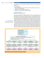
TABLE 39-1
Effect of Antiepileptic Drugs on CYP Isoenzymes or Other Enzyme Systems
DRUG EFFECT ON METABOLISM ENZYMES
Phenobarbital, carbamazepine, Inducers Broad CYP, UGT
phenytoin inducers
Valproic acid Inhibitor CYP 2C19, UGT,
Epoxide hydrolase
Gabapentin, pregabalin No effect
Lamotrigine Weak inducer UGT
Levetiracetam No effect
Oxcarbazepine Inducer (modest) CYP3A4
Tiagabine No effect —
Topiramate Inhibitor (modest) CYP2C19
Inducer (modest) CYP 3A4
Vigabatrin None
Zonisamide No effect
IV • GENERAL PRINCIPLES OF THERAPY
538
weeks and is a reversible process. Addition of an inducer
will cause a lowering of serum concentrations of the tar-
get drug, conceivably resulting in inadequate therapeutic
response. Conversely, removal of an enzyme inducer will
cause a rise in the levels of the target drug, potentially
causing toxicity.
Among the older-generation AEDs, carbamazepine,
phenytoin, and the barbiturates phenobarbital and primi-
done are inducers of both the cytochrome P450 (CYP)
and UGT enzyme systems (18). Combining these agents
with other AEDs that are metabolized by either of these
enzyme systems can result in markedly enhanced sys-
temic clearance, and reduced serum concentrations of the
affected drug, requiring higher doses in order to main-
tain comparable (as compared to monotherapy) steady-
state serum concentrations. An example of this sort of
interaction would be the combination of phenytoin and
lamotrigine.
Lamotrigine is extensively (Ͼ90%) metabolized
hepatically by N-glucuronidation via UGT 1A3 and UGT
1A4. Lamotrigine does not appear to significantly alter
concentrations of carbamazepine or carbamazepine epox-
ide (21, 22) nor any of the other AEDs. However, the half-
life of lamotrigine is reduced from 24 hours to 15 hours
when administered with enzyme-inducing drugs as just
described. Following the withdrawal of the enzyme induc-
ers carbamazepine and phenytoin, lamotrigine plasma
concentrations have been observed to increase by 50%
and 100 %, respectively (23).
Levetiracetam shows limited metabolism in humans,
with 66% of the dose renally excreted unchanged. Its
major metabolic pathway is via hydrolysis of the acet-
amide group to yield a carboxylic derivative, which is
mainly recovered in the urine. Levetiracetam is not sig-
nificantly metabolized by CYPs or UGTs and appears
to be devoid of pharmacokinetic drug interactions
(24, 25). Similarly, the drugs gabapentin and pregabalin
appear to be devoid of enzyme-inducing (or inhibition)
properties.
Oxcarbazepine is converted to 10-hydroxycarbam-
azepine (OHCZ), the metabolite primarily responsible for
pharmacologic activity. This active metabolite is mostly
excreted by direct conjugation to glucuronic acid. Oxcar-
bazepine does not seem to be a broad-spectrum enzyme
inducer, although it does posses modest, specific induc-
tion potential toward the CYP3A subfamily, as evidenced
by the increased metabolism of estrogens and dihydro-
pyridine calcium channel antagonists (1, 2). Clinicians
should be aware that this drug does indeed have modest
potential for causing enzyme induction interactions, but
that this potential may vary among different patients.
Topiramate is approximately 60% excreted
unchanged in the urine. It is also metabolized by hydrox-
ylation and hydrolysis. Two of its metabolites are con-
jugated as glucuronides. While not considered a potent
enzyme inducer, topiramate can increase clearance of val-
proate by approximately 13% and may lower oral con-
traceptive serum concentrations (26, 27). Whether these
changes in valproic serum concentration are clinically
meaningful is unclear. Topiramate metabolic clearance
can be increased when it is administered with enzyme-
inducing AEDs, thereby reducing half-life and lowering
serum concentrations by up to 40%.
Zonisamide is a synthetic 1,2-benzisoaxole deriva-
tive that is metabolized in large part by reduction and
conjugation reactions. Oxidative reactions involving
CYP3A4 and CYP2D6 are also involved. Zonisamide
elimination can be altered by other drugs. Specifically,
enzyme-inducing drugs such as carbamazepine and phe-
nytoin can significantly increase the clearance of this
drug, effectively reducing the half-life of zonisamide by
about half.
Hepatic Inhibition. Hepatic enzyme inhibition can occur
when two drugs compete for the same enzyme site, reduc-
ing the metabolism of the target drug. A resultant increase
in the object drug can occur if a substantial portion of
the target drug is prevented from occupying the enzyme
site. Inhibition is usually a rapid process that is dose/
concentration dependent. Addition of an enzyme inhibitor
may cause a very rapid rise in serum concentrations of the
target drug, potentially leading to acute toxicity (18).
In contrast to enzyme induction, inhibition of
selected CYP and/or UGT enzymes can be caused by
several AEDs of both the older and newer generations.
These combinations may result in unexpectedly high
serum concentrations of the affected AED. An exam-
ple is the interaction of valproic acid and lamotrigine.
Lamotrigine’s half-life is increased to approximately
59–70 hours when it is coadministered with valproate,
resulting from valproate’s inhibition of glucuronidation.
Inhibition of lamotrigine clearance can occur at val-
proate doses as low as 125–250 mg/day and becomes
maximal at dosages approaching 500 mg/day (28). The
clinical implication is that lamotrigine dose and dose
escalation will need to be substantially reduced in order
to reduce the potential for adverse effects (including
perhaps severe rash).
Topiramate may decrease the clearance of phenyt-
oin, suggesting inhibition of CYP2C19. Topiramate has
been shown to increase phenytoin serum concentration
in some patients. While this interaction is not clinically
meaningful in most patients, given the non-linear phar-
macokinetics of phenytoin, the potential does exist for
this interaction to result in phenytoin intoxication.
A significant advancement of oxcarbazepine over
carbamazepine is its lack of susceptibility to inhibitory
interactions. Consistent with its differing metabolism (as
compared to carbamazepine), oxcarbazepine’s pharmaco-
kinetics are not altered by erythromycin. Oxcarbazepine
39 • PRINCIPLES OF DRUG INTERACTIONS: IMPLICATIONS FOR TREATMENT WITH ANTIEPILEPTIC DRUGS
539
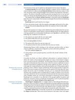
TABLE 39-2
Interactions Between AEDs and Non-AED Medications
NON-AED MEDICATION
TYPE DRUG AED INTERACTION
Adrenergic blockers
Analgesics
Antiarrythmics
Anticoagulants
Antidepressants
Antidiabetic agents
Antimicrobial agents
Antifungal
Alprenolol
Metoprolol
Propranolol
Acetaminophen
Narcotics
Propoxyphene
Salicylates
Disopyramide
Mexiletine
Quinidine
Warfarin
Tricyclics
Tolazamide
Tolbutamide
Acetohexamide
Glibenclamide
Ciprofloxacin
Erythromycin
Fluconazole
PB
CBZ, LTG, PB, PHT
CBZ, PB, PHT
CBZ
PHT, VPA
PB, PHT
CBZ, PB, PHT
PB, PHT
CBZ, PB, PHT
CBZ, PB
CBZ, PB, PHT
PHT
CBZ, BZD, VPA
PHT
PB increases metabolism; dosage of adrenergic
blockers may need to be increased.
Patients on enzyme inducers such as CBZ,
PHT, and PB may be at greater risk of
hepatotoxicity following acetaminophen
overdose. Acetaminophen appears to slightly
increase the elimination of LTG.
Enzyme inducers (CBZ, PHT, PB) increase the
toxicity and decrease the efficacy of
meperidine by increasing the conversation to
normeperidine.
Propoxyphene inhibits CBZ elimination and
may lead to CBZ toxicity. Propoxyphene
should be avoided if possible.
High-dose salicylates displace PHT and VPA
from protein-binding sites and may decrease
VPA elimination.
PB and PHT may increase hepatic metabolism
of disopyramide and require dosage
adjustments.
Enzyme inducers can substantially decrease
mexiletine serum concentrations.
Enzyme inducers decrease serum
concentrations of quinidine.
Inducers increase warfarin metabolism and
decrease hypoprothrombinemic effect.
Induction of tricyclic metabolism. Dosage may
require adjustment.
Enzyme inducers increase elimination and
decrease hypoglycemic effects.
Ciprofloxacin increases serum PHT
concentrations, probably by decreasing
phenytoin elimination.
Erythromycin decreases biotransformation and
can markedly increase serum concentrations.
Fluconazole decreases biotransformation of
PHT and can result in marked increase in
serum concentrations.
IV • GENERAL PRINCIPLES OF THERAPY
540
is a weak inhibitor of CYP2C19, however, and, like
topiramate, it may increase the plasma concentrations
of phenytoin (1).
Because of their primarily renal clearance, and
absence of substantial hepatic metabolism, levetiracetam,
gabapentin, and pregabalin are not subject to inhibition.
In addition, none of these drugs appears to cause inhibi-
tion of metabolism of any other medication.
Interactions Between AEDs and
Other Medications
Traditionally, most attention regarding AED pharma-
cokinetic interactions has been directed toward inter-
actions between various combinations of AEDs. It is
important for the clinician to recognize the potential
impact that AEDs may have on concomitant medications
that a patient receive. For example, many psychotropic
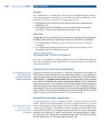
TABLE 39-2
(Continued)
NON-AED MEDICATION
TYPE DRUG AED INTERACTION
Antineoplastics
Antituberculous
agents
Carbonic anhydrase
inhibitors
Corticosteroids
Miscellaneous
Selective serotonin
reuptake inhibitors
Isoniazid
Rifampin
Acetazolamide
Dichlorphenamide
Methazolamide
Dexamethasone
Hydrocortisone
Methylprednisolone
Prednisone
Cimetidine
Clozapine
Enteral feedings
Nafimidone
Ritonavir
Fluoxetine
PHT
CBZ, PHT, VPA
BZD, PHT, VPA
TPM
CBZ, PB, PHT
CBZ, PHT, BZD, ESM
CBZ
PHT
CBZ, PHT
BDZ, ESM
CBZ
Cytotoxic agents appear to decrease oral
absorption of PHT with marked reductions in
serum PHT concentrations.
Isoniazid decreases CBZ, PHT, and VPA
elimination and may lead to toxicity.
Rifampin increases elimination; dosage
adjustments may be necessary.
Concomitant use may lead to increased risk of
nephrolithiasis.
Enzyme inducers increase metabolism of
steroids and decrease efficacy. Decreased
PHT absorption and subsequent decrease in
serum concentrations.
Cimetidine decreases biotransformation of CBZ
and PHT and may lead to toxicity.
May result in increased risk of bone marrow
suppression.
Decreased PHT absorption and marked
decreased in serum concentration.
May result in CBZ toxicity.
Ritonavir decreases biotransformation of BDZ
and ESM and may lead to toxicity.
Fluoxetine has been reported to result in CBZ
toxicity by inhibiting CYP3A3/4.
BDZ ϭ benzodiazepines; CBZ ϭ carbamazepine; LTG ϭ lamotrigine; PB ϭ phenobarbital; PHT ϭ phenytoin; PRM ϭ primidone;
VPA ϭ valproic acid; ESM ϭ ethosuximide; TPM ϭ topiramate; MSM ϭ methsuximide.
Source: McInnes and Brodie 1988 (39).
39 • PRINCIPLES OF DRUG INTERACTIONS: IMPLICATIONS FOR TREATMENT WITH ANTIEPILEPTIC DRUGS
541
agents, including tricyclic antidepressants, selective
serotonin reuptake inhibitors (SSRIs), and antipsychotic
drugs are extensively metabolized by one or more of
the CYP isozymes (29). This would imply that higher
than expected doses of these drugs may be required in
patients receiving enzyme-inducing AEDs such as phe-
nytoin or carbamazepine. Conversely, enzyme-inhibiting
drugs such as valproate may inhibit the clearance of
certain psychotropic drugs such as amitriptyline, nor-
triptyline, or paroxetine (1, 2).
For example, AEDs such as carbamazepine and
phenytoin have been reported to increase the clearance,
and consequently markedly lower the serum concentra-
tion, of a number of antipsychotic medications includ-
ing haloperidol, chlorpromazine, clozapine, risperidone,
ziprazidone, and olanzapine (2, 30). Valproate appears
to have minimal pharmacokinetic interactions impact on
these drugs (31, 32).
Antipsychotic drugs are less likely to cause phar-
macokinetic interactions with AEDs, although both
chlorpromazine and thioridazine have been reported to
result in increases in phenytoin serum concentrations.
Risperidone has been noted to result in modest decreases
in carbamazepine concentrations (33).
Many commonly used antidepressant agents such
as tricyclics and SSRIs are also metabolized via the CYP
system. Consequently, it would be expected that drugs
such as amitriptyline, nortriptyline, imipramine, desip-
ramine, clomipramine, protriptyline, doxepin, sertraline,
paroxetine, mianserin, citalopram, and nefazodone may
display reduced serum concentrations in patients receiv-
ing enzyme-inducing AEDs (1, 2, 34, 35). Conversely,
comedication with the enzyme inhibitor valproate may
cause substantial (50–60%) increases in serum concentra-
tions of drugs such as amitriptyline and nortriptyline.
AED-antidepressant interactions may be bidirec-
tional, and the clinician should recognize that treatment
with certain drugs may result in increased serum con-
centrations of AEDs, particularly the older, extensively
metabolized agents. For example, there are data that
suggest that SSRIs such as fluoxetine and sertraline can
result in increased phenytoin and carbamazepine serum
concentrations.
Examples of other classes of drugs that are exten-
sively metabolized and therefore may be influenced by
enzyme-inducing AEDs include stimulants (i.e., methyl-
phenidate), antineoplastics, immunosuppressants, beta
receptor antagonists, oral contraceptives, and many anti-
viral agents such as indinavir, retonavir, and saqquinavir
(1, 2, 36–38). Table 39-2 provides a representative list of
potential AED–non-AED interactions (39).
SUMMARY
Polypharmacy with multiple concomitant medications is
common in patients of all ages who suffer from epilepsy.
Clinicians should be aware that many of the older, tra-
ditional AEDs such as carbamazepine, phenytoin and
the barbiturates have been consistently associated with
pharmacokinetic interactions, both with other AEDs, as
well as many commonly used medications. In many cases,
these interactions may go unrecognized, as routine serum
concentration monitoring is not available, or practical in
all situations. It would seem prudent therefore for clini-
cians to monitor clinical response to concomitant medi-
cations, and consider potential drug interactions, should
sub-optimal patient response (including the appearance
of adverse effects) be noted.
Alternatively, clinicians may want to consider using
appropriate newer generation AEDs such as that do not
seem to interfere, either with drug metabolism, or oral
absorption/transport, and thereby avoid these potentially
problematic interactions.
References
1. Perucca E. Clinically relevant drug interactions with antiepileptic drugs. Br J Clin Phar-
macol 2005; 61:246–255.
2. Patsalos P, Perucca E. Clinically important drug interactions in epilepsy: Interactions
between antiepileptic drugs and other drugs. Lancet Neurology 2003; 2:473–481.
3. Juurlink DN, Mamdani M, Kopp A, Laupacis A, et al. Drug-drug interactions among
elderly patients hospitalized for drug toxicity. JAMA 2003; 289:1652–1658.
4. Bauer LA. Interference of oral phenytoin absorption by continuous nasogastric feedings.
Neurology 1982; 32:570–572.
5. Krueger KA, Garnett WR, Comstock TJ, et al. Effect of two administration schedules of
an enteral nutrient formula on phenytoin bioavailability. Epilepsia 1987; 28:706–712.
6. Nishimura LY, Armstrong EP, Plezia PM, et al. Influence of enteral feedings on phenytoin
sodium absorption from capsules. Drug Intell Clin Pharm 1988; 22:130–133.
7. Fischer JH, Barr AN, Paloucek FP, Dorociak JV, et al. Effect of food on the serum con-
centration profile of enteric-coated valproic acid. Neurology 1988; 38:1319–1322.
8. Fay MA. Sheth RD. Gidal BE. Oral absorption kinetics of levetiracetam: the effect of mixing
with food or enteral nutrition formulas. Clinical Therapeutics 2005; 27(5):594–598.
9. Gidal BE, Maly MM, Kowalski J, Rutecki P, et al. Gabapentin absorption: effect of mixing
with foods of varying macronutrient content. Ann Pharmacother 1998; 32:405–408.
10. Doose DR, Walker SA, Gisclon LG, Nayak RK. Single-dose pharmacokinetics and effect
of food on the bioavailability of topiramate, a novel antiepileptic drug. J Clin Pharmacol
1996; 36(10):884–891.
11. Degen PH, Flesch G, Cardot JM, Czendlik C, et al. The influence of food on the disposi-
tion of the antiepileptic oxcarbazepine and its major metabolite in healthy volunteers.
Biopharm Drug Dispos 1994; 15(6):519–526.
12. Fay MA. Sheth RD, Gidal BE. Oral absorption kinetics of levetiracetam: the effect of
mixing with food or enteral nutrition formulas. Clinical Therapeutics 2005; 27(5):
594–598.
13. Lin JH, Yamazaki M. Role of P glycoprotein in pharmacokinetics: clinical implications.
Clinical Pharmacokinet 2003; 42:59–98.
14. Ceckova-Novotna M, Pavek P, Staud F. P-glycoprotein in the placenta: Expression, local-
ization, regulation and function. Reprod Toxicol 2006; 22:400–410.
15. Giessmann T, May K, Modess C, et al. Carbamazepine regulates intestinal P-glycoprotein
and multidrug resistance protein MRP2 and influences disposition of talinolol in humans.
Clin Pharmacol Ther 2004; 76:192–200.
16. Anderson GD. A mechanistic approach to antiepileptic drug interactions. Ann Pharma-
cother 1998; 32:554–563.
17. Benet LZ, Hoener BA. Changes in plasma protein binding have little clinical relevance.
Clin Pharmacol Ther 2002; 71:115–121.
18. Anderson GD. Pharmacogenetics and enzyme induction/inhibition properties of antiepi-
leptic drugs. Neurology 2004; 63:(Suppl 4):S3–S8.
19. Xu C, Li CY, Kong AN. Induction of phase I, II, III drug metabolism/transport by xeno-
biotics. Arch Pharm Res 2005; 28:249–269.
IV • GENERAL PRINCIPLES OF THERAPY
542
20. Murray M, Petrovich N. Cytochrome P450: decision-making tools for personalized
therapeutics. Curr Opin Mol Ther 2006; 8:480–486.
21. Pisani F, Xiao B, Faziop A, Spina E, et al. Single-dose pharmacokinetics of CBZ-E in
patients on lamotrigine monotherapy. Epilepsy Res 1994; 19:245–248.
22. Gidal BE, Rutecki P, Shaw R, Maly MM, et al. Effect of lamotrigine on carbamazepine
epoxide/carbamazepine serum concentration ratios in adult patients with epilepsy. Epi-
lepsy Res 1997; 28:207–211.
23. Anderson GD, Gidal BE, Gilliam F, Messenheimer J. Time course of lamotrigine de-induc-
tion: Impact of step-wise withdrawal of carbamazepine or phenytoin. Epilepsy Res 2002;
49:211–217.
24. Gidal BE, Baltes E, Otoul C, Perucca E. Effect of levetiracetam on the pharmacokinetics
of adjunctive antiepileptic drugs: a pooled analysis of data from randomized clinical
trials. Epilepsy Res 2005; 64(1–2):1–11.
25. Perucca E, Gidal BE, et al. Effects of antiepileptic comedication on levetiracetam pharma-
cokinetics: a pooled analysis of data from randomized adjunctive therapy trials. Epilepsy
Res 2003; 53:47–56.
26. Rosenfeld WE, Liao S, Anderson G,et al. Comparison of the steady-state pharmacoki-
netics of topiramate and valproate in patients with epilepsy during monotherapy and
concomitant therapy. Epilepsia 1997; 38:329–333.
27. Zupanc M. Antiepileptic drugs and hormonal contraceptives in adolescent women with
epilepsy. Neurology 2006; 66Suppl 3):37–45.
28. Gidal BE, Sheth R, Parnell J, et al. Evaluation of VPA dose and concentration effects on
lamotrigine pharmacokinetics: implications for conversion to monotherapy. Epilepsy Res
2003; 57:85–93.
29. Spina E, Perucca E. Clinical significance of pharmacokinetic interactions between anti-
epileptic and psychtropic drugs. Epilepsia 2002; 43(Suppl 2):37–44.
30. Jann MW, Ereshefsky L, Saklad SR, et al. Effects of carbamazepine on plasma haloperidol
levels. J Clin Psychopharmacol 1985; 5:106–109.
31. Spina E, Avenoso A, Facciola G, et al. Plasma concentrations of risperidone and
9-hydroxyrespiridone; effect of comedication with carbamazepine or valproate. Ther
Drug Monit 2000; 22:481–485.
32. Hesslinger B, Normann C, Langgosch JM, et al. Effects of carbamazepine and valpro-
ate on haloperidol levels and on psychopathologic outcome in schizophrenic patients.
J Clin Psychopharmacol 1999; 19:310–315.
33. Mula M, Monaco F. Carbamazepine-risperidone interactions in patients with epilepsy.
Clin Neuropharmacol 2002; 25:97–100.
34. Trimble MR, Mula M. Antiepileptic drug interactions in patients requiring psychiatric
drug treatment. In: Majkowski J, Bourgeois B, Patsalos P, Mattson R, eds. Antiepileptic
Drugs. Combination Therapy and Interactions. Cambridge, UK: Cambridge University
Press, 2005:350–368.
35. Pihlsgard M, Eliasson E. Significant reduction of sertraline plasma levels by carbamazepine
and phenytoin. Eur J Clin Pharmacol 2002; 57:915–916.
36. Vecht CJ, Wagner GL, Wilms EB. Treating seizures in patients with brain tumors: drug
interactions between antiepileptic drugs and chemotherapeutic agents. Semin Oncol 2003;
30(6 Suppl 19):49–52.
37. Flockart DA, Tanus-Santos JE. Implications of cytochrome P450 interactions
when prescribing medication for hypertension.
Arch Intern Med 2002; 162:
405–412.
38. Markowitz JS, Morrison SD, DeVane CL. Drug interactions with psychostimulants. Int
Clin Psychopharmacology 1999; 14:1–18.
39. McInnes GT, Brodie MJ. Drug interactions that matter—a critical reappraisal. Drugs
1988; 36:83–110.
ANTIEPILEPTIC DRUGS
AND KETOGENIC DIET
V
545
ACTH and Steroids
he efficacy of adrenocorticotropin
(ACTH) therapy in childhood sei-
zures was first observed by Klein
and Livingston in 1950 in a series of
children with atypical absence seizures (1). In 1958, Sorel
and Dusaucy-Bauloye reported that ACTH was effective
in children with infantile spasms (IS). These authors not
only reported seizure control in children with IS treated
with ACTH but also observed improvements in behav-
ior and electroencephalogram (EEG) (2). Subsequently,
a number of studies appeared that reported on the effi-
cacy of corticosteroids in IS and confirmed the utility
of ACTH in the treatment of this disorder. Both ACTH
and corticosteroids have been used in treating a number
of epilepsy syndromes, including Ohtahara syndrome,
Lennox-Gastaut syndrome and other myoclonic epilep-
sies, and Landau-Kleffner syndrome (3). The epilepsy
syndromes that respond uniquely to ACTH and cortico-
steroid therapy have an age-related onset during a critical
period of brain development, as well as a characteristic
regression or plateau of acquired milestones at seizure
onset, and long-term cognitive impairment (4). In addi-
tion to beneficial effects on the convulsive state, there are
some data to suggest that ACTH, corticosteroids, or both
also can improve the short-term developmental trajectory
and the long-term prognosis for language and cognitive
development in at least some of these patients (5–9).
Rajesh RamachandranNair
O. Carter Snead, III
In this review, we will first discuss the evidence in
support of the use of steroids in IS. This will be fol-
lowed by a review of possible mechanisms of the puta-
tive anticonvulsant effects of ACTH and corticosteroids.
This will be followed by a discussion of the use of these
compounds in epilepsy syndromes other than IS. Finally,
we will review the therapeutic potential of neuroactive
steroids in epilepsy.
INFANTILE SPASMS
In 1841, William West, an English physician, provided the
first description of IS in his own 4-month old son (10).
Later, the association of IS with the sequelae of severe
mental deficiency emerged. In 1952, Gibbs and Gibbs
first described the interictal EEG pattern associated with
infantile spasms and termed it hypsarrhythmia. This pat-
tern was unique and described as showing high-voltage,
chaotic slowing with multifocal spikes, and marked asyn-
chrony (11). Over the years, the triad of infantile spasms,
hypsarrhythmia, and mental retardation became known
as West syndrome (12).
After 1958, studies began to appear in the literature
reporting the effectiveness of corticosteroids in the treat-
ment of this disorder (12). There is a marked variability
of response rates to these therapeutic agents that probably
T
40
V • ANTIEPILEPTIC DRUGS AND KETOGENIC DIET
546
is related to the small cohorts reported and the paucity of
controlled treatment data. Another confound is that the
natural history of IS is poorly understood, particularly
the phenomenon of spontaneous remission. Moreover,
the literature is replete with marked variations in the
dosage of ACTH and/or corticosteroids given, and in
treatment duration, of both drugs. In most studies, an
objective method of documenting spasm cessation has not
been used and response to therapy has been defined in a
graded manner, although there is no convincing evidence
that spasms respond in a graded fashion to any form
of therapy. Usually IS respond in an all-or-none fashion
to treatment with ACTH and/or corticosteroids. Finally,
most studies have been uncontrolled, unblinded, and ret-
rospective, complicating the establishment of evidence-
based recommendations for optimal treatment (12).
The controversies surrounding the treatment of IS
outnumber the areas of agreement and encompass the
following questions: Which is the most effective therapy:
ACTH or corticosteroid? Are other anticonvulsants such
as vigabatrin, valproic acid, benzodiazepines, topira-
mate, zonisamide, or pyridoxine effective against infan-
tile spasms? Is there some other treatment regimen with
newer antiepileptic drugs that is effective against infantile
spasms? What is the impact of treatment with ACTH
compared with corticosteroids on long-term outcome
in recurrence of spasms, evolution into other forms of
intractable epilepsy, and cognitive or behavioral func-
tion? Does treatment change the outcome for a patient
with preexisting mental retardation and a structurally
abnormal brain? What is the optimal dosage of these
drugs, and how long should treatment last? Does the
ultimate outcome depend on timing of treatment? Does
the efficacy of ACTH depend on the formulation (natural
vs. synthetic, sustained vs. short-acting)?
Some of these questions were addressed in a recently
published American Academy of Neurology (AAN)/Child
Neurology Society (CNS) Practice Parameter on the treat-
ment of infantile spasms (13). In the following few para-
graphs we will discuss the key issues addressed by this
practice parameter. Important studies published subse-
quent to the practice parameter also will be discussed in
the relevant sections.
Summary of the AAN/CNS Practice Parameter
on the Treatment of Infantile Spasms
Three major questions were addressed in the practice
parameter.
1. What are the most effective therapies for infantile
spasms, as determined by short-term outcome mea-
sures, including complete cessation of spasms, reso-
lution of hypsarrhythmia, and likelihood of relapse
following initial response?
2. How safe are currently used treatments?
3. Does successful treatment of infantile spasms lead
to long-term improvement of neurodevelopmental
outcome or a decreased incidence of epilepsy?
Articles included for critical analysis pursuant to answer-
ing these questions and formulating treatment recommen-
dations for infantile spasms had the following inclusion
criteria (14–27):
1. A clearly stated diagnosis of infantile spasms
2. An EEG demonstrating hypsarrhythmia or modified
hypsarrhythmia
3. Age of 1 month to 3 years.
Infantile spasms were classified as either symptomatic
or cryptogenic as defined by the International League
Against Epilepsy (ILAE).
Outcome measures included short- and long-term
measures. Short-term outcome measures were defined as
the following:
1. Complete cessation of spasms
2. Resolution of hypsarrhythmia and, where docu-
mented, normalization of EEG
3. Relapse rate
In studies with a mean follow-up of Ͼ2 years, the follow-
ing were considered long-term outcome measures:
1. Nonepileptiform EEG
2. Absence of seizures
3. Normal development
A four tiered classification scheme for diagnostic evidence
approved by the Quality Standards Subcommittee was
utilized as part of the assessment. This schema is outlined
in Table 40-1. Depending on the strength of the evidence
under this classification system, specific recommenda-
tions were made. The strength of these recommendations
is shown in Table 40-2.
Based on this critical analysis it was concluded in
the practice parameter that ACTH was probably effective
in the short-term treatment of IS and in the resolution of
hypsarrhythmia (level B). Time to response was usually
within 2 weeks, and an “all-or-none” response had been
reported in a number of studies. The data were insuf-
ficient to recommend optimal dosage and duration of
treatment with ACTH for the treatment of IS (level U). As
well, data also were insufficient to recommend treatment
of IS with oral corticosteroids (level U). ACTH was more
effective than oral corticosteroids in causing the cessation
of seizures. Side effects reported for ACTH were common
and included hypertension, irritability, infection, revers-
ible cerebral shrinkage, and, rarely, death due to sepsis.
40 • ACTH AND STEROIDS
547
Although vigabatrin (VGB) is not a steroid, its use
in infantile spasms is relevant to this review because ste-
roids and vigabatrin are generally considered to be the
only two groups of drugs that work in this disorder—an
impression borne out by the Practice Parameter (13). In
the analysis that led to the AAN/CNS practice parameter,
the evidence for the therapeutic efficacy of vigabatrin in
IS was weaker than that for ACTH (level C for vigabatrin
vs. level B for ACTH). Hence, vigabatrin was found to be
possibly effective for the short-term treatment of IS.
As for the efficacy of ACTH in improving the long-
term outcomes in terms of seizure freedom and normal
development of children with IS, the data were insuf-
ficient (28–31) in that regard (Level U, class III and IV
evidence). Similarly, there was insufficient evidence to
support the thesis that early initiation of treatment with
ACTH improves the long-term outcome of children with
IS (Level U, class III and IV evidence).
More recently, the United Kingdom Infantile Spasms
Study (32) assessed comparative efficacy of vigabatrin
and hormonal treatment of IS in a randomized controlled
trial. The primary outcome was cessation of spasms on
days 13 and 14. Minimum doses were VGB 100 mg/kg
per day, oral prednisolone 40 mg per day, or intramuscu-
lar tetracosactide depot 0.5 mg (40 IU) on alternate days.
Of 208 infants screened and assessed, 107 were randomly
assigned to VGB (n ϭ 52) or hormonal treatments (pred-
nisolone n ϭ 30, tetracosactide n ϭ 25). Patients with
no spasms on days 13 and 14 consisted of: 40 (73%) of
55 infants assigned hormonal treatments (prednisolone
21/30 [70%], tetracosactide 19/25 [76%]) and 28 (54%)
of 52 infants assigned VGB (difference 19%, CI 1–36%,
P ϭ 0.043). Adverse events were reported in 30 (55%)
of 55 infants on hormonal treatments and 28 (54%) of
52 infants on VGB. This study concluded that cessation
TABLE 40-1
American Academy of Neurology Evidence
Classification Scheme for a Therapeutic Article
Class I Evidence provided by a prospective,
randomized, controlled clinical trial
with masked outcome assessment, in
a representative population. The
following are required: (a) Primary
outcome(s) is/are clearly defined; (b)
exclusion/inclusion criteria are clearly
defined; (c) dropouts and crossovers
are accounted for adequately with
numbers sufficiently low to have
minimal potential for bias; and (d)
relevant baseline characteristics are
presented and substantially
equivalent among treatment groups,
or there is appropriate statistical
adjustment for differences.
Class II Evidence provided by a prospective
matched-group cohort study in a
representative population with
masked outcome assessment that
meets (a)–(d) as defined for Class I
or a randomized controlled trial in a
representative population that lacks
one criterion of (a)–(d).
Class III All other controlled trials (including
well-defined natural history controls
or patients serving as own controls)
in a representative population, where
outcome assessment is independent
of patients’ treatment.
Class IV Evidence from uncontrolled studies,
case series, case reports, or expert
opinion.
TABLE 40-2
American Academy of Neurology System for Translation of Evidence to Recommendations
TRANSLATION OF EVIDENCE TO RECOMMENDATIONS RATING OF RECOMMENDATION
Level A rating requires at least one convincing class I
study or at least two consistent, convincing class II
studies.
Level B rating requires at least one convincing class II
study or at least three consistent class III studies.
Level C rating requires at least two convincing and
consistent class III studies.
A ϭ established as effective, ineffective, or harmful for the
given condition in the specified population.
B ϭ probably effective, ineffective, or harmful (or probably
useful/predictive or not useful/predictive) for the given
condition in the specified population.
C ϭ possibly effective, ineffective, or harmful (or possibly
useful/predictive or not useful/ predictive) for the given
condition in the specified population.
U ϭ data inadequate or conflicting. Given current knowledge,
treatment is unproven.
V • ANTIEPILEPTIC DRUGS AND KETOGENIC DIET
548
of spasms was more likely in infants given hormonal
treatments than in those given VGB.
Infants enrolled in the United Kingdom Infantile
Spasms Study were followed up until clinical assessment
at 12–14 months of age (33). Neurodevelopment was
assessed with the Vineland Adaptive Behavior Scales
(VABS) at 14 months of age. Of 107 infants enrolled,
five died, and 101 survivors reached both follow-up
assessments. Absence of spasms at final clinical assess-
ment (hormone 41/55 [75%] vs. vigabatrin 39/51 [76%])
was similar in each treatment group. Mean VABS score
did not differ significantly (hormone 78.6 vs. vigabatrin
77.5). In infants with no identified underlying etiology,
the mean VABS score was higher in those allocated hor-
mone treatment than in those allocated vigabatrin (88.2
vs. 78.9; difference 9.3, 95% CI 1.2–17.3). This study
reported that better initial control of spasms by hormone
treatment in those with no identified underlying etiology
might lead to improved developmental outcome.
Kivity and coworkers assessed the long-term cogni-
tive and seizure outcomes of 37 patients with cryptogenic
infantile spasms (onset, age 3 to 9 months) receiving a
standardized treatment regimen of high-dose tetracos-
actide depot, 1 mg intramuscularly (IM) every 48 hours
for 2 weeks, with a subsequent 8- to 10-week slow taper
and followed by oral prednisone, 10 mg/day for a month,
with a subsequent slow taper for 5 months or until the
infant reached the age of 1 year, whichever came later (8).
Cognitive outcomes were determined after 6 to 21 years
and analyzed in relation to treatment lag and pretreat-
ment regression. Normal cognitive outcome was found
in all 22 (100%) patients of the early-treatment group
(within 1 month), and in 40% of the late-treatment group
(1–6.5 months). Normal cognitive outcome was found in
all 25 (100%) patients who had no or only mild mental
deterioration at presentation, including four in the late-
treatment group but in only three of the 12 patients who
had had marked or severe deterioration before treatment.
This study indicated that early treatment of cryptogenic
infantile spasms with a high-dose ACTH protocol was
associated with favorable long-term cognitive outcomes.
Once major developmental regression lasted for a month
or more, the prognosis for normal cognitive outcome
was poor.
Practical Considerations Regarding Dosage
Table 40-3 lists the currently available formulations of
ACTH. The biologic activity, expressed in international
units (IU), permits a comparison of potency in terms of
the relative ability of the peptide to stimulate the adrenals,
but may not necessarily reflect the ability of the ACTH
preparation to affect brain function. The biologic activity
of natural ACTH in the brain may differ from that of syn-
thetic ACTH as a result of ACTH fragments and possibly
other pituitary hormones with neurobiologic activity in
the brain that are present in the pituitary extracts (5).
These compounds could enhance the therapeutic efficacy
of natural ACTH (34). Any differences in the biologic
effects of sustained ACTH levels provided by the depot
formulations, as opposed to those of the short-acting
preparations, are unknown. Given in high doses, how-
ever, long-acting depot preparations are associated with
an increased incidence of severe side effects, including
death from overwhelming infection.
The most effective dose of ACTH for remission
of spasms is controversial. Notably, in comparison to
prednisone, no major advantage was demonstrated by
TABLE 40-3
Available Preparations of ACTH
Corticotropin (ACTH 1-39): porcine pituitary extract
(short-acting)
Acthar gel 80 IU/mL 100 IU* ϭ 0.72 mg
Acthar lyophylized powder 100 IU* ϭ 0.72 mg
Cosyntropin/Tetracosactrin (ACTH 1-24): synthetic
(short-acting)
Cortrosyn 100 IU* ϭ 1.0 mg
Cosyntropin/Tetracosactrin (ACTH 1-24): synthetic
(long-acting)
Synacthen depot (CIBA) 100 IU* ϭ 2.5 mg
Cortrosyn-Z (Organon) 100 I U* ϭ 2.5 mg
*Commercial preparations are described in International Units (IU) based on a potency assay
in hypophysectomized rats in which depletion of adrenal ascorbic acid is measured after subcuta-
neous ACTH injection.
40 • ACTH AND STEROIDS
549
low-dose ACTH, whereas high-dose ACTH was reported
to be superior (15, 25). High-dose ACTH (60 IU/day
or 150 IU/m
2
per day) has been associated with excel-
lent short-term response rates (87–93%) in prospective
studies (14, 25). However; in the only randomized, pro-
spective comparison of ACTH, Hrachovy and coworkers
found no difference between high dose and low dose (16).
A prospective study of synthetic ACTH by Yanagaki and
coworkers compared very low-dose ACTH (0.2 IU/kg per
day) to low-dose (1 IU/kg per day) and found equivalent
efficacy, with response and relapse rates comparable to
other studies (18). Heiskala et al. described a protocol
that utilized a stepwise increase in dosage, demonstrat-
ing that some patients can be controlled on lower doses
of carboxymethylcellulose ACTH (3 IU/kg per day) but
others required high doses (12 IU/kg per day). Overall,
spasms were controlled initially in 65% of patients, but
the rate of relapse was high (35).
Some evidence supports a beneficial effect of high-
dose ACTH over low-dose ACTH or oral steroids in
cognitive outcome. Glaze and coworkers found no dif-
ference between low-dose ACTH (20 to 30 IU/day) and
prednisone (2 mg/kg per day) with regard to the cognitive
outcome (31). However, in a comparison of high-dose
ACTH (110 IU/m
2
per day) and steroids, however, Lom-
broso showed a higher rate of normal cognitive outcome
in cryptogenic patients treated with ACTH than in those
treated with prednisone alone (55% vs. 17%) (5). Ito and
coworkers also showed a positive correlation between
dose and developmental outcome comparing different
ACTH dosage regimens retrospectively (36).
The optimal ACTH dose may lie between 85 and
250 IU/m
2
per day. Doses of 400 IU/m
2
per day or higher
are contraindicated because of a high incidence of life-
threatening side effects. The optimal dose of ACTH
required to enhance short-term response and long-term
cognitive outcome is unknown; however, relatively high
doses given early in the disease, accompanied by a second
course in the event of relapse, appear warranted. The
following high-dose ACTH protocol (21, 25) has been
used successfully by us in treating more than 700 children
with infantile spasms.
The child is admitted to a day-care unit to initiate
ACTH therapy and to teach parents to give the injection,
measure urine glucose three times daily with Chemstix,
and recognize spasms to keep an accurate seizure calen-
dar. Any diagnostic workup indicated by clinical circum-
stances is also performed, including screening for occult
congenital infections. Before ACTH is started, an endo-
crine profile, complete blood count, urinalysis, electrolyte
panel, baseline renal function, and calcium, phosphorus,
and serum glucose levels are obtained. Blood pressure and
electrocardiogram are also assessed. The drug is not given
if any of these studies show abnormal results. Diagnostic
neuroimaging is indicated before initiation of ACTH or
steroids because of the association of ACTH treatment
with ventriculomegaly. The initial dose of ACTH is 150 IU/
m
2
/day of ACTH gel, 80 IU/mL, intramuscularly in two
divided doses for 1 week. In the second week, 75 IU/m
2
per day is given, followed by 75 IU/m
2
every other day
in the third week. Over the next 6 weeks, the dose is
gradually tapered. The lot number of the ACTH gel is
carefully recorded. Usually, a response is seen within the
first 7 days; if no response is noted in 2 weeks, the lot
is changed.
Blood pressure must be measured daily at home
during the first week and three times weekly thereafter.
Control of hypertension is attempted with salt restriction
and amlodipine therapy rather than discontinuation of
ACTH. The patient is monitored in the outpatient clinic
weekly for the first month and then biweekly, with appro-
priate blood work at each visit. Waking and sleeping EEG
patterns are obtained 1, 2, and 4 weeks after the start
of ACTH to assess treatment response. As the treatment
response is usually all or none, positive results are sug-
gested when properly trained parents report no seizures
in a child, whose waking and sleeping EEG patterns are
normal. If relapse occurs, the dose may be increased to the
previously effective dose for 2 weeks and another tapering
begun. If seizures continue, the dose may be increased to
150 IU/m
2
per day and the regimen restarted.
If prednisone is chosen because of its oral formu-
lation and lower incidence of serious side effects, the
pretreatment laboratory evaluation described earlier is
performed. The initial dose is 3 mg/kg per day in four
divided doses for 2 weeks, followed by a 10-week taper
(25). A multiple-daily-dose regimen is recommended to
produce the sustained elevations of plasma cortisol dem-
onstrated in high-dose ACTH therapy.
Adverse Effects of ACTH and Steroids
ACTH and steroids, particularly at the high doses recom-
mended for infantile spasms, can produce dangerous side
effects. These are more frequent and more pronounced
with ACTH (37). Cushingoid features and extreme irri-
tability are seen frequently; hypertension is less common
but appears to be associated with higher doses. Vigilance
is required for signs of sepsis; pneumonia; glucosuria;
metabolic abnormalities involving the electrolytes cal-
cium and phosphorus (38–40); and congestive heart
failure (41, 42). Cerebral ventriculomegaly, which is
not always reversible, can lead to subdural hematoma
(43, 44). The cause of the apparent cerebral atrophy is
obscure, but its existence emphasizes the importance of
diagnostic neuroimaging before initiation of ACTH.
Because hypothalamic-pituitary or adrenocortical
dysfunction can result from ACTH therapy, morning lev-
els of cortisol should be monitored during a taper and any
medical stress treated with high-dose steroids (45–47).
V • ANTIEPILEPTIC DRUGS AND KETOGENIC DIET
550
Treatment with ACTH or steroids also can be immu-
nosuppressant and associated with infectious complica-
tions, such as overwhelming sepsis, perhaps as a result
of impaired function of polymorphonuclear leukocytes
(48). Both agents are therefore contraindicated in the face
of serious bacterial or viral infection such as varicella
or cytomegalovirus. Because of the potential for fatal
Pneumocystis pneumonia as an infectious complication
of ACTH therapy, prophylaxis with trimethoprim-
sulfamethoxazole, accompanied by folate supplementa-
tion and frequent blood counts, may be prudent in infants
older than 2 months of age. In rare cases, ACTH can
exacerbate seizures (49).
Potential Mechanisms of Action of ACTH in
Infantile Spasms
ACTH is a 39-amino-acid peptide hormone produced,
through post-translational modification of the larger
peptide pro-opiomelanocortin (POMC), in the anterior
pituitary. POMC expression, processing to ACTH, and
ACTH secretion are stimulated by corticotropin-releasing
factor (CRF) generated in the hypothalamus, and these
processes are under negative feedback control by gluco-
corticoids (50). ACTH secretion is pulsatile and normally
has a pronounced diurnal variation, but secretion also
increases substantially in response to a range of stressors.
The effects of ACTH are mediated via stimulation of the
G-coupled cell surface ACTH receptor, which is expressed
primarily on adrenocortical cells. This receptor is a mem-
ber of the melanocortin family and is alternatively known
as the melanocortin-2 (MC-2) receptor. ACTH acutely
stimulates the synthesis of cortisol in the adrenal gland.
ACTH also increases the long-term capacity of the adre-
nal gland to generate cortisol by inducing a range of ste-
roidogenic enzymes and hypertrophy of the cortex (51).
ACTH additionally has the capacity to cross-react with
other melanocortin receptors (52).
The pathogenesis of infantile spasms, and therefore
the mechanism of action of ACTH and steroids in this
condition, are unknown, principally because there is no
available animal model for this disorder. Infantile spasms
occur within a narrow developmental window in terms of
age of onset and can be found concurrently with a variety
of congenital abnormalities of brain, which may be caus-
ally linked—so-called symptomatic spasms. However, IS
also may occur without apparent cause in children with
no pre-existing neurologic abnormality at the onset of
spasms (i.e., idiopathic spasms). Those children who are
not neurologically normal when the spasms appear, yet
have no demonstrable imaging or metabolic abnormality,
are said to have cryptogenic spasms.
The effect of ACTH and corticosteroids in infan-
tile spasms is frequently all or none, and the steroid-
induced seizure-free state is often sustainable even after
drug withdrawal. These observations support the theory
that due to various etiologies, a significant stress response
is experienced by the developing brain; resulting in this
age-dependent epileptic encephalopathy. Within this very
narrow developmental window, ACTH and steroids may
be able to reset the deranged homeostatic mechanisms of
the brain, thereby reducing the convulsive tendency and
improving the developmental trajectory.
The Brain-Adrenal Axis There is evidence to suggest
that the effects of ACTH in infantile spasms may be
independent of steroidogenesis. Efficacy studies have
demonstrated superiority of ACTH to corticosteroids in
treating infantile spasms and also its efficacy in adrenal-
suppressed patients. Substantial physiologic and phar-
macologic data indicate that ACTH has direct effects
on brain function: increasing dendritic sprouting in
immature animals; stimulation of myelination; regula-
tion of the synthesis, release, uptake, and metabolism
of dopamine, norepinephrine, acetylcholine, serotonin,
and gamma-aminobutyric acid (GABA); regulation of
the binding to glutamatergic, serotoninergic, muscarinic
type 1, opiate, and dopaminergic receptors; and altera-
tion of neuronal membrane lipid fluidity, permeability,
and signal transduction (53–57). Though activation of
glucocorticoid receptors has little direct anticonvulsant
effect, it modulates the expression and release of a num-
ber of neurotransmitters and neuromodulators, including
the proconvulsant neuropeptide corticotropin-releasing
hormone (CRH). High brain CRH levels would be pre-
dicted to reduce the cerebrospinal fluid ACTH and ste-
roids (58). Many authors have reported reduced levels
of ACTH in patients with infantile spasms, compared
to their age-matched controls (59, 60). In infant animal
models CRH causes seizures and death of neurons (61).
These effects of CRH are most marked in developing
brain (62). Suppression of the after-hyperpolarization
and activation of the glutamatergic neurotransmission
are the possible mechanisms by which CRH may mediate
these effects. In animals, ACTH appears to down-regulate
the CRH expression in amygdala. This effect was found
to be independent of glucocorticoid receptor activation
but required melanocortin receptors (63). ACTH reduces
CRH gene expression in specific brain regions. This effect
has been demonstrated in the absence of adrenal steroids
and resides within the 4-10 fragment of ACTH, a frag-
ment that does not release adrenal steroids. Melanocor-
tin receptor antagonists blocked this effect, suggesting
that the melanocortin receptors are the targets of ACTH
action (63).
A hypothesis, therefore, can be generated in which a
stress response results in enhanced CRH expression, lead-
ing to neuronal hyperexcitability and seizures. By sup-
pressing CRH expression, possibly through the action of
peptide fragments of ACTH on melanocortin receptors,
40 • ACTH AND STEROIDS
551
the CRF-induced hyperexcitability may be reduced, hence
ameliorating infantile spasms. Clinical trials of ACTH
fragments that have no activity on the adrenal axis have
been disappointing (64); however, these clinical trials
have utilized the 4-9 peptide fragment rather than the
4-10 peptide fragment studied in animal models.
The events that precipitate this proposed endocrine
abnormality remain unclear.
THE USE OF ACTH AND CORTICOSTEROIDS
IN OTHER SEIZURE DISORDERS
There is limited information concerning treatment of
other intractable seizure disorders with ACTH and/or
steroids. The Ohtahara and Lennox-Gastaut syndromes
are believed to represent earlier and later manifestations,
respectively, of a spectrum of infantile epileptic encepha-
lopathies that include infantile spasms. These conditions
respond poorly to traditional anticonvulsant drug ther-
apies but are sometimes improved by the antiepileptic
drugs used in infantile spasms: ACTH, steroids, benzodi-
azepines, and valproic acid. ACTH or steroids also may
be beneficial in Landau-Kleffner syndrome (65).
Ohtahara Syndrome
The Ohtahara syndrome, also known as early infantile
epileptic encephalopathy (EIEE), is characterized by
spasms beginning within the first three months of life
associated with persistent burst suppression on the EEG
in all stages of the sleep-wake cycle. Despite reports of
improvement in seizures in Ohtahara syndrome follow-
ing ACTH, vigabatrin, and/or zonisamide therapy, the
long-term prognosis is usually unchanged by any treat-
ment (66). Mortality in this epilepsy syndrome is high,
and survivors are usually severely handicapped. If used,
ACTH should be administered as described for infantile
spasms.
Lennox-Gastaut Syndrome and Other
Myoclonic Seizure Disorders
ACTH and steroids have been found to be useful in
younger children with various combinations of severe
and intractable seizures, particularly atypical absence,
myoclonic, tonic, and atonic seizures. This group includes
patients with Lennox-Gastaut syndrome, a disorder
characterized by mental retardation, generalized slow
spike-and-wave discharges, intractable atypical absence,
myoclonus, and frequent ictal falls. Snead and coworkers
treated 64 children who had myoclonic seizures without
EEG evidence of hypsarrhythmia, or other intractable sei-
zures with either prednisone or ACTH. Seventy-three per-
cent of the children treated with ACTH achieved seizure
control, as opposed to none of the prednisone-treated
children; however, there was a relapse rate of Ͼ50%
observed on discontinuation of the ACTH (25).
In 45 cases of Lennox-Gastaut syndrome treated
with ACTH, the immediate and long-term effects and
the various factors affecting them were investigated by
a follow-up study (67). Twenty-three (51.1%) of the 45
children became “seizure free” for over 10 days. Ten
children relapsed into Lennox syndrome within 6 months,
and in the remaining 13 children, seizures were sup-
pressed for over 6 months. Of these 13 patients, seizure
relapse was observed in eight from 9 months to 7 years
later. The other five children followed a favorable course
without relapse. Sinclair treated 10 children with Lennox
Gastaut syndrome and intractable seizures with predniso-
lone at a dose of 1 mg/kg/day for six weeks followed by
withdrawal over the next 6 weeks, and achieved seizure
freedom in 7 and seizure reduction in 3 children. Long-
term outcome was not mentioned (68).
In summary, several uncontrolled, retrospective
studies suggest that ACTH is superior to oral steroids
in Lennox-Gastaut syndrome. If the decision is made to
embark upon such treatment for Lennox-Gastaut syn-
drome, the regimen described in this chapter for ACTH
or prednisone is recommended. Nevertheless, ACTH and
steroids should be reserved for the most severe and intrac-
table patients. Usually, the best result is temporary relief,
because 70% to 90% of patients with multiple seizure
types suffer a relapse during the ACTH taper. As well,
older patients with Lennox-Gastaut syndrome do not
tolerate high dose ACTH as well as those children under
the age of 2 years who are receiving the same regimen
for infantile spasms.
Uncontrolled trials of steroids or adrenocortico-
tropic hormone also have been reported to reduce seizure
frequency in severe myoclonic epilepsy of childhood (69),
but without a favorable impact on the overall outcome.
Myoclonic astatic epilepsy, first described by Doose, is
another age-dependent epileptic disorder, characterized
by the onset of myoclonic and astatic seizures between 7
months and 6 years of age in a previously normal child,
associated with generalized discharges on the EEG. This
disorder is resistant to most conventional antiepileptic
drugs. Oguni and coworkers retrospectively analyzed
81 patients with myoclonic-astatic epilepsy of early child-
hood to investigate the most effective treatment. The
most effective treatments were ketogenic diet, followed
by ACTH and ethosuximide (70).
Landau-Kleffner Syndrome and
Related Disorders
Described in 1957, Landau-Kleffner syndrome, also
known as acquired epileptic aphasia, is characterized
by regression in receptive and expressive language,
V • ANTIEPILEPTIC DRUGS AND KETOGENIC DIET
552
associated with epileptic seizures (71). The usual pre-
sentation occurs between the ages of 2 and 8 years.
Behavioral disturbances are frequent, ranging from
hyperactivity and aggression to autism and global cog-
nitive deterioration. Some children display sustained
agnosia and mutism. Others show a waxing and wan-
ing course that parallels the EEG changes. Spontaneous
resolution also has been reported. The electroencephalo-
gram typically shows 1- to 3-Hz high-amplitude spikes
and slow waves; these may be unilateral, bilateral, unifo-
cal, or multifocal, but often include the temporal region
with or without parietal and occipital involvement, and
are activated during sleep.
Valproate and benzodiazepines may control the
clinical seizures but have only a partial and transient
effect on the EEG abnormalities (72). In 1974, McKinney
and McGreal described the beneficial effect of ACTH
on the characteristic seizures, language regression, and
behavioral changes in Landau-Kleffner syndrome (73).
Since then, although no controlled prospective trials of
ACTH or steroids have been published, case reports and
retrospective series have demonstrated improvements in
seizure control and language in children treated with
varying ACTH or corticosteroid regimens. Marescaux
and coworkers reported that corticosteroid treatment
resulted in improved speech, suppression of seizures,
and normalization of the EEG in three of three children
with Landau-Kleffner syndrome (74). Four children with
Landau-Kleffner syndrome received early and prolonged
ACTH or corticosteroid therapy, with high initial doses
(75). In all four cases the EEG promptly became normal,
with subsequent long-lasting remission of the aphasia
and improvement of seizure control. Three to six years
after discontinuation of hormone therapy the children
were off medication and free from seizures and language
disability. Sinclair and Snyder treated 10 children who
had Landau-Kleffner syndrome (8 patients) and continu-
ous spike wave discharge during sleep (2 patients) with
steroids. Nine children had significant improvement in
language and behavior (76). Use of ACTH or corticoste-
roids in patients with Landau Kleffner syndrome appears
justified; however, further study of dose and duration of
therapy is warranted. If ACTH or corticosteroids are
chosen to treat LKS, a high-dose regimen, as described
in this chapter for infantile spasms, is recommended,
with a longer tapering schedule and concomitant use of
valproic acid.
Rasmussen Encephalitis
Rasmussen encephalitis is a focal progressive inflamma-
tory condition of the brain, of unclear etiology. Rasmussen
encephalitis is characterized by malignant, progressive,
and intractable partial seizures with a high incidence of
epilepsia partialis continua. Treatments advocated in
Rasmussen include anticonvulsants, high-dose steroids,
ACTH, intravenous immunoglobulin G (IV IgG), plas-
mapheresis, antiviral agents, and hemispherectomy (77).
Dulac, in 1992 (78), reported the results of high-dose
IV methylprednisolone (400 mg/m
2
), followed by oral
prednisone, in seven patients with epilepsia partialis con-
tinua. Six of the seven showed an improvement in seizure
control, which was variably sustained over a two-year
follow-up period. Hart (79) reported a benefit of steroids,
with 10 of 17 patients showing a reduction of 25–75%
in seizure frequency. Granata and coworkers reported
positive time-limited responses in 11 of 15 patients with
Rasmussen encephalitis, using variable combinations of
corticosteroids, apheresis, and high-dose IV immuno-
globulins (80).
NEUROSTEROIDS
The term neurosteroid was coined by Etienne Baulieu
(81) and Paul Robel (82) to refer to pregnenolone,
20-alphaOH-pregnenolone, and progesterone synthesized
in the brain. A more general definition would include all
steroids synthesized in the brain. The phrase “neuroac-
tive steroids” refers to steroids that are active on neural
tissue. Therefore, they may be synthesized endogenously
in the brain or may be synthesized by classic endocrine
tissue but act on neural tissues (83).
Anticonvulsant Properties of Neurosteroids
Grosso and coworkers investigated serum allopregnano-
lone levels in 52 children with active epilepsy at pubertal
Tanner stage I. The interictal serum allopregnanolone
levels in the epileptic children were not statistically dif-
ferent from those detected in the control group, whereas
postictal levels were significantly higher than the interictal
ones. In this subgroup of patients, allopregnanolone levels
decreased to the basal values within 12 hours of the sei-
zure. Serum allopregnanolone levels may reflect changes
in neuronal excitability, and allopregnanolone appears
to be a reliable circulating marker of epileptic seizures.
It is possible that increased postictal serum levels of allo-
pregnanolone may play a role in modulating neuronal
excitability and represent an endogenous mechanism of
seizure control (84).
The brain regulates hormonal secretion and is sen-
sitive to hormonal feedback. This is particularly true of
certain highly epileptogenic mesial temporal lobe regions,
such as the amygdala and hippocampus (85). The amyg-
dala, in particular, is linked directly to regions of the
hypothalamus that are involved in the regulation, pro-
duction, and secretion of ovarian steroids (86). Neurons
containing corticotropin-releasing factor are particularly
prominent in the central division of the extended amyg-
40 • ACTH AND STEROIDS
553
dala (87), which shows structural changes in temporal
lobe epilepsy (88). Seizures, if occurring in a repetitive
manner, are stressful events for the organism, which can
cause lack of inhibitory control in the hypothalamus-
pituitary axis system (89, 90). Thus, hypothalamus-
pituitary axis dysfunction might be induced in epileptic
disorders independent of the localization of the focus.
Notably, stress and seizures can alter levels of gonadal,
adrenal, and neuroactive steroids, which may then influ-
ence subsequent seizure activity (91).
Anovulatory cycles are associated with greater
seizure frequency (92, 93). This phenomenon may be
due to high serum estradiol-to-progesterone ratios that
characterize the inadequate luteal phases of anovula-
tory cycles and to the opposing neuroactive properties
of these steroids. Both adult animal models of epilepsy
and clinical evidence suggest that estrogen has excit-
atory and progesterone has inhibitory effects on neuro-
nal excitability and seizures (93). Progesterone protects
against seizures in animals and in open-label clinical
trials (94, 95). There is also evidence from the work of
Lonsdale and Burnham (96) to suggest that an inter-
mediate product of progesterone reduction, 5[alpha]-
dihydroprogesterone, exerts potent antiseizure effects in
the amygdala kindling model of generalized convulsions
in female rats. Androgens also have antiseizure effects.
Aromatization of testosterone produces estradiol, which
is highly epileptogenic in male rodents (97). Reduction
produces androstanediol, which has potent GABAergic
properties and inhibits seizures (98).
Putative Mechanism of Action
of Neurosteroids
Electrophysiologic and ligand binding experiments
showed that the steroids alphaxolone, allopregnanolone,
pregnanolone, allotetrahydrodeoxycorticosterone, and
tetrahydrodeoxycorticosterone could all interact with the
GABA
A
receptor. It is now clear that these neurosteroids
act as allosteric agonists of the GABA
A
receptor and act
to enhance GABAergic inhibition in the brain via a single
site on the GABA
A
receptor. Other neurosteroids (e.g.,
pregnenolone sulfate and DHEA sulfate, but not nonsul-
fated steroids), act as noncompetitive antagonists of the
GABA
A
receptor. Modulation of neurosteroid action can
result from regionally specific differences in neurosteoid
synthesis, as well as from regionally specific differences in
GABA
A
receptor subunit composition (99). Further, data
suggest that the anticonvulsant effects of progesterone
may involve its metabolism to the neuroactive steroid
5-alpha-pregnan-3 alpha-ol-20-one (3 alpha, 5 alpha-
THP) and the subsequent actions of this metabolite at
GABA
A
receptors (91). Although this activity has been
attributed to the reduced progesterone metabolite tetrahy-
droprogesterone (THP), also known as allopregnanolone,
a GABA
A
receptor-modulating neurosteroid with anti-
convulsant properties, a possible role for progesterone
receptors also has been raised (100). However, the potent
antiseizure properties of progesterone do not require
action at the progesterone receptor and can be blocked
by preventing reduction of progesterone to its potent
GABAergic metabolite tetrahydroprogesterone. Reddy
and coworkers used progesterone receptor knockout mice
studies to provide strong evidence that the antiseizure
effects of progesterone result from its conversion to the
neurosteroid THP and not through the actions of proges-
terone on its receptor (100). The anticonvulsant effects
of androgens may be mediated, in part, through actions
of the testosterone metabolite and neuroactive steroid
5 alpha-androstane-3 alpha,17 alpha-diol (3 alpha-diol)
at GABA
A
receptors (91).
Potential for Clinical Use
Since progesterone and 3-reduced pregnane steroids have
potent anticonvulsant effects, attempts to develop novel
antiepileptic drugs with neurosteroidal properties seem
reasonable. In preclinical studies, metabolites of proges-
terone and deoxycorticosterone, as well as the synthetic
neuroactive steroid ganaxolone, exhibit a broad anticon-
vulsant profile in different animal models (101, 102).
Ganaxolone is a member of a novel class of neuroactive
steroids, called epalons, which allosterically modulate
the GABA
A
receptor complex. Ganaxolone is chemically
related to progesterone but is devoid of hormonal activ-
ity. In animal studies, there appears to be no tolerance
to the anticonvulsant activity of ganaxolone when this
drug is administered chronically over the course of up
to 7 days. In humans, ganaxolone showed a promising
pharmacokinetic profile and was well tolerated in a trial
with 96 healthy volunteers (103). The steroid proved to
be well tolerated, and effective in clinical studies with epi-
lepsy patients (104, 105). Kerrigan and associates (105)
found that ganaxolone reduced the frequency of spasms
by at least 50% in 33% of 16 children, with medically
intractable infantile spasms, who completed the study.
Drug-related adverse events (occurring in 10% of the
patients) were generally mild and included somnolence,
diarrhea, nervousness, and vomiting. The tolerability of
ganaxolone at doses up to 36 mg/kg per day was accept-
able (106). Ganaxolone monotherapy was evaluated in
a randomized, double-blind, presurgical clinical trial.
Ganaxolone was administered at a dose of 1,500 mg
per day on day 1 and 1,875 mg per day on days 2–8.
The tolerability of ganaxolone was similar to that of
placebo and the drug showed significant antiepileptic
activity, which was measured by the duration of treat-
ment before withdrawal from the study (104). However,
like all GABAergic drugs, ganaxolone has the potential
to exacerbate absence seizures (107).