Pediatric Epilepsy Diagnosis and Therapy - part 9 pptx
Bạn đang xem bản rút gọn của tài liệu. Xem và tải ngay bản đầy đủ của tài liệu tại đây (1.07 MB, 92 trang )
55 • VITAMINS, HERBS, AND OTHER ALTERNATIVE THERAPIES
713
psychogenic, nonepileptic seizures (39, 40). Cognitive-
behavioral interventions may help epileptic patients with
anxiety and depression (35).
FEEDBACK THERAPY
Andrews-Reiter Method
The Andrews-Reiter method, developed by Donna
Andrews and Joel Reiter, was a new comprehensive
neurobehavioral approach for using biofeedback to treat
epilepsy. Their initial positive results from six patients
with refractory complex partial seizures (41) were rep-
licated at the Victoria Epilepsy Center (MacKinnon J,
personal communication, 2002) and others (42, 43). The
Andrews-Reiter method was developed to treat patients
with refractory epilepsy and antiepileptic drug (AED)
side effects who declined surgery or were not good can-
didates for it. This approach targets the onset of seizures
in developing preventive techniques (44).
An essential part of the Andrews-Reiter treatment
is to change the patient’s response to preseizure warnings
and auras by recognizing them and instituting a new,
seizure-preventing response (6). Triggers may be physical
(e.g., chemical imbalance, disturbed sleep, and missed
medication), external (people, places, or situations that
cause pressure or stress), or internal (emotional reactions
and stressful states). The Andrews-Reiter treatment pro-
gram includes cognitive and behavioral counseling to
reduce seizure activity through enhanced awareness of
premonitory or aura symptoms, identifying emotional,
behavioral, physiologic, or environmental mechanisms
that trigger seizure activity, progressive relaxation and
reinforcement, deep diaphragmatic breathing, and EEG
and electromyographic (EMG) biofeedback (44).
Neurofeedback Therapy
Operant conditioning of the EEG (i.e., neurofeedback
or neurotherapy) is a noninvasive treatment for patients
with refractory epilepsy. Neurofeedback may increase
the seizure threshold through producing EEG changes.
Initially, a quantitative EEG assessment is obtained to iden-
tify abnormal regions that can be targeted during treat-
ment. Neurofeedback is a learning procedure with the goal
of altering recorded EEG patterns. It is based on the law of
effect, that is, provide positive reinforcement of EEG pat-
terns that approach a normal configuration, and negative
reinforcement of abnormal (epileptic) EEG patterns. In
practice, a computer processes the EEG signals, identifies
the critical components, and then modifies a display on
a screen in front of the patient, which provides an inte-
grated response dependent upon the EEG pattern. This
process can be reduced to a simple game involving the
completion of a task followed by the scoring of points.
For children, these points can lead to meaningful rewards,
such as privileges or money. The ultimate reward is learn-
ing to change the underlying circuitry of the brain to raise
the seizure threshold. Although a natural consequence of
neurofeedback is relaxation, it is not a relaxation tech-
nique; on the contrary, neurofeedback is more akin to a
“cerebral workout” (6).
In animal studies, increasing the sensorimotor
rhythm through operant conditioning eliminated or
significantly reduced seizures induced by convulsant
chemical compounds (45). In other animal experiments,
sensorimotor rhythm training increased sleep spindles
and improved sleep organization (46). These findings led
to a pilot study of neurofeedback for a boy with refrac-
tory tonic-clonic seizures; he became seizure free after
3 months (47).
The clinical data on neurofeedback is based on
uncontrolled, nonblinded studies in which operant con-
ditioning reduced seizure frequency by over 50% and also
reduced seizure severity (6). There is no relation between
AED levels and the outcome of the conditioning (6). Stud-
ies using sham feedback, relaxation training, or alternate
EEG criteria for reward showed no benefits.
Neurofeedback is an expensive and time-consuming
process. It can be administered in 1-hour sessions, one to
three times per week for periods ranging from 3 months
to more than 1 year. It is usually about $100 per session
and is often covered by health insurance under the “out-
patient mental health” benefit (6).
Craniosacral Therapy
Craniosacral therapy was created by Dr. William Suther-
land in the early 1900s to examine, assess, and correct
cranial bone movements (6). Current practice focuses on
the cranial and sacral bones and the membrane structures
that connect them. This system is hypothesized to have a
craniosacral rhythm, created by the flow of cerebrospinal
fluid through the membrane complex.
A practitioner of craniosacral therapy assesses this
rhythm and seeks to treat subtle imbalances in the ner-
vous and skeletomuscular systems to restore health. A
minimal amount of force (approximately the weight of a
nickel) over a long period of time (30 seconds to several
minutes) is used during craniosacral therapy. Widespread
regions of the body, including many soft tissue structures,
are often treated (6).
Although craniosacral therapy is used to treat epi-
lepsy, there have been no controlled trials. The technique
can probably reduce stress and muscular tension, but
evidence to support the specific craniosacral rhythm mech-
anism of action is lacking. The reliability of palpating a
craniosacral rhythm is poor between practitioners (48, 49).
This fundamental skill is the basis of therapy. In one
V • ANTIEPILEPTIC DRUGS AND KETOGENIC DIET
714
study (49), two registered osteopaths with postgradu-
ate training in craniosacral techniques simultaneously
palpated the head and sacrum of 11 normal subjects.
Intrarater reliability at either the head or the sacrum was
fair to good (correlation coefficients, 0.52Ϫ0.73). Inter-
examiner reliability was poor to nonexistent (correlation
coefficients, Ϫ0.09Ϫ0.31).
VITAMINS
The use of vitamins, minerals, and other dietary sup-
plements in the care of children with epilepsy takes
multiple forms, including treatment of seizures in the
case of vitamin-deficient or vitamin-dependent seizure
disorders involving, for example, pyridoxine, biotin, and
folinic acid, the replacement or supplementation of vita-
min stores, such as folate and carnitine, depleted by the
adverse effects of AEDs, and the use of multivitamins for
general health maintenance.
Biotin
Biotin, a water-soluble B vitamin, was discovered to be
an essential nutrient in the 1930s, when animal studies
of diets containing large quantities of raw egg whites
resulted in toxicity manifested by severe dermatitis, hair
loss, and poor motor coordination. Biotin administra-
tion reversed the symptoms. Subsequently, avidin, a
glycoprotein in egg whites, was found to irreversibly bind
biotin, preventing its absorption. Cooking eggs destroys
the ability of avidin to bind biotin.
Biotin deficiency, which is rare, can result from eat-
ing raw eggs, total parenteral nutrition without biotin
supplementation, AED use, and prolonged antibiotic use.
Antiepileptic medications associated with biotin deficiency
include phenytoin, primidone, and carbamazepine; these
agents can inhibit transport across the intestinal mucosa
and accelerate the metabolism of biotin. Alteration of
intestinal flora results in biotin deficiency with antibiotic
use. Human biotin deficiency is manifested by seborrheic
dermatitis, fungal infections, a perioral, erythematous,
macular rash, fine brittle hair, hair loss, depression, men-
tal status changes, myalgias, and paresthesias.
In addition to biotin therapy to counteract the
adverse effects of AEDs, biotin is used to treat seizures
secondary to biotinidase deficiency. Biocytin, the product
of proteolysis of biotin-containing proteins and peptides,
is cleaved by biotinidase into lysine and biotin, which
is then free to be absorbed across the intestinal mucosa
and used in multiple carboxylation reactions. Mutation
of the gene that encodes for biotinidase is localized to
chromosome 3p25 and results in seizures, ataxia, neu-
ropathy, auditory dysfunction, breathing irregularities,
and optic atrophy, as well as skin rashes, hair loss, and
chronic candidiasis. Seizures are the presenting symp-
tom in 38% of patients with biotinidase deficiency and
are found in up to 55% of patients at some time before
treatment (50). Generalized seizures (tonic-clonic, clonic,
and myoclonic) and infantile spasms can occur (51, 52).
Biotinidase deficiency is readily screened for by enzyme
assay, and is part of newborn screening in many states
and countries around the world. Recommended daily
treatment is 5 to10 mg of biotin.
Pyridoxine
Pyridoxine, or vitamin B
6
, is a cofactor involved in the
metabolism of amino acids and multiple neurotransmit-
ters, including gamma-aminobutyric acid (GABA). Glu-
tamate decarboxylase, the enzyme responsible for the
conversion of glutamate to GABA, requires pyridoxine.
Insufficient concentrations of pyridoxine or glutamate
decarboxylase dysfunction result in diminished pro-
duction of GABA and increased concentrations of the
excitatory neurotransmitter glutamate, producing cir-
cumstances favoring seizure activity. Seizures associated
with pyridoxine are classified as pyridoxine deficient,
pyridoxine dependent, or pyridoxine responsive.
Pyridoxine-deficient seizures were first reported in
1950 when children given a diet lacking in pyridoxine
experienced seizures that resolved rapidly with intravenous
doses of 50 mg pyridoxine (53). Later case reports were
associated with baby formulas containing insufficient levels
of pyridoxine. Vitamin B
6
-deficient seizures typically begin
in the first 4 months of life. The seizures are refractory to
AEDs, and a family history of seizures is unlikely. These
seizures typically respond to a single dose of pyridoxine
(1–5 mg) and do not recur if dietary intake is adequate.
Neonatal seizures refractory to standard AEDs are
the characteristic manifestation of pyridoxine-dependent
epilepsy, a rare genetic disorder that can present as late
as the second year of life. These neonatal seizures may
take the form of partial, atonic, or generalized myoclonic
episodes, as well as infantile spasms (54). Status epilepti-
cus or seizures may occur in utero. EEG findings include
focal, multifocal, and generalized epileptiform discharges.
If seizures in early life are resistant to standard treat-
ment, consider pyridoxine-dependent seizures and give
an intravenous dose of 50 to 100 mg of pyridoxine. The
response to pyridoxine may be dramatic and rapid; EEG
monitoring during administration may reveal an abrupt
cessation of seizure activity. Children responding to the
intravenous dose of vitamin B
6
should then be maintained
on daily supplementation to ensure seizure control and
promote normal development. Confirmation of the diag-
nosis requires cessation of treatment with recurrence of
seizure activity, then restarting vitamin B
6
and regaining
a seizure-free state. An atypical form of pyridoxine-dependent
seizures characterized by later onset may be manifested by
55 • VITAMINS, HERBS, AND OTHER ALTERNATIVE THERAPIES
715
seizures with febrile illness and episodes of status epilepticus.
Intravenous pyridoxal phosphate, the active form of vitamin
B
6
, may be more effective than the oral form of pyridoxine
in treating pyridoxine-dependent seizures (55).
Pyridoxine-responsive seizures were first described
in 1968 (56). Pyridoxine is used to treat infantile spasms
associated with diminished GABA concentrations in chil-
dren and evidence of pyridoxine deficiency. There are
no randomized, controlled trials of this use, but two
prospective, open-label studies of pyridoxine treatment
for infantile spasms revealed response rates of 13% to
29% (57, 58), raising the question of whether response
to pyridoxine therapy exceeds the spontaneous remis-
sion rate. Data are insufficient to determine whether
vitamin B
6
is effective in treating infantile spasms (59).
Folate
Folate is another vitamin that plays an important role
in the health care of individuals with epilepsy, primarily
in relation to AED side effects and the care of women
of childbearing age, but also in the treatment of seizures
in rare metabolic disorders. Folate is essential for DNA
synthesis, and inadequate concentrations are associ-
ated with an increased risk of fetal neural tube defects.
Certain AEDs (phenytoin, carbamazepine, and barbi-
turates) can decrease folate absorption. The American
College of Obstetricians and Gynecologists in 1996 and
the American Academy of Neurology in 1998 published
statements recommending folate supplementation in
women with a history of a previous pregnancy affected by
a neural tube defect and girls and women of childbearing
age with epilepsy. Dosage recommendations range from
0.4 to 4 mg daily. Folate deficiency is also associated with
elevated homocysteine levels, which increase the risk of
cardiovascular disease in both men and women.
Seizures are associated with two disorders of folate
activity, cerebral folate deficiency, and folinic acid-
responsive seizures. Folinic acid-responsive seizures are
most often noted in the neonatal period. The medically
refractory seizures are at times mistakenly thought to be
related to perinatal hypoxic-ischemic injury because of
the presence of atrophy and abnormalities of the white
matter seen on magnetic resonance imaging (MRI) of the
brain. Analysis of cerebrospinal fluid with high-perfor-
mance liquid chromatography revealed an as yet uniden-
tified compound (60). Seizures responded to treatment
within 24 hours of folinic acid administration.
AMINO ACIDS AND SUPPLEMENTS
Gamma-Aminobutyric Acid
The fact that GABA is an inhibitory neurotransmitter
has led to both the development of aids that enhance
the effect of GABA and attempts to increase its cerebral
concentrations via oral supplementation. GABA is not
well absorbed across the blood-brain barrier, even when
nitric oxide and other free radicals thought to increase the
permeability of the blood-brain barrier are used simulta-
neously, and increased brain GABA levels are not known
to affect seizure activity (61, 62).
Carnosine
Research into the use of carnosine in the treatment of
epilepsy has led to conflicting results. In one study (63),
higher levels of homocarnosine were found in children
with refractory epilepsy than in those with medically
controlled epilepsy. In other studies (64, 65), greater
concentrations of homocarnosine were associated with
better seizure control. The usefulness of carnosine in the
treatment of epilepsy remains uncertain.
Taurine
Taurine is an amino acid that acts as an inhibitory neu-
rotransmitter in multiple metabolic pathways, includ-
ing cell membrane stabilization, regulation of cellular
calcium levels, and detoxification. Genetic variations
of taurine metabolism occur in some epilepsies (66).
Although taurine in higher concentrations is associated
with lower seizure susceptibility, and in lower concentra-
tions with increased seizure activity, there is no conclusive
evidence that taurine supplementation improves seizure
control (67). One unblinded study of 25 children with
intractable epilepsy treated with taurine reported com-
plete seizure control in only 1 patient, a greater than 50%
decrease of seizure frequency in another, and a less than
50% decrease of seizures in 4 patients, but no effect in
18 patients (68). No more than transient effects were noted
in another unblinded study of 9 patients with intractable
seizures. Five patients were seizure free for about 2 weeks,
seizure frequency was temporarily reduced by 25% in
1 patient, and no effect was noted in the remaining 3
patients (69).
Carnitine
Treatment of mice with carnitine before exposure to a pro-
convulsant agent had a protective effect on the brain, with
reduced seizure frequency noted, but no human research
has shown similar results (70). Carnitine- deficient states
can be associated with the use of valproate. Clinically
significant carnitine deficiency is not common, but may
be associated with fatigue, weakness, cardiomyopathy,
hypotonia, poor growth, and hyperammonemia. Carni-
tine supplementation is recommended for patients with
deficiency syndromes, but the use of carnitine prophy-
lactically is not advised (71).
V • ANTIEPILEPTIC DRUGS AND KETOGENIC DIET
716
Glycine
Glycine, another inhibitory neurotransmitter, may reduce
seizure frequency at a dose of 200 mg a day (72), and
one study showed a reduction of seizures provoked by
strychnine in animals (73). However, most reports show
no significant anticonvulsant effect of glycine (74, 75).
HERBS
The use of herbal therapy has dramatically increased
during the last decade. Despite their common use, lit-
tle is known about the efficacy or side effects of these
compounds. This is due not only to the paucity of con-
trolled trials and rigorous research, but also to the lack
of an oversight agency. Moreover, side effects tend to go
unreported, or their incidence can be increased by the
lack of knowledge on proper dosing and administration,
misidentification of a particular herb, or poor manufac-
turing and quality control. New studies are in progress,
but long-term effects will not be known for many years.
The lack of information on these supplements makes it
even more difficult for the practitioner to advise patients
appropriately. Nevertheless, from increasing experience,
knowledge about these substances continues to expand.
Most of the available information must be adapted from
the adult population, keeping in mind the unique dif-
ferences in the physiology and underlying conditions of
the pediatric population. Extreme caution must be taken
when these substances are a component of treatment,
because they can have profound side effects, negatively
affect other conventional medications, or worsen preex-
isting conditions. All herbs have potential risks and side
effects during pregnancy and, in particular, can negatively
affect the unborn fetus; therefore their use during preg-
nancy and lactation is contraindicated (76).
Many popular herbs have been used in patients with
epilepsy, but the mechanisms of their antiseizure activity
are often unknown. Some herbs in toxic doses may actu-
ally provoke seizures. Comorbid conditions should be
taken into account, because many of these supplements
may interact with other medications or may be contra-
indicated in certain individuals. Of note, blue cohosh
(Caulophyllum thalictroides) is an herb that has some
properties similar to those of nicotine and is primarily
used to induce labor. Its seeds are bright blue and eye-
catching to children, who are at particular risk for poison-
ing if they ingest larger than recommended quantities (76).
It is beyond the scope of this chapter to describe each
individual herb in detail. Table 55-2 provides information
about some of the more popular herbs.
Kava (Piper methysticum) is used for the allevia-
tion of anxiety. It can also be used as an antiepileptic
supplement. It potentially exhibits various mechanisms
of action, including the inhibition of L-type calcium
channels and sodium channels, increase of K
ϩ
outward
current, and enhancement of GABAergic inhibitory neu-
rotransmission in animal studies (77). Although many
studies have evaluated its efficacy in anxiety, there are
few large studies involving patients with epilepsy. Kava’s
toxicity is increased when it is combined with alcohol,
benzodiazepines, and barbiturates (78). Cases of hepa-
totoxicity have been reported. Its use is contraindicated
in children less than 12 years of age.
Gotu kola (Centella asiatica) has a variety of uses.
Animal studies suggest that it is protective against sei-
zures via action at D2 receptors and possible cholinergic
mechanisms, and it delays penetylenetetrazol-induced
seizures. Some reports suggest that this herb may also
improve children’s cognitive status, but the sample sizes
were small (76).
More than 30 herbs have been found to block sei-
zures in animal experiments (79). Mistletoe (Viscum),
for example, is protective against pentylenetetrazol- and
bicuculline-induced seizures in animal models (80), but
has no significant effect in the N-methyl-D-aspartic acid
(NMDA) tonic seizure model. Data from human stud-
ies, however, are insufficient to recommend its use. It
is an extremely toxic substance that may cause cardiac,
neurologic, and gastrointestinal side effects.
Some herbs should not be used despite their popu-
larity. Ginkgo biloba has been grown in China for more
than 200 years. Typically, it is used in the treatment of
cognitive deficits such as in Alzheimer’s and multi-infarct
dementia. It is believed to act as a free-radical scavenger.
It reportedly can reduce the seizure threshold, induce
seizures, or both (81). Ginkgo biloba should be avoided
in patients with epilepsy because it can decrease the effec-
tiveness of certain AEDs, including carbamazepine, phe-
nytoin, and phenobarbital (81). The herb should not be
used in individuals taking tricyclic antidepressants, which
can also lower the seizure threshold (81). Ginkgo biloba
must be used with caution in patients taking anticoagu-
lants because of possible bleeding (82, 83).
Valerian (Valeriana officinalis) is commonly used as
an anxiolytic and sleep aid. It is thought to inhibit the deg-
radation and reuptake of GABA. Studies on its efficacy,
safety, and potential drug interactions are sparse. Because
valerian binds to the same receptors as benzodiazepines
and may cause sedation, it should not be combined
with benzodiazepines or sedatives such as barbiturates.
Tremor, headache, cardiac disturbances, and gastrointes-
tinal upset have been reported in patients using valerian
in high doses or for a prolonged period of time (84).
Primrose oil and borage, both used for various con-
ditions, are known to lower the seizure threshold (81).
Another widely used herb, St. John’s wort, which is used
to treat depression, is believed to inhibit GABA and
other neurotransmitters. Theoretically, this mechanism
55 • VITAMINS, HERBS, AND OTHER ALTERNATIVE THERAPIES
717
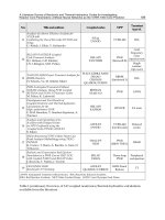
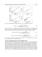
TABLE 55-2
Common Herbs Used as Adjunctive Treatment in Epilepsy
HERB LATIN NAME COMMON USES PLACE OF ORIGIN CHEMISTRY SYSTEMIC EFFECTS CNS EFFECTS PREGNANCY ISSUES
American Veratum viride Antiemetic, United Similar to Blood pressure Paresthesias, Teratogenic
hellebore neuralgia, States steroids alteration, weakness,
pneumonia gastrointestinal and paralysis,
respiratory problems, seizure
salivation; high risk
of side effects, narrow
therapeutic index
Behen Moringa Antimicrobial, India Contains Gastrointestinal Dizziness Possible abortive
oleifera gastrointestinal glucosinolates, problems effect
ailments fatty acids
Betony Stachys Respiratory and Europe, Part of mint Hypotension, — Uterine
officinalis gastrointestinal North Africa, family, related gastrointestinal contractions
ailments, Siberia to tannins problems, hepatic
dysfunction
Black cohosh Cimicifuga Menstrual pain North America Estrogen effect Hypotension, Sedation, Increased risk of
racemosa gastrointestinal headache spontaneous
problems; not for abortion
long-term use
Blue cohosh Caulophyllum Induce labor Midwest and Similar to Hypertension, Seizures Uterine
thalictroides Eastern United nicotine gastrointestinal contractions,
States, Canada problems, increases teratogenic
glucose; poisonous to
children, cardiotoxic
to neonates
Calotropis Calotropis Antineoplastic Asia, India, Related to Gastrointestinal Seizures —
procera Africa, Pakistan, steroids steroids
Sunda Islands bradycardia;
highly toxic
European Paeonia Pain, headache Southern Europe, Contains tannins, Hypotension No Uterine
peony officinalis Asia flavonoids anticonvulsant contractions
effect in studies
Ginkgo Ginkgo biloba Cognitive China Platelet-activating Bleeding Seizures —
impairment factor antagonist
V • ANTIEPILEPTIC DRUGS AND KETOGENIC DIET
718
Goto kola Centella asiatica Antimicrobial, South East Asia, Consists of Contact dermatitis, — Should not be
antineoplastic, India, Sri Lanka, triterpene acids infertility, used in pregnancy
CNS depressant, China, and sugar hyperglycemia,
wound healing Madagascar, residues, affects hyperlipidemia
South Africa, D2 receptors
Southeastern and cholinergic
United States, system
Mexico, parts of
South America
Groundsel Senecio vulgaris Worm infestation Europe, Asia, Contains Hepatic dysfunction, — —
Africa, Australia, alkaloids, carcinogenic;
Americas flavonoids should not be
taken internally
Kava Piper Anxiety South Pacific Inhibits L-type Hypertension, Acute Loss of uterine
methysticum Ca
2ϩ
and Na
ϩ
gastrointestinal and dystonic tone
channels, respiratory problems, reaction
increases K
ϩ
hepatic dysfunction,
outward current, leucopenia,
enhances GABA thrombocytopenia,
transmission; dermatitis; should
member of black not be used in
pepper family children Ͻ12 years
Lily of the valley Convallaria Arrhythmia, — Related to Gastrointestinal Headache, —
majalis cardiac steroids problems, cardiac stupor, changes
insufficiency arrhythmia; many in color
drug interactions, perception
highly toxic, not
recommended
for use
Melatonin* — Sleep disorders, — Derivative of — Drowsiness Should not be
jet lag serotonin used in pregnancy
TABLE 55-2
(Continued)
HERB LATIN NAME COMMON USES PLACE OF ORIGIN CHEMISTRY SYSTEMIC EFFECTS CNS EFFECTS PREGNANCY ISSUES
55 • VITAMINS, HERBS, AND OTHER ALTERNATIVE THERAPIES
719
Mistletoe Viscum album Cancer, England, Contains choline, Blood pressure Coma, seizures, Uterine
seizures, Europe, histamine, alteration, sedation, contractions
heart disease, Asia tyramine gastrointestinal psychosis
headache problems,
bradycardia,
cardiac arrest;
highly toxic
Mugwort Artemisia Change of Northern Europe, Part of daisy Dermatitis, allergy; — Uterine
vulgaris fetal position Asia, family not recommended contractions,
in utero (breech), North America for use increased risk
menstrual of abortion
problems,
depression
Pipsissewa Chimaphilia Seizures, Europe, Asia, — Gastrointestinal — Should not be
umbellale antispasmodic, North America problems, rash; used in
diuretic problems, rash; pregnancy
not recommended
for use
Skullcap Scutellaria Cancer, North America Contains Hepatic dysfunction, Confusion, Should not be
laterifolia sedative flavonoids cardiac arrhythmia seizures used in
pregnancy
Valerian Valeriana Anxiety, sleep Europe, Mexico, Inhibits Gastrointestinal Sedation, Uterine
officinalis India, Japan degradation and problems, hepatic tremor, contractions
reuptake of GABA dysfunction; many headache;
drug interactions should not be
used in children
Ͻ14 years
Yew Taxus baccata Antimicrobial Europe Contains Cardiotoxic, — Causes
taxines, arrhythmia, spontaneous
flavonoids severely toxic abortion
CNS, Central nervous system; GABA, gamma-aminobutyric acid.
*not an herb
V • ANTIEPILEPTIC DRUGS AND KETOGENIC DIET
720
can make seizures worse (84). Herbs containing thu-
jone, such as wormwood and sage, which are used to
treat gastrointestinal disorders, may have proconvul-
sant effects. Table 55-3 lists some herbs that may cause
seizures.
Some herbs can interfere with the hepatic P450 system
(Table 55-4) and, when used together with antiepileptic
medications, produce toxic side effects or decrease their
effectiveness. Other herbs can lower anticonvulsant levels
or otherwise interact with AEDs (Table 55-5).
MELATONIN
Melatonin is an indolamine that is synthesized from
tryptophan in the pineal gland. It is released in a cir-
cadian pattern, with peak levels in the early morning
hours (85). Its apparent main function is to signal the
brain to induce sleep. Melatonin is used for a variety of
conditions, including sleep disorders, jet lag, and autism.
By regulating sleep patterns it appears to be helpful in
attaining better seizure control, and from animal models,
melatonin is helpful in preventing seizure-related brain
damage. A variety of proposed mechanisms are thought
to account for melatonin’s antiepileptic effect. It appears
to increase GABA’s concentration in the brain and pro-
tects against seizure-induced brain damage by inhibiting
calcium influx into neurons and by free-radical scaveng-
ing properties (86–89). When given orally, its blood
concentration peaks within 1 hour, and usually returns
to baseline within 4 to 8 hours (90).
Melatonin’s effectiveness has been shown in ani-
mal models. It inhibits kainic acid-induced seizures
in rats. It also inhibits lipid peroxidation, is a potent
free-radical scavenger, and reduces seizure-induced
brain damage (91). Melatonin also blocks potassium
cyanide-induced seizures in mice (92). Melatonin stimu-
lates brain glutathione peroxidase activity, which is an
antioxidative enzyme that metabolizes the precursor
of the hydroxyl and peroxyl radicals to water. It also
raises the electroconvulsive threshold in animal models
and potentiates the anticonvulsive activity of carbam-
azepine and phenobarbital (93). In addition, melato-
nin significantly reduces neurobehavioral changes in
mice, as well as morphologic changes in association
with seizures, mostly in the CA3 region of the rat hip-
pocampus (94).
Most clinical studies have looked at the use of mela-
tonin in a limited number of subjects. Its effectiveness
and safety profile in epilepsy patients was supported by
several open-label trials. Peled et al (95) found that five
of six children with intractable epilepsy had significant
improvement not only in seizure control but also in their
cognitive function. Bazil et al (96) showed that patients
with epilepsy had a peak level of melatonin that was 50%
of controls’ peak level and that the peak occurred 3 hours
before that of controls. Some studies have shown a lack of
melatonin’s efficacy, which may be related to inadequate
dosing or other factors (97, 98).
TABLE 55-3
Herbs That May Cause Seizures
Bearberry (Arcostaphylos uva-ursi)
Borage (Borago officinalis)
Ephedra (Ephedra sinica)
Gingko (Gingko biloba)
Ginseng (Panax ginseng)
Ma Huang (Herba ephedra)
Monkshood (Aconitum sp.)
Primrose (Oenothera biennis)
Yohimbe (Pausinystalia yohimbe)
TABLE 55-4
Herbs That Inhibit the P450 System
American hellebore
Chamomile
Echinacea
Garlic
Licorice
Milk thistle
Mugwort
Pipsissewa
Pycnogel
St. John’s wort*
Trifolium pratense (red clover)
*Effect on the P450 system is controversial.
TABLE 55-5
Herbs and Their Effects on Antiepileptic Drugs
HERB DRUG EFFECT
Septilin Carbamazepine Decreases drug
level
Sho-seiryu-to Carbamazepine Delays drug
absorption
Paeoniae radix Phenytoin Delays drug
absorption
Thujone Phenobarbital Reduces drug
(wormwood, sage) efficacy
Ginkgo Phenytoin, Reduces
phenobarbital, drug efficacy
carbamazepine
55 • VITAMINS, HERBS, AND OTHER ALTERNATIVE THERAPIES
721
PHYTOTHERAPY
Practitioners of phytotherapy believe that there is an
imbalance in the body and that specific herbs may restore
this balance. Many plants are known for their anticon-
vulsant properties. Approximately 150 preparations of
plants have been investigated. Individual plants are usu-
ally used but can be combined if necessary. In most cases,
the active compound has not yet been identified. Studies
have shown that some natural plant coumarins and tri-
terpenoids exhibit anticonvulsant properties (99, 100).
Several show promise against seizures (see Table 55-6),
but further study is needed before their routine use.
Albizia lebbek increases levels of GABA in the
brain. Piper nigrum may have antagonistic actions at
NMDA receptors. The efficacy of Casimiroa edulis has
been shown in animal studies to be similar to that of
phenytoin and phenobarbital. Ipomoea stans is similar
in effectiveness to valproic acid. The action of Piper
guineese and Psidium guyanensis is similar to that of
phenobarbital (101). The toxicity or side effects of these
plants are largely unknown. Some plants may interact
with antiepileptic medications; Ruta chalepensis, for
example, may increase the hypnotic effects of pheno-
barbital. More work on the use of phytotherapy in
epilepsy is needed.
ASIAN MEDICINE
Traditional Chinese medicine has been used for thousands
of years and has been gaining interest in the Western
world for quite some time. It is partly based on the view
that the body is closely related to its surrounding outside
world. The organs inside of the body are themselves con-
sidered to be interconnected via an interlacing network of
channels and collaterals (102). Most therapies are com-
posed of several herbs. The use of combination therapy is
thought to improve the effectiveness and lessen any pos-
sible side effects. From the Chinese perspective, certain
types of seizures are considered to be due to an exogenous
or endogenous “wind.” In children, the pathogenesis is
attributed to the insufficiency of the spleen, stagnation
of phlegm, and reversed flow of qi (known as the vital
forces of the body), and stirring up of the endogenous
wind (103). Some open-label studies of traditional Chinese
herbal mixtures have shown a reduction in seizures and
fewer side effects compared with standard AEDs, but
well-controlled double-blind studies are lacking. Numer-
ous combinations of Chinese herbs are used to combat
seizures; only a few will be discussed.
Tianma gouteng yin is composed of amino acids,
alkaloids, and fatty acids in addition to other compounds.
Interestingly, it has been found to act as an NMDA-receptor
antagonist. Not only does it have direct influences at
the receptor, but it also helps to prevent neuronal injury
and death (102). When quingyangsen (root) was given as
an adjunct to standard AED treatment, almost 30% of
patients had seizure control ranging from 2 to 9 months
after initiating therapy (104). This compound has also
been shown to block seizures in animal models (105).
Zhenxianling contains different flowers, animal parts,
and human placenta in addition to other substances. A
study using Zhenxianling in 239 patients with refractory
epilepsy, of whom 147 were aged 1.5 to 20 years, showed
that 66% had a greater than 75% seizure reduction and
an additional 30% had a greater than 50% reduction of
their seizures (106). These effects were seen 1 to 5 days
after treatment. In 15 patients with absence seizures, 11
had their seizure frequency reduced by 50% to 75%. A
few studies have been performed using longdanxiegan
tang, or a modified version, in absence epilepsy. Approxi-
mately 90% of patients taking this herb showed signifi-
cant clinical or EEG improvement (107, 108).
In a study using capsules composed of a variety
of Chinese supplements, a significant improvement
was found among children with different types of epi-
lepsy (103). More than 900 children were treated with
these capsules, and their response was compared with
that of only 160 patients treated with phenobarbital.
In children taking the capsules, 57% had their seizures
reduced by more than 75%. An additional 26% had a
seizure reduction of 50% to 75%. The duration of indi-
vidual seizures was also significantly diminished. In the
control group, 40% of patients achieved a 75% seizure
decrease and 12% had a 50% reduction in seizures.
Approximately 1% had worsening seizure control. Fifty
percent of children with absence and benign rolandic
seizures had a 75% decrease in seizures. Two cases of
infantile spasms were included in this study. One patient
had a 75% reduction of seizures and the other a 50%
to 75% decrease. Of those in the treatment group who
previously had abnormal EEGs, 54% had normal EEGs
at the end of the study period.
In Japan, kampo medicines are herbal remedies used
to combat various medical conditions, including epilepsy.
Most of these therapies are mixtures of different herbs,
TABLE 55-6
Some Homeopathic Remedies Used for Seizues
FEBRILE SEIZURES NONFEBRILE SEIZURES
Aconitum napellus Atropa belladonna
Aethusa cynapium Chamomilla vulgaris
Cuprum metallica
Glonoinum
Ignatia amara
Zincum metallicum
V • ANTIEPILEPTIC DRUGS AND KETOGENIC DIET
722
some of which are similar to those used in traditional
Chinese medicine. Sho-saiko-to is an herbal formula
commonly used to treat liver disorders; it also is recom-
mended as a possible treatment for intractable epilepsy.
Another formula similar to this compound, the Chinese
bupleurum–cinnamon combination (chai-hu-keui-chi-
tang), has shown some preliminary benefit in epilepsy.
These formulas contain the same nine herbs with minor
variations in their relative amounts. They appear to have
equivalent effects (109). Sho-saiko-to has been adminis-
tered to adults and children. The pediatric dose depends
on the child’s weight (110). There are no well-designed
clinical studies on the benefit of this formula in epilepsy.
In one study (111), it was given to 24 patients who were
taking multiple drugs for uncontrolled epilepsy. Six of
the 24 patients had no seizures within 10 months of the
herbal therapy. An additional 13 patients were improved,
three had no change, and two did not complete the study.
Improvements were seen as soon as 1 month. Tonic-clonic
seizures seemed to have the best response rate. Another
study (112) revealed possible cognitive improvements
with the use of this supplement, but the study was flawed
and not optimally designed. Animal studies (113) have
shown that it can inhibit pentylenetetrazol-induced sei-
zures as well as cobalt-induced seizures and neuronal
damage. Other studies (114, 115) revealed that there were
no changes in barbiturate potentiation.
Adverse effects have rarely been reported. The for-
mula has caused pneumonitis or hepatitis, or both, in
a number of patients with liver disease, and has caused
fatalities. Patients using this supplement must be advised
to report coughs and fevers to their health care providers;
prompt and careful follow-up is necessary (116, 117).
Occasionally, gastrointestinal upset or mild transient
symptoms are present. In addition to side effects, some
of these supplements have been known to contain toxic
ingredients that are not named on the label. These herbal
remedies have been found to contain such elements as
lead, arsenic, and mercury, which, if consumed in greater
than safe amounts, can lead to serious consequences (118,
119). Although some report significant success with the
use of these products, extreme caution should be main-
tained.
ACUPUNCTURE
Acupuncture has been practiced in Asia for more than
two thousand years. In the United States, where it has
increased in popularity over the past 30 to 40 years, it
is used by approximately one million individuals. This
ancient therapy is used mostly for pain management,
but also for a number of different conditions, includ-
ing epilepsy. Up to 70% of people who undergo acu-
puncture treatments do not inform their health care
providers (120). Acupuncture involves the use of fine
needles (now made of stainless steel) that are inserted
into the skin at defined points of the body. For epilepsy,
points along the scalp are key, as acupuncturists consider
the scalp a direct projection of the cerebral cortex. Differ-
ent points are selected depending on the different types
and symptoms of seizures (121).
Acupuncture is presumed to restore balance to the
disruption of the natural flow of energy that the body
requires to function normally. Acupuncture releases endor-
phins, adrenocorticotropic hormone (ACTH), and other
neurochemicals, such as GABA (122). Some studies show
that afterward there is an increase of the serotonin level,
which may lead to improved cognitive function (123).
Specific sites are used to combat different conditions. The
point naokong (GB19) is located near the occipital pro-
tuberance. It is selected for acupuncture in a variety of
medical conditions in addition to epilepsy, and has been
used in children. It is said to have tranquilizing effects,
regulate blood flow, and calm “endopathic wind.”
In terms of efficacy, there is a paucity of well-
performed and well-controlled clinical trials for evalu-
ating the usefulness of acupuncture in epilepsy, as well
as other medical conditions. This is due in part to the
individualization of therapy. Acupuncture differs between
individuals, making standardization difficult. Even sham
acupuncture is difficult to assess, because nonspecific
needling can lead to the stimulation of neurohormonal
responses (124).
Kloster et al (125) compared the effects of sham
acupuncture and actual acupuncture in patients with
intractable epilepsy. There was a small but statistically
nonsignificant reduction in seizure frequency in both
groups, perhaps due to the small sample size. No signifi-
cant EEG changes were appreciated. Stavem et al (126)
also failed to show that acupuncture significantly reduced
seizures or had any effect on the patients’ quality of life.
Adverse effects are rare, the most common being infection
and trauma. Other rare complications such as pneumo-
thorax, cardiac tamponade, hepatitis, and spinal cord
injuries have also been reported. The transmission of
human immunodeficiency virus (HIV) has rarely been
reported. The importance of sterilization and universal
precautions cannot be emphasized enough (120).
In a child who had almost continuous simple par-
tial seizures, acupuncture improved the seizures after
seven sessions, and almost completely eliminated them by
30 sessions (127). Six months later, the patient was
reported to be seizure free. In another study (121) involv-
ing almost 100 children and adults, 66% had a greater
than 75% reduction in seizures, and an additional 24%
had a 50% to 75% reduction. Yang (128) reported the
use of acupuncture in eight children with status epilepti-
cus. Different acupoints were used depending on the case.
Seizures ceased within 10 minutes in all cases. No
55 • VITAMINS, HERBS, AND OTHER ALTERNATIVE THERAPIES
723
recurrences were reported in three patients for up to
2 years and in one patient for 8 years. Acupuncture may be
a useful adjunctive therapy in epilepsy, but better designed
studies are needed to fully evaluate its effectiveness.
HOMEOPATHY
There is little scientific evidence for the efficacy of
homeopathy in epilepsy, and even less information is
available on its use in children. It has been used for
more than 200 years, and more than 500 homeopathic
remedies have been used to treat seizures. Homeopa-
thy is based on the principle that substances causing
medical conditions can also be used to combat them.
Symptoms are believed to represent the body’s attempt
to restore itself to health, and homeopathy aims to treat
the patient’s symptoms. It relies on the body’s own pow-
ers for self-healing; therefore an individual’s mental
and physical state is important and taken into account
prior to the administration of remedies. The identifi-
cation of imbalances within the person in conjunction
with his or her symptoms aids the homeopathist in
choosing supplements that will restore the body’s abil-
ity to heal itself. When dealing with children, choos-
ing the correct treatment is further complicated by the
fact that the homeopathist must rely on parental obser-
vations instead of directly obtaining information from
the child.
The remedies used are derived mainly from plants,
minerals, and animals. Remedies derived from tox-
ins that are believed to cause illness are diluted before
administration (129). Formulas containing Aconitum
napellus and Aethusa cynapium are thought to be help-
ful in febrile seizures and other seizures associated with
illnesses (Table 55-6). There are supplements that may be
helpful in febrile and nonfebrile seizures (130). Reliable
information on the risks associated with homeopathic
remedies is lacking.
NATUROPATHY
Naturopathic medicine was established more than a
hundred years ago and uses forms of Western medicine
in addition to natural therapies. It uses noninvasive
techniques and is based on the principle that natural
substances can help the body’s intrinsic ability to heal
itself. Naturopath practitioners place a large emphasis on
attempting to remove the underlying cause of the disease.
Consuming the proper foods for sufficient nutrition of
the body is one way of using natural substances. Nutri-
tional supplements in the form of vitamins and minerals
are often used (6). Certain herbs are prescribed to sup-
port the liver and kidneys, through which many AEDs
are metabolized. In addition, some vitamins can interfere
with certain AEDs. Information on the efficacy of natu-
ropathic medicine in epilepsy is limited.
CONCLUSION
Alternative medicine is a growing field that comprises
several different approaches. Most facets have not been
well studied, making recommendations for their use in
patients with epilepsy difficult. Side effects and compli-
cations are mostly underreported. Open communication
between patients and health care professionals is vital in
ensuring the well-being of patients. Patients should be
encouraged to make their physicians aware of any other
treatments they are receiving. They should also be advised
not to discontinue their conventional medications with-
out discussing their plans with their primary caregivers.
Further research is required to better evaluate the role of
alternative medicine in the treatment of epilepsy.
References
1. Eisenberg DM, Davis RB, Ettner SL, Yager A, et al. Trends in alternative medicine
use in the United States, 1990-7: results of a follow-up national survey. JAMA 1998;
280:1569–1575.
2. Barnes PM, Powell-Griner E. Complementary and alternative medicine use among adults:
United States, 2002. Advance data from vital and health statistics. US Department of
Health and Human Services: Centers for Disease Control and Prevention, National Center
for Health Statistics. No. 343, Hyattsville, MD. May 27, 2004.
3. Ottolini MC, Hamburger EK, Loprieato JO, et al. Complementary and alternative
medicine use among children in the Washington, DC area. Ambul Pediatr 2001;
1:122–125.
4. Sibinga EM, Ottolini MC, Duggan AK, Wilson MH. Parent–pediatrician communica-
tion about complementary and alternative medicine use for children. Clin Pediatr 2004;
4:367–373.
5. Lim A, Cranswick N, Skull S, South M. Survey of complementary and alternative medi-
cine use at a tertiary children’s hospital. J Pediatr Child Health 2005; 41:424–427.
6. Devinsky O, Schachter S. Pacia S, eds. Complementary and alternative therapies for epi-
lepsy. New York: Demos, 2005:33–42, 53–6, 65–80, 81–94, 113–9, 165–76, 285–90.
7. Losier A, Taylor B, Fernandez CV. Use of alternative therapies by patients presenting
to a pediatric emergency department. J Emerg Med 2005; 28:267–271.
8. Soo I, Mah JK, Barlow K, Hamiwka L, et al. Use of complementary and alternative
medical therapies in a pediatric neurology clinic. Can J Neurol Sci 2005; 32:524–528.
9. Spigelblatt L, Laine-Ammara G, Ples IB, Guyer A. The use of alternative medicine by
children. Pediatrics 1994; 94:811–814.
10. Hurvitz EA, Leonard C, Ayyangar R, Nelson VS. Complementary and alternative
medicine use in families of children with cerebral palsy. Dev Med Child Neurol 2003;
45:364–370.
11. Waaler PE, Blom BH, Skeidsvoll H, Mykletun A. Prevalence, classification and severity
of epilepsy in children in western Norway. Epilepsia 2000; 41:802–810.
12. Fearon J. Complementary therapies: knowledge and attitudes of health professionals.
Pediatr Nurs 2003; 15:24–27.
13. Kemper KJ, O’Connor KG. Pediatricians’ recommendations for complementary and
alternative medical (CAM) therapies. Ambul Pediatr 2004; 4:482–487.
14. Haut SR, Vouyiouklis M, Shinnar S. Stress and epilepsy: a patient perception survey.
Epilepsy Behav 2003; 4:511–514.
15. Spatt J, Langbauer G, Mamoli B. Subjective perception of seizure precipitants: results
of a questionnaire study. Seizure 1998; 7:391–395.
16. Spector S, Cull C, Goldstein LH. Seizure precipitants and perceived self-control of
seizures in adults with poorly-controlled epilepsy. Epilepsy Res 2000; 38:207–216.
17. Lai C-W, Trimble MR. Stress and epilepsy. J Epilepsy 1997; 10:177–186.
18. Jones JE, Hermann BP, Barry JJ, Gilliam F, et al. Clinical assessment of Axis I psychi-
atric morbidity in chronic epilepsy: a multicenter investigation. J Neuropsychiatry Clin
Neurosci 2005; 17:172–179.
V • ANTIEPILEPTIC DRUGS AND KETOGENIC DIET
724
19. Nakken KO, Loyning A, Loyning T, Gloerson G, et al. Does physical exercise influence the
occurrence of epileptiform EEG discharges in children? Epilepsia 1997; 38:279–284.
20. Panjwani U, Gupta HL, Singh SH, Selvamurthy W, et al. Effect of Sahaja yoga prac-
tice on stress management in patients of epilepsy. Indian J Physiol Pharmacol 1995;
39:111–116.
21. Lehrer PM, Carr R, Sargunaraj D, Woolfolk RL. Stress management techniques: are
they all equivalent, or do they have specific effects? Biofeedback Self Regul 1994;
19:353–401.
22. Denio LS, Drake ME Jr, Pakalnis A. The effect of exercise on seizure frequency. J Med
1989; 20:171–6.
23. Eriksen HR, Ellertsen B, Gronningsaeter H, Nakken KO, et al. Physical exercise in
women with intractable epilepsy. Epilepsia 1994; 35:1256–1264.
24. Nakken KO, Bjorholt PG, Johannessen SI, Loyning T, et al. Effects of physical training
on aerobic capacity, seizure occurrence, and serum level of antiepileptic drugs in adults
with epilepsy. Epilepsia 1990; 31:88–94.
25. Esquivel E, Chaussain M, Plouin P, Ponsot G, et al. Physical exercise and voluntary
hyperventilation in childhood absence epilepsy. Electroencephalogr Clin Neurophysiol
1991; 79:127–132.
26. Ogunyemi AO, Gomez MR, Klass DW. Seizures induced by exercise. Neurology 1988;
38:633–634.
27. Schmitt B, Thun-Hohenstein L, Vontobel H, Boltshauser E. Seizures induced by physical
exercise: report of two cases. Neuropediatrics 1994; 25:51–53.
28. Sturm JW, Fedi M, Berkovic SF, Reutens DC. Exercise-induced temporal lobe epilepsy.
Neurology 2002; 59:1246–1248.
29. Panjwani U, Selvamurthy W, Singh SH, Gupta HL, et al. Effect of Sahaja yoga practice
on seizure control & EEG changes in patients of epilepsy. Indian J Med Res 1996;
103:165–172.
30. Ogata A, Amano K. A psychosocial approach to epileptic patients. Epilepsia 2000; 41
Suppl 9:36–38.
31. Olley BO, Osinowo HO, Brieger WR. Psycho-educational therapy among Nigerian
adult patients with epilepsy: a controlled outcome study. Patient Educ Couns 2001;
42:25–33.
32. Snead K, Ackerson J, Bailey K, Schmitt MM, et al. Taking charge of epilepsy: the
development of a structured psychoeducational group intervention for adolescents with
epilepsy and their parents. Epilepsy Behav 2004; 5:547–556.
33. Chmelarova D. [Use of psychotherapy in patients with combined epileptic and nonepi-
leptic seizures]. Cas Lek Cesk 2005; 144:557–559; discussion 559.
34. Martinovic Z. Adjunctive behavioural treatment in adolescents and young adults with
juvenile myoclonic epilepsy. Seizure 2001; 10:42–47.
35. Au A, Chan F, Li K, Leung P, et al. Cognitive-behavioral group treatment program for
adults with epilepsy in Hong Kong. Epilepsy Behav 2003; 4:441–446.
36. Goldstein LH, McAlpine M, Deale A, Toone BK, et al. Cognitive behaviour therapy
with adults with intractable epilepsy and psychiatric co-morbidity: preliminary observa-
tions on changes in psychological state and seizure frequency. Behav Res Ther 2003;
41:447–460.
37. Ramaratnam S, Baker GA, Goldstein L. Psychological treatments for epilepsy. Cochrane
Database Syst Rev 2003; (4):CD002029.
38. Schmid-Schonbein C. Improvement of seizure control by psychological methods in
patients with intractable epilepsies. Seizure 1998; 7:261–270.
39. Goldstein LH, Deale AC, Mitchell-O’Malley SJ, Toone BK, et al. An evaluation of
cognitive behavioral therapy as a treatment for dissociative seizures: a pilot study. Cogn
Behav Neurol
2004; 17:41–49.
40. Zaroff CM, Myers L, Barr WB, Luciano D, et al. Group psychoeducation as treatment
for psychological nonepileptic seizures. Epilepsy Behav 2004; 5:587–592.
41. Reiter JM, Lambert RD, Andrews DJ, et al. Complex-partial epilepsy: a therapeutic
model of behavioral management and EEG biofeedback. Self-Control Epilepsy
1990;
1:27–38.
42. Andrews DJ, Schonfeld WH. Predictive factors for controlling seizures using a behavioral
approach. Seizure 1992; 1:111–116.
43. Meencke HJ, Schmid-Schonbein C, Heinen G. Methods and results of a treatment
program on self-control of epileptic seizures. Presented at the 54th Annual Meeting of
the American Epilepsy Society, Los Angeles, CA, December 2000.
44. Reiter JM, Andrews DJ, Janis C. Taking control of your epilepsy: a workbook for
patients and professionals. Available from Andrews/Reiter Epilepsy Research Program,
1103 Sonoma Avenue, Santa Rosa, CA 95405; 1987.
45. Sterman MB. Studies of EEG biofeedback training in man and cats. In: Highlights of
the 17th Annual Conference. VA Cooperative Studies in Mental Health and Behavioral
Sciences. Washington D.C.: U.S. Government, 1972:50–60.
46. Sterman MB, Howe RD, Macdonald LR. Facilitation of spindle-burst sleep by condition-
ing of electroencephalographic activity while awake. Science 1970; 167:1146–1148.
47. Sterman MB, Friar L. Suppression of seizures in an epileptic following sensorimotor
EEG feedback training. Electroencephalogr Clin Neurophysiol 1972; 33:89–95.
48. Hanten WP, Dawson DD, Iwata M, Seiden W, et al. Craniosacral rhythm: reliability
and relationships with cardiac and respiratory rates. J Orthop Sports Phys Ther 1998;
27:213–218.
49. Moran RW, Gibbons P. Intraexaminer and interexaminer reliability for palpation of the
cranial rhythmic impulse at the head and sacrum. J Manipulative Physiol Ther 2001;
24:183–190.
50. Salbert B, Pellock J, Wolf B. Characterization of seizures associated with biotinidase
deficiency. Neurology 1993; 43:1351–1354.
51. Kalayci O, Coskun T, Tokatli A, Demir E, et al. Infantile spasms as the initial symptom
of biotinidase deficiency. J Pediatr 1994; 124:103–104.
52. Wolf B, Heard G, Wissbecker K, McVoy JR, et al. Biotinidase deficiency: initial clinical
features and rapid diagnosis. Ann Neurol 1985; 18:614–617.
53. Snyderman SE, Carretero R, Holt LE. Pyridoxine deficiency in the human being. Fed
Proc 1950; 9:372–373.
54. Hunt AD, Stockes J, McCrory WW. Pyridoxine dependency: report of a case of intrac-
table convulsions in an infant controlled by pyridoxine. Pediatrics 1954; 13:140–145.
55. Wang HS, Kuo MF, Chou ML, Hung PC, et al. Pyridoxal phosphate is better than pyridox-
ine for controlling idiopathic intractable epilepsy. Arch Dis Child 2005; 90:512–515.
56. Hansson O, Hagberg B. Effect of pyridoxine treatment in children with epilepsy. Acta
Soc Med Uppsala 1968; 73:35–43.
57. Ohtsuka Y, Matsuda M, Ogino T, Kobayashi K, et al. Treatment of the West syndrome
with high-dosage pyridoxal phosphate. Brain Dev 1987; 9:418–421.
58. Pietz J, Benninger C, Schafer H, Sontheimer D, et al. Treatment of infantile spasms with
high-dosage vitamin B
6
. Epilepsia 1993; 34:757–763.
59. Mackay MT, Weiss SK, Adams-Webber T, Ashwal S, et al; American Academy of Neu-
rology; Child Neurology Society. Practice parameter: medical treatment of infantile
spasms. Report of the American Academy of Neurology and the Child Neurology Society.
Neurology 2004; 62:1668–1681.
60. Torres OA, Miller VS, Buist NM, Hyland K. Folinic acid-responsive neonatal seizures.
J Child Neurol 1999; 14:529–532.
61. Shyamaladevi N, Jayakumar AR, Sujatha R, Paul V, et al. Evidence that nitric oxide
production increases gamma-amino butyric acid permeability of blood-brain barrier.
Brain Res Bull 2002; 57:231–236.
62. Oztas B, Kilic S, Dural E, Ispir T. Influence of antioxidants on the blood-brain barrier
permeability during epileptic seizures. J Neurosci Res 2001; 66:674–678.
63. Takahashi H. Studies on homocarnosine in cerebrospinal fluid in infancy and childhood.
Part II. Homocarnosine levels in cerebrospinal fluid from children with epilepsy, febrile
convulsions or meningitis. Brain Dev 1981; 3:263–270.
64. Petroff OA, Hyder F, Collins T, Mattson RH, et al. Acute effects of vigabatrin on brain GABA
and homocarnosine in patients with complex partial seizures. Epilepsia 1999; 40:958–964.
65. Petroff OAC, Hyder F, Rothman DL, Mattson RH. Brain homocarnosine and seizure
control of patients taking gabapentin or topiramate. Epilepsia 2006; 47:495–498.
66. Collins BW, Goodman HO, Swanton CH, Remy CN. Plasma and urinary taurine in
epilepsy. Clin Chem 1988; 34:671–675.
67. Durelli L, Mutani R. The current status of taurine in epilepsy. Clin Neuropharmacol
1983; 6:37–48.
68. Fukuyama Y, Ochiai Y. Therapeutic trial by taurine for intractable childhood epilepsies.
Brain Dev 1982; 4:63–69.
69. Konig P, Kriechbaum G, Presslich O, et al. Orally-administered taurine in therapy-
resistant epilepsy. Wien Klin Wochenschr 1977; 89:111–113.
70. Igisu H, Matsuoka M, Iryo Y. Protection of the brain by carnitine. Sangyo Eiseigaku
Zasshi 1995; 37:75–82.
71. De Vivo DC, Bohan TP, Coulter DL, Dreifuss FE, et al. L-Carnitine supplementation in
childhood epilepsy: current perspectives. Epilepsia 1998; 39:1216–1225.
72. Department of Neurology, Massachusetts General Hospital. Epilepsy and nutritional
supplementation [pamphlet]. 1994.
73. Gascon G, Patterson B, Yearwood K, Slotnick H. N,N Dimethylglycine and epilepsy.
Epilepsia 1989; 30:90–93 (letter).
74. Haidukewych D, Rodin EA. N,N-Dimethylglycine shows no anticonvulsant potential. Ann
Neurol 1984; 15:405 (letter).
75. Roach ES, Gibson P. Failure of N,N-dimethylglycine in epilepsy. Ann Neurol 1983;
14:347 (letter).
76. Groenwald J, ed. PDR for herbal medicines. 3rd ed. Montvale, NJ: Thomson PDR. 2004.
77. Grunze H, Langosch J, Schirrmacher K, Bingmann D, et al. Kava pyrones exert effects on
neuronal transmission and transmembranous cation currents similar to established mood
stabilizers: a review. Prog Neuropsychopharmacol Biol Psychiatry 2001; 25:1555–1570.
78. Almeida JC, Grimsley EW. Coma from the health food store: interaction between kava
and alprazolam. Ann Intern Med 1996; 125:940–941.
79. Tyagi A, Delanty N. Herbal remedies, dietary supplements, and seizures. Epilepsia 2003;
44:228–235.
80. Amabeoku GJ, Leng MJ, Syce JA. Antimicrobial and anticonvulsant activities of Viscum
capense. J Ethnopharmacol 1998; 61:237–241.
81. Miller LG. Herbal medicinals: selected clinical considerations focusing on known or
potential drug–herb interactions.
Arch Intern Med 1998; 158:2200–2211.
82. Rosenblatt M, Mindel J. Spontaneous hyphema associated with ingestion of Ginkgo
biloba extract. N Engl J Med 1997; 336:1108 (letter).
83. Vale S.
Subarachnoid hemorrhage associated with Ginkgo biloba. Lancet 1998;
352:36 (letter).
84. Mar C. Clinical evidence: an evidence based review of the ten most commonly used
herbs. West J Med 1999; 171:168–171.
85. Brzezinski A. Melatonin in humans. N Engl J Med 1997; 336:186–195.
86. Kabuto H, Yokoi I, Ogawa N. Melatonin inhibits iron-induced epileptic discharges in
rats by suppressing peroxidation. Epilepsia 1998; 39:237–243.
87. Niles LP, Pickering DS, Arciszewski MA. Effect of chronic melatonin administration of
GABA and diazepam binding in rat brain. J Neurol Transm 1987; 70:117–124.
88. Castroviejo DA, Rosenstein RE, Romeo HE, Cardinali DP. Changes in gaba-amino-
butyric acid high affinity binding to cerebral cortex membranes after pinealectomy or
melatonin administration to rats. Neuroendocrinology 1986:43:24–31.
89. Ross C, Davies P, Whitehouse W. Melatonin’s role as an anticonvulsant and neuronal
protector: experimental and clinical evidence. Neurology 1998:13:501–519.
90. Ross C, Whitehouse W. Melatonin treatment for sleep disorders in children with neurodevel-
opmental disorders: an observational study. Dev Med Child Neurol 2002; 44:339–344.
55 • VITAMINS, HERBS, AND OTHER ALTERNATIVE THERAPIES
725
91. Mohanan PV, Yamamoto HA. Preventative effect of melatonin against brain mitochon-
dria DNA damage, lipid peroxidation and seizures induced by kainic acid. Toxicol Lett
2002; 129:99–105.
92. Yamamoto H, Tang H. Antagonistic effect of melatonin against cyanide induced seizures
and acute lethality in mice. Toxicol Lett 1996; 87:19–24.
93. Borowicz KK, Kaminski R, Gasior M, Kleinrok Z, et al. Influence of melatonin upon
the protective action of conventional anti-epileptic drugs against maximal electroshock
in mice. Eur Neuropsychopharmacol 1999; 9:185–190.
94. Tan DX, Manchester LC, Reiter RJ, Qi W, et al. Melatonin protects hippocampal
neurons in vivo against kainic acid-induced damage in mice. J Neurosci Res 1998;
54:382–389.
95. Peled N, Shorer Z, Peled E, Pillar G. Melatonin effect on seizures in children with severe
neurologic deficit disorders. Epilepsia 2001; 42:1208–1210.
96. Bazil CW, Short D, Crispin D, Zheng N. Patients with intractable epilepsy have low
melatonin, which increases following seizures. Neurology 2000; 55:1746–1748.
97. Sheldon SH. Pro-convulsant effects of oral melatonin in neurologically disabled children.
Lancet 1998; 351:1254 (letter).
98. Camfield P, Gordon K, Dooley J, Camfield C. Melatonin appears ineffective in children
with intellectual deficits and fragmented sleep: six “N of 1” trials. J Child Neurol
1996:11:341–343.
99. Chaturvedi AK, Parmar SS, Bhatnagar SC, Misra G, et al. Anticonvulsant and antiin-
flammatory activity of natural plant coumarins and triterpenoids. Res Commun Chem
Pathol Pharmacol 1974; 9:11–22.
100. Chaturvedi AK, Parmar SS, Nigam SK, Bhatnagar SC, et al. Anti-inflammatory and
anti-convulsant properties of some natural plant triterpenoids. Pharmacol Res Commun
1976; 8:199–210.
101. Nsour WM. Review on phytotherapy in epilepsy. Seizure 2000; 9:96–107.
102. Sucher NK. Insights from molecular investigations of traditional Chinese herbal stroke
medicines: implications for neuroprotective epilepsy therapy. Epilepsy Behav 2006;
8:350–362.
103. Ma R, Li S, Li X, Hu S, et al. Clinical observation on 930 child epilepsy cases treated
with anti-epilepsy capsules. J Tradit Chin Med 2003; 23:109–112.
104. Ding Y, Xiaoxian H. Traditional Chinese herbs in treatment of neurological and neu-
rosurgical disorders. Can J Neurol Sci 1986; 13:210–213.
105. Guo Q, Kuang P. Studies of Qingyangshen (II): modulatory effect of co-treatment with
qingyangshen and diphenylhydantoin sodium on rat hippocampal c-fos expression dur-
ing seizures. J Tradit Chin Med 1996; 16:48–50.
106. Tiancai W. Effects of Chinese medicine Zhenxianling in 239 cases of epilepsy. J Tradit
Chin Med 1996; 16:94–97.
107. Li X. The herbal treatment of petit mal epilepsy based on traditional Chinese medicine
liver and spleen therapies. Liaoning J Tradit Chinese Med 1998; 25:310.
108. Deng S. Clinical observation on 36 petit mal cases treated by tonifying method. J Tradit
Chin Med 1997; 7:418.
109.
Sugaya E. Introduction. In: Hosoya E, Yamamura Y, eds. Recent Advances in
the Pharmacology of Kampo (Japanese Herbal) Medicine: Proceedings of Satellite Meet-
ing on Kampo Medicine of the 10th International Congress of Pharmacology (August
19–21, 1988). Auckland and Amsterdam: Excerpta Medica, 1988:54.
110. Tajiri H, Kozaiwa K, Ozaki Y, Miki K, et al. Effect of Sho-saiko-to (xiao-chai-hu-tang) on
HBeAg clearance in children with chronic hepatitis B virus infection and with sustained
liver disease. Am J Chin Med 1991; 19:121–129.
111. Narita Y, Satowa H, Kokubu T, Sugaya E. Treatment of epileptic patients with the
Chinese herbal medicine “saiko-keshi-to.” IRCS Med Sci 1982; 10:88–89.
112. Nagakubo S, Niwa S, Kumagai N, Fukuda M, et al. Effects of TJ-960 on Sternberg’s
paradigm results in epileptic patients. Jpn J Psychiatry Neurol 1993; 47:609–619.
113. Sugimoto A, Ishige A, Sudo K, et al. Protective effect of “Sho-siko-to-go-keishi-ka-
shaduyaku-to” (TJ-960) against cerebral ischemia. In: Hosoya E, Yamamura Y, eds. Recent
Advances in the Pharmacology of Kampo (Japanese Herbal) Medicine: Proceedings of
Satellite Meeting on Kampo Medicine of the 10th International Congress of Pharmacology
(August 19–21, 1988). Aukland and Amsterdam: Excerpt Medica, 1988:112–119.
114. Takato M, Takamura K, Sugaya A, Tsuda T, et al. Sugaya E. Effect of the Chinese medi-
cine “saiko-keishi-to” on audiogenic seizure mice, kindling animals, and conventional
pharmacological screening procedures. IRCS Med Sci 1982; 10:85–87.
115. Sugaya E, Ishige A, Sekiguchi K, Iizuka S, et al. Inhibitory effect of a mixture of herbal
drugs (TJ-960, SK) on pentyenetetrazole-induced convulsions in E1 mice. Epilepsy Res
1988; 2:337–339.
116. Yarnell E, Abascal K. An herbal formula for treating intractable epilepsy. Alternative
& Complementary Therapies 2000; 6:203–206.
117. Kamiyama T, Nouchi T, Kojima S, Murata N, et al. Autoimmune hepatitis triggered by
administration of an herbal medicine. Am J Gastroenterol 1997; 92:703–704.
118. Haung WF, Wen KC, Hsiao ML. Adulteration by synthetic therapeutic substances of
tradition Chinese medicines in Taiwan. J Clin Pharmcol 1997; 37:344–350.
119. Espinoza EO, Mann MJ, Bleasdell B. Arsenic and mercury in traditional Chinese herbal
balls. N Engl J Med 1995; 333:803–804.
120. Pearl D, Schillinger E. Acupuncture: its use in medicine. West J Med 1999;
171:176–180.
121. Shi ZY, Gong BT, Jia YW, Huo ZX The efficacy of electroacupuncture on 98 cases of
epilepsy. J Tradit Chin Med 1987; 7:21–22.
122. Wu D. Mechanism of acupuncture in suppressing epileptic seizures. J Tradit Chin Med
1992; 12:187–192.
123. Dos Santos J Jr, Tabosa A, do Monte FH, Blanco MM, et al. Electoacupuncture prevents
cognitive deficits in pilocarpine-epileptic rats. Neurosci Lett 2005; 384:234–238.
124. Le Bars D, Dickenson AH, Besson JM. Diffuse noxious inhibitory controls (DNIC). I.
Effects on dorsal horn convergent neurons in the rat. Pain 1979; 6:283–304.
125. Kloster R, Larsson PG, Lossius R, Nakken KO, et al. The effect of acupuncture in
chronic intractable epilepsy. Seizure 1999; 8:170–174.
126. Stavem K, Kloster R, Rossberg E, Larsson PG, et al. Acupuncture in intractable epilepsy:
lack of effect on health-related quality of life. Seizure 2000; 9:422–426.
127. Lu F. Experience in the clinical application of Naokong (GB19). J Tradit Chin Med
2005; 25:10–12.
128. Yang J. Treatment of status epilepticus with acupuncture. J Tradit Chin Med 1990;
10:101–102.
129. Koehler G. The handbook of homeopathy. London: Thursons, 1983.
130. Lockie A. Family guide of homeopathy: symptoms and natural solutions. New York:
Fireside/Simon Schuster, 1998.
727
Zonisamide
onisamide (ZNS) was first synthe-
sized by Dainippon Pharmaceuti-
cal Company in Osaka, Japan,
in 1974 (1). ZNS was originally
developed in an effort to discover medications for
psychiatric illness (2–4). However, screening for anti-
convulsant effectiveness in the maximal electroshock
seizures (MES) model showed positive results, and
ZNS subsequently entered human epilepsy trials, with
Phase I studies in Japan in 1979 and Phase II trials in
1985. ZNS was approved for marketing in Japan in
1989. Enthusiasm for ZNS in the United States was
dampened by the occurrence of renal calculi in early
trials, which was attributed to the mechanism of car-
bonic anhydrase (CA) inhibition. However, subsequent
pivotal trials led to approval for marketing by the
U.S. Food and Drug Administration (FDA) in March
2000, with an indication for adjunctive therapy for
adults with partial epilepsy. As of 2006, the estimated
worldwide exposure to ZNS exceeds 2 million patient-
years (5).
Very few controlled trials examining the efficacy of
ZNS in children have been performed. However, pub-
lished open-label reports describing ZNS effectiveness
with various seizure types and epilepsy syndromes that
occur in the pediatric age range have demonstrated that
John F. Kerrigan
John M. Pellock
ZNS should be included among the broad-spectrum
group of antiepileptic drugs (AEDs).
CHEMISTRY, ANIMAL PHARMACOLOGY,
AND MECHANISMS OF ACTION
Chemistry
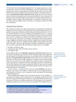
ZNS (1,2-benzisoxazole-3-methanesulfonamide) is a syn-
thetic amine sulfonamide compound (Figure 56-1(A)) (2).
ZNS is the only compound in this chemical class among
AEDs. Several features of the chemical structure of ZNS
deserve comment. All sulfonamide antibiotic compounds
include an arylamine domain at the N4-position, which
contributes to allergic reactions in susceptible individuals
(Figure 56-1(B)) (6). However, ZNS is a nonarylamine
sulfonamide and therefore lacks the chemical domain
with the greatest potential to produce hypersensitivity
reactions. ZNS shares structural similarity with acet-
azolamide (Figure 56-1(C)) and likewise shares an abil-
ity to inhibit the function of CA. Although the role of
CA inhibition has been questioned as a mechanism of
anticonvulsant action, there appears to be little debate
that it contributes significantly to the side effect profile
of ZNS. Lastly, ZNS bears structural similarity to other
Z
56
V • ANTIEPILEPTIC DRUGS AND KETOGENIC DIET
728
compounds of neurologic interest, including serotonin
(Figure 56-1(D)) and sumatriptan (Figure 56-1(E)).
Mechanisms of Action: Seizure Protection
Like many of the new AEDs, a number of different mech-
anisms of action have been proposed for ZNS. The mech-
anism or mechanisms that are of greatest importance in
inhibiting seizure activity in humans remain unknown.
ZNS inhibits repetitive neuronal firing in spinal
cord neurons that are depolarized during microelectrode
recordings (7). This effect occurred at concentrations
(3 g/mL) that are less than the blood levels typically
achieved in human subjects taking ZNS. The mechanism
of this effect may be partial blockade of activity-dependent
sodium channels, which has been shown in the giant axon
of Myxicola infundibulum (8). (It is of interest that this
critical observation was made in an invertebrate fanworm
Zonisamide
N
CH
2
NH
2
O
O
S
O
A
Sulfamethoxazole
N
NH
CH
3
H
2
N
O
O
S
O
B
FIGURE 56-1
Chemical structures of (A) the AED zonisamide; (B) the antimicrobial sulfamethoxazole; (C) the CA inhibitor acetazolamide;
(D) the neurotransmitter serotonin; (E) the migraine medication sumatriptan.
Sumatriptan
N
H
CH
2
NH
CH
3
CH
2
CH
2
N
H
3
C
H
3
C
O
O
S
E
Acetazolamide
H
N
NH
2
N
S
N
O
S
O
O
Serotonin
CH
2
NH
2
N
H
HO
CH
2
C
D
56 • ZONISAMIDE
729
inhabiting the intertidal zone; it does not appear to have
been reported in any other experimental system.)
ZNS also reduces voltage-dependent calcium currents
by blocking the T-type calcium channel in a concentration-
dependent fashion (9, 10). A methylated analog of ZNS,
shown to be ineffective in blocking MES seizures, was
likewise ineffective in blocking calcium currents (9). The
relevance of T-type calcium channels as potential thera-
peutic targets of ZNS is suggested by studies in an animal
model that examined the consequences of a single episode
of status epilepticus, in which hippocampal CA1 pyramidal
cells are converted into an abnormal burst-firing mode by
up-regulation of T-type calcium channels (11). In addition,
the study of dentate granule cells derived from temporal
lobe tissue from patients undergoing surgery for refrac-
tory epilepsy has shown the presence of calcium currents
mediated by T-type calcium channels (12).
In-vitro and animal studies also suggest that ZNS
may modulate synaptic transmission as well. ZNS inhibited
potassium-mediated glutamate release in the hippocampus
in a microdialysis model in rats (13). Also in microdialysis
experiments, ZNS functions to increase extracellular levels
of dopamine and serotonin (possibly by enhancing synaptic
release) in rat hippocampus and striatum (14–18).
ZNS may affect synaptic transmission by altering
gene expression of neurotransmitter transporter proteins.
In a rat model of hippocampal seizures elicited by injection
of FeCl
3
into the amygdala, ZNS caused (1) up-regulation
of excitatory amino-acid carrier-1 (EAAC-1), which has
the potential effect of enhancing the removal of excitatory
amino acids such as glutamate from the synaptic cleft,
and (2) down-regulation of the expression of gamma-
aminobutyric acid (GABA) transporter-1 (GAT-1), which
could enhance inhibitory neurotransmission by increasing
synaptic levels of GABA (19). This change in EAAC-1
and GAT-1 expression was present in both epileptic and
control animals.
Although chemically related to acetazolamide, it appears
that the mechanism for treating seizures may not be by CA
inhibition, because drug concentrations for ZNS needed to
be 100- to 1,000-fold higher than that of acetazolamide to
reach an equivalent inhibitory effect on CA (20, 21). On the
other hand, the profile of side effects of ZNS includes signs and
symptoms attributable to CA inhibition.
ZNS has also been studied in seizure and epilepsy
models in intact animals (22). ZNS can prevent the tonic
extensor components of MES in several different species (3).
ZNS inhibited the focal cortical discharge provoked by
acute electrical stimulation of the visual cortex in cats (23),
as well as seizures provoked by electrical stimulation in cats
that have undergone kindling of the visual cortex (24). ZNS
also prevents spread from the ictal focus in cats that have
undergone focal freezing lesions of the cortex (23).
In addition to antiseizure effects, ZNS may have
effectiveness as an antiepileptogenic compound as well
(see also the discussion in the following section on pos-
sible neuroprotective effects). ZNS suppressed the kin-
dling process, as well as inhibiting seizures resulting
from kindling, in adult rats (25). ZNS also inhibited the
development of elevated levels of chemical markers for
oxidative damage in iron-induced focal epileptogenic foci
in rat brain (26). This finding requires further study, but
it raises the possibility that ZNS may help prevent the
emergence of seizure foci following traumatic brain injury
or intracranial hemorrhage.
Mechanisms of Action: Neuroprotection
ZNS has also been a subject of investigation for pos-
sible neuroprotective effects. In an in vitro model, ZNS
demonstrates dose-dependent reductions in hydroxyl
and nitric oxide free radicals (27). The same group
has also shown that ZNS has reduced nitric oxide syn-
thase activity in the hippocampus of rats exposed to
N-methyl-
D-aspartate (NMDA) (28). Recent studies
have shown that pretreatment with ZNS reduces the
half-life of free radicals in the hippocampus of rats.
This finding was present in normal animals and in
those undergoing an acute episode of status epilepticus
induced by kainic acid (29, 30). These results suggest
that ZNS has the capacity to protect the brain from
free-radical-mediated injury.
ZNS reduces hypoxic-ischemic brain injury in
neonatal rats (31). In this model, the carotid artery was
ligated on one side, and the animal was then exposed
to prolonged hypoxia (8% O
2
for 2.5 hours). ZNS was
administered by intraperitoneal injection (75 mg/kg)
prior to hypoxemia. Cortical infarction size, as a per-
centage of volume, was 6% in the ZNS-treated animals
compared to 68% in control animals. Neuronal loss in
the hippocampus was also reduced in ZNS-treated ani-
mals. Seizure recordings showed no significant difference
between the two groups, suggesting that the neuropro-
tective effect was independent of any impact of the drug
on seizure activity.
ZNS reduced cerebral infarction in a transient mid-
dle cerebral artery occlusion model in adult rats (32).
Pretreatment with ZNS has shown neuroprotective effects
following transient cerebral ischemia in the adult gerbil,
as determined by memory performance with water maze
and subsequent histologic study. These findings correlated
with reduced extracellular glutamate in the hippocampus
of the ZNS-treated animals (33).
BIOTRANSFORMATION,
PHARMACOKINETICS, AND INTERACTIONS
ZNS has several properties that are desirable for an
AED. These include a long half-life (making once daily
V • ANTIEPILEPTIC DRUGS AND KETOGENIC DIET
730
dosing a feasible option), limited plasma-protein bind-
ing (minimizing displacement of other AEDs because of
competition for protein binding sites), and an absence of
autoinduction of the hepatic enzymes responsible for its
metabolism (34).
Absorption
Absorption of ZNS following oral administration appears
to be highly efficient, probably approaching 100%, based
on recovery of radiolabeled drug in urine (35, 36). Absorp-
tion is rapid, with peak blood levels occurring 2–6 hours
following dosage administration (37). When taken with
food, peak levels are delayed slightly (4–6 hours), but the
bioavailability is no different (37).
Distribution
The volume of distribution following oral administration
is 1.0 to 1.9 L/kg in healthy adult volunteers. ZNS is
only approximately 50% noncovalently bound to plasma
proteins, which limits competition for binding sites with
highly protein-bound medications such as phenytoin and
valproic acid (37). Accordingly, drug-drug interaction
resulting from alterations of free drug levels because of
competition for binding sites is minimized.
On the other hand, ZNS has a particularly high
binding affinity to the intracellular compartment of
erythrocytes. The tendency of ZNS to sequester in eryth-
rocytes has potential practical clinical importance, with
a risk of false elevations of reported ZNS serum levels in
hemolyzed specimens (38).
Distribution in the brain has been studied in rats with
autoradiography following injection of
14
C-ZNS), showing
preferential uptake in cerebral cortex relative to subcortical
structures, including striatum, thalamus, hypothalamus,
and cerebellum (39). However, further specificity with
respect to binding sites is not yet available.
Metabolism and Drug-Drug Interactions
Among the drugs used to treat epilepsy, ZNS has a relatively
long elimination half-life, with a mean of approximately
60 hours in healthy adult volunteers (range 52 to 69 hours)
(40, 41). This favorable serum half-life enables twice-daily
or even once-daily dosing schedules, but it also implies a pro-
longed time to steady state, as much as 10–14 days (22).
Zonisamide is partially metabolized by either
acetylation, to produce N-acetyl ZNS, or by reduction
to 2-sulfamoylacetylphenol (SMAP), then followed by
glucuronidation and urinary excretion (40, 42–45) (see
Figure 56-2). Study of the disposition of radio-labeled
ZNS in human adult volunteers has shown that 62%
of ZNS was recovered in the urine, of which 35% was
unmetabolized ZNS, 15% N-acetyl ZNS, and 50% as
the glucuronide derivative of SMAP (46).
N
CH
2
NH
2
O
S
O
O
CH
2
NH
2
O
O
S
OH
C
O
N
CH
2
N
H
O
O
S
O
CH
3
C
O
Zonisamide 2-sulfamoylacetylphenol (SMAP)
N-acetylzonisamide
Glucuronidation
Percent of Total ZNS Metabolites Recovered in Urine
15% 35% 50%
FIGURE 56-2
Metabolic pathways for zonisamide.
56 • ZONISAMIDE
731
ZNS is metabolized by the hepatic cytochrome P450
system, specifically by the 3A subfamily, and predominately
by the 3A4 isoenzyme (45, 47). However, ZNS does not
induce its own metabolism, nor does it induce or inhibit
other elements of the cytochrome P450 system (46). Because
of the relatively low plasma-protein binding of ZNS, as well
as its lack of hepatic enzyme induction, ZNS has little or no
effect on the blood levels of other AEDs (48).
Other AEDs, however, can affect ZNS blood levels
by virtue of their cytochrome P450-inducing or -inhibiting
features. The elimination half-life of ZNS is decreased to
27 hours in adult patients fully induced by coadministra-
tion of phenytoin, to 38 hours in patients taking carbam-
azepine or phenobarbital, and to 46 hours in patients taking
sodium valproate (46, 48–50). Non-AED compounds such
as cimetidine can inhibit hepatic metabolism of ZNS (51).
Other non-AED compounds, such as erythromycin, mid-
azolam, and nifedipine, are also preferentially metabolized
by the 3A4 isoenzyme and can therefore compete with
ZNS for metabolism, resulting in increased ZNS blood lev-
els (52). Certain dietary items, such as grapefruit juice, may
have this same effect. Dexamethasone can act to induce
the P450 reduction pathway for ZNS (47).
Detailed pharmacokinetic studies of ZNS within the
pediatric age range have not been performed. However,
as the metabolizing capacity of young children generally
exceeds that of adults on a per-kilogram basis, higher
doses may be required to reach the same target drug
level (53). ZNS is likely to have a shorter serum half-life
in children compared to adults.
Serum Levels
The significance of ZNS serum levels with respect to
efficacy appear limited (37, 54). However, levels greater
than 40 g/mL were more likely to be associated with
dose-related side effects, specifically drowsiness (54). This
study also showed relatively small changes between blood
levels at the peak (4 hours after once-daily morning dose)
and the trough (prior to once-daily morning dose). The
ratio of the peak to the trough levels showed a mean of
1.28 Ϯ 0.15 in this sample of 72 children on initial ZNS
monotherapy, which is consistent with the relatively long
half-life of this AED.
A therapeutic ZNS level of 15 to 40 g/mL has been
suggested (50, 55).
CLINICAL EFFICACY
Pivotal Adult Trials
Not unexpectedly, Class I studies demonstrating the
efficacy of ZNS for treating epilepsy enrolled predomi-
nantly adult subjects with partial seizures. In 2005 Brodie
reported the results of a multicenter, randomized, double-
blind, placebo-controlled trial in patients with refractory
partial seizures (56). The group consisted of patients aged
12 to 77 years, who were taking one to three preexisting
AEDs at the time of enrollment. Subjects were random-
ized to placebo or ZNS at one of three doses (100 mg/day,
300 mg/day, or 500 mg/day).
Efficacy varied directly with ZNS dose. The median
reduction in seizure frequency for complex partial sei-
zures was 51.2% for the study group taking 500 mg/day
and 16.3% for the placebo group (P Ͻ 0.0001). The
responder rate (the percentage of patients having at least
a 50% improvement in seizure frequency) for complex
partial seizures was 52.3% for ZNS at 500 mg/day and
21.3% for the placebo group (P Ͻ 0.001). The most fre-
quent treatment-emergent adverse events during titration to
500 mg/day were somnolence (14.4%), headache (6.8%),
dizziness (11.9%), and nausea (7.6%). The frequency of
adverse events was generally lower at lower ZNS doses (and
with placebo) and during the fixed-dose phase of the trial.
Additional pivotal studies with ZNS utilizing a ran-
domized, placebo-controlled, add-on design have been
performed in adult patients with refractory, partial-onset
epilepsy (57–59). Representative results of these four
studies are shown in Table 56-1.
Continued observation of these refractory partial-
epilepsy patients with open-label extensions of the pivotal
trials has provided further evidence that ZNS is effective
and well tolerated with long-term follow-up (5, 60, 61).
Early Pediatric Trials in Japan
The initial observations concerning the use of ZNS in
children with epilepsy arises from open-label clinical
trials and practice experience in Japan, subsequently
published in either Japanese or English, and reviewed
by Glauser and Pellock (53). In this detailed meta-analysis
examining efficacy and safety of ZNS in children in the
Japanese publications, the available studies were broken
down into those employing ZNS monotherapy (either
newly diagnosed or previously treated cases) (62–67),
adjunctive therapy with ZNS for treatment-resistant cases
(68–72), or studies with a mixed group of patients receiv-
ing either ZNS monotherapy or ZNS adjunctive therapy
(73–75). Results were then further analyzed by seizure
type (partial-onset or generalized-onset seizures), and the
percentage of responders (those with an improvement of
at least 50% in seizure frequency) was reported. These
results are shown in Table 56-2.
A component of one study included a group of
32 children that was randomized to monotherapy treat-
ment with either ZNS (n ϭ 16) or valproate (n ϭ 16)
(53, 73). These patients had generalized seizures and had
previously failed seizure control with one to three AEDs.
No significant difference in efficacy was observed. The
V • ANTIEPILEPTIC DRUGS AND KETOGENIC DIET
732
responder rate was 50% for ZNS treatment (at a mean
dose of 7.3 mg/kg/day) and 44% for valproate (at a mean
dose of 27.6 mg/kg/day).
Recent Studies on ZNS Efficacy in Children
Initial Monotherapy. In an uncontrolled trial of initial
monotherapy in 72 children (mean age, 8.3 years; range,
3 months to 15 years) with newly diagnosed cryptogenic
partial epilepsy, Miura treated patients with ZNS at an
initial dose of 2 mg/kg/day, escalating at weekly inter-
vals to a maintenance dose of 8 mg/kg/day (54). Dosing
was provided once daily. At this initial target dose, 49 of
72 cases (68%) were completely controlled. After further
dosage adjustment, 57 of 72 cases (79%) were completely
controlled. The mean duration of ZNS treatment was
27 months (range, 6 to 43 months). Although a clear
relationship between ZNS blood levels and clinical effec-
tiveness was not seen, patients who were symptomatic
with dose-related side effects (predominantly drowsiness)
had blood levels greater than 40 g/mL.
Seki and colleagues studied 77 children (ages 8 months
to 15 years) with various seizure types, 68 of whom were
included for analysis of ZNS efficacy (76). Fifty had
TABLE 56-2
Anti-Epilepsy Treatment with Zonisamide in Children
INCLUSION CRITERIA FOR
PARTIAL-ONSET GENERALIZED-ONSET
SEIZURES SEIZURES
OPEN-LABEL STUDY RR N RR N
Patients on ZNS 78% 209 71% 49
Monotherapy Only
Patients on ZNS as 34% 137 15% 54
Adjunctive Therapy Only
Patients on Either Monotherapy 60% 718 42% 291
or Adjunctive Therapy
Summary results of open-label trials in Japan as reviewed by Glauser and Pellock 2002 (53),
noting the responder rate for children with either partial-onset or generalized-onset seizures accord-
ing to the inclusion criteria for the study (ZNS monotherapy only, ZNS adjunctive therapy only, or
combined ZNS monotherapy and adjunctive therapy).
n ϭ Number of patients in each summary category. RR ϭ Responder rate.
TABLE 56-1
Seizure Efficacy for ZNS in Randomized, Placebo-Controlled, Add-On Trials in Adults with Refractory
Partial Epilepsy
MEDIAN REDUCTION MEDIAN REDUCTION
IN TOTAL SEIZURE IN TOTAL SEIZURE
STUDY FREQUENCY FREQUENCY
(FIRST NUMBER OF AGE RANGE MAXIMUM (% CHANGE FROM (% CHANGE FROM
AUTHOR) SUBJECTS (YEARS) ZNS DOSE BASELINE) BASELINE) P VALUE
ZNS GROUP PLACEBO GROUP
Schmidt 139 18–59 20 mg/kg/day 22.5% Ϫ3.0% Ͻ0.05
1993 (57)
Faught 203 13–68 400 mg/day 40.5% 9.0% ϭ 0.009
2001 (58)
Sackellares 152 17–67 600 mg/day 25.5% Ϫ6.6% ϭ 0.0005
2004 (59)
Brodie 351 12–77 500 mg/day 51.3% 18.1% Ͻ0.0001
2005 (60)
56 • ZONISAMIDE
733
previously not taken any AEDs. All patients were on ZNS
monotherapy. Doses were initiated at 2 mg/kg/day (divided
twice daily), and increased by 1–2 mg/kg/day at 1–2 week
intervals, up to a dose of 12 mg/kg/day. Forty-eight patients
(of 68 patients, 62%) had localization-related epilepsy,
and 20 (38%) had generalized epilepsies. Of the patients
with localization-related epilepsy, 40 of 48 (83%) showed
an “excellent” response (at least 3 months seizure-free);
for patients with generalized epilepsy, 18 of 20 (90%)
were seizure-free for at least 3 months.
Follow-up Monotherapy. Wilfong has described an
uncontrolled case series of 131 children and adoles-
cents treated with open-label ZNS monotherapy (77).
Patients with both partial and generalized seizure types
were included, although limited detail regarding seizure
type and epilepsy syndrome was provided. Eighty-nine
patients (68%) had previously been treated with at least
one AED. When all patients are grouped together, 30%
were completely seizure free, and an additional 47% had
an improvement of at least 50% in seizure frequency.
Forty-three patients (33%) reported at least one adverse
effect while on ZNS, but only 3 (2.3%) had to discon-
tinue ZNS therapy (sleeplessness and increased seizures,
failure to gain weight, and behavioral changes in one
patient each).
Add-on Polytherapy. Kim and colleagues have
reported an uncontrolled, retrospective study of ZNS
use in 68 children and adolescents treated for epilepsy
(median age, 6.9; range, 1.9 to 18.1 years) (78). ZNS was
used initially as monotherapy in 11.8% and as adjunc-
tive polytherapy in 88.2%. The median ZNS dose was
8.0 mg/kg/day (range, 1.5 to 23.2 mg/kg/day). Of the
68 patients, 69.1% had exclusively generalized seizures,
10.3% exclusively partial seizures, 14.7% both general-
ized and partial, and 5.9% had seizures of undetermined
type. Data to determine efficacy were available in
62 patients: 25.8% were completely seizure free, 21.0%
had a reduction of at least 50% in overall seizure fre-
quency, 16.1% were improved in seizure frequency by
less than 50%, 22.6% showed no change, and 14.5%
were reported as having increased seizure frequency.
There were no clear trends with regard to efficacy and
seizure type or etiology. Adverse events were reported by
61.8% of patients, predominantly during dose escala-
tion. These were generally central nervous system (CNS)
related, with behavioral or psychiatric symptoms (such as
aggression, agitation, and decreased attention) reported
in 23.5%, cognitive dysfunction in 12.0%, and sedation
in 10.3%.
A retrospective study by Santos and Brotherton
included 50 patients (range, 9 months to 20 years; mean
age, 9.1 years) (79). With one exception, all had pre-
viously failed at least one other AED, and 47 subjects
(94%) were taking at least one other AED with ZNS. The
study group experienced a wide diversity of seizure types.
For the entire population, 16% became seizure free, and
an additional 22% had an improvement of at least 50%
in seizure frequency. Efficacy was not broken down by
seizure type. Adverse events were experienced by 62%
of patients, including loss of appetite in 14 (28% of total
study population), weight loss in 5 (10%), and kidney
stones in 2 (4%). Fourteen patients (28% of total study
population) discontinued ZNS because of adverse effects.
Mean dosage for ZNS was 15.9 mg/kg/day (range, 3.3
to 35 mg/kg/day).
In another retrospective chart review of ZNS add-on
therapy in 35 children with refractory epilepsy, Mandel-
baum and colleagues determined that 11% of the study
population was completely seizure-free, and 31% had an
improvement in seizure frequency of at least 50% (80).
When efficacy was analyzed by seizure type (generalized,
partial, or mixed), there were no clear trends. The authors
conclude that ZNS is a broad-spectrum AED.
Efficacy in Specific Seizure Types or
Epilepsy Syndromes in Children
Partial Seizures. Miura has reported an uncontrolled
series of 72 children (mean age, 8.3 years; range, 0.3–
15 years) treated with initial ZNS monotherapy for
cryptogenic localization-related epilepsy (54). The initial
maintenance dose of ZNS was increased incrementally
to 8 mg/kg/day and could be increased thereafter for
patients not responding with complete seizure control.
At 8 mg/kg/day of ZNS, 49 patients (68.1%) were com-
pletely controlled. Of the remaining patients, an additional
8 achieved complete seizure control with an increased dose
of ZNS (79.2% of study group completely controlled on
ZNS monotherapy). CNS-related side effects (drowsiness)
tended to occur with blood levels greater than 40 g/mL.
A therapeutic range of 15–40 g/mL was suggested.
Absence. T-type calcium channels have been implicated
as an important mechanistic component of absence sei-
zures (81). The ability of ZNS to functionally block these
channels makes ZNS a potentially attractive AED for
treating this seizure type (9, 10). Wilfong and Schultz have
retrospectively reviewed the charts of 45 patients under
18 years of age treated with ZNS for absence seizures (82).
Study subjects included both typical and atypical absence
seizures and were not further analyzed according to epi-
lepsy syndrome. However, 88.9% of patients had failed
prior AED therapy. The mean ZNS dose was 9.0 mg/kg/day
(range, 2–24 mg/kg/day). Twenty-three of 45 patients
(51.1%) were 100% seizure-free for absence seizures, and
14 (31.1%) had a reduction in absence seizures of at least
50%. The efficacy with regard to treating other seizure
types in this series of patients was not provided.
V • ANTIEPILEPTIC DRUGS AND KETOGENIC DIET
734
Juvenile Myoclonic Epilepsy(JME). Kothare examined
the use of ZNS for seizure control in 15 patients with
JME, utilizing a retrospective chart review method (83).
There was no control group. ZNS was used as mono-
therapy in 13 patients (87%) and as add-on therapy in 2
patients. Dosing for ZNS ranged from 200 to 500 mg/day
(2.0 to 8.5 mg/kg/day). For patients taking ZNS mono-
therapy, 80% had a decrease of at least 50% in total
seizure frequency. When broken down by the individual
seizure types commonly seen with JME, 69% of patients
were seizure free for generalized tonic-clonic seizures,
62% were seizure-free for myoclonic seizures, and 38%
were seizure free for absence seizures. One patient on
monotherapy discontinued ZNS because of lack of efficacy,
and 3 patients reported adverse events (weight loss, head-
ache, dizziness), which resolved with continuing therapy.
West Syndrome or Infantile Spasms (IS). Several uncon-
trolled, open-label studies have reported the use of ZNS
in patients with IS (West syndrome) (77, 80, 84–90).
Suzuki published a prospective, multicenter, open-
label, uncontrolled trial with treatment of 13 infants
with newly diagnosed IS (84). By protocol, all patients
were initially treated with high-dose pyridoxine. Two
responded, and the remaining 11 patients were then
treated with ZNS, with a dose escalation program to
reach seizure control or a maximum of 10 mg/kg/day.
Eight of 11 patients (73%) had symptomatic IS. A
positive response was defined as complete cessation of
seizures and disappearance of hypsarrhythmia on elec-
troencephalogram (EEG).
Four of 11 patients (36%) were initial responders.
Interestingly, all had resolution of spasms within days of
taking the initial dose at 3–5 mg/kg/day. However, 2 of
these patients later relapsed. There were no adverse events
reported during ZNS treatment. Of the 7 nonresponders,
5 of 7 later responded to an intramuscular adrenocorti-
cotropic hormone (ACTH) treatment protocol.
Yanagaki treated 23 infants with West syndrome,
ages 4–11 months, as either initial or adjunctive ther-
apy, with a dose-ranging protocol to study an increased
titration rate. Initial doses varied from 3 mg/kg/day to
10 mg/kg/day, to achieve a final target dose of 9–11
mg/kg/day for the entire group (90). A positive response
(defined as a complete cessation of spasms and disap-
pearance of hypsarrhythmia for at least 3 months) was
observed in 7 patients (30%). Of these, 4 were crypto-
genic and 3 were symptomatic cases. A mild degree of
hyperthermia (temperature above 37.5°C), but without
obvious signs of infection, was observed in 3 of the
10 patients in the highest initial dose treatment group,
but the medication was continued in all, with simple
environmental cooling.
Based on this collective open-label experience,
it has been noted that the likelihood of success with
ZNS for IS is higher in patients with cryptogenic IS in
comparison to those with symptomatic IS. Notably, those
who responded often did so quickly and at relatively low
doses of 4–8 mg/kg/day (87).
Lennox-Gastaut Syndrome (LGS). In comparison to
IS, there is relatively little published information directly
addressing the use of ZNS for treating patients with LGS.
Some of the retrospective, tertiary clinic-based surveys
previously noted contain large numbers of patients with
cryptogenic or symptomatic generalized epilepsy, but LGS
is not broken out as an identified subgroup (78, 80).
Yamauchi has recently described a large cadre of
patients followed prospectively as part of an uncontrolled,
multicenter, postmarketing surveillance study consisting
of 1,631 patients, including enrollment of 774 children
(ages 15 years or less) (91). Within the entire study popu-
lation, 79 patients are described as having LGS. Details
regarding ZNS dosing, including whether ZNS was used
as monotherapy or polytherapy, are not broken out for
the LGS subgroup. However, in response to ZNS treat-
ment, 27.9% of LGS patients experienced an improve-
ment in total seizure frequency of at least 50%, whereas
51.9% are described as unchanged.
Myoclonic Seizures and Progressive Myoclonus
Epilepsy. Yamauchi (see preceding discussion) also
describes open-label efficacy of ZNS broken down
by seizure type in a large postmarketing surveillance
study (91). Fifty-six subjects experienced myoclonic sei-
zures. Of these, 19.6% are described as completely free
of myoclonic seizures, and an additional 32.2% showed
an improvement of at least 50% in myoclonic seizure
frequency. Details regarding ZNS dosing are not provided
for this subgroup.
Generally favorable results have been described in
several small, uncontrolled, open-label reports of ZNS
treatment of the refractory seizures associated with
progressive myoclonus epilepsies, including Unverricht-
Lundborg disease (92, 93) and Lafora disease (93, 94).
Importantly, ZNS is not one of the AEDs that may
exacerbate myoclonic seizures, which is a list that includes
lamotrigine, carbamazepine, phenytoin, gabapentin, pre-
gabalin, and vigabatrin (95).
ADVERSE EFFECTS
Dose-Related Effects
Premarketing Phase II and Phase III trials in Japan in
1,008 patients (of whom, 403 patients [40%], were
ages 15 years or less) demonstrated that the most fre-
quent treatment-emergent adverse events were drowsi-
ness (24%), ataxia (13%), decreased appetite (11%),
56 • ZONISAMIDE
735
gastrointestinal symptoms (7%), decrease in spontaneity
(6%), and slowing of mental activity (5%) (96).
Ohtahara has described the side effect profile of
ZNS in 928 children participating in an open-label post-
marketing surveillance study (97). The children had a sta-
tistically significant lower likelihood of all adverse events
(24.3% of pediatric population) in comparison to adult
patients in the study (40.1%; P Ͻ 0.001).
Not surprisingly, the likelihood of adverse events
increased with polypharmacy. In one large multicenter
trial of ZNS in Japanese children (72), the incidence of
adverse events of any kind was 14% in those taking ZNS
monotherapy, 37% in those taking one other AED in
addition to ZNS, and 53% in those taking two additional
AEDs [reviewed in (53)]. The likelihood of adverse effects
appeared to correlate with serum level of ZNS; mean
levels of ZNS for symptomatic patients were generally
greater than 20 g/mL. Target doses of 8 mg/kg/day and
lower were well tolerated.
Idiosyncratic Effects
Hyperthermia and Oligohydrosis. Decreased sweating
and increased core temperature, a potential consequence of
CA inhibition, is a recognized adverse event with ZNS ther-
apy. This side effect appears to be more common in children
and more likely to occur in hot weather (98). Postmarket-
ing assessment of the likelihood of oligohidrosis, hyperther-
mia, or both in the United States suggested an incidence
of 1 case per 4,590 patient-years (0.02% per patient-year)
(98). Surveillance of the Japanese market for 11 years after
ZNS approval suggested an incidence of 1 case per 10,000
pediatric-years (0.01% per pediatric-year) (98). Knudsen
analyzed the Adverse Events Reporting System of the FDA
for cases with oligohidrosis, fever, or both (99). Six cases
were identified, all in children or adolescents. The report-
ing rate was calculated to be 1 case per 769 pediatric-years
(0.13% per pediatric-year). Ascertainment of the cases in
these reports depends on physician report and is biased
in favor of medically serious episodes. Conversely, one
Japanese report identified decreased sweating in 12 of 70
patients (17%) taking ZNS, based on historical reports by
the patients’ parents (100). In summary, the preponderance
of data suggests that the incidence of serious episodes of
oligohidrosis and hyperthermia is low, but milder instances
are encountered during routine clinical practice.
Renal Calculi. Renal stones occurred in 1.9% of 700
adult patients treated with ZNS in U.S. and European
studies (101). A more recent study, also in adult patients,
identified stones in 4% of 750 patients (1.2% symptom-
atic and 2.8% asymptomatic) (102). The incidence of
renal calculi in children taking ZNS is unknown. An
increased risk of renal stones is most likely attributable
to the effect of ZNS as a CA inhibitor, resulting in alka-
linization of the urine and an increased risk of calcium
phosphate stones. An increased risk for kidney stones is
likely in those with a prior history of stones or in those
with a family history of renal calculi.
Rash. Although ZNS is a sulfonamide, it does not
include the arylamine group that increases risk for hyper-
sensitivity skin reactions (6). Accordingly, the risk of aller-
gic drug rash appears to be substantially lower with the
use of ZNS in comparison to the sulfonamide antibiotics,
for which hypersensitivity can occur in as much as 6%
of the population (6). Postmarketing surveillance reports
show an incidence of serious rash associated with ZNS
treatment of approximately 0.25% (5). Even so, caution
and careful observation with use of ZNS in a patient with
a recognized history of hypersensitivity to sulfonamide
drugs are warranted.
Cognitive and Behavioral Side Effects. There is a lack
of research into the potential impact of ZNS on cogni-
tion, particularly in children. A pilot study in nine adult
patients with refractory partial-onset epilepsy, undergo-
ing psychometric testing prior to and 12 weeks follow-
ing the addition of ZNS, showed impairments in verbal
learning without overt signs of overmedication (103).
The degree of impairment correlated directly with ZNS
plasma levels, particularly with blood levels greater than
30 g/mL. However, retesting at 24 weeks failed to show
significant changes in cognition function relative to base-
line, suggesting a drug tolerance effect.
Adverse effects of ZNS on behavior and psychiatric
functioning have been reported. In a retrospective report
based on open-label use of ZNS for epilepsy in 68 pediat-
ric patients, Kim and colleagues determined behavioral or
psychiatric side effects in 23.5% of patients (78). Difficul-
ties described included aggression, agitation, irritability,
poor attention, hallucinations, hyperactivity, dysphoria,
paranoia, and psychosis. All of these behavioral side
effects resolved with lowering the dose or discontinu-
ing ZNS. Only 5 patients in the entire study population
(7.4%) discontinued because of side effects, suggesting
that behavioral problems were handled successfully by a
reduction in dose in most instances.
A postmarketing surveillance study in Japan of
1,512 patients (including 928 children ages more than
16 years) revealed irritable or excitable behavioral epi-
sodes in 45 patients (3.0%), depression in 8 (0.5%), and
anxiety or hypochondria in 14 (0.9%) (97).
Teratogenicity. There is insufficient data regarding ZNS
and any associated risk of fetal malformations (104).
A small prospective registry study of pregnant epileptic
women taking ZNS examined 26 offspring (105). The
4 children born to mothers taking ZNS monotherapy
V • ANTIEPILEPTIC DRUGS AND KETOGENIC DIET
736
were normal. The remaining 22 offspring were exposed
to AED polytherapy, including ZNS: 2 had significant
malformations, 1 with anencephaly, and 1 with an atrial
septal defect. Both of these mothers had low ZNS serum
concentrations (6.1 g/mL and 6.3 g/mL, respectively)
during the first trimester.
CLINICAL USE
A recent survey of recommendations for AED manage-
ment by selected pediatric epilepsy specialists in the
United States shows that ZNS is favorably regarded as a
first-line option for initial monotherapy in children with
myoclonic and generalized tonic-clonic seizures and as a
second-line option for initial treatment of patients with
IS, LGS, symptomatic generalized tonic-clonic seizures,
and for children with partial seizures who have failed
prior AED treatment (106).
Formulations for ZNS currently available in the
United States include 25-mg, 50-mg, and 100-mg cap-
sules. Liquid suspensions can be specially formulated, but
there are no data regarding stability and shelf-life.
Initial dosing is usually at 1–2 mg/kg/day, and the
conventional recommendation is to increase the dose by
1–2 mg/kg/day every 2 weeks. ZNS is correctly regarded
as an AED for which it is suggested to “start low and go
slow.” Clinical experience has shown that tolerance is
improved and the likelihood of dose-related side effects
is lower with incremental dosing. The maximum dose
is determined by patient tolerance and seizure control;
however, a total daily dose of 10 mg/kg/day is a reason-
able initial target, and doses up to 20 mg/kg/day are com-
monly used. In light of the long serum half-life, ZNS can
be administered once or twice daily.
Anticipatory guidance should include a recommen-
dation about the need for the child to stay well hydrated,
in an effort to reduce the likelihood of renal calculi, and
the need to be aware of the importance of environmen-
tal heat as a possible risk factor for oligohidrosis and
hyperthermia.
SUMMARY
ZNS is properly regarded as a broad-spectrum AED.
Although not often chosen as the drug of first choice for
the newly diagnosed patient, ZNS is an important option
for treating children with epilepsy who have failed initial
therapy. It may be a particularly good choice for patients
with myoclonic seizures as a component of their epilepsy.
Although evidence from open-label trials and practice
experience is compelling, controlled studies examining
the efficacy of ZNS in children and its impact on cogni-
tion and behavior would be most welcome.
References
1. Seino M. Review of zonisamide development in Japan. Seizure 2004; 13 (Suppl 1):
S2–S4.
2. Uno H, Kurokowa M, Masuda Y, Nishimura H. Studies on 3-substituted 1,2-
benzisoxazole derivatives. VI. Synthesis and their anticonvulsant properties. J Med
Chem 1979; 22:180–183.
3. Masuda Y, Karasawa T, Shiraishi Y, Hori M, et al. 3-Sulfamoylmethyl-1,2-benzisoxazole,
a new type of anticonvulsant drug: pharmacological profile. Arzneimittel-Forschung
1980; 30:477–483.
4. Masuda Y, Ishizaki I, Shimizu M. Zonisamide: pharmacology and clinical efficacy in
epilepsy. CNS Drugs Rev 1998; 4:341–360.
5. Leppik IE. Practical prescribing and long-term efficacy and safety of zonisamide. Epilepsy
Res 2006; 68(Suppl 2); S17–S24.
6. Brackett CC, Singh H, Block JH. Likelihood and mechanisms of cross-allergenicity
between sulfonamide antibiotics and other drugs containing a sulfonamide functional
group. Pharmacotherapy 2004; 24:856–870.
7. Rock DM, Macdonald RL, Taylor CP. Blockade of sustained repetitive action potentials
in cultured spinal cord neurons by zonisamide (AD 810, CI 912), a novel anticonvulsant.
Epilepsy Res 1989; 3:138–143.
8. Schauf CL. Zonisamide enhances slow sodium inactivation in Myxicola. Brain Res 1987;
413:185–188.
9. Suzuki S, Kawakami K, Nishimura S, Watanabe Y, et al. Zonisamide blocks T-type calcium
channels in cultured neurons of rat cerebral cortex. Epilepsy Res 1992; 12:21–27.
10. Kito M, Maehara M, Watanabe K. Mechanisms of T-type calcium channel blockade by
zonisamide. Seizure 1996; 5:115–119.
11. Su H, Sochivko D, Becker A, Chen J, et al. Upregulation of a T-type Ca
2ϩ
channel causes
a long-lasting modification of neuronal firing mode after status epilepticus. J Neurosci
2002; 22:3645–3655.
12. Beck H, Steffens R, Heinemann U, Elger CE. Properties of voltage-activated Ca
2ϩ
currents in acutely isolated human hippocampal granule cells. J Neurophysiol 1997;
77:1526–1537.
13. Okada M, Kawata Y, Mizuno K, Wada K, et al. Interaction between Ca
2ϩ
, K
ϩ
, carba-
mazepine and zonisamide on hippocampal extracellular glutamate monitored with a
microdialysis electrode. Br J Pharmacol 1998; 124:1277–1285.
14. Okada M, Kaneko S, Hirano T, Ishida M, et al. Effects of zonisamide on extracellular
levels of monoamine and its metabolite, and on Ca
2ϩ
dependent dopamine release.
Epilepsy Res 1992; 13:113–119.
15. Okada M, Kaneko S, Hirano T, Mizuno K, et al. Effects of zonisamide on dopaminergic
system. Epilepsy Res 1995; 22:193–205.
16. Kawata Y, Okada M, Murakami T, Mizuno K, et al. Effects of zonisamide on K
ϩ
and
Ca
2ϩ
evoked release of monoamine as well as K
ϩ
evoked intracellular Ca
2ϩ
mobilization
in rat hippocampus. Epilepsy Res 1999; 35:173–182.
17. Okada M, Hirano T, Kawata Y, Murakami T, et al. Biphasic effects of zonisamide on
serotonergic system in rat hippocampus. Epilepsy Res 1999; 34:187–197.
18. Gluck MR, Santana LA, Granson H, Yahr MD. Novel dopamine releasing response of
an anti-convulsant agent with possible anti-Parkinson’s activity. J Neural Transm 2004;
111:713–724.
19. Ueda Y, Doi T, Tokumaru J, Willmore LJ. Effect of zonisamide on molecular regulation
of glutamate and GABA transporter proteins during epileptogenesis in rats with hip-
pocampal seizures. Brain Res Mol Brain Res 2003; 116:1–6.
20. Masuda Y, Karasawa T. Inhibitory effect of zonisamide on human carbonic anhydrase
in vitro. Arzneimittel-Forschung 1993; 43:416–418.
21. Masuda Y, Noguchi H, Karasawa T. Evidence against a significant implication of
carbonic anhydrase inhibitory activity of zonisamide in its anticonvulsant effect.
Arzneimittel-Forschung 1994; 44:267–269.
22. Leppik IE. Zonisamide: chemistry, mechanism of action, and pharmacokinetics. Seizure
2004; 13(Suppl 1):S5–S9.
23. Ito T, Hori M, Masuda Y, Yoshida K, et al. Sulfamoylmethyl-1,2-benzisoxazole, a new
type of anticonvulsant drug: electroencephalographic profile. Arzneimittel-Forschung
1980; 30:603–609.
24. Wada Y, Hasegawa H, Okuda H, Yamaguchi N. Anticonvulsant effects of zonisamide
and phenytoin on seizure activity of the feline visual cortex. Brain Dev 1990; 12:
206–210.
25. Hamada K, Song HK, Ishida S, Yagi K, et al. Contrasting effects of zonisamide and
acetazolamide on amygdaloid kindling in rats. Epilepsia 2001; 42:1379–1386.
26. Komatsu M, Hiramatsu M, Willmore LJ. Zonisamide reduces the increase in 8-hydroxy-
2’-deoxyguanosine levels formed during iron-induced epileptogenesis in the brains of
rats. Epilepsia 2000; 41:1091–1094.
27. Mori A, Noda Y, Packer L. The anticonvulsant zonisamide scavenges free radicals.
Epilepsy Res 1998; 30:153–158.
28. Noda Y, Mori A, Packer L. Zonisamide inhibits nitric oxide synthase activity induced by
N-methyl-D-aspartate and buthionine sulfoximine in the rat hippocampus. Res Commun
Mol Pathol Pharmacol 1999; 105:23–33.
56 • ZONISAMIDE
737
29. Tokumaru J, Ueda Y, Yokoyama H, Nakajima A, et al. In vivo evaluation of hippocampal
anti-oxidant ability of zonisamide in rats. Neurochem Res 2000; 25:1107–1111.
30. Ueda Y, Doi T, Tokumaru J, Nakajima A, et al. In vivo evaluation of the effect of
zonisamide on the hippocampal redox state during kainic acid-induced seizure status
in rats. Neurochem Res 2005; 30:1117–1121.
31. Hayakawa T, Higuchi H, Nigami H, Hattori H. Zonisamide reduces hypoxic-ischemic
brain damage in neonatal rats irrespective of its anticonvulsive effect. Eur J Pharmacol
1994; 257:131–136.
32. Minato H, Kikuta C, Fujitani B, Masuda Y. Protective effect of zonisamide, an anti-
epileptic drug, against transient focal cerebral ischemia with middle cerebral artery
occlusion-reperfusion in rats. Epilepsia 1997; 38:975–980.
33. Owen AJ, Ijaz S, Miyashita H, Wishart T, et al. Zonisamide as a neuroprotective agent
in an adult gerbil model of global forebrain ischemia: a histological, in vivo microdialysis
and behavioral study. Brain Res 1997; 770:115–122.
34. Biton V. Zonisamide: newer antiepileptic agent with multiple mechanisms of action.
Expert Rev Neurotherapeutics 2004; 4:935–943.
35. Perucca E, Bialer M. The clinical pharmacokinetics of the newer antiepileptic drugs.
Focus on topiramate, zonisamide and tiagabine. Clin Pharmacokinet 1996; 31:29–46.
36. Perucca E. Pharmacokinetic variability of new antiepileptic drugs at different ages. Ther
Drug Monit 2005; 27:714–717.
37. Mimaki T. Clinical pharmacology and therapeutic drug monitoring of zonisamide. Ther
Drug Monitor 1998; 29:593–597.
38. Willmore LJ. Commentary on Leppik. Seizure 2004; 13(Suppl 1):S10.
39. Mimaki T, Tanoue H, Matsunaga Y, Miyazaki H, et al. Regional distribution of 14C-
zonisamide in rat brain. Epilepsy Res 1994; 17:233–236.
40. Ito T, Yamaguchi T, Miyazaki H, Sekine Y, et al. Pharmacokinetic studies of AD-810, a new
antiepileptic compound: phase I trials. Arzneimittel-Forschung 1982; 32:1581–1586.
41. Kochak GM, Page JG, Buchanan RA, Peters R, et al. Steady-state pharmacokinetics of
zonisamide, an antiepileptic agent for treatment of refractory complex partial seizures.
J Clin Pharmacol 1998; 38:166–171.
42. Stiff DD, Zemaitis MA. Metabolism of the anticonvulsant agent zonisamide in the rat.
Drug Metab Dispos 1990; 18:888–894.
43. Stiff DD, Robicheau JT, Zemaitis MA. Reductive metabolism of the anticonvulsant
agent zonisamide, a 1,2-benzisoxazole derivative. Xenobiotica 1992; 22:1–11.
44. Nakasa H, Komiya M, Ohmori S, Rikihisa T, et al. Characterization of human liver
microsomal cytochrome P450 involved in the reductive metabolism of zonisamide. Mol
Pharmacol 1993; 44:216–221.
45. Nakasa H, Ohmori S, Kitada M. Formation of 2-sulphamoylacetylphenol from zonisamide
under aerobic conditions in rat liver microsomes. Xenobiotica 1996; 26:495–501.
46. Eisai Inc., Zonegran (zonisamide) FDA Approved Labeling Text, March 27, 2000.
47. Nakasa H, Komiya M, Ohmori S, Rikihisa T, et al. Rat liver microsomal cytochrome
P-450 responsible for reductive metabolism of zonisamide. Drug Metab Dispos 1993;
21:777–781.
48. Ojemann LM, Shastri RA, Wilensky AJ, Friel PN, et al. Comparative pharmacokinetics
of zonisamide (CI-912) in epileptic patients on carbamazepine or phenytoin mono-
therapy. Ther Drug Monit 1986; 8:293–296.
49. Wagner JG, Sackellares JC, Donofrio PD, Berent S, et al. Nonlinear pharmacokinetics
of CI-912 in adult epileptic patients. Ther Drug Monit 1984; 6:277–283.
50. Sackellares JC, Donofrio PD, Wagner JG, Abou-Khalil B, et al. Pilot study of zonisamide
(1,2-benzisoxazole-3-methanesulfonamide) in patients with refractory partial seizures.
Epilepsia 1985; 26:206–211.
51. Nakasa H, Komiya M, Ohmori S, Kitada M, et al. Formation of reductive metabolite,
2-sulfamoylacetylphenol, from zonisamide in rat liver microsomes. Res Commun Chem
Pathol Pharmacol 1992; 77:31–41.
52. Eisai Inc., data on file.
53. Glauser TA, Pellock JM. Zonisamide in pediatric epilepsy: review of the Japanese lit-
erature. J Child Neurol 2002; 17:87–96.
54. Miura H. Zonisamide monotherapy with once-daily dosing in children with cryptogenic
localization-related epilepsies: clinical effects and pharmacokinetic studies. Seizure
2004;
13(Suppl 1):S17–S23.
55. Wilensky AJ, Friel PN, Ojemann LM, Dodrill CB, et al. Zonisamide in epilepsy: a pilot
study.
Epilepsia 1985; 26:212–220.
56. Brodie MJ, Duncan R, Vespignani H, Solyom A, et al. Dose-dependent safety and efficacy
of zonisamide: a randomized, double-blind, placebo-controlled study in patients with
refractory partial seizures. Epilepsia 2005; 46:31–41.
57. Schmidt D, Jacob R, Loiseau P, Deisenhammer E, et al. Zonisamide for add-on treat-
ment of refractory partial epilepsy: a European double-blind trial. Epilepsy Res 1993;
15:67–73.
58. Faught E, Ayala R, Montouris GG, Leppik IE. Randomized controlled trial of zonisamide
for the treatment of refractory partial-onset seizures. Neurology 2001; 57:1774–1779.
59. Sackellares JC, Ramsay RE, Wilder BJ, Browne TR III, et al. Randomized, controlled
clinical trial of zonisamide as adjunctive treatment for refractory partial seizures.
Epilepsia 2004; 45:610–617.
60. Brodie MJ. Zonisamide clinical trials: European experience. Seizure 2004; 13(Suppl 1):
S66–S70.
61. Faught E. Review of United States and European clinical trials of zonisamide in the
treatment of refractory partial-onset seizures. Seizure 2004; 13(Suppl 1):S59–S65.
62. Shuto H, Sugimoto T, Yasuhara A, et al. Efficacy of zonisamide in children with refractory
partial seizures. Curr Therapeut Res 1989; 45:1031–1036. [As cited in reference (53)]
63. Hosoda N, Miura H, Takanashi S, et al. Clinical efficacy and blood concentration
with once-daily zonisamide monotherapy in pediatric patients with partial seizures [in
Japanese]. TDM Kenkyu 1993; X:240–241. [As cited in reference (53)]
64. Hosoda N, Miura H, Takanashi S, et al. Clinical effects and blood concentration of
zonisamide in epileptic children with partial seizures treated with a once-daily dose of
zonisamide monotherapy. Jpn J Ther Drug Monit 1994; O-18:68. [As cited in reference
(53)]
65. Hosoda N, Miura H, Takanashi S, et al. Once-daily dose of zonisamide monotherapy
in the control of partial seizures in children with cryptogenic localization-related epi-
lepsies: clinical effects and their pharmacokinetic basis. Jpn J Psychiatry Neurol 1994;
48:335–337. [As cited in reference (53)]
66. Kumagai N, Seki T, Yamawaki H, Suzuki N, et al. Monotherapy for childhood epilepsies
with zonisamide. Jpn J Psychiatry Neurol 1991; 45:357–359.
67. Hayakawa T, Nejihashi Y, Kishi T, et al. Serum zonisamide concentration in fresh cases
of childhood epilepsy following zonisamide monotherapy [in Japanese]. J Jpn Epilepsy
Soc 1994; 12:249–254. [As cited in reference (53)]
68. Fukushima K, Yagi K, Seino M, et al. Phase II clinical trial of a new antiepileptic drug,
zonisamide (ZNA) in pediatric epilepsy patients [in Japanese]. Jpn J Pediatr 1987;
40:3389–3397. [As cited in reference (53)]
69. Kanazawa K, Segoku A, Kawai I. A clinical trial of zonisamide, a new antiepileptic
drug, on adults and children with refractory epilepsy [in Japanese]. J Clin Therapeut
Med 1987; 3:1181–1186. [As cited in reference (53)]
70. Takahashi I, Yamamoto N, Furune S, et al. Efficacy of zonisamide for intractable epilepsy in
childhood [in Japanese]. J Jpn Epilepsy Soc 1987; 5:100–105. [As cited in reference (53)]
71. Tagawa T, Mimaki T, Yabuuchi H, et al. Treatment of childhood epilepsies with
zonisamide (AD-810) [in Japanese]. J Pediatr Pract 1988; 51:539–543. [As cited in
reference (53)]
72. Oguni H, Hayakawa T, Fukuyama Y. Clinical trial of zonisamide, a new antiepileptic
drug, in cases of refectory childhood epilepsy [in Japanese]. J Jpn Epilepsy Soc 1989;
7:43–50. [As cited in reference (53)]
73. Oguni H, Hayashi K, Fukuyama Y, et al. Phase III clinical study of the new antiepileptic
drug AD-810 (zonisamide) in patients with childhood epilepsy [in Japanese]. Jpn J
Pediatr 1988; 41:439–450. [As cited in reference (53)]
74 Suzuki N, Seki T, Yamawaki H, et al. Therapy of partial epilepsy with AD-810
(zonisamide). Jpn J Pediatr 1987; 40:3147–3152. [As cited in reference (53)]
75.
Sakamoto K, Kurokawa T, Tomita S, et al. Effects of zonisamide on children with
epilepsy. Curr Therapeut Res 1988; 13:378–383. [As cited in reference (53)]
76. Seki T, Kumagai N, Maezawa M. Effects of zonisamide monotherapy in children with
epilepsy. Seizure
2004; 13(Suppl 1):S26–S32.
77. Wilfong AA. Zonisamide monotherapy for epilepsy in children and young adults. Pediatr
Neurol 2005; 32:77–80.
78. Kim HL, Aldridge J, Rho JM. Clinical experience with zonisamide monotherapy and
adjunctive therapy in children with epilepsy at a tertiary care referral center. J Child
Neurol 2005; 20:212–219.
79. Santos CC, Brotherton T. Use of zonisamide in pediatric patients. Pediatr Neurol 2005;
33:12–14.
80. Mandelbaum DE, Bunch M, Kugler SL, Venkatasubramanian A, et al. Broad-spectrum
efficacy of zonisamide at 12 months in children with intractable epilepsy. J Child Neurol
2005; 20:594–597.
81. Kim D, Song I, Keum S, Lee T, et al. Lack of the burst firing of thalamocortical relay
neurons and resistance to absence seizures in mice lacking alpha (1G) T-type Ca(2ϩ)
channels. Neuron 2001; 31:35–45.
82. Wilfong A, Schultz R. Zonisamide for absence seizures. Epilepsy Res 2005; 64:
31–34.
83. Kothare SV, Valencia I, Khurana DS, Hardison H, et al. Efficacy and tolerability of
zonisamide in juvenile myoclonic epilepsy. Epileptic Disord 2004; 6:267–270.
84. Suzuki Y, Nagai T, Ono J, Imai K, et al. Zonisamide monotherapy in newly diagnosed
infantile spasms. Epilepsia 1997; 38:1035–1038.
85. Yanai S, Hanai T, Narazaki O. Treatment of infantile spasms with zonisamide. Brain
Dev 1999; 21:157–161.
86. Kishi T, Nejihashi Y, Kajiyama M, Ueda K. Successful zonisamide treatment for infants
with hypsarrhythmia. Pediatr Neurol 2000; 23:274–277.
87. Suzuki Y. Zonisamide in West syndrome. Brain Dev 2001; 23:658–661.
88. Suzuki Y, Imai K, Toribe Y, Ueda H, et al. Long-term response to zonisamide in patients
with West syndrome. Neurology 2002; 58:1556–1559.
89. Lotze TE, Wilfong AA. Zonisamide treatment for symptomatic infantile spasms. Neurol-
ogy 2004; 62:296–298.
90. Yanagaki S, Oguni H, Yoshii K, Hayashi K, et al. Zonisamide for West syndrome:
a comparison of clinical responses among different titration rate. Brain Dev 2005;
27:286–290.
91. Yamauchi T, Aikawa H. Efficacy of zonisamide: our experience. Seizure 2004; 13(Suppl 1):
S41–S48.
92. Henry TR, Leppik IE, Gumnit RJ, Jacobs M. Progressive myoclonus epilepsy treated
with zonisamide. Neurology 1988; 38:928–931.
93. Kyllerman M, Ben-Menachem E. Zonisamide for progressive myoclonus epilepsy: long-
term observations in seven patients. Epilepsy Res 1998; 29:109–114.
94. Yoshimura I, Kaneko S, Yoshimura N, Murakami T. Long-term observations of
two siblings with Lafora disease treated with zonisamide. Epilepsy Res 2001; 46:
283–287.
95. Wheless JW, Sankar R. Treatment strategies for myoclonic seizures and epilepsy syn-
dromes associated with myoclonic seizures. Epilepsia 2003; 44(Suppl 11):27–37.
96. Yagi K. Overview of Japanese experience—controlled and uncontrolled trials. Seizure
2004; 13(Suppl 1):S11–S15.
97. Ohtahara S, Yamatogi Y. Safety of zonisamide therapy: prospective follow-up survey.
Seizure 2004; 13(Suppl 1):S50–S55.