Báo cáo y học: " Disrupted postnatal lung development in heme oxygenase-1 deficient mice" doc
Bạn đang xem bản rút gọn của tài liệu. Xem và tải ngay bản đầy đủ của tài liệu tại đây (959.19 KB, 10 trang )
RESEARC H Open Access
Disrupted postnatal lung development in heme
oxygenase-1 deficient mice
Tiangang Zhuang
1
, Monica Zhang
1
, Huayan Zhang
1,2
, Phyllis A Dennery
1,2
, Qing S Lin
1,2*
Abstract
Background: Heme oxygenase (HO) degrades cellular heme to carbon monoxide, iron and biliverdi n. The HO-1
isoform is both inducible and cyto-protective during oxidative stress, inflammation and lung injury. However, little
is known about its precise role and function in lung development. We hypothesized that HO-1 is required for
mouse postnatal lung alveolar development and that vascular expression of HO-1 is essential and protective during
postnatal alveolar development.
Methods: Neonatal lung development in wildtype and HO-1 mutant mice was evaluated by histological and
molecular methods. Furthermore, these newborn mice were treated with postnatal dexamethasone (Dex) till
postnatal 14 days, and evaluated for lung development.
Results: Compared to wildtype littermates, HO-1 mutant mice exhibited disrupted lung alveolar structure including
simplification, disorganization and reduced secondary crest formation. These defects in alveolar development were
more pronounced when these mice were challenged with Dex treatme nt. Expression levels of both vascular
endothelial and alveolar epithelial markers were also further decreased in HO-1 mutants after Dex treatment.
Conclusions: These experiments demonstrate that HO-1 is required in normal lung development and that HO-1
disruption and dexamethasone exposure are additive in the disruption of postnatal lung growth. We speculate that
HO-1 is involved in postnatal lung development through modulation of pulmonary vascular development.
Background
Despite the dramatic advances in modern neonatal care
for premature infants, bronchopulmonary dysplasia (BPD)
remains a major cause for morbidity and mortality in
extremely premature infants born at 23-28 weeks of age.
The central pathophysiological hallmarks of BPD include
arrested alveolar development and impaired pulmonary
vascularization, which result in a simplified alveolar struc-
ture with reduced surface gas exchange area and compro-
mised pulmonary function. Normal lung development is a
complex process, highly coordinated by growth factors,
signaling molecules, transcrip tion factors, hormones and
antioxidant enzymes to direct cell fate determination,
branching morphogenesis, vascularization and alveolariza-
tion [1,2]. Disruption of alveolarization correlates directly
with decreased lung compliance in pulmonary function
tests both in patients with bronchopulmonary dysplasia
(BPD) and in rodent models [3,4]. Certain conditions such
as hypoxia, hyperoxia, or treatment with corticosteroids
inhibit lung alveolarization, whereas treatment with reti-
noic acid and vitamin D promote alveolar development
[5-8]. However, the exact mechanisms regulating alveolar
development are not completely understood, as it requires
interactions between multiple cell types, each of which
responds to a variety of growth factors, hormones, and
environmental conditions [9].
Increased oxidative stress contributes significantly to the
development of BPD in preterm infants who often require
ventilation and oxygen therapy. Heme oxygenase (HO) is
an anti-oxidant molecule that catalyzes the degradation of
cellular heme to carbon monoxide (CO), free iron, and
bilirubin [10,11]. Two isoforms, the inducible HO-1 and
the constitutively expressed HO-2, have been identified in
a wide range of tissues including the lung [12,13]. In th e
lung, expression of the inducible HO-1 isoform peaks in
the perinatal period, a critical phase for alveolar develop-
ment, then decreases to adult levels [14,15]. At the cellular
level, HO-1 is expressed in multiple lung cell types
* Correspondence:
1
Division of Neonatology, Children’s Hospital of Philadelphia, Philadelphia, PA
19104 USA
Full list of author information is available at the end of the article
Zhuang et al. Respiratory Research 2010, 11:142
/>© 2010 Zhuang et al; licensee BioMed Central Ltd. This is an Open Access article distributed under the terms of the Creative Commons
Attribution License (http://creativecom mons.org/licenses/by/2.0), which permits unrestricted use, distribu tion, and reproduction in
any medium, provided the original work is properly cited.
including alveolar type II epithelial cells, macrophages,
vascular smooth muscle and endothelial cells [16-18].
HO-1 gene expression can be dramatically induced by
hyperoxia, hypoxia, heavy metals, oxidized LDL and
inflammation amongst other injuries [13,19]. Induction of
HO-1 has been reported in patients with impaired lung
alveolar structure, such as acute respir atory di stress syn-
drome (ARDS), chronic obstruc tive pulmonary disease
(COPD) and cystic fibrosis (CF) [20-22]. In cell and animal
models, the induction of HO-1 plays a cyto-protective role
in response to oxidative stress, inflammation, and lung
injury [23-28]. HO-1 is also involved in vascular develop-
ment as it facilitates blood vessel formation in tumors,
wounds, and experimental models of angiogenesis [19,29].
Mice with a targeted HO-1 mutation show par tial
penetrance of embryonic l ethality, growth ret ardation
and deficiency in iron metabo lism [19,30,31]. Embryonic
fibroblasts generated from these mice have high free oxy-
gen radical production and display hypersensitivity to cel-
lular toxins, indicating that the lack of HO-1 may make
mutants more susceptible to injury and str ess [31]. The
exact cause of this lethality and growth retardation, how-
ever, has not yet been determine d. To date, only one
human patient with HO-1 deficiency has been reported.
This individual displayed severe and persistent endothe-
lial cell damage, which was dramati cally enhanced by
further oxidative stress [32].
Antenatal and postnatal steroid therapy benefits preterm
infants by accelerating lung maturation, reducing lung
inflammation and facilitating extubation from the ventila-
tor [33,34]. However, the adverse effects associated with
glucocorticoid usage are significant and there also may
be detrimental long-term damage to t he brain and lung
[35,36]. In the lung, dexamethasone can impair lung septa-
tion and alveolar formation in early postnatal age. Molecu-
lar m echanisms of the hormonal effects and its interactions
with other signa ling molecules are not well understood ye t.
To better understand the mechanism by which HO-1
affects lung development in the neonatal period, we eval-
uated the lung histology and gene expression of alveolar
type II epithelial cell and vascular cell markers in wild-
type and HO-1 null littermates. Furthermore, we also
compared the effect of postnatal dexamethasone on these
parameters between wild type and HO-1 null neonates.
Results of these studies suggest that there is abnormal
alveolar devel opment and expression of cel l specific
genes in mice lacking HO-1 and that this disruption of
lung development is additive to the effects of postnatal
dexamethasone.
Methods
Animals and Dexamethasone treatment
Mice were housed at the Stokes Institute Laboratory
Animal Facility under pathogen-free conditions on a
12:12 h dark-light cycle with unlimited access to food
and water. All protocols were reviewed and approved by
the Stokes Institutional Animal Care and Use Commit-
tee and in accordance with the Animal Welfare Act and
the National Institutes of Health guidelines for the care
and use of animals in biomedical research.
Wildtype C57BL/6 mice were purchased from com-
mercial vendors. Within 12 hours of birth, newborn
mice were randomly split into two groups and injected
subcutaneously each day from postnatal day 3 (P3) to
P14 with 20 μl of saline (0.9% NaCl) with or without
dexamethasone (Dex, 1 μg/pup/day in saline).
In each experiment with HO-1 knockout mutants, at
least two litters of newborn mice from HO-1 +/- breeding
were randomly selected for control and Dex treatment.
The newborn animals were injected subc utaneously each
day from P3 to P14 with 20 μl of saline with or without
Dex (0.25 μg/pup/day ). Genotypes of the animals were
deter mined by PCR with tail biopsies obtained at time of
sacrifice [30].
Lung tissue collection and histology
Mice were sacrificed at two time points, P10 and P14.
Mice were anesthetized by intraperitoneal injection of
Ketamine/Xylazine. After the pulmonary artery was per-
fused with 1X PBS, the right lung was excised and snap-
frozen with liquid nitrogen, providing samples for protein
and RNA analysis. The left lung was inflated to 20 cm
H
2
O pressure and fixed with 4% formaldehyde overnight.
Lung tissue was paraffin-embedded and 5- μmthick
sections were mounted on glass slides and stained with
hematoxylin and eosin (H&E).
Radial alveolar count (RAC)
Alveolarization was quantified by performing radial
alveolar counts (RAC), as described [37,38]. Briefly,
respiratory bronchioles wer e identified as bronchioles
lined by epithelium in one part of the wall. A perpendi-
cular line was drawn from the center of the respira tory
bronchiole to the distal acinus (either the pleura or the
nearest c onnective tissue septu m). A minimum of fort y
lines for each lung was drawn and the number of septae
intersected by each line counted. In addition, at least
three sections from several levels within the tissue block
were used for each animal.
Determination of HO protein levels
HO protein levels in the lung were examined for wildtype
animals treated with saline or Dex. Whole lung homoge-
nates from the snap-frozen right lungs were sub jected to
Western blot analysis with pri mary antibodies (Stressgen,
895 for HO-1 and 897 for H O-2), secondary antibodies
and ECL reagents (Amersham Biosciences). Equal load-
ing was verified with Western blot analysis using actin
Zhuang et al. Respiratory Research 2010, 11:142
/>Page 2 of 10
antibod ies (SC-7210, Santa Cru z Biotechnology). Protein
levels were quantified by densitometric analysis (BioRad
Quantity One).
RNA and Quantitative real-time PCR (qRT-PCR) Analysis
Total RNA was extracted from the snap-frozen lung tis-
sues using Trizol reagent (Invitrogen). 200 ng of total
RNA were reverse transcribed with random primers and
Superscript III enzyme (Invitrogen). Real-time PCR was
performed in 384-well format with ABI P rism SDS 7900
HT (Applied Biosystems) according to manufacturer’s
instructions. 5% of each reverse transcription reaction was
used in real-time PCR with gene specific Taqman assays
(Applied Biosystems). These assays are: 18 S (Assay ID:
Hs99999901_s1), SP-A (Assay ID: Mm00499170_m1),
SP-B (Assay ID: Mm00455681_m1), SP-C (Assay I D:
Mm00488144_m1), SP-D (Assay ID: Mm00486060), Flk-1
(Assay ID: Mm01222419_m1), and Tie2 (Assay ID:
Mm0001256904_m1). SDS 2.3 program was used to calcu-
late delta Ct values normalized to 18 S. Relative quantifica-
tion of mRNA expression was determined by the delta
delta CT method, and presented as ratio to the wildtype,
or control treatment group level.
Statistical Analysis
Data from three or more independent experiment s were
collected and analyzed as mean ± SEM. For comparison
between treatment groups, the Null hypothesis that
there is no difference between treatment means will be
tested by a single factor analysis of variance (ANOVA)
for multiple groups or unpaired t-test for two groups.
The significance of the results was assessed by a paired t
test between two groups. A p value <0.05 was consid-
ered significant.
Results
HO-1 mutants displayed partial embryonic lethality
To generate HO-1 homozygous null mutants (HO1-/-),
HO-1 heterozygous animals (HO1+/-) were time-mated.
Genotypes of the offspring were determined by PCR
using wildtype and HO-1 mutant allele specific primers.
Compared to the wildtype littermates, the viable
HO-1-/-neonatesweresmallerinbodysizeandless
active. Instead of the expected Mendelian ratio of 25%,
we identified only 9.9% of the offspring as homozygous
mutants (n = 221), indicating that HO-1 knockout mice
display partial embryonic lethality. To determine the cri-
tical stage when HO-1-/- embryos were dying, staged
embryos were harvested f rom HO-1+/- breeding pairs.
Viable embryos identified by a visible beating heart at
dissection were genotyped with the same PCR strategy.
At E15.5, we recovered viable HO-1-/- embryos at a
similar ratio as wildtype littermates. However, at E18.5,
viable HO-1-/- embryos had decreased to 16.2%, and
live P1 pups represented only 11.6% of the offspring
(Table 1). These results suggest that the lethality of
HO-1-/- embryos occurs in late gestation stage and dur-
ing birth.
Lung alveolar defects in HO-1 knockout mice
Lungs from the wildtype and mutant littermates were
harvested and processed for histologic and molecular
analysis. At postnatal day 10, lungs from the wildtype
animals had developed well-organized terminal airways
consistent with alveolar sacs with secondary septations.
These structures are critical to the efficient gas-exchange
function of the lung (Figure 1A). The HO-1+/- lung
did not reveal visible differences compared to wildtype
(Figure 1B). However, the HO-1 -/- lung displayed a dis-
organized and simplified alveolar structure, ranging in
lung defect severity (Figure 1C, D). Figure 1C represents
a lung from a HO-1 -/- animal with only mild defects
including slightly enlarged alveolar spaces and thinning
of the alveolar wall. Figure 1D represents the lung of
another HO-1 -/- animal with much more severe defects,
including dramatically disorganized alveolar structure,
largely missing secondary septations, enlarged alveolar
spaces, and thickened interst itial regions. The HO-1 -/-
animals displayed significantly decreased radial alveolar
counts (RAC), a quantitative measurement of the devel-
opment of the alveolar structure (Figure 1E). These data
support a role for HO-1 during early postnatal alveolar
formation.
Postnatal glucocorticoid treatment caused disruption of
alveolar development
Previous studies documente d that postnatal corticoster-
oid treatment in rodent causes impaired alveolar devel-
opment with inhibited secondary septation formation
[39-41]. We first tested the effects of postnatal Dex treat-
men t in newborn wildtype mice development and exam-
ined HO-1 expression in the treated lungs. Newborn
wildtype C57BL6 pups were injected subcutaneously with
Table 1 Genotypes of offspring from HO-1 heterozygous
mice mating indicating partial embryonic lethality in
HO-1 homozygous mutants
Stage Wildtype HO-1 (+/-) HO-1 (-/-) HO-1 (-/-)
Expected
Total (n)
P10 68 (30.8%) 131 (59.3%) 22 (9.9%)* 55.25 (25%) 221
P1 13 (30.3%) 25 (58.1%) 5 (11.6%) 10.75 (25%) 43
E18.5 10 (27.0%) 21 (56.8%) 6 (16.2%) 9.25 (25%) 37
E15.5 10 (24.4%) 22 (53.7%) 9 (21.9%) 10.25 (25%) 41
Genotypes of offspring from HO-1 heterozygous mice mating were
determined by genomic PCR. Percentage of HO-1 null animals among the
offspring at postnatal day 10 (P10) was significantly lower than the expected
Mendelian ratio (p = 0.0001, Chi-square test). P1 animals were genotyped
between 12-24 hours after birth.
Zhuang et al. Respiratory Research 2010, 11:142
/>Page 3 of 10
dexamethasone from postnatal day 3 (P3) to P10 using a
dose of 1 μg/pup/day, as previously published [42]. At
P10, Dex-treated pups weighed approximately 10% less
than the control groups, which included pups receiving
no injection or saline (diluents for Dex) injections. In
Dex-treated lungs, alveolar walls were thin, secondary
septations were incompl ete, distal airspaces were signifi-
cantly larger and simplified, resulting in loss of alveolar
surface area for gas exchange (Figure 2 A, B). The RAC
measurements in the Dex group were significantly
decreased compared to controls (Figure 2C), indicat-
ing enlarged alveoli. Intriguingly, HO-1 protein level
decreased by approximately 45% in the lungs from the
Dex-treatment animals at P10. Protein levels of the non-
inducible HO-2 isoform did not change after Dex injec-
tion (Figure 2D). These results demonstrate that Dex
significantly inhibits postnatal alveolar formation and
that HO-1 expression is dramatically repressed by Dex
treatment, suggesting that negative regulation of HO-1
protein by Dex might contribute to the alveolar defects
observed.
Dex treatment in HO-1 knockout animals exacerbated
lung defects
We further evaluated the effect of postnatal Dex treatment
on HO-1 -/- newborn mice. In our previous experiment
with wildtype C57BL6 mice (Figure 2), a Dex dose of 1
μg/pup was used. This was based on published studies
[42] as well as work in our own lab. However, none of the
HO-1 mutant animals survived the 1 μg/pup dose. We
therefore reduced the Dex dose to 0.25 μg/pup and treated
the entire litter from P3 to P14. This protocol resulted in
no difference of survival rate in the treated and control
HO-1 mutants. Consistent with our data at earlier time
point P10, HO-1 mutant lungs at P14 displayed defective
alveolar structure at baseline, without Dex treatment. The
phenotypes include bigger alveolar space, reduced
complexity, and reduced secondary septation formation
(Figure 3D vs. 3A). RAC measurement demonstrated a
significantly reduced alveolar number in HO-1 mutant
lung (RAC = 7.4), 37% less of the wildtype value (RAC =
11.7) (Figure 3G). After Dex treatment, the HO-1 mutant
lung had much worse disruption of alveolarization than
that of the wildtype littermates (Figure 3B-F). Dex treat-
ment in wildtype animals resulted in inhibition of alveolar
formation. With the reduced dose of Dex, the effect
(Figure 3B, C) is milder than the effect shown in Figure 2.
However, in the Dex-treated HO-1 -/- lung, the alveolar
space was dramatically enlarged; the alveolar lining was
thinning, and the overall alveolar architecture was simpli-
fied. Most strikingly, the formation of secondary septation
in the alveoli, an event essential in generating a sufficient
gas exchange area, was largely abolished in the mutant
lung, suggesting a significant loss of gas exchange surface
and compromised pulmonary function (Figure 3E, F).
Quantification of alveolar formation by RAC displayed
that the Dex treatment further lowered the alveolar count
to 4.7 in the HO-1 mutants, a 60% decrease from the wild-
type no treatment group (Figure 3G). To determine if HO-
1 deficiency and Dex treatment change the regulation of
cell death, we performed TUNEL assay on P14 lung
sections. There was no significant difference between the
different genotyp es and different treatm ent gr oup s (data
not shown).
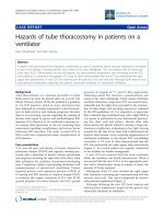
Figure 1 HO-1 homozygous mutant mice display disrupted alveolar development. A-D: H & E staining of lung sections at P10. A: Wildtype;
B: HO-1 +/-; C, D: HO-1 -/ In A and B, normal organized alveolar sac and formation of secondary septations are shown. In HO-1 homozygous
mutants, alveolar development was disrupted at various severities. Panel C represents a mutant with mildly enlarged alveolar airspaces, and
Panel D illustrates another mutant with more severe defects including a dramatically disorganized alveolar sac, missing septation, and a
thickened interstitial region. E: Radial alveolar counts (RAC) of lung sections from wildtype (WT) and HO-1 -/- littermates at P10 (n = 4 in each
group).
*
P < 0.05 vs. WT. The HO-1 mutants demonstrated significant decreased RAC.
Zhuang et al. Respiratory Research 2010, 11:142
/>Page 4 of 10
Down-regulation of lung epithelial and vascular genes in
Dex-treated HO-1 mutants
To evaluate the maturation of the lung alveolar cells, we
further examined the mRNA levels of pulmonary type II
epithelial cell markers in these animals. Surfactant Pro-
teins (SP) genes are a family of genes specific for type II
cells and essential for type II cell function. We assessed all
four surfactant protein genes by quantitative real-time
PCR with gene specific probes and primers. At baseline of
P14, expression levels of three surfactant proteins, -A, -C,
and -D, were significantly lower in HO-1 mutant lungs.
No difference in SP-B expression was detected between
HO-1 mutant and WT littermates. Dex treatment in
wildty pe resu lted in decreased levels of SP-A, -B, and -C,
and an increase in SP-D. However, after Dex treatments
in the HO-1 null mutant, all surfactant gene mRNAs
were decreased compared to the untreated group
(Figure 4A-D).
It is well reported that pulmonary vascular develop-
ment is critical to postnatal alveolar formation. Previously
published results also demonstrated that the VEGF recep-
tor-2 (KDR/Flk-1) was down-regulated in Dex-treated
neonatal mice with reduce d alveolarization [40]. We
further examine the expression of two endothelial cell
markers, Flk-1 and Tie-2, in the different animal groups.
At baseline, Flk-1 and Tie-2 expression in the HO-1
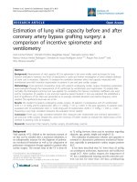
Figure 2 Dexamethasone treatment disrupts postnatal alveolar development. A, B: Representative H&E staining of mouse lung sections at
P10. Wildtype newborn mice were injected daily with saline (Control in A) or 1 ug/pup dexamethasone (Dex, in B). Note the enlarged alveolar
airspace and thinning of the alveolar wall in Dex-treated animals (B). C. Lung alveolar counts in control and Dex-treated mice.
*
P < 0.05 vs.
Control. D. HO protein levels in control and Dex-injected lungs. Upper panel: representative Western blot of P10 lungs from Control and Dex-
injected mice with antibodies against HO-1, HO-2 and ß-actin (loading control). Samples from two animals for each group are shown. HO-1
protein levels were visibly decreased in Dex-injected samples, whereas HO-2 protein levels remained unchanged. Lower panel: densitometric
values for HO-1 protein levels normalized with ß-actin, and expressed as ratio to control.
*
P < 0.05 vs. Control.
Zhuang et al. Respiratory Research 2010, 11:142
/>Page 5 of 10
mutant have 26% and 29% decrease compared to wild-
type littermates. Consistent with the published report,
Flk-1 and Tie-2 expression both decreased significantly
after Dex treatment in the wildtype, 74% and 70% respec-
tively, from the values of the untreated group. Although
it is well known that Dex has inhibitory effects on alveo-
lar formation, these data demonstrate that pulmonary
angiogenesis is also significantly inhibited by Dex via
down-regulation of VEGF and Ang-mediated pathways.
Most interestingly, the Dex-mediated decrease of mRNA
expression levels of endothelial cell markers, including
both Flk-1 and Tie-2, further decreased in HO-1 mutant
after Dex treatment. The expression levels of Flk-1 and
Tie-2 in the Dex treated HO-1 mutant group decreased
to 7% and 15% of the values observed in the wildtype
untreated group (Figure 4).
In summary, these data demonstrated that HO-1 is
critical to postnatal alveolardevelopmentandthatitis
involved in ep ithelial cel l growth regula tion. The effects
of HO-1 disruption are also additive to those of postna-
tal corticosteroid exposure.
Discussion
In this paper we show with histology and molecular
analysis that postnatal lung development is alte red in
HO-1 knockout mice. We also document that Dex
treatment exacerbates the alveolar defects seen with
HO-1 disruption.
HO-1 null mice display partial penetrance of embryo-
nic lethality. Although the exact cause and the underlin-
ing mechanism are not yet determined, our prelim inary
data (Q. Lin, unpublished) suggest that defects in the
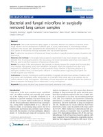
Figure 3 Dexamethasone treatment exacerbates the alveolar defects in HO-1 mutant mice. A-F: Representative H&E staining of mouse lung
sections at P14 at 5X and 20X magnifications. A-C: Wildtype; D-F: HO-1 -/ A, D: lungs from untreated animals. B, C, E, F: lungs from Dex-treated
animals (P3-P14, 0.25 ug/pup/day). In untreated group, lungs from HO-1-/- animals showed simplified, enlarged, and disorganized alveolar structure
(A, D). Postnatal Dex treatment in wildtype animals resulted in alveolar simplification and loss of secondary septation (A, and B, C). Dex treatment in
HO-1 -/- animals resulted in more dramatic disruption of the alveolar structure with larger alveolar space, thinning of the alveolar wall, and lack of
secondary septation. V: pulmonary vasculature. A: airway. Arrowhead indicates the normal secondary septae. Arrows indicate the elongated and
thinning of the alveolar wall. G: Quantification of alveolar development by RAC of the lung sections at P14.
*
P < 0.05 vs. wildtype, † P < 0.05 vs.
untreated group of same genotype. n = 3-4 for each group.
Zhuang et al. Respiratory Research 2010, 11:142
/>Page 6 of 10
embryonic vasculature might significantly contribute to
the early lethality. In the present manuscript, we studied
lung development in the HO-1 -/- mutants that survive
to the postnatal period. Compared to the wildtype litter-
mates, HO-1 -/- mutants display defects in lung alveolar
development with a range of severi ty, including disorga-
nized alveolar structure, thickening of the interstitial
section, and loss of air exchange surface area. These
loss-of-function phenotypes indicate that HO-1 is essen-
tial to normal postnatal lung development in vivo.Anti-
oxidant enzymes have been shown to protect the lung
from oxidativ e injury. For example, in Type II epithelial
cells from newborn mice, over-expression of extracellu-
lar superoxide d ismutase (EC-SOD) preserved type I I
cell proliferation and protected the lung from hyperoxic
injury [43]. Mice deficient in endothelial nitric oxide
synthase (e-NOS) d isplayed defectiv e lung vascu lar
development, which resembles the alveolar capillary dys-
plasia in infants with Persistent Pulmonary Hypertension
of the Newborn (PPHN). It is intriguing that the antiox-
idant HO-1 is not only protective against stress and
injury, but is also required for normal embryonic
development and postnatal alveolar formation. The
exact mechanism by which this occurs is not yet
elucidated.
The HO-1 null mice display its phenotypic defects
with a range of severity, from the viability rate to the
lung and vascular disruptions. In our data analysis, this
partial penetrance is the cause of more variation among
the KO samples compared to the WT samples. This
sometimes resulted in not achieving the statistical signif-
icance threshold of p < 0.05, even with large differences
in mean values. Partial or incomplete penetrance of phe-
notypes is not rare and the exact molecular mechanism
for this is unknown. However, this may indicate that
HO-1 protein plays an important role in maintenance of
the delicate homeostasis of many cellular events.
Postnatal steroid therapy benefits preterm infants by
accelerating lung maturation, reducing lung inflamma-
tion and facilitating extubation from the ventilator.
However, the adverse effects associated with glucocorti-
coid usage are significant and there also may be detri-
mental long-term damage to the brain and lung. In the
lung, dexamethasone can impair lung septation and
Figure 4 Expression of lung epithelial and vascular genes in wildtype and HO-1 -/- after dexamethasone treatment. Gene expression of
surfactant protein (SP)-A, -B, -C, and D, as well as Flk-1 and Tie-2 was determined by realtime PCR and normalized to 18 S mRNA levels. Data
represents relative quantification of mRNA to wildtype littermates at baseline (no treatment).
*
P < 0.05 vs. WT, † P < 0.05 vs. untreated group of
same genotype. n = 3-4 for each group.
Zhuang et al. Respiratory Research 2010, 11:142
/>Page 7 of 10
alveolar formation in the early postnatal period. Further-
more, the interrupted alveolar development does not
resume normally even after treatment stops. In our
experiment, we have shown that Dex treatment in the
postnatal period cause significant loss of alveolar com-
plexity and decreased HO-1 protein levels in the wild-
type lung. This reduction of the cytoprotective molecule
HO-1 may contribute to the abnormal alveolar growth
in the treated animals. Previous studies have also
reported that HO-1 expression can be suppressed by
Dex i n cultured endothelial cells [44,45]. Genomic ana-
lysis of the HO-1 gene promoter and enhancer regions
reveals at least four putative glucocorticoid receptor
(GR) binding sites, indicating transcriptional repression
via direct binding of GR to the HO-1 gene regulatory
regions. Nonetheless, repression can be achieved
through many mechanisms including epigenetic regula-
tion o r chromatin r emodeling, posttranslational modifi-
cations, a nd protein-protein interactions. However, this
was not specifically tested in the current work.
HO-1 mutant mice have reduced viability during
embryonic development and postnatally. The cause of
the postnatal lethality is not fully understood yet. In this
paper, we reported lung structural defects in HO-1
mutant animals including enlarged alveolar spaces, and
simplified alveolar structure with less secondary septae.
These defects would result in reduced gas exchange sur-
face area of the lung and lead to compromised pulmon-
ary function, hence the postnatal mortality.
When we subjected the HO-1 mutant to Dex treat-
ment, the dosage previously used on wildtype animal at
the same age was lethal to HO-1 mutants. We then
reduced the Dex do se to 25% of the original. This
resulted in milder alveolar simplification in the WT, but
dramatic defects in the HO-1 mutants with decreased
radial alveolar counts, thinner alveolar wall, and inhib-
ited secondary septae formation. This suggested that the
effects of HO-1 disruption and dexamethasone treat-
ment are additive. Since mice lacking HO-1 show
further disrupted lung development when treated with
steroids, the data suggest that HO-1 and steroid-
mediated disruption of lung development are indepen-
dent but additive.
Gene array experiments identified that critical vascular
genes, including Flk-1, were down-regulated in the
Dex-treated animals, suggesting that pulmonary vascu-
larization in the developing lung is critical to postnatal
alveolarization [40]. We have observed a similar
decrease in Flk-1 after Dex treatment in the current
study. In addition, we have examined another endothe-
lial marker Tie-2, and confirmed that Dex treatment
caused significant decrease in endothelial gene expres-
sion. In BPD and in other neonatal lung diseases with
arrested and impaired alveolar development, defects in
pulmonary vasculature, such as dec reased blood vessel
density, abnormal v essel branching p atterns and down-
regulation of vascular growth factors, have also been
identified. Furthermore, studies also found that in addi-
tion to decreased endothelial content, proliferation,
migration and survival of these cells may also be com-
promised in BPD [46]. Altogether, increasing evidence
suggests that the proliferation, differentiation, and pat-
terning of vascular endothelial and epithelial lineages in
the lung may exert a reciprocal influence on lung mor-
phogenesis and growth.
Previous studies demonstrated that HO-1 is involved
in vascular development by facilitating blood vessel for-
mation in tumors, wounds, and experimental models of
angiogenesis [19,29]. In cultured endothelial cells, induc-
tion of HO-1 and i ncreased CO levels up-regulate the
expression of VEGF and VEGF receptors, increase
endothelial cell proliferation, migration and sprouting,
and promote angiogenesis [47-49]. HO-1 induction or
CO exposure in vascular smooth muscle cells also up-
regulates the expression of VEGF [50]. These data indi-
cate that induced HO-1 may function in the vascular
system by counteracting the deleterious effects o f reac-
tive oxygen species (ROS) and by producing CO as a
vasorelaxant and regulator of vascular growth. In our
current study, we have shown that Dex-treated HO-1
knockout mice have dramatically disrupted alveolar
development. Interestingly, vascular gene expression was
even more significantly decreased in HO-1 mutant mice
aft er Dex treatment. In addition, severe vascular defects
are found in HO-1 mutant embryos (unpublished data).
Thus, we spe culate that the exacerbate d lung alv eolar
defects observed after Dex treatment in HO-1 null
mutant mice might result from the disrupted pulmonary
vasculature.
Postnatal glucocorticoid usage in preterm infants can
facilitate lung maturation and reduce lung inflammation,
yet it can have detrimental effects on lung and neural
development. During postnatal lung growth, Dex treat-
ment causes loss of alveolar septation, which results in a
large, simplified alveolar structure with decreased gas
exchange surface area. In addition, i ncreased oxidative
stress contributes to neonatal lung disease by aff ecting
alveolar growth. Our findings, that Dex treatment
decreases HO-1 expression and that disruption of HO-1
protein results in more severe vascular and alveolar
def ects after Dex treatment, suggest that enhancing this
important antioxidant system might be a beneficial
strategy to obviate neonatal lung disease.
There are several limitations of the current study.
Firstly, the sample sizes of HO-1 mutant animals in
each experiment group in the study were small. This is
due to the difficulty of o btaining viable HO-1 null new-
borns. With the partial penetrance of the phenotype, in
Zhuang et al. Respiratory Research 2010, 11:142
/>Page 8 of 10
some assays, the data variation is bigger than the WT
control group. With a bigger bree ding colony or in vitro
fertilization techniques using gametes from homozygous
and heterozygous animals, we will be able to generate
more HO-1 mutant animals for future stud ies. Secondly,
we have demonstrated structural defects as well as gene
expression alterations, but no functional assessment was
conducted. In future studies, we can measure pulmonary
function in the animals as a correlate. Thirdly, further
genetic analysis with more tools is needed to establish
the molecular mechanism connecting glucocorticoids to
HO-1. For example, it would be useful to evaluate the
effect of Dex in the HO-1 transgenic over-expressors.
Conclusions
In summary, we show evidence that HO-1 deficiency in
the mouse results in disrupted postnatal alveolar devel-
opment including abnormal a lveolar structure and
decreased epithelial and endothelial marker expression.
These defects were further exacerbated when the HO-1
mutant animals were treated with glucocorticoids. The
decrease in endothelial gene expression was more dra-
matic than that of the lung epithelial markers. These
experiments demonstrate that HO-1 is require d for nor-
mal lung development and that HO-1 disruption and
dexamethasone have additive detrimental effects on
postnatal lung growth. We speculate that HO-1 is
involved in postnatal lung development through modu-
lation of pulmonary vascular development.
Acknowledgements
This study was supported by Institutional Development Funds (Children’s
Hospital of Philadelphia) to QSL and NIH grant HL058 752 to PAD. We would
also like to thank Dr. Guang Yang, Dr. Clyde Wright, and Dr. Ping La for
helpful discussions.
Author details
1
Division of Neonatology, Children’s Hospital of Philadelphia, Philadelphia, PA
19104 USA.
2
Department of Pediatrics, Division of Neonatology, University of
Pennsylvania School of Medicine, Philadelphia, PA 19104 USA.
Authors’ contributions
TZ performed the molecular biology experiments in the manuscript and
participated in its design. MZ performed part of the animal studies and
radial alveolar counts. HZ supervised lung morphology analysis and assisted
in data analysis. PAD participated in study design, data interpretation and
manuscript editing. QSL conceived of the study, participated in its design
and execution, performed animal studies, and wrote the manuscript. All
authors read and approved the final manuscript.
Competing interests
The authors declare that they have no competing interests.
Received: 16 December 2009 Accepted: 10 October 2010
Published: 10 October 2010
References
1. Groenman F, Unger S, Post M: The molecular basis for abnormal human
lung development. Biol Neonate 2005, 87(3):164-177.
2. Warburton D, Schwarz M, Tefft D, Flores-Delgado G, Anderson KD,
Cardoso WV: The molecular basis of lung morphogenesis. Mech Dev 2000,
92(1):55-81.
3. Warner BB, Stuart LA, Papes RA, Wispe JR: Functional and pathological
effects of prolonged hyperoxia in neonatal mice. Am J Physiol 1998,
275(1 Pt 1):L110-117.
4. Wagenaar GT, ter Horst SA, van Gastelen MA, Leijser LM, Mauad T, van der
Velden PA, de Heer E, Hiemstra PS, Poorthuis BJ, Walther FJ: Gene
expression profile and histopathology of experimental
bronchopulmonary dysplasia induced by prolonged oxidative stress. Free
Radic Biol Med 2004, 36(6):782-801.
5. Massaro D, Massaro GD: Dexamethasone accelerates postnatal alveolar
wall thinning and alters wall composition. Am J Physiol 1986, 251(2 Pt 2):
R218-224.
6. Massaro D, Massaro GD: Pulmonary alveolus formation: critical period,
retinoid regulation and plasticity. Novartis Found Symp 2001, 234:229-236,
discussion 236-241.
7. Massaro D, Massaro GD: Invited Review: pulmonary alveoli: formation, the
“call for oxygen,” and other regulators. Am J Physiol Lung Cell Mol Physiol
2002, 282(3):L345-358.
8. Massaro D, Massaro GD: Retinoids, alveolus formation, and alveolar
deficiency: clinical implications. Am J Respir Cell Mol Biol 2003,
28(3):271-274.
9. Massaro GD, Massaro D: Formation of pulmonary alveoli and gas-
exchange surface area: quantitation and regulation. Annu Rev Physiol
1996, 58:73-92.
10. Yoshida T, Kikuchi G: Purification and properties of heme oxygenase from
rat liver microsomes. J Biol Chem 1979, 254(11):4487-4491.
11. Yoshinaga T, Sassa S, Kappas A: The oxidative degradation of heme c by
the microsomal heme oxygenase system. J Biol Chem 1982,
257(13):7803-7807.
12. Maines MD: Heme oxygenase: function, multiplicity, regulatory
mechanisms, and clinical applications. FASEB J 1988, 2(10):2557-2568.
13. Choi AM, Alam J: Heme oxygenase-1: function, regulation, and
implication of a novel stress-inducible protein in oxidant-induced lung
injury. Am J Respir Cell Mol Biol 1996, 15(1):9-19.
14. Dennery PA, Rodgers PA: Ontogeny and developmental regulation of
heme oxygenase.
J Perinatol 1996, 16(3 Pt 2):S79-83.
15. Dennery PA, Lee CS, Ford BS, Weng YH, Yang G, Rodgers PA:
Developmental expression of heme oxygenase in the rat lung. Pediatr
Res 2003, 53(1):42-47.
16. Morita T, Kourembanas S: Endothelial cell expression of vasoconstrictors
and growth factors is regulated by smooth muscle cell-derived carbon
monoxide. J Clin Invest 1995, 96(6):2676-2682.
17. Weng YH, Tatarov A, Bartos BP, Contag CH, Dennery PA: HO-1 expression
in type II pneumocytes after transpulmonary gene delivery. Am J Physiol
Lung Cell Mol Physiol 2000, 278(6):L1273-1279.
18. Li N, Venkatesan MI, Miguel A, Kaplan R, Gujuluva C, Alam J, Nel A:
Induction of heme oxygenase-1 expression in macrophages by diesel
exhaust particle chemicals and quinones via the antioxidant-responsive
element. J Immunol 2000, 165(6):3393-3401.
19. Duckers HJ, Boehm M, True AL, Yet SF, San H, Park JL, Clinton Webb R,
Lee ME, Nabel GJ, Nabel EG: Heme oxygenase-1 protects against vascular
constriction and proliferation. Nat Med 2001, 7(6):693-698.
20. Mumby S, Upton RL, Chen Y, Stanford SJ, Quinlan GJ, Nicholson AG,
Gutteridge JM, Lamb NJ, Evans TW: Lung heme oxygenase-1 is elevated
in acute respiratory distress syndrome. Crit Care Med 2004,
32(5):1130-1135.
21. Tsoumakidou M, Tzanakis N, Chrysofakis G, Siafakas NM: Nitrosative stress,
heme oxygenase-1 expression and airway inflammation during severe
exacerbations of COPD. Chest 2005, 127(6):1911-1918.
22. Zhou H, Lu F, Latham C, Zander DS, Visner GA: Heme oxygenase-1
expression in human lungs with cystic fibrosis and cytoprotective effects
against Pseudomonas aeruginosa in vitro. Am J Respir Crit Care Med 2004,
170(6):633-640.
23. Minamino T, Christou H, Hsieh CM, Liu Y, Dhawan V, Abraham NG,
Perrella MA, Mitsialis SA, Kourembanas S: Targeted expression of heme
oxygenase-1 prevents the pulmonary inflammatory and vascular
responses to hypoxia. Proc Natl Acad Sci USA 2001, 98(15):8798-8803.
Zhuang et al. Respiratory Research 2010, 11:142
/>Page 9 of 10
24. Stocker R, Yamamoto Y, McDonagh AF, Glazer AN, Ames BN: Bilirubin is an
antioxidant of possible physiological importance. Science 1987,
235(4792):1043-1046.
25. Dore S, Takahashi M, Ferris CD, Zakhary R, Hester LD, Guastella D,
Snyder SH: Bilirubin, formed by activation of heme oxygenase-2, protects
neurons against oxidative stress injury. Proc Natl Acad Sci USA 1999,
96(5):2445-2450.
26. Dennery PA, Sridhar KJ, Lee CS, Wong HE, Shokoohi V, Rodgers PA,
Spitz DR: Heme oxygenase-mediated resistance to oxygen toxicity in
hamster fibroblasts. J Biol Chem 1997, 272(23):14937-14942.
27. Otterbein LE, Kolls JK, Mantell LL, Cook JL, Alam J, Choi AM: Exogenous
administration of heme oxygenase-1 by gene transfer provides
protection against hyperoxia-induced lung injury. J Clin Invest 1999,
103(7):1047-1054.
28. Soares MP, Lin Y, Anrather J, Csizmadia E, Takigami K, Sato K, Grey ST,
Colvin RB, Choi AM, Poss KD, Bach FH: Expression of heme oxygenase-1
can determine cardiac xenograft survival. Nat Med 1998, 4(9):1073-1077.
29. Bussolati B, Ahmed A, Pemberton H, Landis RC, Di Carlo F, Haskard DO,
Mason JC: Bifunctional role for VEGF-induced heme oxygenase-1 in vivo:
induction of angiogenesis and inhibition of leukocytic infiltration. Blood
2004, 103(3):761-766.
30. Poss KD, Tonegawa S: Heme oxygenase 1 is required for mammalian iron
reutilization. Proc Natl Acad Sci USA 1997, 94(20):10919-10924.
31. Poss KD, Tonegawa S: Reduced stress defense in heme oxygenase 1-
deficient cells. Proc Natl Acad Sci USA 1997, 94(20):10925-10930.
32. Yachie A, Niida Y, Wada T, Igarashi N, Kaneda H, Toma T, Ohta K,
Kasahara Y, Koizumi S: Oxidative stress causes enhanced endothelial cell
injury in human heme oxygenase-1 deficiency. J Clin Invest 1999,
103(1):129-135.
33. Merrill JD, Ballard RA: Antenatal hormone therapy for fetal lung
maturation. Clin Perinatol 1998, 25(4):983-997.
34. Grier DG, Halliday HL: Corticosteroids in the prevention and management
of bronchopulmonary dysplasia. Semin Neonatol 2003, 8(1):83-91.
35. Barrington KJ: The adverse neuro-developmental effects of postnatal
steroids in the preterm infant: a systematic review of RCTs. BMC Pediatr
2001, 1:1.
36. Friedman S, Shinwell ES: Prenatal and postnatal steroid therapy and child
neurodevelopment. Clin Perinatol 2004, 31(3):529-544.
37. Emery JL, Mithal A: The number of alveoli in the terminal respiratory unit
of man during late intrauterine life and childhood. Arch Dis Child
1960,
35:544-547.
38. Cooney TP, Thurlbeck WM: The radial alveolar count method of Emery
and Mithal: a reappraisal 1–postnatal lung growth. Thorax 1982,
37(8):572-579.
39. Massaro GD, Massaro D: Postnatal treatment with retinoic acid increases
the number of pulmonary alveoli in rats. Am J Physiol 1996, 270(2 Pt 1):
L305-310.
40. Clerch LB, Baras AS, Massaro GD, Hoffman EP, Massaro D: DNA microarray
analysis of neonatal mouse lung connects regulation of KDR with
dexamethasone-induced inhibition of alveolar formation. Am J Physiol
Lung Cell Mol Physiol 2004, 286(2):L411-419.
41. Maden M: Retinoids have differing efficacies on alveolar regeneration in
a dexamethasone-treated mouse. Am J Respir Cell Mol Biol 2006,
35(2):260-267.
42. Massaro GD, Massaro D: Retinoic acid treatment partially rescues failed
septation in rats and in mice. Am J Physiol Lung Cell Mol Physiol 2000,
278(5):L955-960.
43. Auten RL, O’Reilly MA, Oury TD, Nozik-Grayck E, Whorton MH: Transgenic
extracellular superoxide dismutase protects postnatal alveolar epithelial
proliferation and development during hyperoxia. Am J Physiol Lung Cell
Mol Physiol 2006, 290(1):L32-40.
44. Deramaudt TB, da Silva JL, Remy P, Kappas A, Abraham NG: Negative
regulation of human heme oxygenase in microvessel endothelial cells
by dexamethasone. Proc Soc Exp Biol Med 1999, 222(2):185-193.
45. Lavrovsky Y, Drummond GS, Abraham NG: Downregulation of the human
heme oxygenase gene by glucocorticoids and identification of 56b
regulatory elements. Biochem Biophys Res Commun 1996, 218(3):759-765.
46. Thebaud B: Angiogenesis in lung development, injury and repair:
implications for chronic lung disease of prematurity. Neonatology 2007,
91(4):291-297.
47. Deramaudt BM, Braunstein S, Remy P, Abraham NG: Gene transfer of
human heme oxygenase into coronary endothelial cells potentially
promotes angiogenesis. J Cell Biochem 1998, 68(1):121-127.
48. Jozkowicz A, Huk I, Nigisch A, Weigel G, Dietrich W, Motterlini R, Dulak J:
Heme oxygenase and angiogenic activity of endothelial cells:
stimulation by carbon monoxide and inhibition by tin protoporphyrin-IX.
Antioxid Redox Signal 2003, 5(2):155-162.
49. Abraham NG, Scapagnini G, Kappas A: Human heme oxygenase: cell
cycle-dependent expression and DNA microarray identification of
multiple gene responses after transduction of endothelial cells). J Cell
Biochem 2003, 90(6):1098-1111.
50. Dulak J, Jozkowicz A, Foresti R, Kasza A, Frick M, Huk I, Green CJ,
Pachinger O, Weidinger F, Motterlini R: Heme oxygenase activity
modulates vascular endothelial growth factor synthesis in vascular
smooth muscle cells. Antioxid Redox Signal
2002, 4(2):229-240.
doi:10.1186/1465-9921-11-142
Cite this article as: Zhuang et al.: Disrupted postnatal lung development
in heme oxygenase-1 deficient mice. Respiratory Research 2010 11:142.
Submit your next manuscript to BioMed Central
and take full advantage of:
• Convenient online submission
• Thorough peer review
• No space constraints or color figure charges
• Immediate publication on acceptance
• Inclusion in PubMed, CAS, Scopus and Google Scholar
• Research which is freely available for redistribution
Submit your manuscript at
www.biomedcentral.com/submit
Zhuang et al. Respiratory Research 2010, 11:142
/>Page 10 of 10