Báo cáo y học: "RhoA signaling modulates cyclin D1 expression in human lung fibroblasts; implications for idiopathic pulmonary fibrosis" ppsx
Bạn đang xem bản rút gọn của tài liệu. Xem và tải ngay bản đầy đủ của tài liệu tại đây (725.26 KB, 14 trang )
BioMed Central
Page 1 of 14
(page number not for citation purposes)
Respiratory Research
Open Access
Research
RhoA signaling modulates cyclin D1 expression in human lung
fibroblasts; implications for idiopathic pulmonary fibrosis
KL Watts*, E Cottrell, PR Hoban and MA Spiteri
Address: Lung Research, Institute of Science and Technology in Medicine, University Hospital of North Staffordshire/Keele University,
Staffordshire, UK
Email: KL Watts* - ; E Cottrell - ; PR Hoban - ;
MA Spiteri -
* Corresponding author
Abstract
Background: Idiopathic Pulmonary Fibrosis (IPF) is a debilitating disease characterized by
exaggerated extracellular matrix deposition and aggressive lung structural remodeling. Disease
pathogenesis is driven by fibroblastic foci formation, consequent on growth factor overexpression
and myofibroblast proliferation. We have previously shown that both CTGF overexpression and
myofibroblast formation in IPF cell lines are dependent on RhoA signaling. As RhoA-mediated
regulation is also involved in cell cycle progression, we hypothesise that this pathway is key to lung
fibroblast turnover through modulation of cyclin D1 kinetic expression.
Methods: Cyclin D1 expression was compared in primary IPF patient-derived fibroblasts and
equivalent normal control cells. Quantitative real time PCR was employed to examine relative
expression levels of cyclin D1 mRNA; protein expression was confirmed by western blotting.
Effects of Rho signaling were investigated using transient transfection of constitutively active and
dominant negative RhoA constructs as well as pharmacological inhibitors. Cellular proliferation of
lung fibroblasts was determined by BrdU incorporation ELISA. To further explore RhoA regulation
of cyclin D1 in lung fibroblasts and associated cell cycle progression, an established Rho inhibitor,
Simvastatin, was incorporated in our studies.
Results: Cyclin D1 expression was upregulated in IPF compared to normal lung fibroblasts under
exponential growth conditions (p < 0.05). Serum deprivation inhibited cyclin D1 expression, which
was restored following treatment with fibrogenic growth factors (TGF-β1 and CTGF). RhoA
inhibition, using a dominant negative mutant and a pharmacological inhibitor (C3 exotoxin),
suppressed levels of cyclin D1 mRNA and protein in IPF fibroblasts, with significant abrogation of
cell turnover (p < 0.05). Furthermore, Simvastatin dose-dependently inhibited fibroblast cyclin D1
gene and protein expression, inducing G1 cell cycle arrest. Similar trends were observed in control
experiments using normal lung fibroblasts, though exhibited responses were lower in magnitude.
Conclusion: These findings report for the first time that cyclin D1 expression is deregulated in
IPF through a RhoA dependent mechanism that influences lung fibroblast proliferation. This
potentially unravels new molecular targets for future anti-IPF strategies; accordingly, Simvastatin
inhibition of Rho-mediated cyclin D1 expression in IPF fibroblasts merits further exploitation.
Published: 15 June 2006
Respiratory Research 2006, 7:88 doi:10.1186/1465-9921-7-88
Received: 13 February 2006
Accepted: 15 June 2006
This article is available from: />© 2006 Watts et al; licensee BioMed Central Ltd.
This is an Open Access article distributed under the terms of the Creative Commons Attribution License ( />),
which permits unrestricted use, distribution, and reproduction in any medium, provided the original work is properly cited.
Respiratory Research 2006, 7:88 />Page 2 of 14
(page number not for citation purposes)
Background
Idiopathic pulmonary fibrosis (IPF) is an insidious fibro-
proliferative disorder, characterised by interstitial alveolar
fibrosis thought to be consequent on aberrant responses
to undefined microinsults. Lung injury maybe exacer-
bated by concurrent failure of re-epithelialisation and
excessive fibroblast differentiation [1,2], underpinned by
erratic deposition of extracellular matrix (ECM) proteins
and progressive lung tissue remodelling. Although a
number of scientific advances have been made in under-
standing disease pathogenesis, no efficacious therapy is
available to halt or alter these exaggerated pro-fibrotic
processes.
It follows that IPF pathogenesis must involve aberrations
within regulatory pathways critical to the described cellu-
lar – biomolecular events. Under such conditions, fibrob-
lasts acquire an aggressive, contractile myofibroblast
phenotype, with potent capability for ECM protein pro-
duction [3]. Fibroblast-myofibroblast differentiation, is
driven by an upregulated pool of growth factors, of which
connective tissue growth factor (CTGF) is a key player [4].
CTGF induction primarily, but not exclusively, is medi-
ated by TGF-β1 through a TGF-β response element in the
CTGF promoter [5]. CTGF modulates IPF fibroblast differ-
entiation through a signalling pathway involving RhoA
[6,7]. Interestingly, RhoA is also known to be instrumen-
tal in the kinetics of cyclin D1 expression, specifically in
G1 phase of the cell cycle [8]. It follows that as relentless
proliferation and differentiation of fibroblasts are crucial
to IPF progression, deregulated expression of key cell cycle
genes and transcription factors may be pivotal to disease
pathogenesis.
The cell cycle regulator cyclin D1 is a critical factor in the
development of proliferative disease [9], including spe-
cific organ oncogenesis [10-12]. This 36-kDa protein has
a widely accepted role in positive regulation of G1-S pro-
gression [13]. Functioning as a 'mitogenic sensor', in the
presence of growth factors, cyclin D1 gene (CCND1)
drives target cells through the restriction point in the G1
phase of their cycle (thus committing them to cell divi-
sion). This function is facilitated through binding and
activation of cyclin-dependent kinases (CDK) 4 and 6,
with phosphorylation of the retinoblastoma protein (Rb),
and release of sequestered transcription factors such as
E2F [14,15]. Furthermore, in vitro induction of CCND1
augments cellular proliferation and transformation of
mammalian cells [16]; which in rodent cells is character-
ised by a shortened G1 phase with reduced dependence
on mitogens [17].
A key histological feature of IPF lungs is presence of
fibroblast proliferation, with fibroblastic foci formation.
We hypothesise that cyclin D1 plays an instrumental role
in these pro-fibrogenic processes, augmented by in situ
growth factor overproduction and exaggerated extracellu-
lar matrix deposition [18]. We contend that cyclin D1
influence in fibroblasts is mediated via a RhoA signalling
pathway, especially as RhoA is known to regulate G1 pro-
gression of cells [19]. Accordingly, our study explores for
the first time expression levels of cyclin D1 in IPF patient-
derived fibroblasts (and equivalent controls) and identi-
fies the influence of Rho, using constitutively active and
dominant negative RhoA constructs as well as pharmaco-
logical inhibitors, including the agent Simvastatin. This
agent selectively blocks a key cascade enzyme, 3-hydroxy-
3-methylglutaryl coenzyme A reductase (HMG CoA),
inhibiting essential post-translational modification of
RhoA, thus inactivating its signalling function.
Methods
Human lung fibroblast cell culture
Three separate human lung fibroblast cell lines isolated
from IPF patients (LL29 and LL97a both ATCC, Manassas,
USA; and HIPF – a generous gift from R.J. McAnulty, UCL
London,) and normal control equivalents (CCD8LU,
ATCC, Manassas, USA). The control cell line (CCD8LU) is
an adult lung fibroblast cell line, derived from a 48 year
old male with cerebral thrombosis, which are a good rep-
resentative control cell line for analysis of IPF specific
effects. All cells were cultured in Dulbecco's modified
Eagles medium (DMEM, Sigma Aldrich, Dorset, UK).
Media was supplemented with penicillin/streptomycin
(100 U/ml) and L-glutamine (2 mM) (both Gibco BRL,
Paisley, Scotland) with 10% fetal calf serum (FCS,
Labtech, Sussex, UK). All cell lines were cultured and uti-
lized at passages 5–8 to limit passage dependent effects on
the observed effects. For experiments, medium was
replaced with serum free DMEM (SF-DMEM), for 48
hours to induce quiescence before treatment.
Treatment with fibrogenic growth factors
Following serum depravation for 48 hours the fibroblasts
were stimulated with fibrogenic growth factors; human
recombinant TGF-β1 (R&D systems, Oxford, UK) dose of
1 ng/ml and 5 ng/ml; and human recombinant CTGF
(Fibrogen, CA, USA) doses of 10 ng/ml and 100 ng/ml.
Fibroblasts were treated with the above-mentioned
growth factors for 8 hours for gene expression analysis
and 24 hours for protein expression studies. The chosen
time points and concentrations of growth factors were
determined and established in previous and ongoing
studies within our laboratories [6,7].
C3 exotoxin treatment of lung fibroblasts
Quiescent lung fibroblasts were incubated overnight (16
hours) with Clostridium botulinum C3 exotoxin (Upstate
cell signalling solutions, NY, USA) in SF-DMEM. C3 exo-
toxin was used at concentrations of 0.5 µg/ml, 1 µg/ml
Respiratory Research 2006, 7:88 />Page 3 of 14
(page number not for citation purposes)
and 5 µg/ml; these doses have been previously shown to
inhibit Rho signalling pathways in similar fibroblast lines
[6].
Simvastatin treatment
Simvastatin is used clinically for the treatment of hyperc-
holesterolaemia due its ability to abrogate the cholesterol
synthesis pathway via HMG CoA inhibition. The statins
also possess a range of secondary effects arising from dis-
ruption of guanosine triphosphatase (GTPase) signalling,
including members of the Rho and Ras family. Simvasta-
tin (Merck Sharp and Dohme, Hertfordshire, UK) was dis-
solved and filter sterilised before use in cell culture studies
[20]. Quiescent lung fibroblasts were then incubated with
physiological concentrations of Simvastatin (0.1 µM, 1
µM 10 µM) for 16 hours in serum free cell culture media.
Following Simvastatin pre-conditioning, cells were stimu-
lated with human recombinant TGF-β1 (R&D systems,
Oxford, UK) at a dose of 5 ng/ml, cells were harvested at
8 hours for mRNA studies and 24 hours for protein anal-
ysis.
Transient transfection of dominant negative/constitutively
active RhoA constructs
Transfection of dominant-negative and constitutively
active RhoA (accession number L25080) constructs into
human lung fibroblasts (IPF-derived and CCD8LU cells)
were performed using Transfast mammalian transfection
system (Promega, Southampton, UK). Transfection was
performed in lung fibroblasts at 90% confluency follow-
ing the manufacturer's recommendations. 0.75 µg of DNA
was transfected per well (18 mm diameter) using a 1:1
ratio of DNA/Transfast reagent in serum-negative cultures.
90% confluent cells were incubated in the transfection
mix containing the RhoA plasmid for 1 hour; DMEM con-
taining 10% FCS was added up to a volume of 1 ml, and
cultures were left for 4 hours. Following this, the trans-
fected cells were serum deprived for 48 hours before treat-
ment with TGF-β1 (5 ng/ml) for 8 hours. RhoA G14V (a
construct containing a mutation at G14V to render it con-
stitutively active) and RhoA T19N (a construct containing
a mutation at T19N, giving it a dominant negative pheno-
type) constructs were utilized in a cDNA3.1+ vector and
were obtained from the Guthrie research institute http://
www.cdna.org.
Real time PCR
Stored cDNA samples isolated from normal and IPF iso-
lated lung fibroblasts were used to assess CTGF and α-
SMA gene expression. 2 µl of undiluted cDNA was used
per 25 µl reaction; the primer and probe sets were 'pre-
designed assay on demand' probes (Applied Biosystems,
Foster City, CA); these pre-designed primers are tested and
standardised for reproducible expression analysis. Primer
and cDNA were added to the TaqMan universal PCR mas-
ter mix (Applied Biosystems, Foster City, CA), containing
all the reagents for PCR. Absolute quantification of the
PCR products was carried out with an ABI prism 7000
(Applied Biosystems, Foster City, CA) utilising the relative
standard curve method. cDNA that positively expresses
the target gene is used to create a dilution series with arbi-
trary units. To ensure reproducibility, quantitative data
were taken at a point in which each sample was in the
exponential phase of amplification. The mean quantity of
target gene expression was determined from the generated
standard curve; then all samples were normalised against
an internal standard β actin or 18s in all quantitative PCR
reactions. All data are presented as the fold-change over
control in cyclin-D1 gene expression.
Western blotting
Total cell proteins were extracted in lysis buffer compris-
ing 1% (v/v) Triton X-100, 20 mM Tris HCL (pH 8.0),
10% (v/v) glycerol, 1 mM sodium orthovanadate, 2 mM
EDTA, 1 mM phenylmethylsulfonyl fluoride (PMSF), 20
µM leupeptin and 0.15 U/ml aprotinin. Recovered cells
were lysed in above lysis buffer and placed on ice for 20
minutes. The lysates were then centrifuged at 10 000 g,
4°C to pellet cell debris. The supernatant containing the
protein was recovered and assayed for total protein using
a commercial microplate assay (Bio-Rad, Hemel Hemp-
sted, UK). 25 µg of total protein was combined with sam-
ple buffer and boiled prior to gel loading. In addition full-
length, recombinant human cyclin D1 protein a 61 Kda
tagged fusion protein corresponding to amino acids 1–
295 (Santa Cruz Biotechnology, CA, USA) was also
loaded onto the gels to ensure detection of the protein of
interest. Proteins were resolved on a 12.5% polyacryla-
mide gel by electrophoresis at 120 V in reducing buffer
and transfer was carried out at 100 V. Membranes were
blocked with 5% (w/v) BSA in TBS-T buffer overnight. For
detection of the cyclin D1 protein DCS-6 (Santa Cruz Bio-
technology, CA, USA) antibody was used at 1:100 dilu-
tion in TBS-T and 1% BSA. Secondary detection was
carried out with horseradish peroxidase-conjugated
(HRP) Affinipure goat anti-mouse IgG antibody (Jackson
Immunoresearch) at 1:25,000 in TBS-T containing 1%
BSA. The cyclin D1 band was visualised by enhanced
chemiluminescence (ECL; Amersham Pharmacia Biotech,
Buckinghamshire, UK) according to the manufacturer's
recommendations and blots were quantified by densito-
metrical analysis, which involved correcting each blot for
background density on each gel. Ponceau S staining of
blots after transfer revealed equal loading of total protein;
additionally the membranes were reprobed for GAPDH
using rabbit polyclonal antibody to GAPDH (1:1000 dilu-
tion, Abcam, UK) to ensure equal loading.
Respiratory Research 2006, 7:88 />Page 4 of 14
(page number not for citation purposes)
DNA synthesis of proliferating cells
DNA synthesis was assessed by colorimetric cell prolifera-
tion Biotrak ELISA method according to the manufac-
turer's recommendations (Amersham Biosciences, UK)
based on the measurement of 5-bromo-2'-deoxyuridine
(BrdU) incorporation during DNA synthesis of proliferat-
ing cells. Briefly 30,000 cells were seeded per well of a 96
well plate and left for 24 hours. Cells were then synchro-
nised in situ by incubation with serum-depleted media for
48 hours. Cells were then treated with the recognised Rho
inhibitor Clostridium botulinum C3 exotoxin, (0.5–5 µg/
ml) (Upstate cell signalling solutions, Lake Placid, NY)
overnight prior to treatment with recombinant human
TGF-β1 (5 ng/ml) for up to 5 days. BrdU incorporation
was measured daily, during which cells were subjected to
BrdU incorporation for 4 hours. The colorimetric change
was measured at 450 nm on a Dynatech MR50000 micro-
plate reader (Dynex Laboratories, UK).
FACS analysis
LL97a lung fibroblasts were grown to approximately 60%
confluency prior to serum deprivation for 48 hours (this
ensures the cells become quiescent and are synchronised
in the cell cycle). The lung fibroblasts were then treated,
accordingly with Simvastatin (0.1 µg/ml or 10 µg/ml)
with or without TGF-β1 (5 ng/ml) for 24 hours. The cells
were then harvested and the cell suspension fixed in 70%
ice-cold ethanol. The cell suspension was centrifuged at
200 rpm and the cell pellet resuspended in PBS. RNase (1
mg/ml) and propidium iodide (0.5 mg/ml) were added
and incubated for 30 minutes at 37°C. To ensure no
clumping of the cells the suspension was passed through
a 25 g needle. The cells were analysed on a MOFLO cell
sorter (Dakocytomation, Glostrup, Denmark) at a wave-
length of 488 nm and speed of 100 events per second
(eps). A minimum of 20,000 events per data profile was
collected.
Statistical analysis
Data are shown as a mean ± SEM. An unpaired student's t
test was employed for comparing 2 groups of data. Multi-
ple comparisons were made using analysis of variance
(ANOVA) followed by Tukeys pairwise comparison. All p
values < 0.05 were considered significant.
Results
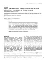
Cyclin D1 gene expression is upregulated in IPF fibroblasts
The expression of the cyclin D1 gene was quantified in 3
IPF-derived lung fibroblast cell lines (HIPF, LL29, LL97a)
and the adult normal lung fibroblast cell line CCD8LU
using a real time PCR approach (Fig 1). Under exponen-
tial growth conditions (cells grown in 10% FCS, i.e.
actively dividing cells), IPF-derived lung fibroblasts dem-
onstrated a 4.72 to 11.29 fold elevation of cyclin D1
mRNA expression (average of 10.10 fold increase) com-
pared to the CCD8LU normal lung fibroblast cell line (p
< 0.05). We compared these data to A431 cells, a human
epithelial squamous carcinoma cell line with a known 5
fold amplification of cyclin D1 [21]; the IPF fibroblast cell
lines studies significantly exceeded the amplified cyclin
D1 mRNA expression of A431 by an average of 2.45 fold.
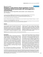
Cyclin D1 gene and protein levels are augmented by
growth factors
Cyclin D1 gene expression was measured in the normal
lung fibroblasts and the 3 IPF-derived lung fibroblast cell
lines following growth factor treatment (Fig 2a), Cells
were serum deprived for 48 hours to ensure quiescence
and to synchronise cell proliferation; cells were then
exposed to physiologically relevant concentrations of
growth factors (CTGF and TGF-β1) known to be impli-
cated in IPF pathogenesis [4,5]. Serum deprivation inhib-
ited cyclin D1 expression (as expected); however
expression was restored upon treatment with recom-
binant growth factors. Cyclin D1 augmentation was more
pronounced in the IPF-derived lung fibroblasts, especially
in the presence of TGF-β1 (1 ng/ml and 5 ng/ml) and
CTGF (10 ng/ml) (p < 0.05). Interestingly, in cultures con-
taining the higher concentration of CTGF (100 ng/ml), we
observed fibroblast apoptosis especially in IPF-related cell
line (data not shown); and no further increase in cyclin
Expression levels of cyclin D1 mRNA in human lung fibrob-lasts during exponential growthFigure 1
Expression levels of cyclin D1 mRNA in human lung
fibroblasts during exponential growth. Quantitative
real-time RT-PCR was performed on three separate human
lung fibroblast cell lines from IPF patients (HIPF, LL29, and
LL97a) and normal control equivalents CCD8LU. Quantifica-
tion of mRNA was performed by determining the threshold
cycle; and standard curves were constructed using the values
obtained from serially diluted positively expressing human
cDNA. All cells were under conditions of exponential
growth (10% FCS supplemented media). 3 PCR reactions
were performed from 3 independent cell culture experi-
ments, graph represents mean cyclin D1 expression ± S.E.M;
* = p < 0.05.
0
2
4
6
8
10
12
14
16
CCD8 HIPF LL29 LL97a A431
Cell Line
*
*
Fold change in CCDN1 expression
*
Respiratory Research 2006, 7:88 />Page 5 of 14
(page number not for citation purposes)
Expression of cyclin D1 mRNA and protein in human lung fibroblasts; response to fibrogenic growth factorsFigure 2
Expression of cyclin D1 mRNA and protein in human lung fibroblasts; response to fibrogenic growth factors.
Figure 2a: Cyclin D1 mRNA levels were determined by quantitative real-time PCR on three separate human lung fibroblast cell
lines from IPF patients (HIPF, LL29, and LL97a) and normal control equivalents CCD8LU exposed to fibrogenic mediators
TGF-β1 (5 ng/ml and 10 ng/ml) and CTGF (10 ng/ml and 100 ng/ml) for 8 hrs. Data shown demonstrates analysis from LL97a
and CCD8LU fibroblasts, no significant difference was observed in baseline cyclin D1 expression between the cell lines. Data
are representative of 3 independent experiments, within each of which PCRs were performed in triplicate. Data represents
mean cyclin D1 expression ± S.E.M; * = p < 0.05 compared to serum free, † = p < 0.05 compared to normal lung fibroblasts.
Figure 2b: Quantification of cyclin D1 protein expression was performed by western blotting in all three IPF fibroblast lines and
normal equivalents. Quiescent serum deprived lung fibroblasts were stimulated with fibrogenic growth factors for 24 hours.
Cyclin D1 protein levels found in 25 µg of total protein from normal and IPF derived lung fibroblasts was determined by west-
ern blotting. Data shown demonstrates analysis from LL97a and CCD8LU fibroblasts. Data are representative of 3 independ-
ent westerns and represented as mean density ± SEM. * = p < 0.05 compared to normal lung fibroblasts. Figure 2c:
Representative western blots for cyclin D1 and GAPDH protein expression in a representative IPF lung fibroblast cell line
(LL97a). (i) cyclin D1 blot-lane 1 = marker lane 2 = control (serum deprived); lane 3 = 10% FCS; lane 4 = TGF-β1 1 ng/ml; lane
5 = TGF-β1 5 ng/ml; lane 6 = CTGF 10 ng/ml; lane 6 = CTGF 100 ng/ml. (ii) GAPDH blot-lane 1 = control (serum deprived);
lane 2 = 10% FCS; lane 3 = TGF-β1 1 ng/ml; lane 4 = TGF-β1 5 ng/ml; lane 5 = marker; lane 6 = CTGF 10 ng/ml; lane 6 = CTGF
100 ng/ml. Ponceau S staining of blots after transfer revealed equivalent loading of total protein.
(a)
0
5
30
35
40
10% FCS serum free TGF 1ng/ml TGF 5ng/ml CTGF 10ng/ml CTGF 100ng/ml
Treatment
Normal IPF
†
†
†
*
*
*
10
15
20
25
Fold change in CCND1
(b)
Cyclin D1
0
0.5
1
.5
2
2.5
3
3.5
10% FCS serum free TGF 1ng/ml TGF 5ng/ml CTGF 10ng/ml CTGF 100ng/ml
Treatment
Normal
IPF
*
*
*
*
1
Density
(c)
Cyclin D1
34Kda
GAPDH
36Kda
Lane 1 2 3 4 5 6 7
Respiratory Research 2006, 7:88 />Page 6 of 14
(page number not for citation purposes)
D1 expression. It is also of interest to note that in the
absence of mitogens the levels of cyclin D1 mRNA are not
significantly different between the cell lines studied and
expression lies are within are narrow range (2.10–6.65 ×
10
-4
).
To further confirm above findings, cyclin D1 protein
expression was determined in the same cell lines under
the same experimental conditions using western blot
analysis (Fig 2b). We observed the same patterns of
expression and induction in cyclin D1 protein, reflecting
results of cyclin mRNA expression. A representative blot
from one of the IPF derived cell lines is shown in Fig 2c.
Data shown in fig 2a, 2b and 2c is taken from the patient
cell line LL97a; these mRNA and protein data reflect
results obtained with the other 2 IPF fibroblast cell lines
studied.
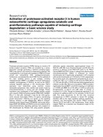
RhoA modulates cyclin D1 gene and protein levels in lung
fibroblasts
Transient transfection of dominant-negative RhoA (RhoA
T19N) and constitutively active (RhoA G14V) constructs
were utilised to confirm the regulatory role of Rho in cyc-
lin D1 induction (Fig 3a). These data revealed that cyclin
D1 mRNA expression levels are of comparable magnitude
between cells stimulated with TGF-β1 (5 ng/ml) alone
compared to those expressing constitutively active RhoA
(G14V RhoA transfected). When G14V active, RhoA cul-
tures were subsequently conditioned with TGF-β1, signif-
icant upregulation (p < 0.05) in cyclin D1 gene expression
was observed in LL97a; this trend was replicated in the
other 2 IPF derived cell lines studied. These data support
a role for Rho in cyclin D1 induction; which is further
confirmed by the use of a dominant-negative RhoA con-
struct. Transfection with Rho T19N induced significant
reduction (p < 0.05) in cyclin D1 gene expression produc-
ing a 25.5% and a 33% reduction in normal and IPF
derived lung fibroblasts respectively compared to cells
treated with 5 ng/ml TGF-β1 alone, further supporting
involvement of RhoA in cyclin D1 expression. In our
experiments, we did not observe complete inhibition of
cyclin D1 gene, as expected of the transient transfection
method used. As the average transfection efficiency
achieved was about 40%, thus a proportion of the cells in
our culture will not have inhibited RhoA signalling. The
above result trends were consistent throughout the 3 IPF
cell lines analysed.
To confirm above findings, cyclin D1 protein expression
was analysed following pharmacological inhibition of
RhoA utilising C3 exotoxin (a recognised inhibitor of
RhoA) (Fig 3b). Compared to TGF-β1 treatment alone (5
ng/ml), both test concentrations of C3 exotoxin signifi-
cantly (p < 0.05) abrogated cyclin D1 protein expression
in both normal and IPF lung fibroblasts, irrespective of
subsequent TGF-β1 exposure.
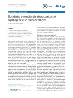
DNA synthesis is suppressed by RhoA inhibition
We used a sensitive BrdU incorporation ELISA that meas-
ures DNA synthesis to determine if Rho inhibition would
alter cell proliferative capability. Firstly DNA synthesis in
response to growth factor treatment was determined (Fig
4a). Actively dividing cells (cultured in media supple-
mented with 10% FCS) had the fastest DNA synthesis rate.
As expected, which was almost halted in serum-deprived
(quiescent) cells; but exhibited some restoration over the
120-hour time course on exposure to fibrogenic factors
TGF-β1 (5 ng/ml) and CTGF (10 ng/ml). At the end time
point (120 hr), TGF-β1 and CTGF induced a 44.85% and
36.88% increase respectively (p < 0.05) in BrdU incorpo-
ration IPF fibroblasts compared to serum-starved equiva-
lent controls. This data is representative of all 3 IPF cell
lines studied. Similar trends are replicated in the normal
fibroblast equivalents although the magnitude of BrdU
incorporation was approximately 3-fold lower in the con-
trols (data not shown).
Involvement of RhoA in above DNA synthesis was deter-
mined using C3 exotoxin, which specifically ADP ribo-
sylates and inactivates Rho. The inhibitor was used at
optimal concentrations of 0.5 µg/ml and 5 µg/ml with or
without additional TGF-β1 (5 ng/ml) stimulation (Fig
4b). Exposure to C3 exotoxin inhibited BrdU incorpora-
tion, even in the presence of TGF-β1 at both 0.5 and 5 µg/
ml concentrations. Both C3 exotoxin treatments sup-
pressed DNA synthesis over the 120 hour time course
compared to control IPF lung fibroblasts treated with 5
ng/ml TGF-β1 alone; becoming significant at p < 0.05 over
time from 72 hours onwards.
Simvastatin inhibits fibroblast cyclin D1 expression via a
Rho signalling pathway
The effect of cell pre-incubation with varying concentra-
tions of Simvastatin (0.1 µM, 1 µM and 10 µM) on cyclin
D1 gene expression in the IPF derived lung fibroblast cell
lines and equivalent normal controls was analysed by real
time PCR (Fig 5). The physiological concentrations of
Simvastatin used abrogated cyclin D1 gene expression,
irrespective of TGF-β1 presence (p < 0.05). Although 0.1
µM Simvastatin had little effect on cyclin D1 expression,
10 µM Simvastatin was efficacious enough to reduce even
basal levels of cyclin D1 mRNA in test fibroblasts, induc-
ing a 1.66 fold and 2.1 fold respective inhibition of the
gene compared to untreated cells and TGF-β1-lone treated
cells respectively. Furthermore the inhibition of cyclin D1
was further confirmed at the protein level by western blot-
ting (data not shown). The data in fig 5 is from LL97a IPF
derived lung fibroblasts; these data are also representative
of the other 2 IPF lung fibroblast cell lines studied. Again
Respiratory Research 2006, 7:88 />Page 7 of 14
(page number not for citation purposes)
RhoA signalling directly influences cyclin D1 mRNA and protein expression in lung fibroblastsFigure 3
RhoA signalling directly influences cyclin D1 mRNA and protein expression in lung fibroblasts. Fig 3a. Human lung
fibroblasts (3 IPF fibroblast lines and normal equivalents) were transfected with RhoA T19N (dominant negative) and RhoA
G14V (constitutively active) constructs. Cells were serum deprived for 48 hours post transfection before incubation with TGF-
β1 (5 ng/ml) for 8 hours. Data shown demonstrates analysis from LL97a and CCD8LU fibroblasts. Data are representative of
transfection performed in triplicate from three independent experiments. Data are expressed as mean fold change in cyclin D1
transcript ± SEM. * = p < 0.05 relative to control, † = p < 0.05 relative to normal lung fibroblasts, †† = p < 0.05 relative to
TGF-β1 treated fibroblasts. Fig 3b. Human lung fibroblasts (normal CCD8LU and IPF-derived HIPF, LL29 and LL97a) were
treated with 0.5 µg/ml and 5 µg/ml of C3 exotoxin with or without subsequent TGF-β1 (5 ng/ml) stimulation. The control
shown represents fibroblasts not exposed to C3 exotoxin and/or TGF-β1. Cyclin D1 protein levels found in 25 µg of total pro-
tein was determined by western blotting. Data shown demonstrates analysis from LL97a and CCD8LU fibroblasts. Data is rep-
resentative of westerns performed in triplicate and shown as mean density ± SEM, * = p < 0.05.
(a)
0
0.5
1
1.5
2
2.5
3
3.5
Control TGF G14V G14V +TGF T19N T19N +TGF
Treatment
normal IPF
*
†
Fold change in CCDN1
*
†
††
††
(b)
0
0.5
1
2
2.5
3
treatmen
t
normal
IPF
*
1.5
Density
***
C3 exotoxin - - 0.5
P
g0.5
P
g5
P
g5
P
g
TGF-
E
1 (5ng/ml) - + - + - +
Respiratory Research 2006, 7:88 />Page 8 of 14
(page number not for citation purposes)
Cell proliferation is enhanced in IPF lung fibroblasts and can be abrogated by RhoA inhibitionFigure 4
Cell proliferation is enhanced in IPF lung fibroblasts and can be abrogated by RhoA inhibition. 4a. Cell prolifera-
tion was determined by incorporation of BrdU in three separate human lung fibroblast cell lines from IPF patients (HIPF, LL29,
and LL97a) and normal control equivalents CCD8LU data represents analysis in the IPF lung fibroblast cell line LL97a. Cells
were subjected to BrdU incorporation at 24-hour intervals as described. Data are representative of the mean of 3 independent
experiments (standard error bars have been omitted to simplify the figure). * = p < 0.05 significant elevation relative to serum
free controls. 4b. Cell proliferation in response to the recognised Rho inhibitor C3 exotoxin (0.5–5 µg/ml) with or without
subsequent TGF-β1 (5 ng/ml) stimulation was measured by BrdU incorporation in three separate human lung fibroblast cell
lines from IPF patients (HIPF, LL29, and LL97a) and normal control equivalents CCD8LU. Data shows analysis in the IPF cell
line LL97a. Data are representative of the mean of 3 independent experiments (standard error bars have been omitted to sim-
plify the figure). * = p < 0.05 relative to TGF-β1 stimulated cells.
Response to growth factors
0
0.1
0.2
0.3
0.4
0.5
0.6
0.7
0.8
24 48 72 96 120
Time (hours)
serum free
10% FCS
TGF 5ng/ml
CTGF 10ng/ml
Optical density
*
*
*
*
*
(b)
Response to C3 exotoxin
0
0.1
0.2
0.3
0.4
0.5
0.6
24 48 72 96 120
Time (hours)
TGF
0.5ug C3 - TGF
0.5ug C3 +TGF
5ug C3 - TGF
5ug C3 +TGF
Optical density
*
*
(a
)
Respiratory Research 2006, 7:88 />Page 9 of 14
(page number not for citation purposes)
trends were replicated within the normal fibroblast equiv-
alents but with a lower magnitude of cyclin D1 expression
compared to patient samples.
Simvastatin induces G1 arrest in IPF lung fibroblasts
To explore the influence, as yet unrecognised, of Simvas-
tatin on IPF lung fibroblast proliferation, we analysed
DNA content in Simvastatin-treated patient-derived lung
fibroblasts (LL97a) using FACS analysis of propidium
iodide stained of cells (Fig 6). Fibroblasts grown in
DMEM containing 10% FCS (6a) showed their progres-
sion through the cell cycle; whereas serum deprivation
limited G1 progression and entry in S phase by 51% (6b).
Compared to serum-depleted samples, cells incubated in
5 ng/ml TGF-β1 (6c) presented a profile similar to that of
cells grown in 10% FCS; with 5.04% of cells entering G1
phase and 9.73% of cells in S phase transition. Further
analysis revealed that fibroblasts were G1 arrested follow-
ing treatment with Simvastatin; small responses were
observed at a dose of 0.1 µM (6d) and a more pronounced
response is seen at the higher concentration of 10 µm
(6e), irrespective of TGF-β1 treatment (5 ng/ml). Such
cells were prevented from entering S phase of the cell
cycle, thus reducing the percentage of cells in G2 phase of
the cell cycle by 40.8% and 76.2% respectively. These
findings are summarised in Fig 7 where Simvastatin is
observed to induce a decrease in the percentage number of
fibroblasts in G2 phase of the cell cycle with concurrent
Simvastatin abrogates cyclin D1 gene expression levels in a dose dependent fashion in human lung fibroblastsFigure 5
Simvastatin abrogates cyclin D1 gene expression levels in a dose dependent fashion in human lung fibroblasts.
Serum deprived cells were incubated with Simvastatin (Sim 0.1–10 µM) for 16 hours. Subsequent TGF-β1 stimulation (5 ng/ml)
was carried out for 8 hours. Experiments were performed in three separate human lung fibroblast cell lines from IPF patients
(HIPF, LL29, and LL97a) and normal control equivalents CCD8LU. The control shown represents fibroblasts not exposed to
Simvastatin and/or TGF-β1. The gene expression of cyclin D1 was then determined by real time PCR. Data shown demon-
strates analysis from LL97a and CCD8LU fibroblasts. Data is representative of the mean of triplicate PCRs obtained from 3
independent experiments. Data are expressed as the mean fold change in cyclin D1 expression ± SEM. * = p < 0.05 compared
to control untreated, † = p < 0.05 compared to TGF-β1 treatment.
0.0
0.5
1.0
1.5
2.0
2.5
3.0
1 2 3 4 5 6 7 8
Treatment
Normal IPF
Simvastatin (
P
M) - - 0.1 1 10 0.1 1 10
TGF-1 (5ng/ml) - + - - - + + +
†
*
Fold change in CCND1
Respiratory Research 2006, 7:88 />Page 10 of 14
(page number not for citation purposes)
Simvastatin influences cell cycle progression in human lung fibroblasts by inducing G1 arrestFigure 6
Simvastatin influences cell cycle progression in human lung fibroblasts by inducing G1 arrest. LL97a lung fibrob-
lasts at 60% confluency were serum deprived for 48 hours; cells were then harvested following treatment (6a) DMEM contain-
ing 10% FCS (exponential growth) (6b) serum free (quiescent cells) (6c) TGF-β1 alone (5 ng/ml) (6d) Simvastatin (10 µM) alone
(6e) Simvastatin (10 µM) and TGF-β1 (5 ng/ml) stimulation. FACS sorting was used to assess cell cycle progression using pro-
pidium iodide staining of cellular DNA content. Data are representative of FACS analysis performed in triplicate.
Figure 6
6a 10% FCS 6b Serum Free
6c +TGF-
β
1 (5ng/ml) 6d 0.1
µ
M Simvastatin + TGF-
β
1 (5ng/ml)
6e 10
µ
M Simvastatin + TGF-
β
1 (5ng/ml)
G1
S
G2
Respiratory Research 2006, 7:88 />Page 11 of 14
(page number not for citation purposes)
increase in cells remaining within G1 phase of the cell
cycle.
Discussion
Cyclin D1 is a critical regulator in progression of the cell
cycle, specifically passage through the G1 phase and entry
into S phase, beyond which cells are committed to mito-
sis. CCND1 is a recognised oncogene; thus, when CCND1
is over-expressed pathologically such as in oncogenesis,
affected cells enter S phase more rapidly resulting in accel-
erated speed and frequency of proliferation [22]. There is
increasing evidence that Rho family members promote
cell cycle progression by regulating cyclin D1 and associ-
ated genes such as p21cip1, p27kip1 [23]. We have previ-
ously demonstrated that Rho is a key driver in fibroblast-
mediated growth factor expression and myofibroblast for-
mation [6,7]. In this study we have explored the role of
cyclin D1 and interaction with RhoA signalling to deter-
mine key influences in observed fibroblast over-prolifera-
tion in IPF.
Our study data demonstrate for the first time that cyclin
D1 gene and protein are upregulated in IPF-derived lung
fibroblasts under basal proliferating conditions (media
supplemented with 10% FCS). Indeed, levels of cyclin D1
mRNA expression greatly exceed those of the control cell
line A431 that has a known 5-fold amplification of the
gene [21]. The reason for the observed elevated levels of
cyclin D1 in IPF cells lines is as yet unknown and will be
addressed in separate lung tissue studies; however possi-
bilities include amplification of gene copy number,
hyper-stimulation of the RhoA pathway through an aber-
rant disease-associated mutation (or pathogenic mutation
causing abrogation of pathway inhibitors) or simply, fac-
tor/s within the profibrogenic milieu. Nonetheless, the
findings to date support our hypothesis that cyclin D1
deregulation could explain exaggerated fibroblast prolif-
eration observed in IPF lungs, and possibly propagate,
albeit partly, associated formation of fibroblastic foci.
Interestingly, we observed that specific pro-fibrogenic
growth factors, known to be associated with IPF patho-
genesis [5], can induce cyclin D1 expression in serum-
Simvastatin modulation of cell cycle progression as determined by FACS analysisFigure 7
Simvastatin modulation of cell cycle progression as determined by FACS analysis. Data is summarised from the
same treatments and FACS data from Fig 6 in LL97a lung fibroblasts. The % number of cells present in G1, S and G2 phase of
the cell cycle are presented ± SEM and is representative of 3 independent experiments. * = p < 0.05 compared to serum free
control, † = p < 0.05 compared to 10% FCS treatment.
cell cycle progression
0
20
40
60
80
100
120
stage of cell cycle
serum free control
92.22 4.6 3.18
10% FCS
83.34 9 7.66
TGF-beta
85.23 9.73 5.04
0.1uM Sim + TGF
91.92 5.1 2.98
10 uM Sim + TGF
94.08 4.72 1.2
% G1 phase % S phase % G2 phase
†
*
*
*
*
% cells
Respiratory Research 2006, 7:88 />Page 12 of 14
(page number not for citation purposes)
deprived fibroblasts. Cells treated with TGF-β1 show gene
upregulation at both 1 ng/ml and 5 ng/ml, with the great-
est response seen at the higher dose. CTGF at 10 ng/ml
also induced cyclin D1 mRNA; however this trend was not
replicated at the higher dose of 100 ng/ml in IPF fibrob-
lasts. This result could be explained by CTGF-induced cell
apoptosis in these cells at high concentrations [24].
We also believe that the growth factor effect on cyclin D1
expression in fibroblasts is not only dependent on the
concentration of the particular mediator, but may also be
factor-specific. Preliminary data in our laboratory reveals
that another known pro-fibrogenic mediator, thrombin
(1 ng/ml and 2.5 ng/ml) only induces small, insignificant
responses in same fibroblast cyclin D1 expression. Thus
not all fibrogenic growth factors have similar effects on
CCND1 expression profiles; known differential effects of
the test growth factors on the Rho signalling pathway may
explain such discrepancy. Specifically, TGF-β1 and CTGF
act via a Rho signalling pathway to induce changes in cyc-
lin D1. However, thrombin has recently been shown to
suppress RhoA activity by inducing tyrosine phosphoryla-
tion coinciding with a decrease in Rho activity [25];
accounting for its limited observed response on fibroblast
cyclin D1 expression (in-house data).
Taken together, these observations support a crucial func-
tion for RhoA signalling in cyclin D1 expression in IPF
lung fibroblasts, with consequence on their proliferative
activity. We have demonstrated that inhibition of RhoA
signalling (using both dominant negative transfection
and pharmacological inhibitors) downregulates cyclin D1
expression in lung fibroblasts, reflected functionally,
albeit indirectly, by altered cell turnover. There is evidence
that there are 2 opposing mechanisms for Rho mediated
control of cyclin D1; a stimulatory axis mediated through
ERK signaling and a concurrent inhibitory axis acting
through Rac/cdc42 [8]. These 2 mechanisms may account
for some of the findings in this manuscript. We observe
that constitutively active RhoA (G14V) augments cyclin
D1 expression, however in separate experiments we also
show that C3 exotoxin a Rho inhibitor is also able to
increases cyclin D1 expression; thus suggesting that these
2 pathways may be active in the lung fibroblasts studied.
Further experiments are needed to further identify the
presence and role of ERK and Rac/cdc42 dependent path-
ways in relation to lung fibroblasts and IPF mechanisms.
Also of interest is that the constitutively active RhoA con-
struct (G14V) in the presence of TGFβ1 (5 ng/ml) is able
to further elevate cyclin D1 mRNA expression in the IPF
cell line with only little or no further effect in the control
fibroblasts. Thus this may highlight a deregulated mecha-
nism specific to the IPF cohort and thus present a suitable
target for therapeutic intervention. We feel that this obser-
vation may be related to deregulation of pathways
involved in suppression of cytokine signalling (SOCS)
genes, which may increase IPF fibroblasts susceptibility to
growth factors such as TGFβ1. This is a potential mecha-
nism that has be highlighted in liver fibrosis [29] and
emerging findings from our own experiments support the
concept of deregulated SOCS 3 expression in IPF lung
fibroblasts (in house data).
Experiments using the specific HMG CoA inhibitor agent,
Simvastatin also support the concept that RhoA modu-
lates cyclinD1 expression. Interestingly such statin agents
possess increasingly recognised pleiotropic effects beyond
that of cholesterol lowering, including CTGF inhibition,
preventing myofibroblast formation and anti-fibrotic
effects in kidney disease and heart disease [26,27]. These
additional effects are due to Simvastatin's ability to mod-
ulate RhoA signalling; occurring as a result of inhibited
post-translational modification of the RhoA molecule (a
pre-requisite for its activation). Using Simvastatin we
achieved abrogation of cyclin D1 mRNA and protein
expression in a concentration dependent manner, irre-
spective of TGF-β1 conditioning. Simvastatin treatment
was able to lower IPF fibroblast cyclin D1 levels to basal
expression of normal cells. Functionally, Simvastatin also
induced G1 arrest in the IPF fibroblasts, again overriding
inductive effects of TGF-β1, resulting in suppressed cell
proliferation. An alternative mechanism for the observed
changes in cell cycle progression and cyclin D1 expression
is Simvastatin-mediated disruption of lipid raft localisa-
tion. The lipid rafts are essential for efficient signal trans-
duction by a number of cell types including B and T cells
[28] resulting in altered growth factor and GTPase signal-
ling such as Ras. However our data is consistent with Rho
being the central mechanism for CCND1 disruption as
the specific Rho inhibitor C3 exotoxin is able to influence
expression, in addition we have preliminary data (in
house data) in which we have utilised Simvastatin to
inhibit GTPase activity, Rho activity can be restored by
introducing geranylgeranylpyrophosphate (GGPP) with
associated augmented cyclin D1 and growth factor expres-
sion. However restoring Ras activity by the incorporation
of farnesylpyrophospahe (FPP) is unable to have the same
effects and expression of cyclinD1 and other key growth
factors is not returned. These observations may suggest
that selective inhibitory manipulation of Rho signalling
pathway components could be exploited to attempt ther-
apeutic reversal of the fibroproliferative processes associ-
ated IPF.
Conclusion
Our studies further enhance understanding of the patho-
genic events within IPF lungs, highlighting fibroblast cell
cycle deregulation via a cyclin D1 mechanism as a key fac-
tor in disease progression. Tentatively, we provide evi-
dence to support future exploitation of direct RhoA
Respiratory Research 2006, 7:88 />Page 13 of 14
(page number not for citation purposes)
inhibition (using HMG CoA inhibitor agents) as a novel
strategic option for fibroproliferative abrogation in lung
fibrosis.
Abbreviations
α-Smooth Muscle Actin α-SMA
5-bromo-2'-deoxyuridine BrdU
cyclin D1 gene CCND1
Connective Tissue Growth Factor CTGF
Extracellular Matrix ECM
Fetal Calf Serum FCS
Fluorescence Activated Cell Sorting FACS
Farnesylpyrophosphate FPP
Geranylgeranylpyrophosphate GGPP
Glyceraldehyde-3-phosphate dehydrogenase GAPDH
Guanine nucleotide-binding regulatory protein G protein
Guanosine triphosphatase GTPase
3 hydroxy3methylglutaryl Coenzyme A HMG CoA
Idiopathic Pulmonary Fibrosis IPF
Phosphate buffered saline PBS
Reverse Transcription Polymerase Chain Reaction RT-PCR
Serum-free DMEM media SF-DMEM
Suppressor of cytokine Signalling SOCS
Transforming Growth Factor-β1 TGF-β1
Competing interests
None of the authors are aware of any competing interests
regarding submission/publication of this manuscript.
Authors' contributions
KW has worked full time as a post-doctoral researcher on
this project (funded by the British Lung Foundation)
including its design, experimental work and data analysis;
she has led production of this manuscript.
EC worked as a project student on the study under the
guidance of KW and PH. EW helped perform the Simvas-
tatin experiments and subsequent analysis that appears in
Fig 5.
PH has given guidance to KW on experimental design and
has helped in manuscript preparation.
MS is director of the lung fibrosis programme, closely
supervising and advising KW; and has extensively revised
manuscript drafts.
Acknowledgements
This work was generously supported by the British Lung Foundation (grant
number P02/5); K.W is a BLF research fellow. We thank Dr Robin McAn-
ulty (UCL, London, UK) for provision of the HIPF cell line used in this study.
We would also like to acknowledge Philip Whitby for technical assistance
on the FACS experiments and Dr Sarah Holley for technical guidance on
cyclin D1.
References
1. Selman M, King TE, Pardo A: Idiopathic pulmonary fibrosis: pre-
vailing and evolving hypotheses about its pathogenesis and
implications for therapy. Ann Intern Med 2001, 134(2):136-151.
2. Nicholson AG, Colby TV, du Bois RM, Hansell DM, Wells AU: The
prognostic significance of the histologic pattern of intersti-
tial pneumonia in patients presenting with the clinical entity
of cryptogenic fibrosing alveolitis. Am J Respir Crit Care Med
2000, 162:2213-2217.
3. Phan SH: The myofibroblast in pulmonary fibrosis. Chest 2002,
122(6 Suppl):286S-289S.
4. Allen JT, Knight RA, Bloor CA, Spiteri MA: Enhanced insulin-like
growth factor binding protein-related protein 2 (Connective
tissue growth factor) expression in patients with idiopathic
pulmonary fibrosis and pulmonary sarcoidosis. Am J Respir Cell
Mol Biol 1999, 21(6):693-700.
5. Grotendorst GR, Okochi H, Hayashi N: A novel transforming
growth factor beta response element controls the expres-
sion of the connective tissue growth factor gene. Cell Growth
Differ 1996, 7(4):469-80.
6. Watts KL, Spiteri MA: Connective tissue growth factor expres-
sion and induction by transforming growth factor-beta is
abrogated by simvastatin via a Rho signaling mechanism. Am
J Physiol Lung Cell Mol Physiol 2004, 287(6):L1323-32.
7. Watts KL, Sampson EM, Schultz GS, Spiteri MA: Simvastatin inhib-
its growth factor expression and modulates profibrogenic
markers in lung fibroblasts. Am J Respir Cell Mol Biol 2005,
32(4):290-300.
8. Welsh CF, Roovers K, Villanueva J, Liu Y, Schwartz MA, Assoian RK:
Timing of cyclin D1 expression within G1 phase is controlled
by Rho. Nat Cell Biol 2001, 3(11):950-7.
9. Fu M, Wang C, Li Z, Sakamaki T, Pestell RG: Minireview: Cyclin
D1: normal and abnormal functions. Endocrinology 2004,
145(12):5439-47.
10. Caldon CE, Daly RJ, Sutherland RL, Musgrove EA: Cell cycle con-
trol in breast cancer cells. J Cell Biochem 2005 in press.
11. Holley SL, Parkes G, Matthias C, Bockmuhl U, Jahnke V, Leder K,
Strange RC, Fryer AA, Hoban PR: Cyclin D1 polymorphism and
expression in patients with squamous cell carcinoma of the
head and neck. Am J Pathol 2001, 159(5):1917-24.
12. Ratschiller D, Heighway J, Gugger M, Kappeler A, Pirnia F, Schmid RA,
Borner MM, Betticher DC: Cyclin D1 overexpression in bron-
chial epithelia of patients with lung cancer is associated with
smoking and predicts survival. J Clin Oncol 2003,
21(11):2085-93.
13. Sherr CJ, Roberts JM: CDK inhibitors: positive and negative
regulators of G1-phase progression. Genes Dev 1999,
13(12):1501-12.
14. Qin XQ, Livingston DM, Kaelin WG Jr, Adams PD: Deregulated
trasnscription factor E2F-1 expression leads to S-phase entry
and p53-mediated apoptosis. Proc Natl Acad Sci 1994,
91:10918-19022.
Publish with BioMed Central and every
scientist can read your work free of charge
"BioMed Central will be the most significant development for
disseminating the results of biomedical researc h in our lifetime."
Sir Paul Nurse, Cancer Research UK
Your research papers will be:
available free of charge to the entire biomedical community
peer reviewed and published immediately upon acceptance
cited in PubMed and archived on PubMed Central
yours — you keep the copyright
Submit your manuscript here:
/>BioMedcentral
Respiratory Research 2006, 7:88 />Page 14 of 14
(page number not for citation purposes)
15. He S, Cook BL, Deverman BE, Weihe U, Zhang F, Prachand V, Zheng
J, Weintraub SJ: E2F is required to prevent inappropriate S-
phase entry of mammalian cells. Mol Cell Biol 2000,
20(1):363-71.
16. Bartkova J, Lukas J, Bartek J: Aberrations of the G1- and G1/S-
regulating genes in human cancer. Prog Cell Cycle Res 1997,
3:211-20.
17. Quelle DE, Ashmun RA, Shurtleff SA, Kato JY, Bar-Sagi D, Roussel MF,
Sherr CJ: Overexpression of mouse D-type cyclins accelerates
G1 phase in rodent fibroblasts. Genes Dev 1993, 7(8):1559-71.
18. Schwartz MA, Assoian RK: Integrins and cell proliferation: reg-
ulation of cyclin-dependent kinases via cytoplasmic signaling
pathways. J Cell Sci 2001, 114(Pt 14):2553-60.
19. Welsh CF: Rho GTPases as key transducers of proliferative
signals in g1 cell cycle regulation. Breast Cancer Res Treat 2004,
84(1):33-42.
20. Lamprecht J, Wojcik C, Jakobisiak M, Stoehr M, Schrorter D,
Paweletz N: Lovastatin induces mitotic abnormalities in vari-
ous cell lines. Cell Biol Int 1999, 23(1):51-60.
21. Kurzrock R, Ku S, Talpaz M: Abnormalities in the PARD1 (CYC-
LIN D1/BCL-1) oncogene are frequent in the cervical and vul-
val squamous cell carcinoma cell lines. Cancer 1995,
75:584-590.
22. Diehl JA: Cycling to cancer with cyclin D1. Cancer Biol Ther 2002,
1(3):226-31.
23. Keely PJ: Rho GTPases as early markers for tumour progres-
sion. Lancet 2001, 358(9295):1744-5.
24. Hishikawa K, Nakaki T, Fujii T: Connective tissue growth factor
induces apoptosis via caspase 3 in cultured human aortic
smooth muscle cells. Eur J Pharmacol 2000, 392(1–2):19-22.
25. Holinstat M, Knezevic N, Broman M, Samarel AM, Malik AB, Mehta D:
Suppression of RhoA activity by focal adhesion kinase-
induced activation of p190RhoGAP: role of regulation of
endothelial permeability. JBC 2005 in press.
26. Goppelt-Struebe M, Hahn A, Iwanciw D, Rehm M, Banas B: Regula-
tion of connective tissue growth factor (ccn2; ctgf) gene
expression in human mesangial cells: modulation by HMG
CoA reductase inhibitors (statins). Mol Pathol 2001,
54(3):176-9.
27. Porter KE, Turner NA, O'Regan DJ, Balmforth AJ, Ball SG: Simvas-
tatin reduces human atrial myofibroblast proliferation inde-
pendently of cholesterol lowering via inhibition of RhoA.
Cardiovasc Res 2004, 61(4):745-55.
28. Matallanas D, Sanz-Moreno V, Arozarena I, Calvo F, Agudo-Ibanez L,
Santos E, Berciano MT, Crespo P: Distinct utilization of effectors
and biological outcomes resulting from site specific Ras acti-
vation: Ras functions in lipid rafts and golgi complex are dis-
pensable for proliferation and transformation. Molecular and
Cellular Biology 2006, 26(1):100-116.
29. Ogata H, Chinen T, Yoshida T, Kinjyo I, Takaesu G, Shiraishi H, Iida
M, Kobayashi T, Yoshimura A: Loss of SOCS3 in the liver pro-
motes fibrosis by enhancing STAT3-mediated TGF-beta1
production. Oncogene 25(17):2520-2530.