Báo cáo y học: " Proinflammatory cytokine responses induced by influenza A (H5N1) viruses in primary human alveolar and bronchial epithelial cells" ppt
Bạn đang xem bản rút gọn của tài liệu. Xem và tải ngay bản đầy đủ của tài liệu tại đây (845.62 KB, 13 trang )
Respiratory Research
BioMed Central
Open Access
Research
Proinflammatory cytokine responses induced by influenza A
(H5N1) viruses in primary human alveolar and bronchial epithelial
cells
MCW Chan1, CY Cheung1, WH Chui2, SW Tsao3, JM Nicholls4, YO Chan1,
RWY Chan1, HT Long5, LLM Poon1, Y Guan1 and JSM Peiris*1
Address: 1Department of Microbiology, The University of Hong Kong, Queen Mary Hospital, Hong Kong Special Administrative Region of China,
2Department of Cardiothoracic Surgery, Grantham Hospital, Wong Chuk Hang, Aberdeen, Hong Kong Special Administrative Region of China,
3Department of Anatomy, The University of Hong Kong, Pokfulam, Hong Kong Special Administrative Region of China, 4Department of
Pathology, The University of Hong Kong, Queen Mary Hospital, Hong Kong Special Administrative Region of China and 5National Institute of
Hygiene and Epidemiology, Hanoi, Vietnam
Email: MCW Chan - ; CY Cheung - ; WH Chui - ;
SW Tsao - ; JM Nicholls - ; YO Chan - ;
RWY Chan - ; HT Long - ; LLM Poon - ;
Y Guan - ; JSM Peiris* -
* Corresponding author
Published: 11 November 2005
Respiratory Research 2005, 6:135
doi:10.1186/1465-9921-6-135
Received: 16 June 2005
Accepted: 11 November 2005
This article is available from: />© 2005 Chan et al; licensee BioMed Central Ltd.
This is an Open Access article distributed under the terms of the Creative Commons Attribution License ( />which permits unrestricted use, distribution, and reproduction in any medium, provided the original work is properly cited.
avianchemokinesIP-10pathogenesis
Abstract
Background: Fatal human respiratory disease associated with influenza A subtype H5N1 has been documented in Hong
Kong, and more recently in Vietnam, Thailand and Cambodia. We previously demonstrated that patients with H5N1
disease had unusually high serum levels of IP-10 (interferon-gamma-inducible protein-10). Furthermore, when compared
with human influenza virus subtype H1N1, the H5N1 viruses in 1997 (A/Hong Kong/483/97) (H5N1/97) were more
potent inducers of pro-inflammatory cytokines (e.g. tumor necrosis factor-a) and chemokines (e.g. IP-10) from primary
human macrophages in vitro, which suggests that cytokines dysregulation may play a role in pathogenesis of H5N1 disease.
Since respiratory epithelial cells are the primary target cell for replication of influenza viruses, it is pertinent to investigate
the cytokine induction profile of H5N1 viruses in these cells.
Methods: We used quantitative RT-PCR and ELISA to compare the profile of cytokine and chemokine gene expression
induced by H5N1 viruses A/HK/483/97 (H5N1/97), A/Vietnam/1194/04 and A/Vietnam/3046/04 (both H5N1/04) with
that of human H1N1 virus in human primary alveolar and bronchial epithelial cells in vitro.
Results: We demonstrated that in comparison to human H1N1 viruses, H5N1/97 and H5N1/04 viruses were more
potent inducers of IP-10, interferon beta, RANTES (regulated on activation, normal T cell expressed and secreted) and
interleukin 6 (IL-6) in primary human alveolar and bronchial epithelial cells in vitro. Recent H5N1 viruses from Vietnam
(H5N1/04) appeared to be even more potent at inducing IP-10 than H5N1/97 virus.
Conclusion: The H5N1/97 and H5N1/04 subtype influenza A viruses are more potent inducers of proinflammatory
cytokines and chemokines in primary human respiratory epithelial cells than subtype H1N1 virus. We suggest that this
hyper-induction of cytokines may be relevant to the pathogenesis of human H5N1 disease.
Page 1 of 13
(page number not for citation purposes)
Respiratory Research 2005, 6:135
Background
Influenza pandemics arise from genetic reassortment
between avian and human influenza viruses or alternatively by the direct adaptation of a avian influenza viruses
to efficient human-to-human transmission [1]. Avian
influenza A subtype H5N1 transmitted from poultry to
humans in Hong Kong in 1997 (H5N1/97) causing fatal
human respiratory disease [2,3]. The subsequent re-emergence of human H5N1 disease in southern China [4],
Vietnam [5], Thailand and Cambodia [6] has raised the
specter of a new influenza pandemic. While human-tohuman transmission of the H5N1 subtype influenza virus
appears to be inefficient so far, the disease has exceptional
severity in those affected with reported mortality rates
ranging from 33% in Hong Kong in 1997 to 55% in Thailand and Vietnam in 2004. The reasons for this unusual
severity of human disease have remained unclear.
While dissemination outside the respiratory tract was not
demonstrated in human H5N1 disease in 1997 and 2003
[4,7], there is some evidence that more recent H5N1
viruses may occasionally disseminate to multiple organs
contributing to unusual disease manifestations such as
meningo-encephalitis [8]. However, most patients with
H5N1 disease had a primary viral pneumonia complicated by the syndromes of acute respiratory distress and
multiple organ dysfunction [4-7,9] with lymphopenia
and haemophagocytosis being notable findings. The syndromes of acute respiratory distress and multiple organ
dysfunction as well as haemophagocytosis have previously been associated with cytokine dysregulation
[10,11].
/>
lar macrophages were found to contain viral antigen [16].
Virus infection of alveolar pneumocytes was also demonstrated in the lung of a patient with fatal H5N1 disease
[17]. Human alveolar epithelial cells are vital for the
maintenance of lung function and the pulmonary airblood barrier. In addition, human respiratory epithelial
cells respond to viral infections by mounting a cytokine
response that contributes both to the innate and adaptive
host defenses [18]. Furthermore, type II pneumocytes
express class II major histocompatibility complex (MHC)
molecules in vivo [19]. Expression of class II MHC is usually limited to specialized cells of the immune system
whose role is to present foreign antigen to helper T cells
[20,21]. The expression of these molecules on alveolar
epithelial cells is likely to be of relevance to the adaptive
immune response. Therefore it is important to study
cytokine responses induced by infection of epithelial cells
with influenza viruses including H5N1 viruses.
Human influenza A viruses have been previously reported
to induce interleukin 6 (IL-6), interleukin 8 (IL-8) and
RANTES (regulated on activation, normal T cell expressed
and secreted) in vitro from the transformed bronchial epithelial cell line (NCI-H292) [18]. However, the physiological relevance of findings from transformed cell lines is
uncertain and primary alveolar epithelial cell cultures
would be a more relevant model [22]. Here, we have compared the cytokine profiles induced by H5N1/97 and
H1N1 viruses in human primary type II pneumocytes and
bronchial epithelial cells in vitro to test the hypothesis that
H5N1/97 and H5N1/04 viruses differentially hyperinduce pro-inflammatory cytokines in respiratory epithelial cells.
Influenza virus infection of blood-monocyte-derived
murine and human [12,13] macrophages and porcine
alveolar macrophages [14] have been shown to result in
induction of pro-inflammatory cytokines. Furthermore,
we have previously demonstrated that, when compared to
human H1N1 and H3N2 influenza viruses, infection of
H5N1/97-like viruses lead to the hyper-induction of
proinflammatory cytokines in human primary macrophage cultures in vitro [12]. We also reported that patients
with H5N1 disease have unusually high serum concentrations of chemokines IP-10 (interferon-gamma-inducible
protein-10) and MIG (monokine induced by interferon γ)
[4]. We have therefore hypothesized that this differential
hyper-induction of cytokines and chemokines may contribute to the unusual severity of human H5N1 disease
[4,12].
Viruses
An influenza virus isolated from a patient with fatal influenza A H5N1 disease in Hong Kong in 1997, A/Hong
Kong/483/97 (H5N1/97), viruses from patients with
H5N1 disease in Vietnam in 2004, A/Vietnam/1194/04
and A/Vietnam/3046/04 (both abbreviated as H5N1/04)
and a human H1N1 virus A/Hong Kong/54/98 (H1N1)
were studied. Viruses were initially isolated in MadinDarby canine kidney (MDCK) cells. They were cloned by
limiting dilution, and seed virus stocks were prepared in
MDCK cells. Virus infectivity was assessed by titration of
tissue culture infection dose 50% (TCID50) in MDCK
cells. The H5N1 influenza viruses used in this study were
handled in a BL3 biocontainment facility.
While macrophages are a key sentinel cell of the immune
system and are permissive to influenza virus replication,
the primary target cell for the virus are respiratory epithelial cells [15]. In primates experimentally infected with
H5N1/97 virus, the type I and II pneumocytes and alveo-
Cells
Primary human bronchial epithelial cells (NHBE) were
obtained from Cambrex Bio Science (Walkersville, Inc.,
Maryland, USA). NHBE cells were grown according to the
suppliers instructions in serum-free and hormone supple-
Materials and methods
Page 2 of 13
(page number not for citation purposes)
Respiratory Research 2005, 6:135
mented bronchial epithelial growth media (BEGM) which
included supplements of 13 g/l bovine pituitary extract,
0.5 g/l hydrocortisone, 0.5 mg/l human recombinant epidermal growth factor, 0.5 g/l epinephrine, 10 g/l transferrin, 5 g/l insulin, 0.1 mg/l retinoic acid, 6.5 mg/l 3,3',5triiodo-L-thryonine, 50 g/l gentamicin, and 50 mg/l
amphotericin B (Cambrex Bio Science, Walkersville, Inc.,
Maryland, USA). Medium was changed daily starting from
the day after seeding. Cells reached confluency in approximately 9 to 10 days, and nearly confluent cells were subcultured using trypsin/EDTA (Cambrex) at a ratio of 1:5.
Experiments were carried out on the same batch of cells at
passage 3 to 4. The cells were incubated in a humidified
atmosphere (5% CO2, 37°C) under liquid-covered conditions.
Primary human alveolar epithelial cells (type II pneumocytes) were isolated from human non-tumor lung tissue
obtained from 13 patients (mean age 65 yr [range, 46–77
yr], 10 males and 3 females) undergoing lung resection in
Grantham Hospital, Hong Kong. The research protocol
was approved by the ethics committee of the University of
Hong Kong and Hospital Authority Hong Kong West
Cluster. Human type II pneumocytes were isolated using
a modification of the methods previously described
[19,23]. Briefly, after removing visible bronchi, the lung
tissue was chopped into pieces of >0.5 mm thickness
using a tissue chopper, washed with balanced salt solution (BSS, 137 mM NaCl, 5 mM KCl, 0.7 mM Na2HPO4,
10 mM HEPES, 5.5 mM glucose, pH 7.4) for 30 min at
37°C three times to partially remove macrophages and
blood cells. The tissue was digested using a combination
of trypsin (0.5%, GIBCO BRL, Gaithersburg, MD, USA)
and elastase (2 units/ml, Worthington Biochemical Corporation, Lakewood, NJ, USA) twice for 15 min at 37°C
in a shaking water-bath. The partially digested tissue was
minced in the presence of 40% fetal bovine serum (FBS)
in DMEM/F12 medium and DNase I (350 units/ml)
(GIBCO BRL, Gaithersburg, MD, USA), and cell clumps
dispersed by repeatedly pipetting the cell suspension for
10 minutes. After filtration through gauze and a 40 µm
cell strainer to ensure a single cell suspension, the cells
were incubated with a 1:1 mixture of DMEM/F12 medium
and small airway growth medium (SAGM, Cambrex Bio
Science Walkersville, Inc., Maryland, USA) containing 5%
FBS and 350 units/ml DNase I, on tissue-culture treated
plastic Petri dishes in a humidified incubator (5% CO2,
37°C) for 2 hours in order to let macrophage attach on
the plastic surface. The non-adherent cells were layered on
a discontinuous Percoll density gradient (densities 1.089
and 1.040 g/ml) and centrifuged at 25 × g for 20 min. The
cell layer at the interface of the two gradients was collected
and washed four times with BSS to remove the Percoll. To
remove remaining alveolar macrophages, the cell suspension was incubated with magnetic beads coated with anti-
/>
CD-14 antibodies at room temperature for 20 min under
constant mixing. After the removal of the beads using a
magnet and assessment of cell viability by trypan-blue
exclusion, the purified type II pneumocyte suspension
was suspended in SAGM supplemented with 1% FBS, 100
units/ml penicillin and 100 µg/ml streptomycin, and
plated at a cell density of 300,000 cells/cm2. The cells were
maintained in a humidified atmosphere (5% CO2, 37°C)
under liquid-covered conditions, and growth medium
was changed daily starting from 60 hours after plating the
cells.
Characterization of human type II pneumocytes
Staining for alkaline phosphatase
Human type II pneumocytes were identified by staining
for alkaline phosphatase. Freshly isolated cells were spun
down on glass slides, air-dried, and stained for 20 min at
room temperature. The stain was prepared by dissolving
10 mg naphthol AS bi-phosphate (Sigma) in 40 µl DMSO
and was diluted in 10 ml of 0.125 M 2-amino-2-methyl
propanol buffer (pH 8.9, Sigma) containing 10 mg fast
red (Sigma). The slide was washed and counterstained in
1% methylene green (Sigma) for 30 seconds and was
mounted in aqueous medium [19].
Transmission electron microscopy
Cells were fixed in 2% glutaraldehyde (Electron Microscopy Sciences, Washington, PA, USA), washed three times
in phosphate buffered saline and serially dehydrated in
acetone. The tissue was post-fixed in 1% osmium tetroxide and embedded in an Araldite resin (Polysciences, Inc.,
Washington, PS, USA). Semi-thin sections (1 µm) were
cut using an ultra-microtome (Reichert Ultracut S, Leica
Aktiengesellscharft, Wien, Australia) with a diamond
knife and were stained with toluidine blue for light microscopic examination. Ultra-thin sections (80 nm) mounted
on copper grids were electron contrasted with uranyl acetate (1.5 hours, 30°C, Electron Microscopy Sciences) and
lead citrate (40 minutes, 20°C, Electron Microscopy Sciences, Washington, PA, USA), and were examined with a
transmission electron microscope (EM 208S, FEI Company, Hillsboro, Oregon, USA).
Flow cytometry
The expression of cell surface antigen was measured by
staining purified type II pneumocytes with optimal dilution of rabbit anti-human surfactant protein-C (SP-C)
(Upstate, Lake Placid, NY, USA) monoclonal antibodies
(24°C, 30 minutes) followed by a fluorescein isothiocyanate (FITC-conjugated goat anti-mouse IgG antibody;
Sigma, F-0257, 24°C, 30 minutes). Each cell preparation
was also stained with antibody specific for monocyte/
macrophage surface antigen (CD14 conjugated with FITC,
MCA2185F; Serotec. Oxford, UK). The cells were examined by the flow cytometry (FACSSCalibur; Becton Dick-
Page 3 of 13
(page number not for citation purposes)
Respiratory Research 2005, 6:135
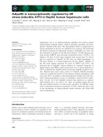
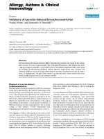
Figure 1(unshaded curve) to (shaded curve) stained
antibody surfactant protein-C confirm their identity with
(A) Primary human type II pneumocytes were and control
(A) Primary human type II pneumocytes were stained with
antibody surfactant protein-C (shaded curve) and control
antibody (unshaded curve) to confirm their identity. (B)
Human type II pneumocytes isolated were stained with antiCD14 FITC-conjugated antibodies (shaded curve) specific for
macrophage surface antigen to check for any contaminant
macrophage.
inson), and the FITC-stained cells were detected by
measuring green light emitted at 530 nm (FL1 channel).
The percentage of cells expressing the epithelial and macrophage makers were determined.
/>
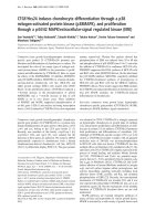
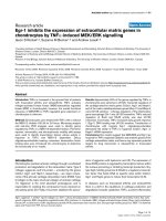
Figure
1 µm and demonstrated using
cytoplasm 50 electron micrographs human type (B) the
mocytes2cultured in vitro (A) and theofmagnification II pneuTransmission nm respectively) higher lamellar bodies in (Bars:
Transmission electron micrographs of human type II pneumocytes cultured in vitro (A) and the lamellar bodies in the
cytoplasm demonstrated using higher magnification (B) (Bars:
1 µm and 50 nm respectively). The cells were scraped off the
culture flask, fixed in 2% glutaraldehyde and embedded in
Araldite resin.
Influenza virus infection of type II pneumocytes and
bronchial epithelial cells
Human type II pneumocytes and bronchial epithelial cells
(seeded at 1 × 10⁶ cells per well in 24-well tissue-culture
plates) were infected at a multiplicity of infection (MOI)
of two unless otherwise indicated. After 60 min of virus
adsorption, the virus inoculum was removed, and the
cells were washed with warm culture medium (SAGM for
Page 4 of 13
(page number not for citation purposes)
Respiratory Research 2005, 6:135
/>
cells for analysis of cytokine gene expression. Ten hours
after infection, replicate cell monolayers were fixed and
analyzed by immuno-fluorescent staining specific for
influenza virus nucleoprotein (DAKO Imagen, Dako
Diagnostics Ltd, Ely, UK) to determine the proportion of
cells
infected.
Quantification of cytokine mRNA by real-time
quantitative RT-PCR
DNase-treated total RNA was isolated by means of RNeasy
Mini kit (Qiagen, Hilden, Germany). The cDNA was synthesized from mRNA with poly(dT) primers and Superscript II reverse transcriptase (Life Technologies, Rockville,
MD, USA) and quantified by real-time PCR analysis with
a LightCycler (Roche, Mannheim, Germany). The mRNA
for IP-10, interferon beta, IL-6, RANTES and tumor necrosis factor (TNF) alpha were quantitated using real-time
RT-PCR. The oligonucleotide primers and methods used
for real-time quantification of cytokines, viral matrix gene
and the housekeeping gene product γ-actin mRNA have
been described previously [12,24].
Quantification of cytokine proteins by ELISA
The concentrations of IP-10, RANTES, interleukin 6 and
interferon beta proteins in the primary human bronchial
and alveolar epithelial cell supernatants were measured
by a specific ELISA assay (R&D Systems, Minneapolis,
MN, USA). Samples of culture supernatant were irradiated
with ultraviolet light (CL-100 Ultra Violet Cross linker)
for 15 min to inactivate any infectious virus before the
ELISA assays were done. Previous experiments had confirmed that the dose of ultraviolet light used did not affect
cytokine concentration as measured by ELISA (data not
shown).
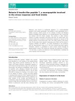
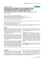
thelialand 0.5 µm at low (A) and of human bronchial epiTransmission vitro respectively) high (B) magnification (Bars:
2 µm cells in electron micrographs
Figure 3
Transmission electron micrographs of human bronchial epithelial cells in vitro at low (A) and high (B) magnification (Bars:
2 µm and 0.5 µm respectively). The cells were scraped off
the culture flask, fixed in 2% glutaraldehyde and embedded in
Araldite resin.
type II pneumocytes and BEBM for bronchial epithelial
cells) and incubated in medium supplemented with 0.6
mg/L penicillin, 60 mg/L streptomycin, and 2 mg/L N-ptosyl-L-phenylalanine chloromethyl ketone-treatedtrypsin (Sigma, St Louis, MO, USA). Aliquots of culture
supernatant were collected and frozen at -80°C for subsequent virus titration and cytokine analysis. The supernatants were titrated on MDCK cells and the viral titre was
quantitated as log10TCID50/ml. RNA was extracted from
Statistical analysis
The quantitative cytokine and chemokine mRNA and protein expression profile were compared using one-way
ANOVA, followed by Bonferroni multiple-comparison
test. Differences were considered significant at p < 0.05.
Results
In vitro infection of human type II pneumocytes
Primary human type II pneumocyte yields were 3.5 ± 0.9
× 106 cells/g lung tissue at 92 ± 5% cell purity as demonstrated by the expression of the type II pneumocyte specific marker surfactant protein C (SP-C), lack of the
monocyte/macrophage cell surface antigen (CD14) (Fig.
1A and 1B), and by staining for alkaline phosphatase
activity. The contaminating cells were predominantly
fibroblasts with monocyte/macrophage cells being less
than 2%. Cell viability was 91 ± 7% (n = 13). Differences
in age and sex of the lung donor had no apparent effects
on cell isolation yields and the performance of the cells in
culture. The isolated cells spread to form a confluent mon-
Page 5 of 13
(page number not for citation purposes)
Respiratory Research 2005, 6:135
/>
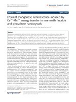
Figure 4
Infection of human type II pneumocytes with human influenza viruses
Infection of human type II pneumocytes with human influenza viruses. (A) Purified alveolar epithelial cells were fixed and analyzed by immunofluorescent staining specific for influenza virus nucleoprotein (×150). (B) The influenza M-gene mRNA profiles
were assayed after infection. The concentrations of M-gene mRNA were normalized to those of β-actin mRNA in the corresponding sample. Means of duplicate assays are shown. (C) Alveolar epithelial cells were infected with human influenza viruses
and the infectious virus yield (log10TCID50/ml) was determined in aliquots of supernatant collected at various times. Data are
the means and the standard errors of independent experiments from three separate donors.
Page 6 of 13
(page number not for citation purposes)
Respiratory Research 2005, 6:135
/>
Table 1: mRNA profile of cytokine and chemokine gene expression of primary culture of human type II pneumocytes 3 h and 6 h after
infection with A/Hong Kong/483/97 (H5N1/97), A/Vietnam/1194/04, A/Vietnam/3046/04 (both H5N1/04) and A/Hong Kong 54/98
(H1N1) influenza viruses denoted as fold increase compared to mock infected cells.
Gene products
Ratio of expression over mock-infected cells
3 hours post infection
483/97
(H5N1/97)
(-■-)c
Interleukin 1
Interleukin 6
Interleukin 8
MCP-1
TNF alpha
RANTES
Interferon-alpha
Interferon-beta
IP-10
-1.2
9.3*a
1
1.1
-1
1.1
3.7*a
3.9*a
1194/04
(H5N1/04)
(-◆-)c
3046/04
(H5N1/04)
(-×-)c
6 hours post infection
54/98
(H1N1)
(-▲-)c
483/97
(H5N1/97)
(-■-)c
1.1
7.4*a
-1.2
1
1
17.4*b
-1.2
-1.2
1.55
1.2
-0.8
-3.5
18.7*b
-1.3
22.1*b
37.9*b
0.3
0.9
15.1*b
9.9*a
0.9
1.1
0.8
1.5
Not detectable
2.2*
9.5*a
0.8
0.6
8.5*a
4.7*a
7.9*a
6.3*a
1194/04
(H5N1/04)
(-◆-)c
3046/04
(H5N1/04)
(-×-)c
0.9
0.8
19.2*b
15.4*b
1.6
1.3
1.7
1.3
Not detectable
24.1*b
16.9*b
1.3
0.9
26.3*b
18.7*b
46.8*b
29.7*b
54/98 (H1N1)
(-▲-)c
-1.3
8.8*a
1.3
1.3
6.9*a
1.2
13.3*b
8.1*a
Signals were normalized to the housekeeping gene, β-actin and expressed as a ratio over mock infected cells.
*Upregulation by two or more times over that of mock infection.
a p < 0.01 and b p < 0.001 (Bonferroni multiple-comparison test).
c Corresponding character symbols as shown in Figure 3 and 4.
olayer, exhibiting protruding nuclei surrounded by thin
cytoplasmic extensions. The identity of the cells in culture
as human type II pneumocytes was confirmed by demonstrating the presence of lamellar bodies and microvilli by
thin section electron microscopy (Figure 2).
Previous studies have demonstrated that avian influenza
viruses can infect human airway epithelial cells [25]. We
first wanted to determine whether alveolar epithelial cells
that constitutively reside in the lung can be infected with
avian and human influenza viruses in vitro. The cells were
infected with influenza A subtypes H5N1 (483/97, 1194/
04 and 3046/04) and H1N1 (54/98) at a MOI of 2 and the
proportion of cells expressing influenza A virus protein
was analyzed at 10 hours post-infection by immunofluorescent staining using an antibody specific for the virus
nucleoprotein (DAKO Imagen, Dako Diagnostics Ltd, Ely,
UK). Similar proportions (93–100%) of type II pneumocytes infected with H5N1 and H1N1 virus had evidence of
viral antigen (nucleoprotein) (Figure 4A). The quantification of influenza M-gene copies at 3 and 6 hours after
infection in cells infected with H5N1 and H1N1 viruses
showed comparable results at 3 and 6 hours post-infection (Figure 4B). Similarly, the infectious viral yield at 24
and 48 hours post-infection from alveolar epithelial cells
infected with H5N1 and H1N1 viruses were not significantly different (Figure 4C).
Induction of pro-inflammatory cytokines and chemokines
in type II pneumocytes
We investigated the cytokine induction profile induced by
H1N1 and H5N1 viruses in primary human type II pneumocytes. Specifically, we also wanted to determine if the
two viruses differed qualitatively or quantitatively in the
profile of cytokines induced. The mRNA of several
cytokines and chemokines were quantified using quantitative RT-PCR at 3 hr and 6 hr post-infection (Table I). The
mRNA levels of IP-10, interferon beta, RANTES and IL-6
were significantly up-regulated by influenza virus when
compared with the mock infected cells, the genes for IP-10
and interferon beta being the most highly induced. There
was no detectable TNF alpha induction in these epithelial
cells (data not shown). Inactivation of the virus by ultraviolet irradiation prior to infection of the alveolar epithelial cells abolished cytokine induction (data not shown)
suggesting that virus replication was required for cytokine
induction.
When compared with human H1N1 influenza virus, the
H5N1/97 and H5N1/04 viruses differentially up-regulated the transcription of IP-10, interferon beta, RANTES
and IL-6 to significantly higher levels (p < 0.001) (Figure
5). These differences were not explainable by a difference
in proportion of cells infected as indicated by immunofluorescence for viral antigen or differences in virus titre (Figure 4). Furthermore, an increase in the multiplicity of
infection of 54/98 (H1N1) virus from 2 to 10 did not
Page 7 of 13
(page number not for citation purposes)
Respiratory Research 2005, 6:135
/>
(Figure 3). The overall gene expression profile was comparable to that seen with type II pneumocytes. The M-gene
transcript copy numbers (Figure 6A) and infectious viral
yields (Figure 6B) from bronchial epithelial cells infected
with H5N1 and H1N1 viruses at an MOI of 2 were comparable. The H5N1/97 and H5N1/04 viruses differentially
up-regulated the transcription of IP-10, interferon beta,
RANTES and IL-6 to significantly higher levels than the
human H1N1 virus (p < 0.001 for IP-10, RANTES and IL6 and p < 0.01 for interferon beta) (Figure 7). In addition,
the two H5N1/04 viruses (1194/04 and 3046/04) differentially up-regulated the transcription of monocyte chemotactic protein 1 (MCP-1) and IL-8 to significantly higher
levels than the human H1N1 and H5N1/97 viruses (p <
0.05). None of the viruses induced TNF alpha in these
cells.
Figure 5
tive RT-PCR
enza-virus-infected human type II pneumocytes by quantitaCytokine and chemokine gene expression profile of influCytokine and chemokine gene expression profile of influenza-virus-infected human type II pneumocytes by quantitative RT-PCR. Cytokine and chemokine mRNA concentration
were assayed 3 h and 6 h after infection with A/Hong Kong/
483/97 (H5N1/97), A/Vietnam/1194/04, A/Vietnam/3046/04
(both H5N1/04) and A/Hong Kong 54/98 (H1N1) influenza
viruses or in mock infected cells. H5N1/97 and both H5N1/
04 influenza viruses induced significantly higher levels of IP10, interferon-beta, RANTES and IL-6 when compared to
H1N1 infected cells at 6 hours post-infection (p < 0.001,
Bonferroni multiple comparison test). The mRNA concentrations of cytokine and chemokine mRNA were normalized
to those β-actin mRNA in the corresponding samples. Means
and standard deviation from experiments from five different
donors are shown
result in cytokine mRNA concentrations similar to those
induced by H5N1/97 and H5N1/04 (data not shown).
Broadly, there were two patterns of kinetics of cytokine
gene transcription. Cytokines up-regulated from 3 hr postinfection onwards included IP-10, interferon beta and IL6 whereas RANTES mRNA was only up-regulated at 6 hr
post-infection (Table 1). The observations remained valid
whether the cytokine mRNA expression data were analyzed with or without normalization for γ-actin mRNA
concentrations.
Infection and cytokine induction profile of primary human
bronchial epithelial cells
The cytokine and chemokine profiles induced by H1N1,
H5N1/97 and H5N1/04 viruses in primary human bronchial epithelial cells were similarly investigated. The identity of the cells in culture as human bronchial epithelial
cells was confirmed by thin section electron microscopy
Secretion of cytokine proteins from bronchial and alveolar
epithelial cells
To confirm that the observed differences of mRNA are
reflected in levels of cytokine and chemokine secreted, the
concentrations of the IP-10, RANTES, interleukin 6 and
interferon-beta proteins were measured by ELISA in culture supernatants of infected bronchial and alveolar epithelial cells. The amount of IP-10 and IL-6 secreted by
bronchial and alveolar epithelial cells infected with all
three H5N1 viruses at 24 hours post infection were significantly higher (p < 0.01) than that secreted by cells
infected with H1N1 virus (Figure 8 and 9). At 24 hours
post infection, levels of IP-10 induced by H5N1/97 and
both H5N1/04 viruses were comparable. However, at 6
hours post-infection, the recent H5N1/04 viruses 1194/04
and 3046/04 appeared to be even more potent at inducing
IP-10 than H5N1/97 virus (p < 0.05) (Figure 8). RANTES
protein secreted from bronchial and alveolar epithelial
cells in response to H5N1/97 and 1194/04 (H5N1/04)
were significantly higher than that induced by H1N1
virus. Although the level RANTES mRNA in 3046/04
(H5N1/04) infected cells at 6 hours post infection was significantly higher than those H1N1 infected cells, the
RANTES protein secreted by these cells at 24 hours post
infection was only increased 4 fold (p = 0.062; not significant) (Figure 5 and 10). We failed to detect any interferon-beta proteins secreted from the supernatants of
bronchial and alveolar epithelial cells after influenza
viruses infection (data not shown) but it should be noted
that the limit of detection of the interferon-beta ELISA was
high (250 pg/ml).
Discussion
We found that the replication efficiency of the H5N1 and
H1N1 viruses was similar in both primary human alveolar
(Figure 4) and bronchial epithelial cells (Figure 6). Both
influenza virus subtypes induced an IP-10, interferon
beta, RANTES, and IL-6 responses. The cytokine induction
Page 8 of 13
(page number not for citation purposes)
Respiratory Research 2005, 6:135
Figure 6
enza viruses
Infection of human bronchial epithelial cells with human influInfection of human bronchial epithelial cells with human influenza viruses. (A) The influenza M-gene mRNA profiles were
assayed after infection. The concentrations of M-gene mRNA
were normalized to those of β-actin mRNA in the corresponding sample. Means of duplicate assays are shown. (B)
Virus yields (log10TCID50/ml) were determined in aliquots of
supernatant collected from influenza-infected bronchial epithelial cells at various times. Data are the means and the
standard errors of two independent experiments.
was dependent on viral replication since UV-inactivated
virus did not induce any effect. Interestingly, we found
that H5N1/97 and 1194/04 (H5N1/04) viruses were
more potent inducers of IP-10, interferon-beta, RANTES
and IL-6 mRNA and protein than the human H1N1 virus
(Figure 5, 7, 8 to 10). Thus, the observed differences of
mRNA are reflected in levels of cytokine and chemokine
proteins secreted (Figure 8 to 10). The results with 3046/
04 (H5N1/04) were generally similar to 1194/04 (H5N1/
04) with the exception that the levels of RANTES protein
in type II pneumocytes was not significantly elevated
when compared with H1N1 virus infected cells (Figure
10) although the mRNA levels were (Figure 5). Our inabil-
/>
Figure 7
tative RT-PCR
enza-virus-infected human bronchial epithelial cells influCytokine and chemokine gene expression profile ofby quantiCytokine and chemokine gene expression profile of influenza-virus-infected human bronchial epithelial cells by quantitative RT-PCR. Cytokine and chemokine mRNA
concentration were assayed 3 h and 6 h after infection with
A/Hong Kong/483/97 (H5N1/97), A/Vietnam/1194/04, A/
Vietnam/3046/04 (both H5N1/04) and A/Hong Kong 54/98
(H1N1) influenza viruses or in mock infected cells. When
compared with H1N1 infected cells, H5N1/97 and both
H5N1/04 influenza viruses significantly up-regulated IP-10,
RANTES and IL-6 (p < 0.001) and interferon beta (p < 0.01)
at 6 hours post-infection (Bonferroni multiple comparison
test). Both H5N1/04 viruses significantly up-regulated MCP-1
and IL-8 to levels higher than H1N1 and H5N1/97 infected
cells (p < 0.05, Bonferroni multiple comparison test). The
mRNA concentrations of cytokine and chemokine mRNA
were normalized to those β-actin mRNA in the corresponding samples. Means and standard deviation of duplicate cultures and assays are shown.
ity to detect any interferon-beta proteins in our experiments in spite of marked induction of mRNA is probably
related to the limited sensitivity of the interferon beta
ELISA. A more sensitive bioassay for interferon-beta may
be required for this purpose. The type II pneumocytes
used in these experiments were derived from a total of 13
donors and each set of experimental data is based on the
results of at least three separate experiments from three
donors therefore excluding a donor specific artifact. The
Page 9 of 13
(page number not for citation purposes)
Respiratory Research 2005, 6:135
Figure influenza viruses or in A/Vietnam/1194/04,
(H1N1) 8
nam/3046/04 (both and RANTES production cells with
Hong bronchial and alveolar mock infected by 54/98
humanInterleukin-6 H5N1/04)epithelial cells infectedA/VietIP-10, Kong/483/97 (H5N1/97),and A/Hong Kongprimary A/
IP-10, Interleukin-6 and RANTES production by primary
human bronchial and alveolar epithelial cells infected with A/
Hong Kong/483/97 (H5N1/97), A/Vietnam/1194/04, A/Vietnam/3046/04 (both H5N1/04) and A/Hong Kong 54/98
(H1N1) influenza viruses or in mock infected cells. Culture
supernatants from influenza virus-infected human respiratory
epithelial cells collected at 3 h, 6 h and 24 h after infection
with H5N1 and H1N1 viruses were tested by ELISA for IP-10
(Figure 8), Interleukin-6 (Figure 9) and RANTES (Figure 10).
The IP-10, Interleukin-6 and RANTES mRNA levels were
assayed at 3 h and 6 h post infection (data not shown) with
results comparable with that shown in figure 5 and 7. The
results from bronchial epithelial cells represent the means
and standard deviations of three independent experiments
(from the same donor). The means and standard deviations
of the results from alveolar epithelial cells are based on
experiments from six separate donors. * indicates p < 0.01
compared with mock and ** indicates p < 0.05 compared
with H5N1/97 and H1N1 infected cells using the Bonferroni
multiple comparison test.
/>
Figure influenza viruses or in A/Vietnam/1194/04,
(H1N1) 9
nam/3046/04 (both and RANTES production cells with
Hong bronchial and alveolar and A/Hong infectedA/ViethumanInterleukin-6 H5N1/04)epithelial cellsKong 54/98
IP-10, Kong/483/97 (H5N1/97),mock infected by primary A/
IP-10, Interleukin-6 and RANTES production by primary
human bronchial and alveolar epithelial cells infected with A/
Hong Kong/483/97 (H5N1/97), A/Vietnam/1194/04, A/Vietnam/3046/04 (both H5N1/04) and A/Hong Kong 54/98
(H1N1) influenza viruses or in mock infected cells. Culture
supernatants from influenza virus-infected human respiratory
epithelial cells collected at 3 h, 6 h and 24 h after infection
with H5N1 and H1N1 viruses were tested by ELISA for IP-10
(Figure 8), Interleukin-6 (Figure 9) and RANTES (Figure 10).
The IP-10, Interleukin-6 and RANTES mRNA levels were
assayed at 3 h and 6 h post infection (data not shown) with
results comparable with that shown in figure 5 and 7. The
results from bronchial epithelial cells represent the means
and standard deviations of three independent experiments
(from the same donor). The means and standard deviations
of the results from alveolar epithelial cells are based on
experiments from six separate donors. * indicates p < 0.01
compared with mock and ** indicates p < 0.05 compared
with H5N1/97 and H1N1 infected cells using the Bonferroni
multiple comparison test.
bronchial epithelial cells were purchased from a commerPage 10 of 13
(page number not for citation purposes)
Respiratory Research 2005, 6:135
/>
cial source and comes from one donor. However, since
the results from these cells are broadly in line with those
from the type II pneumocytes, again, we think that donor
specific artifacts are unlikely to explain the results we have
obtained. Finally, these results are also comparable to our
previous observations from primary human monocyte
derived macrophages [12] with the exception that in contrast to macrophages, no TNF alpha and IL-1 beta was
induced in respiratory epithelial cells by any of the viruses
tested.
This differential hyper-induction of cytokines was not
explained by differences in the replication kinetics
between the two virus subtypes. H5N1 viruses isolated
from patients with H5N1 disease in Hong Kong in 1997,
Vietnam in 2004 and human influenza viruses of the
H1N1 subtype all replicate with similar efficiency.
Increase in the MOI of the H1N1 virus did not result in an
increase of cytokine responses to levels comparable to that
of the H5N1 viruses. The cellular mechanisms underlying
this differential cytokine hyper-induction by H5N1
viruses are presently poorly understood. Studies on the
transformed bronchial epithelial cell line A549 previously
demonstrated that toll-like receptor 3 (TLR-3) is involved
in the influenza virus A initiated cytokine responses [27].
It remains to be determined whether H5N1 viruses also
act via TLR-3 signaling in primary human epithelial cells.
Figure influenza viruses or in A/Vietnam/1194/04,
(H1N1) 10
nam/3046/04 (both and RANTES production cells with
Hong bronchial and alveolar mock infected by 54/98
humanInterleukin-6 H5N1/04)epithelial cells infectedA/VietIP-10, Kong/483/97 (H5N1/97),and A/Hong Kongprimary A/
IP-10, Interleukin-6 and RANTES production by primary
human bronchial and alveolar epithelial cells infected with A/
Hong Kong/483/97 (H5N1/97), A/Vietnam/1194/04, A/Vietnam/3046/04 (both H5N1/04) and A/Hong Kong 54/98
(H1N1) influenza viruses or in mock infected cells. Culture
supernatants from influenza virus-infected human respiratory
epithelial cells collected at 3 h, 6 h and 24 h after infection
with H5N1 and H1N1 viruses were tested by ELISA for IP-10
(Figure 8), Interleukin-6 (Figure 9) and RANTES (Figure 10).
The IP-10, Interleukin-6 and RANTES mRNA levels were
assayed at 3 h and 6 h post infection (data not shown) with
results comparable with that shown in figure 5 and 7. The
results from bronchial epithelial cells represent the means
and standard deviations of three independent experiments
(from the same donor). The means and standard deviations
of the results from alveolar epithelial cells are based on
experiments from six separate donors. * indicates p < 0.01
compared with mock and ** indicates p < 0.05 compared
with H5N1/97 and H1N1 infected cells using the Bonferroni
multiple comparison test.
Cytokine and chemokine responses in vivo result from
autocrine and paracrine interactions involving many cell
types. Chemokines such as IP-10 and MCP-1 are macrophage chemo-attractants and mediate the inflammatory
response by further recruitment of circulating leukocytes
into the inflamed tissue. We have previously demonstrated that IP-10 and MCP-1 are up-regulated in primary
human macrophage by SARS-CoV [28]. The strong induction of chemokines in the lung micro-environment might
explain the prominent macrophage infiltrate observed in
the lungs of patients with fatal H5N1 [4] as well as SARS
[29].
RANTES attracts monocytes, eosinophils, basophils and T
cells, and selectively CD4+ T cells. Its production from the
bronchial epithelial cells contributes to the infiltration of
the inflammatory cells in airway viral infection [18]. IL-6
is a multifunctional cytokine that can regulate immune
and inflammatory responses involved in the activation,
growth and differentiation of T-cells [30] and can contribute to T cell mediated inflammatory reactions. In fact,
autopsy examination showed an increased CD3+ T cells in
the interstitium of the lung from patients with H5N1 diseases [4]. In addition, IL-6 has been shown to be released
by macrophages and epithelial cells during lung injury
[31] and the effects of IL-6 are synergistic with those of IL1 and TNF-alpha [32]. We have previously demonstrated
Page 11 of 13
(page number not for citation purposes)
Respiratory Research 2005, 6:135
that other proinflammatory cytokines such as IL-1, TNFalpha and IL-6 are hyper-induced in H5N1 infected macrophages [12]. Therefore, the differential up-regulation of
IL-6 expression in human respiratory epithelial cells and
the cytokines induced in macrophages by H5N1 viruses
may contribute synergistically to the pathogenesis of
human H5N1 disease.
The H5N1 viruses have continued to reassort, acquiring
different internal genes from other influenza viruses of
avian origin [33,34]. The H5N1/04 viruses, A/Vietnam/
1194/04 and A/Vietnam/3046/04 represent the Z genotype viruses that emerged as the dominant virus genotype
affecting poultry in south-east Asia [27,35]. Thus there
appears to be an association between the property of
hyper-inducing cytokines and high virulence. Additionally, in pig epithelial cells, H5N1/97 viruses were found to
resist the antiviral effects of interferon [36] and this may
also be relevant in pathogenesis. It is notable that patients
with avian influenza (H5N1) disease appeared to have
higher levels of IP-10 in their sera than those with infections with the human influenza viruses [4] providing in
vivo data that parallels our present findings in vitro. Studies
on recombinant viruses bearing the HA and NA of the
1918 "Spanish flu" pandemic virus showed that these
viruses have enhanced virulence for mice and induce
higher levels of macrophage-derived chemokines in vivo
in mice [37]. However, such observations of hyper-induction of cytokines in vivo may simply reflect more extensive
replication of the respective virus. The studies in vitro with
H5N1 viruses exclude such potential confounding factors
and it would be relevant to study the cytokine profiles of
the 1918 recombinant viruses in in vitro models similar to
those described here.
Conclusion
H5N1 subtype influenza A viruses associated with human
disease are more potent than human H1N1 virus at inducing proinflammatory cytokines and chemokines, including IP-10, interferon beta, IL-6 and RANTES, from human
primary alveolar and bronchial epithelial cells infected in
vitro. Previous findings showed that H5N1/97 viruses also
hyper-induce cytokines from macrophages and that
patients with H5N1 disease have high levels of IP-10 and
other chemokines in the serum. These findings may be relevant to the pathogenesis of H5N1 disease. The recent reemergence of H5N1 disease in humans is a cause for
renewed pandemic concern and highlights the need for a
better understanding of the pathogenesis of human H5N1
disease. Such understanding will lead to new strategies for
managing human H5N1 disease and enhance our preparedness to confront pandemic influenza, whether from
H5N1 or other influenza A subtypes.
/>
Competing interests
The author(s) declare that they have no competing interests.
Authors' contributions
JSM Peiris, MCW Chan and CY Cheung conceived the
study, planned the overall experimental design and wrote
the manuscript. MCW Chan carried out the experiments;
MCW Chan, CY Cheung and YO Chan carried out experiments in the BL3 laboratory and RWY Chan assisted in
experiments in the BL2 laboratory. WH Chui provided the
lung biopsy specimens, SW Tsao helped to develop the
methods for primary culture of the human alveolar epithelial cells, JM Nicholls advised on morphogical studies,
and LLM Poon and Y Guan advised in experimental
design. All authors critically reviewed the manuscript.
Acknowledgements
This research was supported by a research grants to MCW Chan from the
Research Fund for the Control of Infectious Diseases (RFCID 03040712)
and the Small Project Funding, CRGC, The University of Hong Kong and
research grants to JSM Peiris from the Research Fund for the Control of
Infectious Diseases (RFCID 01030172), the Research Grants Councils of
Hong Kong (HKU 7459/03M) and The University of Hong Kong Research
Achievement Award, 2005.
References
1.
2.
3.
4.
5.
6.
7.
8.
9.
Webster RG, Bean WJ, Gorman OT, Chambers TM, Kawaoka Y:
Evolution and ecology of influenza A viruses. Microbiol Rev
1992, 56(1):152-79.
Claas EC, Osterhaus AD, van Beek R, De Jong JC, Rimmelzwaan GF,
Senne DA, Krauss S, Shortridge KF, Webster RG: Human influenza
A H5N1 virus related to a highly pathogenic avian influenza
virus. Lancet 1998, 351:472-77.
Subbarao K, Klimov A, Katz J, Regenery H, Lim W, Hall H, Perdue M,
Swayne D, Bender C, Huang J, Hemphill M, Rowe T, Shaw M, Xu X,
Fukuda K, Cox N: Characterization of an avian influenza A
(H5N1) virus isolated from a child with a fatal respiratory illness. Science 1998, 279:393-396.
Peiris JSM, Yu WC, Leung CW, Cheung CY, NG WF, Nicholls JM, NG
TK, Chan KH, Lai ST, Lim WL, Yuen KY, Guan Y: Re-emergence of
fatal human influenza A subtype H5N1 disease. Lancet 2004,
363:617-619.
Tran TH, Nguyen TL, Nguyen TD, Luong TS, Pham PM, Nguyen VC,
Pham TS, Vo CD, Le TQ, Ngo TT, Dao BK, Le PP, Nguyen TT, Hoang
TL, Cao VT, Le TG, Nguyen DT, Le HN, Nguyen KT, Le HS, Le VT,
Christiane D, Tran TT, Menno de J, Schultsz C, Cheng P, Lim W,
Horby P, Farrar J, World Health Organization International Avian
Influenza Investigative Team: Avian influenza A (H5N1) in 10
patients in Vietnam. N Eng J Med 2004, 350:1179-88.
World Health Organization: Avian influenza – cumulative
number of cases – update 18. [ />2005_05_19/en/index.html].
To KF, Chan PKS, Chan KF, Lee WK, Lam WY, Wong KF, Tang NLS,
Tsang DNC, Sung RYT, Buckley TA, Tam JS, Cheng AF: Pathology
of fatal human infection associated with avian influenza A
H5N1 Virus. Journal of Medical Virology 2001, 63:242-246.
De Jong Menno D, Bach Van Cam , Phan Tu Qui , vo Minh Hien , Tran
Tan Thanh , Nguyen Bach Hue , Marcel Beld , Le Thi Phuong , Truong
Huu Khanh , Nguyen Van Vinh chau , Tran Tinh Hien , Do Quang Ha
, Jeremy Farrar : Fatal avian influenza A (H5N1) in a child presenting with diarrhea followed by coma. New England Journal of
Medicine 2005, 352:686-691.
Yuen KY, Chan PK, Peiris M, Tsang DN, Que TL, Shortridge KF, Cheung PT, To WK, Ho ET, Sung R, Cheng AF: Clinical features and
rapid viral diagnosis of human disease associated with avian
influenza A H5N1 virus. Lancet 1998, 351:467-71.
Page 12 of 13
(page number not for citation purposes)
Respiratory Research 2005, 6:135
10.
11.
12.
13.
14.
15.
16.
17.
18.
19.
20.
21.
22.
23.
24.
25.
26.
27.
28.
29.
Fisman DN: Hemophagocytic syndrome and infection. Emerg
Infect Dis 2000, 6:601-08.
Headley AS, Tolley E, Meduri GU: Infections and the inflammatory response in acute respiratory distress syndrome. Chest
1997, 111:1306-21.
Cheung CY, Poon LLM, Lu AS, Luk W, Lau YL, Shortridge KF, Gordon S, Guan Y, Peiris JSM: Induction of proinflammatory
cytokines in human macrophages by influenza A (H5N1)
viruses: a mechanism for the unusual severity of human disease? Lancet 2002, 360:1831-1837.
Fesq H, Bacher M, Nain M, Gemsa D: Programmed cell death
(apoptosis) in human monocytes infected by influenza A
virus. Immunobiology 1994, 190:175-182.
Seo SH, Webby R, Webster RG: No apoptotic deaths and different levels of inductions of inflammatory cytokines in alveolar
macrophages infected with influenza viruses. Virology 2004,
329:270-279.
Ebisawa IT, Kitamoto O, Takeuchi Y, Makino M: Immunocytologic
study of nasal epithelial cells in influenza. Am Rev Respir Dis
1969, 99:507-15.
Kuiken T, Rimmelzwaan GF, Van Amerongen G, Osterhaus AD:
Pathology of human influenza A (H5N1) virus infection in
cynomolgus macaques (Macaca fascicularis). Vet Pathol 2003,
40:304-10.
Uiprasertkul M, Puthavathana P, Sangsiriwut K, Pooruk P, Srisook K,
Peiris M, Nicholls JM, Chokephaibulkit K, Vanprapar N, Auewarakul
P: Influenza A H5N1 replication sites in humans. Emerg Infect
Dis 2005, 11:1036-41.
Adachi M, Matsukura S, Tokunaga H, Kokubu F: Expression of
cytokines on human bronchial epithelial cells induced by
influenza virus A. Int Arach Allergy Immunol 1997, 113:307-311.
Cunningham AC, Milne DS, Wilkes J, Dark JH, Tetley TD, Kirby JA:
Constitutive expression of MHC and adhesion molecules by
alveolar epithelial cells (type II pneumocytes) isolated from
human lung and comparison with immunocytochemical
findings. Journal of Cell Sciences 1994, 107:443-449.
Glanville AR, Tazelaar HD, Theodore J, Imoto E, Rouse RV, Baldwin
JC, Robin ED: The distribution of MHC class I and II antigens
on bronchial epithelium. Amer Rev Respir Dis 1989, 139:330-334.
Peters U, Papadopoulos T, Muller-Hermelink HK: MHC class II
antigens on lung epithelial of human fetuses and neonates.
Ontogeny and expression in lungs with histologic evidence of
infection. Lab Invest 1990, 63:38-43.
Cheek JM, Kim KJ, Crandall ED: Tight monolayers of rat alveolar
epithelial cells: bioelectric properties and active sodium
transport. Am J Physiol 1989, 256:C688-C693.
Elbert KJ, Schafer UF, Schafers HJ, Kim KJ, Lee VHL, Lehr CM: Monolayers of human alveolar epithelial cells in primary culture
for pulmonary absorption and transport studies. Pharmaceutical Research 1999, 16(5):601-08.
Blaschke V, Reich K, Blaschke S, Neumann C: Rapid quantitation
of proinflammatory and chemoattractant cytokine expression in small tissue samples and monocyte-derived dendrite
cells: validation of a new real-time RT-PCR technology. J
Immunol Methods 2000, 246:79-90.
Matrosovich MN, Matrosovich TY, Gray T, Roberts NA, Klent HD:
Human and avian influenza viruses target different cell types
in culture of human airway epithelium. Proc Natl Acad Sci USA
2004, 101(13):4620-4624.
Matrosovich MN, Zhou N, Kawaoka Y, Webster R: The surface
glycoproteins of H5 influenza viruses isolated from humans,
chickens, and wild aquatic birds have distinguishable properties. J Virol 1999, 73:1146-1155.
Guillot L, Le Goffie R, Bloch S, Escriou N, Akira S, Chignard M, SiTahar M: Involvement of Toll-like receptor 3 in the immune
response of lung epithelial cells to double stranded RNA and
influenza A virus. J Biol Chem 2005, 280:5571-5580.
Cheung CY, Poon LL, Ng IH, Luk W, Sia SF, Wu MH, Chan KH, Yuen
KY, Gordon S, Guan Y, Peiris JS: Cytokine responses in severe
acute respiratory syndrome coronavirus-infected macrophages in vitro: possible relevance to pathogenesis. J Virol
2005, 79:7819-26.
Nicholls JM, Poon LLM, Lee KC, Ng WF, Lai ST, Leung CY, Chu CM,
Hui PK, Mak KL, Lim WL, Yan KW, Chan KH, Tsang NC, Guan Y,
Yuen KY, Peiris JSM: Lung pathology of fatal severe acute respiratory syndrome. Lancet 2003, 361:1173-78.
/>
30.
31.
32.
33.
34.
35.
36.
37.
Van Snick J: Interleukin-6: an overview. Annu Rev Immunol 1990,
8:253-78.
Hierholzer C, Kalff JC, Omert L, Tsukada K, Loeffert JE, Watkins SC,
Billiar TR, Tweardy DJ: Interleukin-6 production in hemorrhagic shock is accompanied by neutrophil recruitment and
lung injury. Am J Physiol 1998, 275:611-21.
Le JM, Fredrickson G, Reis LF, Diamantstein T, Hirano T, Kishimoto
T, Vilcek J: Interleukin 2-dependent and interleukin 2-independent pathways of regulation of thymocyte function by
interleukin 6. Proc Natl Acad Sci USA 1988, 85(22):8643-7.
Guan Y, Peiris JS, Lipatov AS, Ellis TM, Dyrting KC, Krauss S, Zhang
LJ, Webster RG, Shortridge KF: Emergence of multiple genotypes of H5N1 avian influenza viruses in Hong Kong SAR.
Proc Natl Acad Sci USA 2002, 99(13):8950-5.
Guan Y, Poon LL, Cheung CY, Ellis TM, Lim W, Lipatov AS, Chan KH,
Sturm-Ramirez KM, Cheung CL, Leung YH, Yuen KY, Webster RG,
Peiris JS: H5N1 influenza: a protean pandemic threat. Proc Natl
Acad Sci USA 2004, 101(21):8156-61.
Li KS, Guan Y, Wang J, Smith GJ, Xu KM, Duan L, Rahardjo AP, Puthavathana P, Buranathai C, Nguyen TD, Estoepangestie AT, Chaisingh A,
Auewarakul P, Long HT, Hanh NT, Webby RJ, Poon LL, Chen H,
Shortridge KF, Yuen KY, Webster RG, Peiris JS: Genesis of a highly
pathogenic and potentially pandemic H5N1 influenza virus
in eastern Asia. Nature 2004, 430:209-13.
Seo SH, Hoffmann E, Webster RG: Lethal H5N1 influenza viruses
escape host antiviral-cytokine responses. Nat Med 2002,
8:950-54.
Kobasa D, Takada A, Shinya K, Hatta M, Halfmann P, Theriault S,
Suzuki H, Nishimura H, Mitamura K, Sugaya N, Usui T, Murata T,
Maeda Y, Watanabe S, Suresh M, Suzuki T, Suzuki Y, Feldmann H,
Kawaoka Y: Enhanced virulence of influenza A viruses with the
haemagglutinin of the 1918 pandemic virus. Nature 2004,
431:703-7.
Publish with Bio Med Central and every
scientist can read your work free of charge
"BioMed Central will be the most significant development for
disseminating the results of biomedical researc h in our lifetime."
Sir Paul Nurse, Cancer Research UK
Your research papers will be:
available free of charge to the entire biomedical community
peer reviewed and published immediately upon acceptance
cited in PubMed and archived on PubMed Central
yours — you keep the copyright
BioMedcentral
Submit your manuscript here:
/>
Page 13 of 13
(page number not for citation purposes)